The Journal of Neuroscience, December 1995, 75( 12): 8156-8166 Expression of p-, 6-, and K-OpiOid Receptor-Like lmmunoreactivities in Rat Dorsal Root Ganglia after Carrageenan-Induced Inflammation Ru-Rong Ji,’ Qin Zhang,’ P.-Y. Law,2 H. H. Low,* R. Elde,3 and T. Hiikfeltl ‘Department of Neuroscience, Karolinska Institute, S-171 77 Stockholm, Sweden and Departments of 2Pharmacology and 3Cell Biology and Neuroanatomy, University of Minnesota, Minneapolis, Minnesota 55455 Recently, antisera that recognize unique epitopes of the cloned p-, S-, and K-opioid receptors (MOR, DOR, KOR, re- spectively) have been developed. In the present study MOR-, DOR-, and KOR-like immunoreactivities (Lls) were examined in rat dorsal root ganglia (DRGs, L4-5) after in- jection of carrageenan (CAR) into the hindpaw. In normal control rats, 20.9%, 13.5%, and 9% of the DRG neurons contained MOR- , DOR-, KOR-LI, respectively. A marked upregulation in MOR-LI was observed in DRG neurons 1 and 3 d after inflammation. In contrast, CAR induced a dis- tinct downregulation in DOR- and KOR-Lls. MOR-, DOR-, and KOR-Lls were preferentially localized in small DRG neurons. MOR-LI was often located in patches in the cy- toplasm, and in some cells close to the somatic plasma- lemma. However, DOR- and KOR-Lls mainly showed a dif- fuse staining pattern within cytoplasm. Two or even all three receptors could sometimes be found to coexist in DRG neurons. In the spinal cord, these receptors were mainly confined to the superficial dorsal horn, with a some- what diffuse staining which was strong for MOR-LI, and weak for KOR-LI. DOR-LI had a distinctly punctate, vari- cose distribution. CAG induced-alterations in opioid recep- tor staining in spinal cord were much less pronounced than those in the DRGs with a small increase in MOR-LI and a slight decrease in DOR-LI ipsilaterally. There was an accumulation of all three types of receptors in the sciatic nerve both proximal and distal to the ligation site as early as 2 hr, indicating both antero- and retrograde transport of multiple opioid receptors. However, DOR-LI accumulation was stronger than that of MOR- and KOR-Lls. Taken to- gether, these results suggest that all three opioid receptors are involved in the response to inflammation and that they may play different roles in this pathological state. The co- existence of MOR, DOR, and KOR in at least some primary sensory neurons provides a substrate for functional inter- actions between these receptors. Received Mar. 20, 1995; revised Aug. 1, 1995; accepted Aug. 14, 1995. This work is supported by the Swedish MRC (04X-2887), the Bank of Swe- den Tercentenary Foundation, Marianne and Marcus Wallenbergs Stiftelse and Gustaf V:s and Drottning Victorias Stiftelse. We thank Dr. Zs. Wiesenfeld- Hallin, Huddinge Hospital, Karolinska Institute for valuable advice. Correspondence should be addressed to Dr. Tomas HGkfelt, Department of Neuroscience/Histology, Karolinska Institute, S-17177 Stockholm, Sweden. Copyright 0 1995 Society for Neuroscience 0270-6474/95/158 156-11$05.00/O [Key words: multiple opioid receptors, pain, primary sen- sory neurons, spinal cord, coexistence, axonal transport] Multiple receptor sitesrecognizing exogenous opiate drugs and endogenous opioid peptideshave been defined basedon phar- macological,behavioral and receptor binding experiments. Thus, there are p, 6, and K receptors,which display high affinities for morphine, enkephalin, and dynorphin, respectively (Martin et al., 1976;Lord et al., 1977; Changand Cuatrecasas, 1979; Smith and Simon, 1980; Corbett et al., 1993; Simon and Gioannini, 1993). Thesereceptors are involved in regulation of, for exam- ple, nociception, respiration, cardiovascularfunctions, gastroin- testinal motility, and mood (Basbaum and Fields, 1984; Paster- nak, 1988; Herz, 1993). Recently a selective loss of 6 opioid analgesiaand binding has been demonstrated after intrathecal administration of antisense oligonuclotides to a Sopioid receptor (Standifier et al., 1994). Using ligand binding autoradiography, all three types of opioid receptors have been demonstrated in the superficial spinal cord (Atweh and Kuhar, 1977; Iadarola et al., 1988; Besseet al., 1990; Stevens et al., 1991b; see also Mansour et al., 19SS), a region of the dorsal horn enriched in opioid peptides, and a known site of termination of peptide con- taining and nociceptive primary afferents (Hiikfelt et al., 1977; Besson and Chaouch, 1987; Willis and Coggeshall,1991; Dado et al., 1993; Arvidsson et al., 1995b).In autoradiographic ligand binding studies Ninkovic et al. (1981, 1982) demonstrated pres- ence of opiate receptors on primary sensory neurons.Transec- tion of the sciatic nerve or dorsal root afferents results in a marked decrease in all three types of opioid binding sites within the dorsal horn, suggesting a partly presynaptic localization of the receptors (Fields et al., 1980; Besseet al., 1990). A com- parable decrease is observed after capsaicin treatment (Gamse et al., 1979; Besson and Chaouch, 1987). However, ligand bind- ing autoradiography is generally limited by a low resolution mostly not allowing unequivocal identification of cellular and of subcellular structures.With the recent cloning of the 6 (Evans et al., 1992; Kieffer et al., 1992;Bzdegaet al., 1993), lt (Thomp- son et al., 1993; Wang et al., 1993; Chen et al., 1993a), and K (Chen et al., 1993b; Meng et al., 1993; Minami et al., 1993; Yasuda et al., 1993) receptors,it has becomepossible not only to study opioid receptor expressionusing in situ hybridization (Mansour et al., 1993; Schafer et al., 1933; Mansour et al., 1994a-c), but one can now generateantibodiesto theserecep- tors. In fact, such antibodieshave recently been usedin immu- nohistochemical studies to localize opioid receptor protein at the
Transcript
The Journal of Neuroscience, December 1995, 75( 12): 8156-8166
Expression of p-, 6-, and K-OpiOid Receptor-Like lmmunoreactivities in Rat Dorsal Root Ganglia after Carrageenan-Induced Inflammation
Ru-Rong Ji,’ Qin Zhang,’ P.-Y. Law,2 H. H. Low,* R. Elde,3 and T. Hiikfeltl
‘Department of Neuroscience, Karolinska Institute, S-171 77 Stockholm, Sweden and Departments of 2Pharmacology and 3Cell Biology and Neuroanatomy, University of Minnesota, Minneapolis, Minnesota 55455
Recently, antisera that recognize unique epitopes of the cloned p-, S-, and K-opioid receptors (MOR, DOR, KOR, re- spectively) have been developed. In the present study MOR-, DOR-, and KOR-like immunoreactivities (Lls) were examined in rat dorsal root ganglia (DRGs, L4-5) after in- jection of carrageenan (CAR) into the hindpaw. In normal control rats, 20.9%, 13.5%, and 9% of the DRG neurons contained MOR- , DOR-, KOR-LI, respectively. A marked upregulation in MOR-LI was observed in DRG neurons 1 and 3 d after inflammation. In contrast, CAR induced a dis- tinct downregulation in DOR- and KOR-Lls. MOR-, DOR-, and KOR-Lls were preferentially localized in small DRG neurons. MOR-LI was often located in patches in the cy- toplasm, and in some cells close to the somatic plasma- lemma. However, DOR- and KOR-Lls mainly showed a dif- fuse staining pattern within cytoplasm. Two or even all three receptors could sometimes be found to coexist in DRG neurons. In the spinal cord, these receptors were mainly confined to the superficial dorsal horn, with a some- what diffuse staining which was strong for MOR-LI, and weak for KOR-LI. DOR-LI had a distinctly punctate, vari- cose distribution. CAG induced-alterations in opioid recep- tor staining in spinal cord were much less pronounced than those in the DRGs with a small increase in MOR-LI and a slight decrease in DOR-LI ipsilaterally. There was an accumulation of all three types of receptors in the sciatic nerve both proximal and distal to the ligation site as early as 2 hr, indicating both antero- and retrograde transport of multiple opioid receptors. However, DOR-LI accumulation was stronger than that of MOR- and KOR-Lls. Taken to- gether, these results suggest that all three opioid receptors are involved in the response to inflammation and that they may play different roles in this pathological state. The co- existence of MOR, DOR, and KOR in at least some primary sensory neurons provides a substrate for functional inter- actions between these receptors.
Received Mar. 20, 1995; revised Aug. 1, 1995; accepted Aug. 14, 1995.
This work is supported by the Swedish MRC (04X-2887), the Bank of Swe- den Tercentenary Foundation, Marianne and Marcus Wallenbergs Stiftelse and Gustaf V:s and Drottning Victorias Stiftelse. We thank Dr. Zs. Wiesenfeld- Hallin, Huddinge Hospital, Karolinska Institute for valuable advice.
Correspondence should be addressed to Dr. Tomas HGkfelt, Department of Neuroscience/Histology, Karolinska Institute, S-17177 Stockholm, Sweden.
Copyright 0 1995 Society for Neuroscience 0270-6474/95/158 156-11$05.00/O
Multiple receptor sites recognizing exogenous opiate drugs and endogenous opioid peptides have been defined based on phar- macological, behavioral and receptor binding experiments. Thus, there are p, 6, and K receptors, which display high affinities for morphine, enkephalin, and dynorphin, respectively (Martin et al., 1976; Lord et al., 1977; Chang and Cuatrecasas, 1979; Smith and Simon, 1980; Corbett et al., 1993; Simon and Gioannini, 1993). These receptors are involved in regulation of, for exam- ple, nociception, respiration, cardiovascular functions, gastroin- testinal motility, and mood (Basbaum and Fields, 1984; Paster- nak, 1988; Herz, 1993). Recently a selective loss of 6 opioid analgesia and binding has been demonstrated after intrathecal administration of antisense oligonuclotides to a S opioid receptor (Standifier et al., 1994). Using ligand binding autoradiography, all three types of opioid receptors have been demonstrated in the superficial spinal cord (Atweh and Kuhar, 1977; Iadarola et al., 1988; Besse et al., 1990; Stevens et al., 1991b; see also Mansour et al., 19SS), a region of the dorsal horn enriched in opioid peptides, and a known site of termination of peptide con- taining and nociceptive primary afferents (Hiikfelt et al., 1977; Besson and Chaouch, 1987; Willis and Coggeshall, 1991; Dado et al., 1993; Arvidsson et al., 1995b). In autoradiographic ligand binding studies Ninkovic et al. (198 1, 1982) demonstrated pres- ence of opiate receptors on primary sensory neurons. Transec- tion of the sciatic nerve or dorsal root afferents results in a marked decrease in all three types of opioid binding sites within the dorsal horn, suggesting a partly presynaptic localization of the receptors (Fields et al., 1980; Besse et al., 1990). A com- parable decrease is observed after capsaicin treatment (Gamse et al., 1979; Besson and Chaouch, 1987). However, ligand bind- ing autoradiography is generally limited by a low resolution mostly not allowing unequivocal identification of cellular and of subcellular structures. With the recent cloning of the 6 (Evans et al., 1992; Kieffer et al., 1992; Bzdega et al., 1993), lt (Thomp- son et al., 1993; Wang et al., 1993; Chen et al., 1993a), and K
(Chen et al., 1993b; Meng et al., 1993; Minami et al., 1993; Yasuda et al., 1993) receptors, it has become possible not only to study opioid receptor expression using in situ hybridization (Mansour et al., 1993; Schafer et al., 1933; Mansour et al., 1994a-c), but one can now generate antibodies to these recep- tors. In fact, such antibodies have recently been used in immu- nohistochemical studies to localize opioid receptor protein at the
The Journal of Neuroscience, December 1995, 15(12) 8157
cellular level (Dado et al., 1993; Arvidsson et al., 1995a-c; Hill- er et al., 1995).
A number of studies have shown that injection of irritative chemicals in the hindpaw of the rat can produce an intense in- flammation characterized by erythema, edema, and hyperthermia ,limited to the injected paw (Iadarola et al., 1988; Dubner and Ruda, 1992; Ji et al., 1994). Such inflammatory tissues are char- acterized by an increased sensitivity to mechanical stimuli (hy- peralgesia) (Dubner and Ruda, 1992; Levine et al., 1993). It has been shown that peripheral inflammation induces a marked upre- gulation of dynorphin and enkephalin at both mRNA and pep- tide levels in the dorsal horn (Millan, 1986; Ruda et al., 1988; Weihe et al., 1988; Noguchi et al., 1989; Dubner and Ruda, 1992; Ji et al., 1994). Inflammation also induces c-fos expression in the dorsal horn and this can be blocked by morphine (Presley et al., 1990). Moreover, an extensive body of research has ad- dressed the question of plasticity of opioid receptor binding at the spinal level after inflammation or neuropathic pain (Millan et al., 1986, 1988; Iadarola et al., 1988; Stevens et al., 1991a; Besse et al., 1992a,b; Kar et al., 1994).
In the present study we have used immunohistochemistry and recently developed antibodies against three opioid receptors, the p. receptor (MOR-), the S receptor (DOR-), and the K receptor (KOR) to study their expression in primary sensory neurons after unilateral peripheral inflammation. These opioid receptors were also studied in the spinal cord as was their transport in the sciatic nerve. Moreover, since the different types of opioid re- ceptors may be functionally coupled (see Holaday et al., 1985; Rothman et al., 1993) attempts were made to analyze a possible colocalization of three opioid receptors.
Materials and Methods
Induction of injfammation. Adult male Sprague-Dawley rats weighing 190-220 gm were anesthetized with sodium oentobarbital (40-60 rnp/ kg,i.p), and 200 p,l of a 4% carrageenan solution (Sigma, dissolved m saline) were injected into the plantar surface of the left hindpaw. Ani- mals were kept in cages (three or four animals in a cage) at an ambient temperature of 20-25°C under a 12 hd12 hr light/dark cycle and had free access to food and water. The experiments have been approved by “Stockholm norra forsiiksdjursetiska kommitte” and attention was paid to the ethical guidelines for investigation of experimental pain in con- scious animals (Zimmermann, 1983).
Antisera. Antibodies were raised against peptides corresponding to amino acids 384-398 (NHQLENLEAETAPLP, code no. 551; Arvidsson et al., 1995b), 3-17 (LVPSARAELOSSPLV, code no. 442; Dado et al., 1993; Arvidsson et al., 1995b), and 366380 (DPASMRDVGGMNKPV, Arvidsson et al., 1995~) of the predicted sequence of MORI (Chen et al., 1993a; Thompson et al., 1993), DOR (Evans et al., 1992; Kieffer et al., 1992), and KOR (Chen et al., 1993b; Meng et al., 1993), respec- tively. All peptides were conjugated to bovine thyroglobulin, and used to immunize rabbits as described.
Immunohistochemistry. One day or 3 d after injection of CAR , both inflammatory and control rats were deeply anesthetized with sodium pentobarbital (120 mg/kg, i.p) and transcardially perfused with 60 ml warm saline, followed by 400 ml 4% paraformaldehyde with 0.4% pi- cric acid in 0.16 M phosphate buffer solution (pH 7.2) (Pease, 1962; Zamboni and De Martino, 1967). The L5 DRGs and L4-5 segments of the spinal cord were removed, postfixed in the same fixative for 90 min, and then placed in 10% sucrose solution at 4°C overnight. Tissues were embedded in OCT compound (Miles, USA), cut coronally in a cryostat at 4 pm (to study coexistence) or at 14 pm thickness and mounted onto gelatin coated slides. The sections were placed in a humid chamber and processed for immunohistochemistry according to both ABC method (Hsu et al., 1981) or indirect immunofluorescence staining (see Coons, 1958), by incubating the sections overnight at 4°C in the following primary antisera: MOR (1:4000), DOR (1: lOOO), or KOR (1:2000). The antisera were diluted in PBS containing 0.3% Triton X-100 (Hartman et al., 1972), 0.01% sodium azide, and 0.02% Bacitracin. For ABC
staining, the sections were then incubated for 1 hr at 37°C with the biotinylated secondary antibody (1:200) and subsequently with ABC comulex (1:lOO: ABC Kit. Vector Labs. Burlinaame. CA). Finallv the
Y , 2 reaction product was visualized with 0.05% DAB/0.003% hydrogen peroxide in 0.1 M acetate buffer (pH = 6) containing 2% ammonium nickel sulfate for 5 min, and then rinsed in acetate buffer, air dried, dehydrated, and coverslipped. For immunofluorescence, the sections were incubated at 37°C for 30 min with fluorescein isothiocyanate (FITC)-conjugated goat anti-rabbit antisera (1:80; Boehringer Mann- heim Scandinavia, Stockholm, Sweden). Finally the sections were mounted in a mixture of glycerol and PBS (9: 1) containing 0.1% para- phenylenediamine (Johnson and de C Nogeira Araujo, 1981; Platt and Michael, 1983) in order to retard fading and examined in a Nikon Mi- crophot-FX microscope. FITC-induced fluorescence was analyzed with a Nikon B-1E filter cube (excitation at 480 + 10 nm with a bandpass emission filter passing 520-550 nm). The specificity of the immuno- staining was tested by preabsorbing the antibody with synthetic peptides 10mh M; (Peninsula, Belmont, CA), or by replacing the primary antisera with normal serum.
For laser scanning confocal microscopy analysis a BIO-RAD MRC- 600 laser scanning confocal imaging system equipped with a krypton/ argon mixed gas laser was used. The Kl channel was used. FITC stain- ing was detected with exciter 488DFlO (488 nm line). The images were printed using a Tektronix Phaser IICD digital colour printer.
For analysis of axonal transport, the left sciatic nerve was ligated at mid thigh level. The animals were allowed to survive 2, 6, 12, 24, and 48 hr (two rats for each time point). After perfusion the portion (10 mm) of the sciatic nerve surrounding the ligation was removed, cut, and processed for immunofluorescence staining.
Quantijkation. The total number of MOR-, DOR-, and KOR-im- munoreactive (IR) neuronal profiles was divided by the total number of profiles in each DRG section, and the percentage of immunoreative neuronal profiles was calculated. Percentages from four sections from one animal were averaged as the percentage for that animal. Three rats were used for anlysis of each group.
The size of immunoreactive neuronal profiles in the DRGs and the density of fiber networks in the dorsal horn were measured on a Mac- intosh 11x computer (Apple Computer Inc., Cupertino, CA), equipped with a Quick Capture frame grabber board (Data Translation, Marlboro, MA) and a Dage-MT1 72 CCD series camera (DAGE-MTI, Michigan City, IN) connected with a Nikon microscope. Image processing was performed with NIH-IMAGE software (courtesy Dr. W. Rasband, NIMH). The size of 200 neuronal profiles each containing MOR-, DOR-, or KOR-LI of DRGs from both control and inflamed animals were mea- sured. In spinal cord, the relative density levels of immunostaining were measured in the medial half of the superficial layers (laminae I and II) of the dorsal horn. Each image was digitized with 256 gray levels for each picture element. Eighteen sections of the spinal cord from rats subjected to 1 (n = 3) and 3 (n = 3) d of inflammation were analyzed.
Statistics. All data were assessed using the two-tailed t test for group comparison and ANOVA for multiple comparison. The criterion for statistical significance was P < 0.05.
Results
Carrageenan-induced injlammation
Intraplantar injection of 200 pl CAR (4%) led to a visibly ob- vious oedema and erythema, indicative of inflammation. The circumference of the injected paw gradually increased to a mean value of 152.7 -C 6.0 and 167.8 ? 7.0% (mean -C SEM, n = 8, P < 0.001, paired t test) at 1 d and 3 d, respectively, when the paw circumference of the noninjected side (right side) was set as 100%. No marked oedema was observed in the right paw.
Opioid receptors in DRGs
In control (normal) rats, many DRG neurons (20.9%) were MOR-positive. These neurons were mainly small with profiles ranging in size from 169-1170 pm*, the majority lying between 300-500 pm2 (Figs. lb, 2A, 3A). Confocal analysis showed that the MOR-LI was localized close to the somatic plasmalemma, as well as in patches in the cytoplasm (Fig. 4a). However, in some neurons the association of MOR-LI with the plasmalemma
8158 Ji et al. l Opioid Receptors and Inflammation
Figure I. u-f, Bright-field micrographs after ABC immunostaining showing the distribution of p, (a, b), 6 (c, d), and K (e, f) opioid receptor-11s in neurons on the ipsilateral (a, c, e) and contralateral (b, d, f) dorsal root ganglia (L5) in rats 1 d after carrageenan injection into the left hindpaw. Asterisks indicate ipsilateral side. All three types of opioid receptors were preferentially localized in small DRG neurons. Inflammation induced an upregulation of l.~- (a, b), but a downregulation of 6- (c, d) and K- (e, f) opioid receptor-11s. Scale bar, 100 p,m. All micrographs have the same magnification.
2 41 4oy A
2 353
:g 30;
[ 25: 3
B H 20:
E; 15: 3
0 Ipsi 0 Contra
; 10 m z 5 1 go], , ,
L
Cont lday 3 day
delta-R l Ipsi
0 Contra
J I I 1 Cont 1 day 3 day
kapw-R a Ipsi
0 Contra
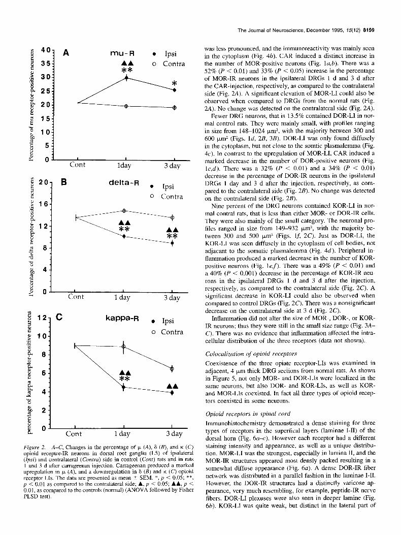
Figure 2. A-C, Changes in the percentage of )” (A), 6 (B), and K (C)
opioid receptor-IR neurons in dorsal root ganglia (L5) of ipsilateral (Ipsi) and contralateral (Contra) side in control (Cant) rats and in rats 1 and 3 d after carrageenan injection. Carrageenan produced a marked upregulation in k (A), and a downregulation in 6 (B) and K (C) opioid receptor Lls. The data are presented as mean ? SEM. *, p < 0.05; **, p < 0.01 as compared to the contralateral side; A, p < 0.05; AA, p < 0.01, as compared to the controls (normal) (ANOVA followed by Fisher PLSD test).
The Journal of Neuroscience, December 1995, 15(12) 8159
was less pronounced, and the immunoreactivity was mainly seen in the cytoplasm (Fig. 4b). CAR induced a distinct increase in the number of MOR-positive neurons (Fig. la,b). There was a 52% (P < 0.01) and 33% (P < 0.05) increase in the percentage of MOR-IR neurons in the ipsilateral DRGs 1 d and 3 d after the CAR-injection, respectively, as compared to the contralateral side (Fig. 2A). A significant elevation of MOR-LI could also be observed when compared to DRGs from the normal rats (Fig. 2A). No change was detected on the contralateral side (Fig. 2A).
Fewer DRG neurons, that is 13.5% contained DOR-LI in nor- mal control rats. They were mainly small, with profiles ranging in size from 148-1024 pm*, with the majority between 300 and 600 pm2 (Figs. Id, 2B, 3B). DOR-LI was only found diffusely in the cytoplasm, but not close to the somtic plasmalemma (Fig. 4~). In contrast to the upregulation of MOR-LI, CAR induced a marked decrease in the number of DOR-positive neurons (Fig. I&). There was a 32% (P < 0.01) and a 34% (P < 0.01) decrease in the percentage of DOR-IR neurons in the ipsilateral DRGs 1 day and 3 d after the injection, respectively, as com- pared to the contralateral side (Fig. 2B). No change was detected on the contralateral side (Fig. 2B).
Nine percent of the DRG neurons contained KOR-LI in nor- mal contra1 rats, that is less than either MOR- or DOR-IR cells. They were also mainly of the small category. The neuronal pro- files ranged in size from 149-932 pm2, with the majority be- tween 300 and 500 pm2 (Figs. 15 2C). Just as DOR-LI, the KOR-LI was seen diffusely in the cytoplasm of cell bodies, not adjacant to the somatic plasmalemma (Fig. 4d). Peripheral in- flammation produced a marked decrease in the number of KOR- positive neurons (Fig. lef). There was a 49% (P < 0.01) and a 40% (P < 0.001) decrease in the percentage of KOR-IR neu- rons in the ipsilateral DRGs 1 d and 3 d after the injection, respectively, as compared to the contralateral side (Fig. 2C). A significant decrease in KOR-LI could also be observed when compared to control DRGs (Fig. 2C). There was a nonsignificant decrease on the contralateral side at 3 d (Fig. 2C).
Inflammation did not alter the size of MOR-, DOR-, or KOR- IR neurons; thus they were still in the small size range (Fig. 3A- C). There was no evidence that inflammation affected the intra- cellular distribution of the three receptors (data not shown).
Colocalization of opioid receptors
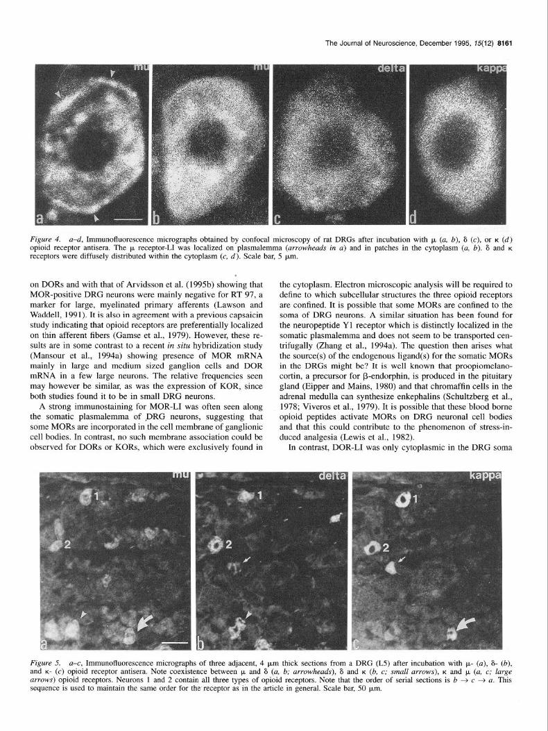
Coexistence of the three opiate receptor-11s was examined in adjacent, 4 pm thick DRG seciions from normal rats. As shown in Figure 5, not only MOR- and DOR-LIs were localized in the same neurons, but also DOR- and KOR-LIs, as well as KOR- and MOR-LIs coexisted. In fact all three types of opioid recep- tors coexisted in some neurons.
Opioid receptors in spinal cord
Immunohistochemistry demonstrated a dense staining for three types of receptors in the superfical layers (laminae I-II) of the dorsal horn (Fig. 6u-c). However each receptor had a different staining intensity and appearance, as well as a unique distribu- tion. MOR-LI was the strongest, especially in lamina II, and the MOR-IR structures appeared most densly packed resulting in a somewhat diffuse appearance (Fig. 6~). A dense DOR-IR fiber network was distributed in a parallel fashion in the laminae I-II. However, the DOR-IR structures had a distinctly varicose ap- pearance, very much resembling, for example, peptide-IR nerve fibers. DOR-LI plexuses were also seen in deeper lamine (Fig. 6b). KOR-LI was quite weak, but distinct in the lateral part of
8160 Ji et al. - Opioid Receptors and Inflammation
lamina I (Fig. 6~). After inflammation no apparent change in MOR-, DOR-, or KOR-LI staining patterns or intensities could be seen in the microscope. However, quantitative evaluation of the staining intensity in the medial part of the ipsilateral super- ficial dorsal horn revealed a moderate increase (21.9% and 13.1%) in MOR-LI and a small decrease in DOR-LI (3.1% and 8.3%) 1 and 3 d after inflammation, respectively, as compared to the contralateral side (Table 1). KOR-LI in spinal cord was too weak to allow acceptable measurements.
Transport of opioid receptors
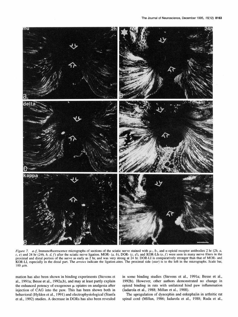
As early as 2 hr after ligation a distinct accumulation of MOR-, DOR-, and KOR-LIs was detected in the proximal part of the sciatic nerve (Fig. 7a,c,e). The accumulation in the distal part,
200 400 600 800 1000 1200 although weaker, could also be observed after two hours (Fig. Neuronal cross-sectional area (square micrometers) 7a,c,e). The staining for all three receptors then increased and
the strongest accumulation was seen at 24 hr, when also the
v1 50 B al 3
g 40 I
q Ipsi
0 Contra
staining distal to the ligation site was most pronounced (Fig. 7b,d,f). Forty-eight hours after ligation the accumulations for the three receptors were weaker, especially for the MORs and KORs (data not shown). At all time intervals the DOR-LI was stronger than MOR- and KOR-LIs, both in the proximal and distal parts (Fig. 7).
Discussion
Taking advantage of the recent cloning of the three opioid re- ceptors (for references, see introductory section), antibodies have been raised against these receptors and used in immuno- histochemical studies (Dado et al., 1993; Arvidsson et al., 1995ax; Hiller et al., 1995), These new data are complementary to previous ligand binding results presented in numerous papers (see Mansour et al., 1988, and introductory section) based on the original autoradiographic methodology described by (Atweh
600 800 1000 and Kuhar, 1977). In particular the cellular resolution of the Neuronal cross-sectional area (square micrometers)
j-J Contra
200 400 600 800 Neuronal cross-sectional area (square micrometers)
Figure 3. A-C, Size range of p. (A), 6 (B), and K (C) opioid receptor- IR neuron profiles in control (Cont) and ipsilateral DRGs (L5) of rats one day after carrageenan injection. Two hundred cells were measured for each group. There were no significant changes in size ranges of cells expressing these receptors after inflammation. The opioid receptors were predominantly distributed in the range of small DRG neurons.
immunohistochemical approach provides a more detailed picture of the receptor protein distribution, for example, with regard to distribution in cell bodies versus processes and to presynaptic versus postsynaptic localization. Moreover, specific antisera may, to some extent, overcome the shortcoming of the fact that a ligand can bind to several receptors albeit with different affin- ity. Also the immunohistochemcial approach may suffer from shortcomings, for example lack of specificity of the antisera. However, the antibodies used are well characterized using a number of criteria (Arvidsson et al., 1995a-c).
Our results show that all three opioid receptors are expressed in DRG neurons, mainly of the small type (see also Dado et al., 1993; Arvidsson et al., 1995b), and that peripheral inflammation differentially regulates the expression of these three types of opioid receptors. This is especially obvious at the DRG level. Moreover, the three opioid receptors were found to coexist in several primary sensory neurons. Finally, although all three opioid receptors to some extent were transported along the sci- atic nerve, only the 6 receptor protein showed a dramatic ac- cumulation central to a crush.
Primary sensory neurons
In the present study MOR-, DOR-, and KOR-LIs were detected in -2O%, - 15%, and -lo%, respectively, of all DRG neurons in normal rats. Size measurement indicated that all these recep- tors were predominantly localized in small neurons, that is neu- rons that give rise to C- or AS-fibers (see Willis and Coggeshall, 1991). This result is consistent with that of Dado et al. (1993)
The Journal of Neuroscience, December 1995, 75(12) 8181
Figure 4. a-d, Immunofluorescence micrographs obtained by confocal microscopy of rat DRGs after incubation with p. (a, b), 6 (c), or K (d) opioid receptor antisera. The p receptor-11 was localized on plasmalemma (arrowheads in a) and in patches in the cytoplasm (a, b). 6 and K receptors were diffusely distributed within the cytoplasm (c, d). Scale bar, 5 pm.
on DORs and with that of Arvidsson et al. (1995b) showing that MOR-positive DRG neurons were mainly negative for RT 97, a marker for large, myelinated primary afferents (Lawson and Waddell, 1991). It is also in agreement with a previous capsaicin study indicating that opioid receptors are preferentially localized on thin afferent fibers (Gamse et al., 1979). However, these re- sults are in some contrast to a recent in situ hybridization study (Mansour et al., 1994a) showing presence of MOR mRNA mainly in large and medium sized ganglion cells and DOR mRNA in a few large neurons. The relative ‘frequencies seen may however be similar, as was the expression of KOR, since both studies found it to be in small DRG neurons.
A strong immunostaining for MOR-LI was often seen along the somatic plasmalemma of ,~DRG neurons, suggesting that some MORs are incorporated in the cell membrane of ganglionic cell bodies. In contrast, no such membrane association could be observed for DORs or KORs, which were exclusively found in
the cytoplasm. Electron microscopic analysis will be required to define to which subcellular structures the three opioid receptors are confined. It is possible that some MORs are confined to the soma of DRG neurons. A similar situation has been found for the neuropeptide Y 1 receptor which is distinctly localized in the somatic plasmalemma and does not seem to be transported cen- trifugally (Zhang et al., 1994a). The question then arises what the source(s) of the endogenous ligand(s) for the somatic MORs in the DRGs might be? It is well known that proopiomelano- cortin, a precursor for P-endorphin, is produced in the pituitary gland (Eipper and Mains, 1980) and that chromaffin cells in the adrenal medulla can synthesize enkephalins (Schultzberg et al., 1978; Viveros et al., 1979). It is possible that these blood borne opioid peptides activate MORs on DRG neuronal cell bodies and that this could contribute to the phenomenon of stress-in- duced analgesia (Lewis et al., 1982).
In contrast, DOR-LI was only cytoplasmic in the DRG soma
Figure 5. a-c, Immunofluorescence micrographs of three adjacent, 4 pm thick sections from a DRG (L5) after incubation with p- (a), S- (b), and K- (c) opioid receptor antisera. Note coexistence between p+ and 6 (a, b; arrowheads), 6 and K (b, c; small arrows), K and p, (a, c; large arrows) opioid receptors. Neurons 1 and 2 contain all three types of opioid receptors. Note that the order of serial sections is b + c + a. This sequence is used to maintain the same order for the receptor as in the article in general. Scale bar, 50 pm.
8182 Ji et al. - Opioid Receptors and Inflammation
Figure 6. a-c, Immunofluorescence micrographs showing the distri- bution of lt- (a), S- (b), and K- (c) opioid receptor-like immunoreactiviry (LI) in the dorsal horn (L4-5). The staining of the three receptor im- munoreactivities was predominantly observed in the superficial layers (laminae I-II). The heaviest labeling was seen for k receptor, especially in lamina II (a). A dense fiber network containing 6 receptor was ob- served in both laminae I and II, with single fibers in deeper laminae (b). The K receptor staining was compartatively weak (c). Scale bar, 100 pm.
Table 1. Relative intensity of p- and b-opioid receptor-like immunoreactivity (MOR-LI and DOR-LI) in ipsilateral (Ipsi) and contralateral spinal dorsal horns (L4-L5) 1 and 3 d after unilateral injection of carrageenan into the hindpaw
The intensity in the contralateral side was set as 100%. Measurements were done in the medial half of the superficial layers (laminae I-II) of the dorsal horn that innervated by the hindpaw. Results are presented as the mean + SEM from three rats. For each rat counts from three to four sections were averaged. All the changes ware significant (P < 0.01) if paired f test is used but not significant (P > 0.05) with the unpaired t test.
to be studied. It seems to be transported somewhat better than MORs, and it could not be detected in association with the cell soma plasmalemma, suggesting a function in the dorsal horn. The KOR staining is somewhat difficult to evaluate, since this antiserum clearly is not as powerful as the MOR and DOR an- tisera.
An interesting finding in the present study is that three opioid receptors are differentially regulated in DRG neurons following inflammation. CAG induced a distinct increase in MOR-LI but a decrease in DOR- and KOR-LIs 1 d and 3 d after inflamma- tion. The changes in receptor levels after inflammation were mainly confined to the ipsilateral side, and therefore the contra- lateral DRG could be used as a control together with the control rats. The time points 1 and 3 d were chosen to detect the CAG- induced changes in the present study, since not only the edema and the hyperalgesia are dramatic at this stage (Iadarola et al., 1988), but inflammation-induced changes in synthesis of neu- ropetides and their receptors are also peaking at these time in- tervals (Iadarola et al., 1988; Ji et al., 1994).
Our results indicate that inflammation does not alter the size category of neurons that express opioid receptors. This is in agreement with our previous results for NPY (Y 1) receptor mRNA (Ji et al., 1994), and galanin mRNA (Ji et al., unpubli- shed data), whereas in contrast there is a distinct shift in ex- pression of SP (Noguchi et al., 1994), NPY (Yl) receptor (Zhang et al., 1994a), and bFGF mRNA (Ji et al., unpublished results) from small to medium sized or large neurons after sciatic nerve transection (axotomy), a process which may also result in neuropathic pain (Wall et al., 1979; Wall and Devor, 1981).
Spinal cord
All three opioid receptors were found in high concentrations distributed in the superficial dorsal horn, with the highest density for the p receptor. The rank order of staining density, that is MOR>DOR>KOR is in agreement with receptor autoradio- graphic studies (Morris and Herz, 1987; Besse et al., 1990; Ste- vens et al., 1991a,b). This may be related to electrophysiological studies showing that the analgesic effects of p agonists are more pronounced than those produced by other agonists (Dickenson et al., 1987). In comparison to the receptor alterations in the DRGs, the inflammation-induced changes in the spinal cord ap- peared much less distinct. Thus, a slight increase in MOR-LI and a small decrease in DOR-LI were observed in the medial part of ipsilateral dorsal horn. These changes in spinal cord par- allel those seen in the DRGs. An increase in MOR after inflam-
and accumulated strongly central to the crush, and was also pres- ent in high levels in CGRP-positive primary afferent fibers in the dorsal horn (Dado et al., 1993). Thus, DORs may be pref- erentially functionally active in the dorsal horn of the spinal cord, that is they represent presynaptic receptors localized on primary afferents. To what extent KOR is targeted to the axonal compartment of neurons to function at a presynaptic site remains
The Journal of Neuroscience, December 1995, 75(12) 6163
Figure 7. a-f, Immunofluorescence micrographs of sections of the sciatic nerve stained with l.r,-, 6-, and a-opioid receptor antibodies 2 hr (2h, a, c, e) and 24 hr (24h, b, d, f) after the sciatic nerve ligation. MOR- (a. b), DOR- (c, d), and KOR-LIs (e, f) were seen in many nerve fibers in the proximal and distal portion of the nerve as early as 2 hr, and was very strong at 24 hr. DOR-LI is comparatively stronger than that of MOR- and KOR-LI, especially in the distal part. The arrows indicate the ligation-sites. The proximal side (star) is to the left in the micrographs. Scale bar, 100 km.
mation has also been shown in binding experiments (Stevens et in some binding studies (Stevens et al., 1991a; Besse et al., al., 1991a; Besse et al., 1992a,b), and may at least partly explain 1992b). However, other authors demonstrated no change in the enhanced potency of exogenous p opiates on analgesia after opioid binding in rats with unilateral hind paw inflammation injection of CAG into the paw. This has been shown both in (Iadarola et al., 1988; Millan et al., 1988). behavioral (Hylden et al., 1991) and electrophysiological (Stanfa The upregulation of dynorphin and enkephalin in arthritic rat et al., 1992) studies. A decrease in DORs has also been revealed spinal cord (Millan, 1986; Iadarola et al., 1988; Ruda et al.,
8184 Ji et al. - Opioid Receptors and Inflammation
1988; Weihe et al., 1988) favors a role for these two peptides in the modulation of sensory input during inflammation. It is at present not clear whether the increased endogenous content of enkephalin and dynorphin in the dorsal horn in fact reflects an increase in opioids available for release and activation of their receptors, or an intracellular accumulation perhaps due to de- creased release. In fact, spontaneous and evoked release of met- enkephalin-11 from the spinal dorsal horn of arthritic rats has been reported to be decreased both in vitro (Cesselin et al., 1984) and in vivo (Bourgoin et al., 1988). Thus, further work is essen- tial to elucidate the precise role of the individual opioid receptor types during inflammatory pain. Interestingly, the present results may have some clinical implications, since earlier studies in hu- mans showed that chronic neurogenic pain is associated with an altered proportion of opioid peptides in the cerebrospinal fluid (Terenius, 1984).
Transport of opioid receptor and peripheral effects
After ligation MOR-, DOR-, and KOR-LIs accumulated in nerve fibers both in the central and the distal portion of the sciatic nerve, providing evidence that all three types of opioid receptors are transported both anterogradely and retrogradely along the sciatic nerve. This corresponds well with the initial binding study of Young et al. (Young et al., 1980) on the transport of opioid receptor in the vagus nerve. Also, lZSI-B-endorphin bind- ing sites accumulate both proximally and distally to a ligature placed on the sciatic nerve, indicating bidirectional axonal trans- port (Hassan et al., 1993). Also in the dorsal roots, opioid re- ceptors accumulate around ligatures (Zarbin et al., 1990) and crushes (Zhang et al., 1994b). The present study provide a time course for both anterograde and retrograde accumulation of the three opioid receptors. Thus, an accumulation of opioid recep- tors could be found as early as 2 hr after ligation, whereby DOR accumulation was stronger than that of MORs and KORs, es- pecially in the distal part. This suggests that the DOR is the main type of the three opioid receptors undergoing axonal trans- port. At 48 hr there was a less pronounced accumulation of all three receptor immunoreactivities, perhaps reflecting the down- regulation of DOR- and MOR-LIs in DRG neurons after axo- tomy (Zhang at al., unpublished data).
Local inflammation induced an enhanced axonal transport of lzsI B-endorphin binding sites in rat sciatic nerve and accumu- lation in paw tissue (Hassan et al., 1993). Peripherally admin- istered opioids produce powerful antinociception in the in- flamed, but not in the noninflamed paw (Levine and Taiwo, 1989; Stein et al., 1989). Opioid receptors were detected on small-diameter cutaneous nerves of noninflamed and inflamed paws (Stein et al., 1990a,b). After inflammation increased amounts of opioid peptides in immune cells was shown to infil- trate the inflamed paw and interact with receptors on sensory nerves to inhibit nociceptive responses in inflammation (Stein et al., 1990a,b). Thus, through axonal transport, the CAG-induced alterations of three opioid receptors in DRG neurons are likely to play roles in regulating peripheral opioid effects under inflam- matory situations.
Coexistence of opioid receptors
A main finding of the present study is that opioid receptors co- exist in some primary sensory neurons. This is in agreement with findings that a subset of MOR-IR axon terminals in the superficial dorsal horn also stain for DOR-LI (Arvidsson et al., 1995b). The present study in DRG neurons demonstrated not
only the coexistence between MOR and DOR, but also between DOR and KOR, and KOR and MOR. Some neurons even con- tained all three types of opioid receptors. A functional interac- tion between opioid receptors was first suggested by Vaught and Takemori (1979) who showed that (Leuj)enkephalin given intra- cerebroventricularly at doses which did not produce antinoci- ception potentiated the antinociceptive potency of morphine. In contrast, Lee et al. (1980) demonstrated that (Met 5)enkephalin, in nonanalgesic doses, antagonized the antinociceptive effects of morphine. Morever, it was shown that intrathecal perfusion with the p agonist DAGO (Tyr-D-Ala-Gly-MePhe-Gly-01) signif- cantly enhanced the spontaneous release of substance P, whereas the K agoinst U 50488 produced no change in the peptide out- flow. However, intrathecal perfusion with DAGO plus U 50488 caused a significant decrease in substance P release (Colhn et al., 1992). A &mu receptor complex has been suggested to co- exist based on a diverse and complex pharmacology (see Ho- laday et al., 1985; Rothman et al., 1993). The further elucidation of coexistence of opioid receptors is important to improve our understanding of the functional coupling among opioid recep- tors.
Concluding remarks
By using recently developed antibodies against three types of opioid receptors and immunohistochemistry, we have compared similarities and differences among these receptors with special reference to DRGs. Similarities include that (1) all three types of receptors are preferentially localized in small DRG neurons, and that inflammation did not affect the size category of neu- ronal profiles containg these receptors, (2) all three receptors immunoreactivities can be found diffusely in the cytoplasma of DRG neurons, (3) they coexist to a certain extent with each other in DRG neurons, (4) the expression of all three is altered fol- lowing inflammation, (5) in the dorsal horn of the spinal cord they are mainly distributed in the superficial layers, and (6) they are transported along the sciatic nerve both anterogradely and retrogradely. The main differences are as follows: (a) They occur in different numbers, whereby -2O%, 15%, and 10% of the DRG neurons contain p, 6, and K receptor, respectively; (2) after inflammation their expression is changed in different directions, with an upregulation of MOR-LI and downregulation of DOR- and KOR-LIs; (3) MOR-Ll in the superficial dorsal horn is very strong, whereas the staining intensity is moderate for DOR-LI, and weak for KOR-LI. However, DOR-LI has a distinct vari- cose/dotted appearance, whereas MOR- and KOR-LIs are more diffuse; (4) DOR- and KOR-LI are only observed in the cyto- plasm of DRG neurons, whereas MOR-LI could also be found on the plasma membrane; and (5) after ligation accumulation of DOR-LI is most pronounced, indicating that DOR to a larger extent than the other two types of receptors undergoes axonal transport.
Taken together, our results provide anatomical evidence for roles of multiple opioid receptors in the regulation of CAG- induced hyperalgesia accompanying peripheral inflammation. The differential regulation of three opioid receptors after CAG injection indicates that they play different roles during inflam- mation. The colocalization of opioid receptors provides a struc- tural basis for a functional interaction between these receptors.
References
Arvidsson U, Dado RJ, Riedl M, Lee JH, Law PY, Loh HH, Elde R, Wessendorf MW (1995a) S-opioid receptor immunoreactivity: dis-
The Journal of Neuroscience, December 1995, 15(12) 8165
tribution in brainstem and spinal cord, and relationship to biogenic amines and enkeuhalin. J Neurosci 15: 12 15-l 235.
Arvidsson U, Riedi M, Chakrabarti S, Lee JH, Nakano AH, Dado RJ, Loh HH, Law PY, Wessendorf MW, Elde R (1995b) Distribution and targeting of a p-opioid receptor (MORl) in brain and spinal cord. J Neurosci 15:3328-3341.
Arvidsson U, Riedl M, Chakrabarti S, Vulchanova L, Lee J-H, Nakano AH. Lin X0. Loh HH. Law P-Y. Wessendorf MW. Elde R (1995~) The K-opioid receptor is primarily postsynaptic: combined immune: histochemical localization of the receptor and endogenous opioids. Proc Nat1 Acad Sci USA 92:506225066.
Atweh SE Kuhar MJ (1977) Autoradiograohic localization of opiate receptors in rat brain. I. Spinal cord aid &lower medulla. Brain- Res 124:53-67.
Basbaum AI, Fields HL (1984) Endogenous pain control systems: brainstem spinal pathways and endorphin circuitry. Annu Rev Neu- rosci 7:309-338.
Besse D, Lombard MC, Zajac JM, Roques BP, Besson JM (1990) Pre- and postsynaptic distribution of p, 6 and K opioid receptors in the superficial layers of the cervical dorsal horn of the rat spinal cord. Brain Res 521:15-22.
Besse D, Lombard MC, Perrot S, Besson JM (1992a) Regulation of opioid binding sites in the superficial dorsal horn of the rat spinal cord following loose ligation of the sciatic nerve: comparison with sciatic nerve section and lumbar dorsal rhizotomy. Neuroscience 50: 921-933.
Besse D, Weil-Fugazza J, Lombard MC, Butler SH, Besson JM (1992b) Monoarthritis induces complex changes in )L-, 6-, and K-opioid bind- ing sites in the superficial dorsal horn of the rat spinal cord. Eur J Pharmacol 223: 123-131.
Besson JM, Chaouch A (1987) Peripheral and spinal mechanisms of nociception. Physiol Rev 67:67-186.
Bourgoin S, Le Bars D, Clot AM, Hamon M, Cesselin F (1988) Spon- taneous and evoked release of met-enkephalin-like material from the spinal cord of arthritic rats in viva. Pain 32: 107-I 14.
Bzdega T, Chin H, Kim H, Jung HH, Kozak CA, Klee WA (1993) Regional expression and chromosomal localization of the 6 opiate receptor gene. Proc Nat1 Acad Sci USA 90:9305-9309.
Cesselin F, Bourgoin S, Artaud E Hamon M (1984) Basic and regu- latory mechanisms of in vitro release of met-enkephalin from the dorsal zone of the rat spinal cord. J Neurochem 43:763-773.
Chang K-J, Cuatrecasas P (1979) Multiple opiate receptors. Enkepha- lins and morphine bind to receptors of different specificity. J Biol Chem 254:2610-2618.
Chen Y, Mestek A, Liu J, Hurley JA, Yu L (1993a) Molecular cloning and functional expression of a mu-opioid receptor from rat brain. Mol Pharmacol 44%12.
Chen Y, Mestek A, Yu L (1993b) Molecular cloning of a rat kappa opioid receptor reveals sequence similarities to the mu and delta opioid receptors. Biochem J 295:625-628.
Collin E, Mauborgne A, Bourgoin S, Mantelet S, Ferhat L, Hamon M, Cesselin F (1992) Kappa-/mu-receptor interactions in the opioid con- trol of the in viva release of substance P-like material from rat spinal cord. Neuroscience 5 1:347-355.
Coons AH (1958) Fluorescent antibody methods. In: General cyto- chemical methods (Danielli JF, eds), pp 3999422. New York: Aca- demic.
Corbett AD, Paterson SJ, Kosterlitz HW (1993) Selectivity of ligands for opioid receptors. In: Handbook of experimental pharmacology, Opioids I (Her; A, ed), pp 645-679. Berlh: Springer.-
-.
Dado RJ. Law PY. Loh HH. Elde R (1993) Immunofluorescent iden- tification of a delta (6).opioid receptor on primary afferent nerve terminals. Neuroreport 5:341-344.
Dickenson AH, Sullivan AF, Knox R, Zajac JM, Roques BP (1987) Opioid receptor subtypes in the rat spinal cord: electrophysiological studies with u.- and &opioid receptor agonists in the control of no- ciception. Brain Res 413:3644. - -
Dubner R. Ruda MA (1992) Activitv-deuendent neuronal olasticitv fol- lowing ‘tissue injury and ‘inflammation. Trends Neurosci 15:96-103.
Eipper BA, Mains RE (1980) Structure and biosynthesis of pro-adren- ocorticotropin/endorphin and related peptides. Endocrine Rev 1: l- 27.
Evans CJ, Keith DE, Morrison H, Magendzo K, Edwards RH (1992) Cloning of a delta opioid receptor by functional expression. Science 258:1952-1955.
Gamse R, Holzer E Lembeck F (1979) Indirect evidence for presyn- aptic location of opiate receptor on chemosensitive primary sensory neurons. Naunyn-Schmiedeberg’s Arch Pharmacol 308:281-285.
Hartman BK, Zide D, Udenfriend S (1972) The use of dopamine B-hy- droxylase as a marker for the noradrenergic pathways of the central nervous svstem in the rat. Proc Nat1 Acad Sci USA 69:2722-2726.
Hassan AH,<Ableitner A, Stein C, Herz A (1993) Inflammation of the rat paw enhances axonal transport of opioid receptors in the sciatic nerve and increases their density in the inflamed tissue. Neuroscience 55:185-195.
Herz A (eds) (1993) Handbook of experimental pharmacology. Opioids II. Berlin, Heidelberg: Springer.
Hiller JM, Zhang Y, Bing G, Gioannini TL, Stone EA, Simon EJ (1995) Immunohistochemical localization of mu-opioid receptors in rat brain using antibodies generated against a peptide sequence present in a purified mu-opioid binding protein. Neuroscience, in press.
Hokfelt T, Ljungdahl A, Terenius L, Elde R, Nilsson G (1977) Im- munohistochemical analysis of peptide pathway possibly related to pain and analgesia. Enkephalin and Substance P Proc Nat1 Acad Sci USA 74:3081-3085.
Holaday JW, Long JB, Tortella FC (1985) Evidence for K, p and 6 opioid-binding site interactions in viva. Fed Proc 44:2860-2862.
Hsu S-M, Raind L, Fanger H (1981) Use of avidin-biotin-peroxidase complex (ABC) in immunoperoxidase technique. A comparison be- tween ABC and unlabeled antibody (PAP) procedures. J Histochem Cytochem 29:577-580.
Hylden JLK, Thomas DA, Iadarola MJ, Nahin RL, Dubner R (1991) Spinal opioid analgesic effects are enhanced in a model of unilateral inflammationn/hyperalgesia: possible involvement of noradrenergic mechanisms. Eur J Pharmacol 194:1355143.
Iadarola MJ, Brady LS, Draisci G, Dubner R (1988) Enhancement of dynorphin gene expression in spinal cord following experimental in- flammation: stimulus specificity, behavioral parameters and opioid receptor binding. Pain 35:3 13-326.
Ji RR, Zhang X, Wiesenfeld-Hallin Z, Hokfelt T (1994) Expression of neuropeptide Y and neuropeptide Y (Yl) receptor mRNA in rat spi- nal cord and dorsal root ganglia following peripheral tissue inflam- mation. J Neurosci 14:6423-6434.
Johnson DG, de C Nogeira Araujo GM (1981) A simple method of reducing the fading of immunofluorescence during microscopy. J Im- munol Methods 43:349-350.
Kar S, Rees RG, Quirion R (1994) Altered calcitonin gene-related pep- tide, substance P and enkephalin immunoreactivities and receptor binding sites in the dorsal spinal cord of the polyarthritic rat. Eur J Neurosci 6:345-354.
Kieffer BL, Befort K, Gaveriaux-Ruff C, Hirth CG (1992) The 6-o- pioid receptor: isolation of a cDNA by expression cloning and phar- macological characterization. Proc Nat1 Acad Sci USA 89:12048- 12052.
Lawson SN, Waddell PJ (199 1) Soma neurofilament immunoreactivity is related to cell size and fiber conduction velocity in rat primary sensory neurons. J Physiol (Lond) 435:41-63.
Lee NM, Leybin L, Chang JK, Loh HH (1980) Opiate and peptide interaction: effect of enkephalins on morphine analgesia. Eur J Phar- macol 68:181-185.
Levine JD, Taiwo YO (1989) Involvement of the mu-opiate receptor in peripheral analgesia. Neuroscience 32:571-575.
Levine JD, Fields HL, Basbaum AI (1993) Peptides and the primary afferent fibers. J Neurosci 13:2273-2286.
Lewis JW, Tordoff MG, Sherman JE, Liebeskind JC (1982) Adrenal medullary enkephalin-like peptides may mediate opioid stress anal- gesia. Science 217:557-559.
Lord J, Waterfield A, Hughes J, Kosterlitz H (1977) Endogenous opioid peptides: multiple agonists and receptors. Nature 267:495499.
Mansour A, Khachaturian H, Lewis ME, Akil H, Watson SJ (1988) Anatomy of CNS opioid receptors. Trends Neurosci 11:308-3 14.
Mansour A, Thompson RC, Akil H, Watson SJ (1993) Delta opioid receptor mRNA distribution in the brain: comparison to delta receptor binding and proenkephalin mRNA. J Chem Neuroanat 6:351-362.
Mansour A, Fox CA, Burke S, Meng F, Thompson RC, Akil H, Watson SJ (1994a) Mu, delta, and kappa opioid receptor mRNA expression in the rat CNS: an in situ hybridization study. J Comp Neurol 350: 412438.
8186 Ji et al. * Opioid Receptors and Inflammation
Mansour A, Fox CA, Meng E Akil H, Watson SJ (1994b) Kappa1 receptor mRNA distribution in the rat CNS: comparison to kappa receptor binding and prodynorphin mRNA. Cell Mol Neurosci 5: 124-144.
Mansour A, Fox CA, Thompson RC, Akil H, Watson SJ (1994~) Mu- opioid receptor mRNA expression in the rat CNS: comparison to mu- receptor binding. Brain Res 643:245-265.
Martin WR, Eades CC, Thompson JA, Huppler RE, Gilbert PE (1976) The effects of morphine- and nalorphine-like drugs in the nondepen- dent and morphine-dependent chronic spinal dog. J Pharmacol Exp Ther 197: 196-203.
Meng E Xie GX, Thompson RC, Mansour A, Goldstein A, Watson SJ, Akil H (1993) Cloning and pharmacological characterization of a rat kappa opioid receptor. Proc Nat1 Acad Sci USA 90:9954-9958.
A, Colpaert FC (1986) A model of chronic pain in the rat:response of multiple opioid system to adjuvant-induced arthritis. J Neurosci 6:899-906.
Millan MJ, Czlonkowski A, Morris B, Stein C, Arendt R, Huber A, Hollt V, Herz A (1988) Inflammation of the hind limb as a model of unilateral localized pain: influence on multiple opioid systems in the spinal cord of the rat. Pain 35:299-312.
Minami M, Toya T, Katao Y, Maekawa K, Nakamura S, Onogi T, Ka- neko S, Satoh M (1993) Cloning and expression of a cDNA for the rat kappa-opioid receptor. FEBS Lett 329:291-295.
Morris BJ, Herz A (1987) Distinct distribution of opioid receptor types in rat lumbar spinal cord. Naunyn-Schmiedebergs Arch Pharmacol 336:240-243.
Ninkovic M, Hunt SP, Kelly JS (1981) Effect of dorsal rhizotomy on the autoradiographic distribution of opiate and neurotensin receptors and neurotensin-like immunoreactivity within the rat spinal cord. Brain Res 230:111-119.
Ninkovic M, Hunt SP, Gleave JRW (1982) Localization of opiate and histamine HI-receptors in the primate sensory ganglia and spinal cord. Brain Res 241: 197-206.
Noguchi K, Morita Y, Kiyama H, Sato M, Ono K, Tohyama M (1989) Preproenkephalin gene expression in the rat spinal cord after noxious stimuli. Mol Brain Res 5:227-234.
Noguchi K, Dubner R, De Leon M, Senba E, Ruda MA (1994) Axo- tomy induces preprotachykinin gene expression in a subpopulation of dorsal root ganglion neurons. J Neurosci Res 37:596-603.
Pasternak GW (1988) The opioid receptors, New Jersey: Humana. Pease PC (1962) Buffered formaldehyde as .a killing agent and primary
fixative for electron microscopy. Anat Ret 142:342. Platt JL, Michael AF (1983) Retardation of fading and enhancement
of intensity of immunofluorescence by p-phenylenediamine. J His- tochem Cytochem 3 1:840-842.
Presley RW, Menetrey D, Levine JD, Basbaum AI (1990) Systemic morphine suppresses noxious-evoked Fos protein-like immunoreac- tivity in the rat spinal cord. J Neurosci 10:323-335.
Rothman RB. Holadav JW. Porreca F (1993) Allosteric couoling . I ” among opioid receptors: evidence for an bpioid receptor complex. In: Handbook of experimental pharmacology, Opioids I (Herz A, ed), pp 217-237. Berlin: Springer.
Ruda MA, Iadarola MJ, Cohen LV, Young WSI (1988) In situ hybrid- ization histochemistry and immunocytochemistry reveal an increase in spinal dynorphin biosynthesis in rat model of peripheral inflam- mation and hyperalgesia. Proc Nat1 Acad Sci USA 85:622-626.
Schultzberg M, Lundberg JM, Hokfelt T, Terenius L, Brandt J, Elde RP, Goldstein M (1978) Enkephalin-like immunoreactivity in gland cells and nerve terminals of the adrenal medulla. Neuroscience 3: 1169- 1186.
Schafer M-H, Nohr D, Krause JE, Weihe E (1993) Localization of kappa-opioid receptor mRNA in neuronal subpopulations of rat sen- sory ganglia and spinal cord. Neurosci Lett 167: 137-140.
Simon EJ, Gioannini TL (1993) Opioid receptor multiplicity: isolation, purification, and chemical characterization of binding sties. In: Hand- book of experimental pharmacology, Opioids I (Herz A, eds), pp 3- 26. Berlin: Springer.
Smith JR, Simon EJ (1980) Selective protection of sterospecific en- kephalin and opiate binding against inactivation by N-ethylmaleimi-
de: Evidence for two classes of opiate receptors. Proc Nat1 Acad Sci USA 77:281-284.
Standifier KM, Chien C-C, Wahlested C, Brown GP, Pasternak GW (1994) Selective loss of 6 opioid analgesia and binding by antisense oligodeoxynucleotides to a 6 opioid receptor. Neuron 12:805-810.
Stanfa LC, Sullivan AE Dickenson AH (1992) Alterations in neuronal excitability and the potency of spinal mu, delta and kappa opioids after carrageenan-induced inflammation. Pain 50:345-354.
Stein C, Gramsch C, Herz A (1990a) Intrinsic mechanisms of antino- ciception in inflammation: local opioid receptors and B-endorphin. J Neurosci 10:1292-1298.
Stein C, Hassan AH, Przewxocki R, Gramsch C, Peter K, Herz A (1990b) Opioids from immunocytes interact with receptors on sen- sory nerves to inhibit nociception in inflammation. Proc Nat1 Acad Sci USA 87:5935-5939.
Stein C, Millan MJ, Shippenberg TS, Peter K, Herz A (1989) Periph- eral opioid receptors mediating antinociception in inflammation. Ev- idence for involvement of mu, delta and kappa receptors. J Pharmacol Exp Ther 248:1269-1275.
Stevens CW, Kajander KC, Bennett GJ, Seybold VS (1991a) Bilateral and differential changes in spinal mu, delta, and kappa opioid binding in rats with a painful, unilateral neuropathy. Pain 46:315-326.
Stevens CW, Lacey CB, Miller KE, Elde RP, Seybold VS (1991b) Bio- chemical characterization and regional quantification of p, 6 and K
opioid binding sites in rat spinal cord. Brain Res 550:77-85. Terenius L (1984) Approaches to neuropeptide and, in particular, en-
dorphin measurements. In: Frontiers in biochemical and pharmaco- logical research in depression (Usdin E, eds), pp 3543. New York: Raven.
Thompson RC, Mansour A, Akil H, Watson SJ (1993) Cloning and phamacological characterization of a rat p, opioid receptor. Neuron 11:903-913.
Vaught JL, Takemori AE (1979) Differential effects of leucine en- kephalin and methionine enkephalin on morphine-induced analgesia, acute tolerance and dependence. J Pharmacol Exp Ther 208:86-90.
Viveros OH, Diliberto JEJ, Hazem E, Chang K-J (1979) Opiate-like materials in the adrenal medulla: evidence for storage and secretion with catecholamines. Mol Pharmacol 16:1101-l 108.
Wall PD, Devor M (1981) The effect of peripheral nerve injury on dorsal root potentials and on transmission of afferent signals into the spinal cord. Brain Res 209:95-l 11.
Wall PD, Devor R, Inbal JW, Scadding D, Schonfeld Z, Seltzer Z, Tomkiewitz MM (1979) Autotomy following peripheral nerve le- sions: experimental anaesthesia dolorosa. Pain 7: 103-I 13.
Wang JB, Imai Y, Eppler CM, Gregor P, Spivak CE, Uhl GR (1993) p. Opiate receptor: cDNA cloning and expression. Proc Nat1 Acad Sci USA 90:10230-10234.
Weihe E, Nohr D, Millan MJ, Stein C, Muller S, Gramsch C, Herz A (1988) Peptide neuroanatomy of adjuvant-induced arthritic inflam- mation in rat. Agents Actions 25:255-259.
Willis WD Jr, Coggeshall RE (1991) Sensory mechanisms of the spinal cord. New York: Plenum.
Yasuda K, Raynor K, Kong H, Breder CD, Takeda J, Reisine T Bell GI (1993) Cloning and functional comparison of K and 6 opioid receptors from mouse brain. Proc Nat1 Acad Sci USA 90:6736-6740.
Zamboni L, De Martin0 C (1967) Buffered picric acid formaldehyde: a new rapid fixative for electron microscopy. J Cell Biol 148A:35.
Zarbin MA, Wamsley JK, Kuhar MJ (1990) Anterograde transport of opioid receptors in rat vagus nerves and dorsal roots of spinal nerves: pharmacology and sensitivity to sodium and guanine nucleotides. Exp Brain Res 81:267-278.
Zhang X, Bao L, Xu ZQ, Jutta K, Arvidsson U, Elde R, Hokfelt T (1994a) Differential transport of Y 1 receptors: a specific distribution of Yl receptors on somatic plasmalemma of small neurons in rat lumbar DRGs. Proc Nat1 Acad Sci USA 91:11738-l 1442.
Zhang X, Wiesenfeld-Hallin Z, Hokfelt T (199413) Effect of peripheral axotomy on expression of neuropeptide Y (Y 1) receptor mRNA in rat lumbar dorsal root ganglia. Eur J Neurosci 6:43-57.
Zimmermann M (1983) Ethical guidelines for investigations of exper- imental pain in conscious animals. Pain 16:109-l 10.