Page 1
External Piercing Technique in Heavily Calcified BTK Lesion in a
Diabetic Foot Patients
Seung-Woon Rha, MD, PhD, FACC, FAHA, FSCAI, FESC, FAPSIC
Division of Cardiovascular Intervention and Research,
Cardiovascular Center,
Korea University Guro Hospital, Seoul, Korea
LINC 2016
Page 2
Disclosure
Speaker name: Seung-Woon Rha
.................................................................................
I have the following potential conflicts of interest to report:
Consulting
Employment in industry
Stockholder of a healthcare company
Owner of a healthcare company
Other(s)
I do not have any potential conflict of interest
Page 3
How to Finalize the Heavily Calcified BTK Lesions?
1. Wire Passage Failure
1) 035 Subintimal angioplasty (1.5J Soft Terumo; Bypass Concept)
2) Guidewire Distal Tip induced Crack (014 Cut Tip, 035 Stiff Terumo)
3) Brockenbrough needle induced Crack
4) Direct Extravascular Needle induced Crack (Ext Piercing Technique)
5) Microcatheter; Corasir PV supported CTO wiring
6) Child in mother catheter supported CTO wiring
7) Retrograde and Bidirectional approach
2. Balloon Passage Failure
1) Multiple parallel CTO wiring
2) Tornus PV
3) Rotablator (1.25mm)
4) CTO Devices; TruePath, Frontrunner, Crosser
5) Special Devices; Orbital atherectomy, Turbohawk, Laser..
6) Excellent balloon; Armada 014 OTW
Page 4
Direct Extravascular Needle induced Crack Technique
(External Piercing Technique) for a heavily calcified BTK lesion
Page 5
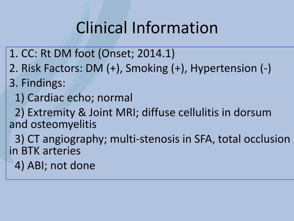
Clinical Information
1. CC: Rt DM foot (Onset; 2014.1) 2. Risk Factors: DM (+), Smoking (+), Hypertension (-) 3. Findings: 1) Cardiac echo; normal 2) Extremity & Joint MRI; diffuse cellulitis in dorsum and osteomyelitis 3) CT angiography; multi-stenosis in SFA, total occlusion in BTK arteries 4) ABI; not done
Page 6
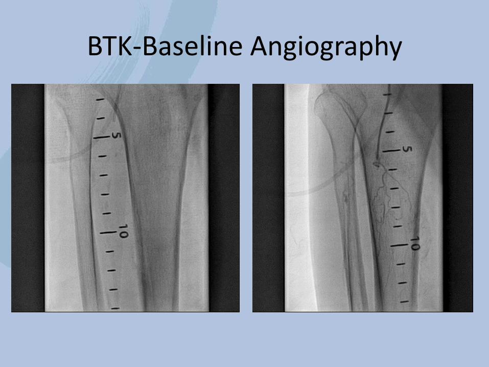
BTK-Baseline Angiography
Page 7
035 Subintimal Approach
035 Soft Terumo Wire (1.5J)
Page 8
Rt ATA-Post 035 Wiring
Delivery of 5F Heartrail Catheter (Terumo)
Page 9
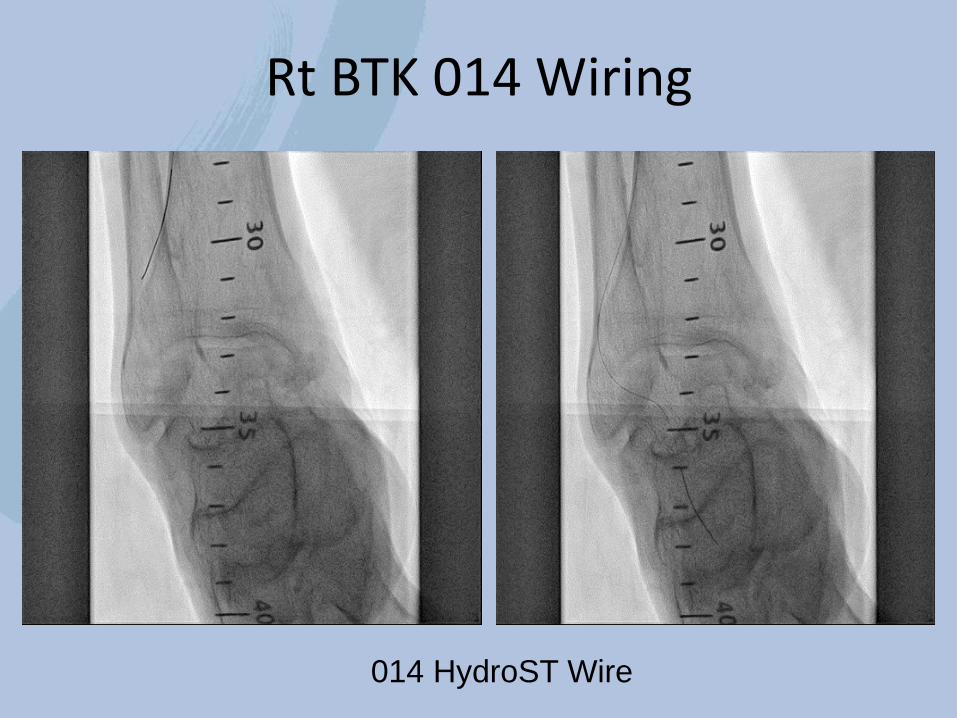
Rt BTK 014 Wiring
014 HydroST Wire
Page 10
Rt BTK Balloon
Advance LP 2.0 and 2.5 X200mm
Page 11
Rt BTK distal-Post 014 long balloon
Page 12
More Aggressive NC Ballooning
More than 10 coronary NC balloons were ruptured…
Page 13
Post Multiple NC Ballooning
Page 14
BTK Stenting
Xpert SES (Medtronic) 3.0X40mm
Page 15
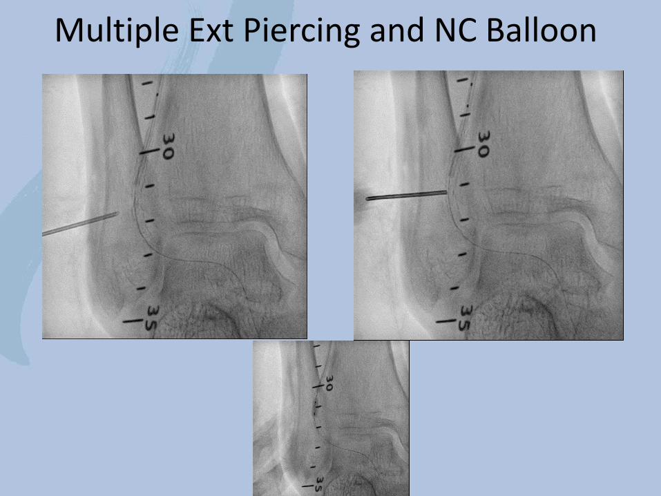
Needle Crack for Adjuvant Ballooning
Page 16
Multiple Ext Piercing and NC Balloon
Page 18
Prolonged Balloon Inflation and Final Angiography
Page 19
CTA FU at 3 months for BTK
Page 20
Conclusion
1. External Piercing Technique was very useful in a un-dilatable heavily calcified BTK lesion despite of risk of extravasation and rupture.
2. External Piercing Technique associated vessel injury can be effectively and safely managed by prolonged balloon inflation.
3. The results appears to be durable once the procedure is successfully finished.
Page 21
Thank You for Your Attention!!
Korea University Guro Hospital