Extracellular acid proteases of wine microorganisms: gene identification, activity characterization and impact on wine by Vernita Jennilee Reid Thesis presented in partial fulfilment of the requirements for the degree of Master of Science at Stellenbosch University Institute for Wine Biotechnology, Faculty of AgriSciences Supervisor: Dr BT Divol Co-supervisor: Prof M duToit March 2012
Transcript
Extracellular acid proteases of wine microorganisms: gene identification, activity
characterization and impact on wine
by
Vernita Jennilee Reid
Thesis presented in partial fulfilment of the requirements for the degree of
Master of Science
at Stellenbosch University
Institute for Wine Biotechnology, Faculty of AgriSciences
Supervisor: Dr BT Divol
Co-supervisor: Prof M duToit
March 2012
ii
Declaration
By submitting this thesis electronically, I declare that the entirety of the work contained therein is my own, original work, that I am the sole author thereof (save to the extent explicitly otherwise stated), that reproduction and publication thereof by Stellenbosch University will not infringe any third party rights and that I have not previously in its entirety or in part submitted it for obtaining any qualification.
Non-Saccharomyces yeasts of oenological origin have previously been associated with spoilage or
regarded as undesired yeasts in wine. However, these yeasts have recently come under investigation for
their positive contribution towards wine aroma especially when used in sequential or co-inoculated
fermentations with Saccharomyces cerevisiae. These yeasts are also known to secrete a number of
enzymes that could be applicable in wine biotechnology. Amongst these enzymes are aspartic proteases.
The secreted proteases from some non-Saccharomyces yeast may play a role in protein haze reduction,
as demonstrated by some authors, while simultaneously increasing the assimilable nitrogen content of
the wine for the utilization and growth of fermentative microorganisms. Moreover, the proteases may have
an indirect effect on wine aroma by liberating amino acids that serve as aroma precursors. Although
many screenings have been performed detecting protease activity in non-Saccharomyces yeasts, no
attempts have been made to characterize these enzymes. This study set out to isolate and characterize
genes encoding extracellular aspartic proteases from non-Saccharomyces yeasts.
An enzymatic activity screening of a collection of 308 Saccharomyces and non-Saccharomyces yeasts,
isolated from grape must, was performed. The aspartic protease-encoding genes of two non-
Saccharomyces yeasts, which showed strong extracellular proteolytic activity on plate assays, were
isolated and characterized by in silico analysis. The genes were isolated by employing degenerate and
inverse PCR. One gene was isolated from Metschnikowia pulcherrima IWBT Y1123 and named MpAPr1.
The other putative gene was isolated from Candida apicola IWBT Y1384 and named CaAPr1. The
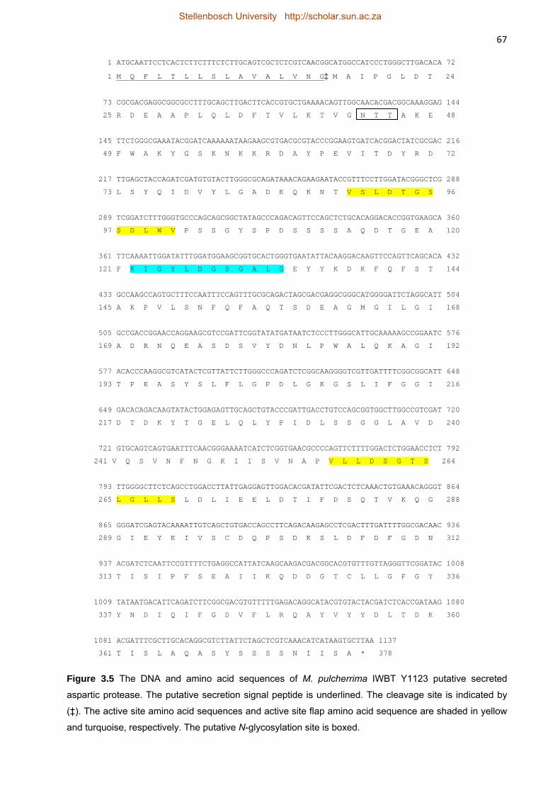
MpAPr1 gene is 1137 bp long, encoding a 378 amino acid putative protein with a predicted molecular
weight of 40.1 kDa. The CaAPr1 putative gene is 1101 bp long and encodes a 367 amino acid putative
protein with a predicted molecular weight of 39 kDa. These features are typical of extracellular aspartic
proteases. The deduced protein sequences showed less than 40% homology to other yeast extracellular
aspartic proteases. By heterologous expression of MpAPr1 in S. cerevisiae, it was confirmed that the
gene encodes an extracellular acid protease. The expression of MpAPr1 was shown to be induced in
media containing proteins as sole nitrogen source and repressed when a preferred nitrogen source was
available. The gene was expressed in the presence of casein, bovine serum albumin (BSA) and grape
juice proteins and repressed in the presence of ammonium sulphate. Expression was most induced in the
presence of grape juice proteins, which was expected since these proteins are present in the natural
habitat of the yeast. A genetic screening confirmed the presence of the MpAPr1 gene in 12 other
M. pulcherrima strains isolated from grape juice. The extracellular protease activity of the strains was also
visualized on plates. As far as we know, this is the first report on the genetic characterization of secreted
aspartic proteases from non-Saccharomyces yeasts isolated from grape must and provides the
groundwork for further investigations.
Stellenbosch University http://scholar.sun.ac.za
iv
Opsomming
Nie-Saccharomyces giste is voorheen met wynbederf geassosieer en hul teenwoordigheid in wyn is
ongewens. Hierdie giste is onlangs ondersoek vir hulle positiewe bydrae tot wyn aroma, in veral
sekwensiële en ko-inokulerings met Saccharomyces cerevisiae. Sommige van die nie-Saccahromyces
giste skei ‘n verskeidenhied ensieme af wat moontlik vir die wynmaker van nut kan wees. Een groep van
hierdie ensieme is die aspartiese suurproteases. Soos deur sommige navorsers aangetoon word, kan die
proteases die vorming van proteïenwaasverlaging, terwyl dit terselfdertyd die assimilerende
stikstofinhoud van die wyn vir die gebruik en groei van fermentasie-mikroörganismes verhoog. Die
proteases kan moontlik ook ‘n indirekte uitwerking op die aromaprofiel van die wyn hê deur die vrystelling
van aminosure wat as aromavoorlopers dien. Alhoewel baie studies gedoen is wat die ekstrasellulêre
teenwoordigheid van proteases bevestig in nie-Saccharomyces giste wat van druiwesap/wyn afkoms is,
is daar geen dokumentasie oor die genetiese karakterisering van hierdie ensieme beskikbaar nie. Die
doel van hierdie studie was om gene wat aspartiese proteases in nie-Saccharomyces giste enkodeer, te
isoleer en gedeeltelik te karakteriseer.
‘n Versameling van 308 Saccharomyces en nie-Saccharomyces giste wat uit druiwe sap geïsoleer is, is
gesif vir ensiematiese aktiwiteit deur plaattoetse uit te voer. Twee gene wat aspartiese protease
enkodeer, is geïsoleer van twee nie-Saccharomyces giste. Dit hetpositief gedurende die aktiwiteitstoetse
getoets en is deur in silico–analise gekarakteriseer. Die gene is deur die uitvoering van gedegenereerde
en inverse PKR geïdentifiseer. Een geen is vanaf Metschnikowia pulcherrima IWBT Y1123 geïsoleer en
is MpAPr1 genoem, terwyl die ander van Candida apicola IWBT Y1384 geïsoleer en CaAPr1 genoem is.
Die MpAPr1-geen is 1137 bp lank en enkodeer ‘n proteïen wat uit 378 aminosure bestaan met ‘n
voorspelde molekulêre massa van 40.1 kDa. Daar teenoor is die CaAPr1-geen 1101 bp lank en enkodeer
vir ‘n proteïen wat uit 367 aminosure met ‘n molekulêre massa van 39 kDa bestaan. Hierdie eienskappe
is kenmerkend van aspartiese protease. Die afgeleide proteïenvolgorde het minder as 40% homologie
met ander ekstrasellulêre aspartiese proteases vertoon, wat dui op die nuwigheid van hierdie ensieme.
Die MpAPr1-geen is heterologies in S. cerevisiae YHUM272 uitgedruk en dit het bevestig dat die geen
inderdaad ‘n ekstrasellulêre aspartiese protease enkodeer. Die MpAPr1-geen is uitgedruk in media wat
alleenlik proteïen as stikstofbron bevat het, terwyl dit onderdruk is in gevalle waar ‘n verkose stikstofbron
beskikbaar was. Die geen is uitgedruk in die teenwoordigheid van kaseïen, BSA en proteïene afkomstig
vanaf druiwesap en in die teenwoordigheid van ammoniumsulfaat onderdruk. Die hoogste uitdrukking
was in die teenwoordigheid van druifproteïene. Hierdie proteïene is teenwoordig in die natuurlike habitat
van die gis en is dus dalk ‘n bekende stikstofbron vir die gis. ‘n Genetiese sifting het die teenwoordigheid
van die MpAPr1-geen in 12 ander M. pulcherrima–rasse, wat ook van wynkundige oorsprong is, bevestig.
Die aspartiese protease-aktiwiteit van die 12 rasse is ook op agarplate waargeneem. Na ons wete, is dit
die eerste verslag oor die genetiese karakterisering van afgeskeide aspartiese proteases van nie-
Saccharomyces giste van wynkundige oorsprong en verskaf die grondslag vir verdere ondersoek.
Stellenbosch University http://scholar.sun.ac.za
v
This thesis is dedicated to
My Mother
Stellenbosch University http://scholar.sun.ac.za
vi
Biographical sketch
Vernita Reid was born in Bloemfontein, South Africa on the 12th of November 1984. She
attended Heide Primary School and completed her matriculation at Oranje Girls’ School in 2002.
She obtained a BSc degree in Food Biotechnology in 2007 and a BSc Honours degree in Food
Science in 2008 from the University of the Free State. She enrolled at Stellenbosch University in
2010 for an MSc in Wine Biotechnology.
Stellenbosch University http://scholar.sun.ac.za
vii
Acknowledgements
I wish to express my sincere gratitude and appreciation to the following persons and institutions:
God, for wisdom and understanding, to God be the glory
Dr Benoit Divol for acting as my supervisor, for his patience, guidance and constructive
criticism throughout this study
Prof Maret du Toit for acting as my co-supervisor, for her advice and guidance
throughout this study
Dr Evodia Setati and Mr Alexis Eschstruth for their invaluable technical guidance
Lab colleagues for their assistance, support and encouragement
My family for their constant enthusiasm and support and for always being there for me
Friends for their support and encouragement
The Institute for Wine Biotechnology, Winetech and the THRIP funding programme
of the National Research Foundation, the Harry Crossley Foundation and
Stellenbosch University (Sub-committee B) for financial support
Stellenbosch University http://scholar.sun.ac.za
viii
Preface
This thesis is presented as a compilation of four chapters.
Chapter 1 General Introduction and project aims
Chapter 2 Literature review
Aspartic proteases and non-Saccharomyces yeasts and their potential
application in wine biotechnology
Chapter 3 Research Results
Identification and characterization of extracellular aspartic protease genes
from Metschnikowia pulcherrima IWBT Y1123 and Candida apicola IWBT
Y1384
Chapter 4 General discussion and conclusions
Stellenbosch University http://scholar.sun.ac.za
ix
Contents
Chapter 1. General introduction and project aims 1
1.1 Introduction 2
1.2 Rationale and scope of the study 3
1.3 References 3
Chapter 2. Literature review: Aspartic proteases of non-Saccharomyces yeasts and their potential applications in wine biotechnology 5
2.1 General introduction 6
2.2 Proteolytic enzymes 6
2.2.1 Definition and characterization of proteolytic enzymes 6
2.2.2 Aspartic proteases 8
2.2.2.1 General description 8
2.2.2.2 Structure of aspartic proteases 10
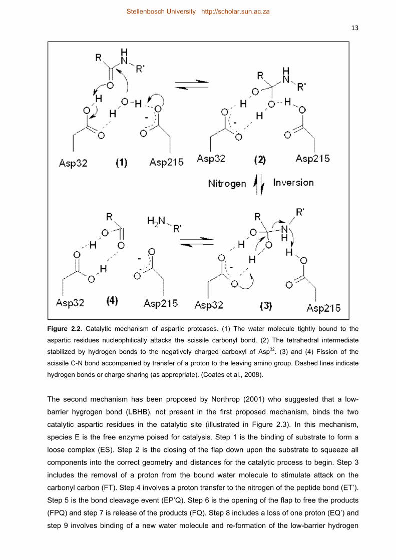
2.2.2.3 Catalytic mechanism of aspartic proteases 11
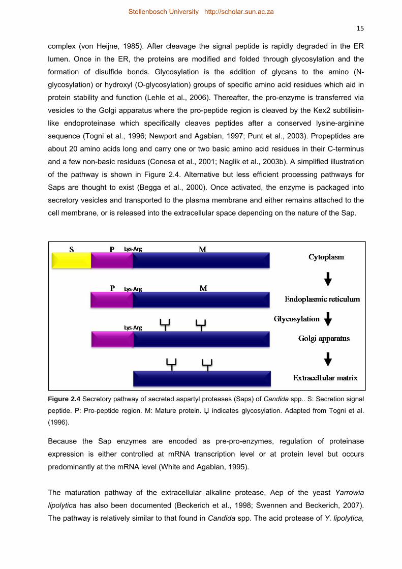
2.2.2.4 Secretion pathway and expression in yeasts 14
2.2.3 Model systems of yeast proteases used in the food industry 16
2.3 Oenological importance of non-Saccharomyces yeasts 17
2.3.1 Wine microbial diversity: spontaneous and inoculated fermentations 17
2.3.2 Growing interest in non-Saccharomyces wine yeasts 21
2.3.3 Non-Saccharomyces yeasts with extracellular enzyme activity 22
2.4 The role of aspartic proteases in wine 23
2.4.1 Production and the risk of protein haze formation 23
2.4.2 Increase in available assimilable nitrogen and wine aroma 26
2.5 References 27
Chapter 3. Research results: Identification and partial characterization of extracellular aspartic protease genes from Metschnikowia pulcherrima IWBT Y1123 and Candida apicola IWBT Y1384 36
3.1 Introduction 37
3.2 Materials and Methods 39
Stellenbosch University http://scholar.sun.ac.za
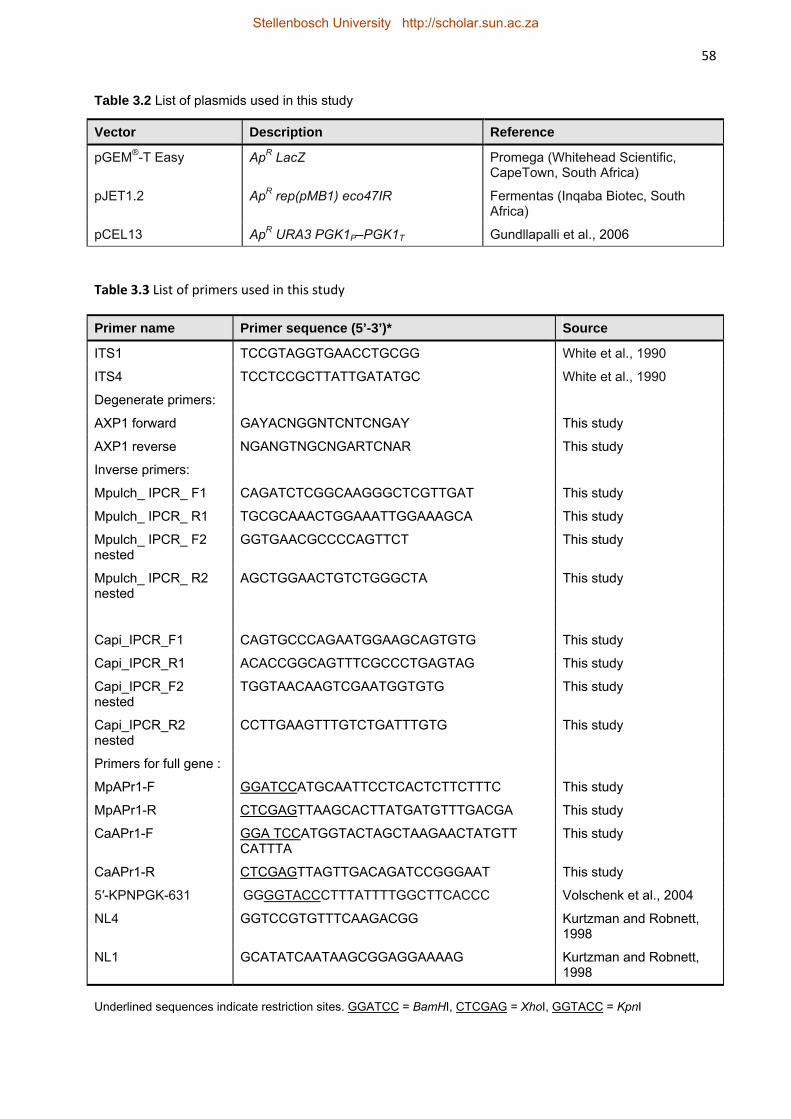
x 3.2.1 Strains, plasmids and culture conditions 39
3.2.2 Molecular biology and Bioinformatics techniques 40
3.2.2.1 Nucleic acid extraction 40
3.2.2.2 In silico analyses 41
3.2.2.3 PCR methods 41
3.2.3 Cloning and heterologous expression in S. cerevisiae YHUM272 42
3.2.4 DNA sequencing 43
3.2.5 Protein work 43
3.2.5.1 SDS-PAGE and zymography 43
3.2.5.2 Protein sequencing 43
3.3 Results 44
3.3.1 Protease activity screening and strain selection 44
3.3.2 Isolation and cloning of protease-encoding genes 44
3.3.3 In silico analysis of the putative gene and deduced protein sequences 45
3.3.4 Putative identification based on homology studies 45
3.3.5 Heterologous expression of the protease-encoding gene of M. pulcherrima IWBT Y1123 in S. cerevisiae YHUM272 46
3.3.6 Induction and substrate specificity investigation 47
3.3.7 Genetic screening of 12 M. pulcherrima strains for the presence of MpAPr1 49
3.4 Discussion 50
3.5 Acknowledgements 54
3.6 References 54
Chapter 4. General discussion and conclusions 79
4.1 Results and general discussion 80
4.2 Conclusions and future prospects 82
4.3 References 83
Stellenbosch University http://scholar.sun.ac.za
Chapter 1
General introduction and project aims
Stellenbosch University http://scholar.sun.ac.za
2
General introduction and project aims
1.1 Introduction
The production of wine is a complex biochemical transformation facilitated by a large pool of
enzymes of plant and microbial origin (Pretorius et al., 1999; Fleet, 2003). The yeast
Saccharomyces cerevisiae plays the predominant role in the transformation of grape juice to
wine, whether the juice is inoculated with commercially available S. cerevisiae strains or left to
ferment spontaneously with the microorganisms present in the grape must (Fleet et al., 1984;
Bisson, 2004). This yeast has high ethanol tolerance and fermentation capacity and releases
secondary metabolites which plays a role in enhancing the aroma and flavour of wine. The
metabolic activities of this yeast are very well characterized (Fleet, 2003). Besides S. cerevisiae,
a range of other yeast species are also present in spontaneously fermenting must and some
may also be present in wine. These yeasts, classified as non-Saccharomyces yeasts, were
thought to be detrimental to wine flavour and aroma and were mostly categorized as wine
spoilage yeasts (Du Toit and Pretorius, 2000; Loureiro and Malfeito-Ferreira, 2003). These
include yeasts of the genera Candida, Metschnikowia, Debaryomyces, Zygosaccharomyces,
Kluyveromyces, and Kloeckera, to name a few (Fleet et al., 1984; Heard and Fleet, 1987).
However, it has been demonstrated that some of these yeasts can confer desirable aroma
nuances to wine when used in conjunction with S. cerevisiae in co-inoculated fermentations
(Ciani and Comitini, 2011; Domizio et al., 2011). It has also been reported by a number of
authors that some non-Saccharomyces yeasts are good secretors of extracellular enzymes e.g.
pectinases, glucosidases and proteases, that could be of interest to the winemaker
(Charoenchai et al., 1997; Fernandez et al., 2000; Strauss et al., 2001). Of particular interest
are the extracellular proteases produced by some non-Saccharomyces wine yeasts.
It has already been reported in literature that the addition of proteases to wine is efficient for
reducing protein haze formation without being detrimental to wine quality (Lagace and Bisson,
1990; Pocock et al., 2003). Protein haze formation in white wine is usually due to the
denaturation of wine proteins during bottle storage (Hsu et al., 1987; Ferreira et al., 2001;
Pocock and Waters, 2006; Marangon et al., 2011). The presence of haze reduces the
commercial value of the wine, making it unacceptable for consumers as it may be perceived as
microbial spoilage (Pocock and Waters, 2006). Winemakers usually add bentonite to their white
wine in order to precipitate the proteins down before bottling. The disadvantages are that such a
treatment is expensive, reduces product yield and may have a negative effect on wine aroma
(Waters et al., 2005). Besides the potential to reduce unsightly protein haze in white wine,
proteases may also liberate peptides and amino acids thereby increasing the assimilable
nitrogen content of wine for the growth of fermentation (and spoilage) microorganisms, which is
essential for efficient fermentation. An increase in assimilable nitrogen may also lead to an
Stellenbosch University http://scholar.sun.ac.za
3 increase in the formation of aroma compounds such as ethyl acetate, acetic acid and other
volatile acids (Bell and Henschke, 2005).
1.2 Rationale and scope of the study
Wine is a unique environment that is characterized by a low pH (2.8 – 4.2) (Somers, 1971), low
temperature (15 - 25˚C), and the presence of inhibitors such as SO2 (160 mg/l), ethanol (10 –
25%) and low sugar content (2.5 – 12 g/l). Organisms and their secretome that are able to
survive or even flourish under these conditions are highly adapted. Certain non-Saccharomyces
yeasts that are able to survive in wine also have the ability to secrete enzymes into the wine
matrix (Bossi et al., 2006). Investigations have been conducted demonstrating the production of
extracellular acid proteases by wine non-Saccharomyces yeasts (Charoenchai et al., 1997;
Fernández et al., 2000; Strauss et al., 2001) but none have focused on characterizing these
enzymes on genetic level or the mechanism involved in the secretion (and regulation) of these
enzymes. The wealth of knowledge and potential regarding non-Saccharomyces yeasts with
hidden potential for oenology is largely untapped.
The aim of this study is to identify and characterize extracellular acid protease encoding genes
from non-Saccharomyces yeast isolated from grape must. Part of the focus of this work is to
better understand the adaptation and the interactions of these microorganisms in the particular
life medium that wine is. It would contribute to the global knowledge of the potential certain wine
microorganisms might possess to survive in wine. The study will provide further insight into
these enzymes on genetic and activity levels.
Specific objectives of the study
1. To identify and isolate new genes encoding aspartic proteases from non-Saccharomyces
yeasts isolated from grape must
2. To characterize the genes and the proteins that they encode
3. To explore the potential applicability of these enzymes in winemaking
1.3 References
Bell, S-J., Henschke P.A., 2005. Implications of nitrogen nutrition for grapes, fermentation and wine. Australian Journal of Grape and Wine Research 11, 242–295. Bisson, L. 2004. The biotechnology of wine yeast. Food Biotechnology 18, 63–96. Bossi, A., Bonizzato, L., Zapparoli, G., 2006. Acidic extracellular proteases from microrganisms of fairly acidic niche. Protein & Peptide Letters 13, 737-741. Charoenchai, C., Fleet, G.H., Henschke, P.A., Todd, B.E.N.T., 1997. Screening of non-Saccharomyces wine yeasts for the presence of extracellular hydrolytic enzymes. Australian Journal of Grape and Wine Research 3, 2-8.
Stellenbosch University http://scholar.sun.ac.za
4 Ciani, M., Comitini, F., 2011. Non-Saccharomyces wine yeasts have a promising role in biotechnological approaches to winemaking. Annals in Microbiology 61, 25–32. Domizio, P., Romani, C., Comitini, F., Gobbi, M., Lencioni, L., Mannazzu, I., Ciani, M., 2011. Potential spoilage non-Saccharomyces yeasts in mixed cultures with Saccharomyces cerevisiae. Annals of Microbiology 61, 137–144. Du Toit, M., Pretorius, I.S., 2000. Microbial spoilage and preservation of wine: Using weapons from nature’s arsenal. A review. South African Journal of Enology and Viticulture 21, 74-96. Fernández, M., Ubeda, J.F., Briones, A.I., 2000. Typing of non-Saccharomyces yeasts with enzyme activities of interest in winemaking. International Journal of Food Microbiology 59, 29-36. Ferreira, R.B., Picarra-Pereira, M.A., Monteiro, S., Loureiro, V.B., Teixeira, A.R., 2001. The wine proteins. Trends in Food Science & Technology 12, 230–239. Fleet, G.H., Lafon-Lafourcade, S., Ribéreau-Gayon, P., 1984. Evolution of yeasts and lactic acid bacteria during fermentation and storage of Bordeaux wines. Applied and Environmental Microbiology 48,1034-1038. Fleet, G.H., 2003. Yeast interactions and wine flavour. International Journal of Food Microbiology 86, 11-22. Heard, G.M., Fleet, G.H., 1987. Occurrence and growth of yeast species during the fermentation of some Australian wines. Food Technology in Australia 38, 22-25. Hsu, J.C., Heatherbell, D. A., Flores, J.H., Watson B.T., 1987. Heat-Unstable Proteins in Grape Juice and Wine. II. Characterization and Removal by Ultrafiltration. American Journal of Enology and Viticulture 38, 17-22. Lagace, L.S., Bisson, L.F., 1990. Survey of yeast acid proteases for effectiveness of wine haze reduction. American Journal of Enology and Viticulture 41, 147-155. Loureiro, V., Malfeito-Ferreira, M., 2003. Spoilage yeasts in the wine industry. International Journal of Food Microbiology 86, 23– 50. Marangon, M., Van Sluyter, S.C., Neilson, K.A., Chan, C., Haynes, P.A., Waters, E.J., Falconer, R.J., 2011. Roles of grape thaumatin-like protein and chitinase in white wine haze formation. Journal Agricultural and Food Chemistry 59, 733–740. Pocock, K.F., Høj, P.B., Adams, K.S., Kwiatkowski, M.J., Waters, E.J., 2003. Combined heat and proteolytic enzyme treatment of white wines reduces haze forming protein content without detrimental effect. Australian Journal of Grape and Wine Research 9, 56-63. Pocock K.F., Waters, E.J., 2006. Protein haze in bottled white wines: How well do stability tests and bentonite fining trials predict haze formation during storage and transport? Australian Journal of Grape and Wine Research 12, 212–220. Pretorius, I.S., Van der Westhuizen, T.J., Augustyn, O.P.H., 1999. Yeast biodiversity in vineyards and wineries and its importance to the South African wine industry. South African Journal of Enology and Viticulture 20, 61-74. Somers, T. C., 1971. The polymeric nature of wine pigments. Phytochemistry 10, 2175-2186. Strauss, M.L.A., Jolly, N.P., Lambrechts, M.G., Van Rensburg, P., 2001. Screening for the production of extracellular hydrolytic enzymes by non-Saccharomyces wine yeasts. Journal of Applied Microbiology 91, 182-190. Waters, E.J., Alexander, G., Muhlack, R., Pocock, K.F., Colby, C., O’Neill, B.K., Høj, P.B. Jones, P., 2005. Preventing protein haze in bottled white wine. Australian Journal of Grape and Wine Research 11, 215–225.
Stellenbosch University http://scholar.sun.ac.za
5
Chapter 2
Literature review
Aspartic proteases of non-Saccharomyces
yeasts and their potential application in wine
biotechnology
Stellenbosch University http://scholar.sun.ac.za
6
2. Literature review
2.1 General introduction
Winemaking involves the biochemical conversion of grape juice to wine where microorganisms,
mainly yeasts, in the juice convert glucose to ethanol, carbon dioxide and a range of other
secondary metabolites (Fleet, 2003). The conversion is facilitated by a large pool of enzymes of
both plant and microbial origin. Winemakers reinforce this pool of indigenous enzymes by
adding a variety of industrially produced enzymes such as pectinases, hemicellulases,
glucanases and glycosidases in order to help enhance clarification, juice yield, as well as the
release of aroma compounds, tannins and colour.
The yeast Saccharomyces cerevisiae is the predominant yeast responsible for fermentation
(Fleet et al., 1984). In recent years however, researchers have been investigating the impact
non-Saccharomyces yeasts have on wine production. Some of the non-Saccharomyces yeasts
have been shown to secrete hydrolytic enzymes including proteases that might be of interest to
the winemaker (Esteve-Zarzoso et al., 1998; Dizy and Bisson, 2000; Fernández et al., 2000).
This review consists of three main sections. The first will focus on aspartic proteases with a
short introduction into proteolytic enzymes followed by a more detailed discussion into aspartic
proteases, i.e. their structure, catalytic mechanisms and the secretion of extracellular aspartic
proteases in yeasts. The last two sections will discuss the oenological importance and the role
of non-Saccharomyces yeasts in winemaking, and the potential of aspartic proteases in
winemaking, respectively.
2.2 Proteolytic enzymes
Proteolytic enzymes catalyse the cleavage of peptide bonds within peptides and proteins. They
are encoded by about 2% of genes in all kinds of organisms. These enzymes regulate most
physiological processes (Tyndall et al., 2005). Some of the important medical roles that
proteolytic enzymes fulfil include food digestion, protein turnover, blood coagulation, embryonic
development and cell division. Approximately 14% of the five hundred human peptidases are
under investigation as drug targets and include the β-secretase that play a role in Alzheimer’s
disease. The human immunodeficiency virus (HIV) protease is another well-known drug target.
They are thus an important group of enzymes in scientific, medical research and biotechnology
(Rawlings et al., 2009).
2.2.1 Definition and categorization of proteolytic enzymes
Proteolytic enzymes are also known as proteinases or proteases, however the Enzyme
Commission (EC) and the Nomenclature Committee of the International Union of Biochemistry
and Molecular Biology (NC-IUBMB) prefer the term peptidases be used for all enzymes that
Stellenbosch University http://scholar.sun.ac.za
7 hydrolyse peptide bonds (subclass E.C.3.4). Nevertheless, proteolytic enzymes are perhaps the
most generally understood term in the current usage. Exopeptidases cleave one or a few amino
acids from the N- or C-terminus while endopeptidases act internally in the polypeptide chains.
Exopeptidases that hydrolyse at a free N-terminus to release a single amino acid residue are
called aminopeptidases, while those that release dipeptides and tripeptides are named
dipeptidyl-peptidases and tripeptidyl-peptidases, respectively. Those hydrolysing at a free C-
terminus to release a single residue are named carboxypeptidases and those releasing
dipeptides are named peptidyl-dipeptidases. Other exopeptidases remove terminal residues that
are substituted, cyclized or linked by isopeptide bonds (peptide linkages other than those of α-
carboxyl to α-amino groups) (Barrett et al., 1998).
Proteases are categorized based on their catalytic mechanism, the amino acid residues present
in the catalytic site and their three-dimensional structure. According to the NC-IUBMB,
proteases can be categorized into four mechanistic classes which include the serine
endopeptidases, cysteine endopeptidases, aspartic endopeptidases and metallo-
endopeptidases. Each type of protease is specific in its ability to break a certain peptide bond
and exhibits a characteristic set of functional amino acid residues arranged in a specific
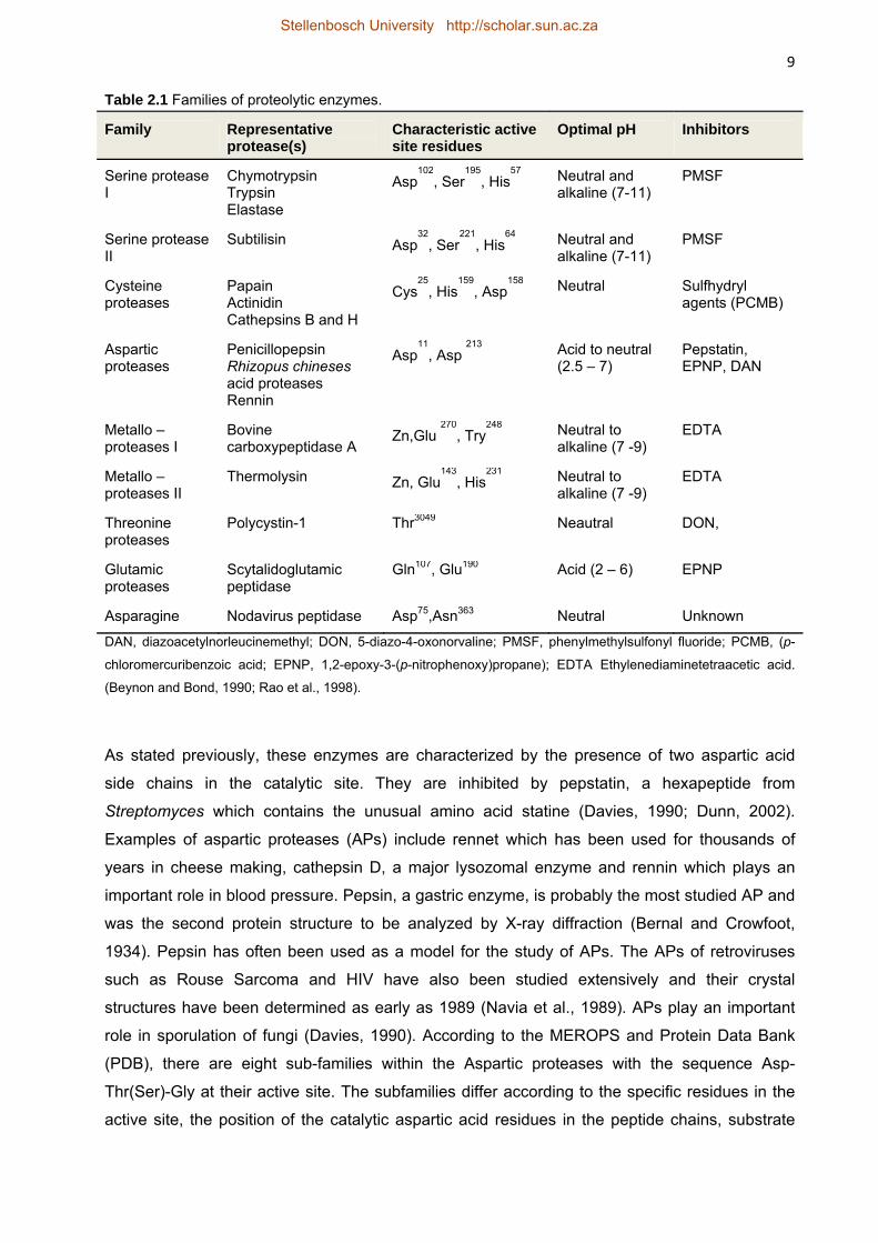
configuration to produce its catalytic site (Barrett et al., 2004; Tyndall et al., 2005). Table 2.1
shows the different protease families, some common examples and the amino acid residues
present in each catalytic domain. Proteases commonly recognize the extended or α-strand
backbone conformation in substrates, inhibitors, and products (Tyndall et al., 2005).
The MEROPS database is a manually curated information resource dedicated solely to
peptidases, their substrates and inhibitors. It can be found at http://merops.sanger.ac.uk. The
MEROPS database divides peptidases into protein species which are then sub-divided into
families according to statistically significant similarities in their amino acid sequences.
Homologous families are then grouped into clans.
The protein species are the Aspartic peptidases, Cysteine peptidases, Glutamic peptidases,
Metallopeptidases, Asparagine peptidases, Serine peptidases, and Threonine peptidases.
The Serine proteases have the catalytic triad aspartic acid, histidine, and serine and play
important roles in digestion. They have one of two structural folds: the trypsin-like type (serine
protease l) which is made up of two β-barrels and the subtilisin-like type (serine protease ll)
made up of a three-layer αβα sandwich fold.
The Cysteine proteases have similar folds as the serine type but are more V-shaped and have
the catalytic dyad histidine and cysteine or triad with an aspartic acid residue. A common
example is papain which is used as a meat tenderiser.
Stellenbosch University http://scholar.sun.ac.za
8 The Metalloproteases have a characteristic divalent zinc metal ion in their catalytic site and are
important for wound healing and tissue morphogenesis (Rao et al., 1998).
The Aspartic proteases, which will be the main focus of this review and in particular those
secreted by non-Saccharomyces yeasts, have a tertiary structure consisting of two
approximately symmetric lobes with each lobe carrying an aspartic acid residue to form the
catalytic site. Unlike the other types of proteases, the activity of the aspartic proteases is
dependent on low pH conditions (Northrop, 2001; Cascella et al., 2005; Borelli et al., 2008).
Threonine proteases contain a threonine nucleophile at their N-terminus and sometimes a
serine residue as well. Glutamic proteases, which were formerly known as pepstatin-insensitive
carboxyl proteases, have a glutamic acid and a glutamine residue in their catalytic sites. They
are also active at acidic pH and are found in some bacterial and fungal species (Tanokura et al.,
1992; Fujinaga et al., 2004; Tyndall et al., 2005).
Asparagine proteases were recently discovered and are found in certain pathogenic viruses and
bacteria (e.g. Escherichia coli) (Rawlings et al., 2011). The catalytic site may consist of a single
residue, asparagine or may contain asparagine with serine, asparagines or cysteine.
Besides these families there have been discoveries of proteases with unidentified catalytic
mechanism. This indicates that novel types of proteases may exist (Tanokura et al., 1992;
Tyndall et al., 2005; Rawlings et al., 2009).
2.2.2 Aspartic proteases
2.2.2.1 General description
Aspartic endopeptidases (E3.4.23.x) are widely distributed in living organisms from vertebrates
to fungi, plants and retroviruses. Most of these enzymes are composed of approximately 323 to
340 amino acid residues, with molecular weights ranging between 35 000 to 50 000 Daltons
(Da) and isoelectric points (pI) ranging between 3 and 4.5 because of the high percentage of
acidic amino acid residues (about 13%) in the proteins. They have optimum function at pH 3 to
4. They show substrate specificity towards extended peptide substrates and residues with large
hydrophobic side chains on either side of the scissile bond (Barrett et al., 1998; Rawlings et al.,
nitrogen, pyrazines, vitamins and nitrate (Henschke and Jiranek, 1993; Cramer et al., 2002). In
wine, concentrations of these compounds are found in a broad range. Yeasts use a mechanism
called nitrogen catabolite repression (NCR), which mediates the selection of good nitrogen
sources by the expression of appropriate transport system (permeases) and the degradation of
non appropriate permeases (Bell and Henschke, 2005). S. cerevisiae, the principal yeast used
for fermentation, preferentially utilizes simple nitrogen sources such as ammonium ions and free
alpha amino nitrogen compounds present in the form of primary amino acids such as glutamine
and asparagine (Henschke and Jiranek, 1993). Arginine is quantitatively the most important
amino acid utilizable by Saccharomyces in grapes and, subsequently unfermented juice. This
amino acid is rapidly incorporated by the yeast at the start of fermentation and subsequently
released back into the wine during autolysis. Secondary amino acids, such as proline and
hydroxyproline are not metabolised to any great extent under winemaking conditions. Only low
molecular weight peptides can also be utilized but grape proteins cannot be used as a source of
nitrogen since S. cerevisiae lacks significant extracellular proteolytic enzymes to hydrolyse
these proteins during fermentation. Proteinase A, a vacuolar protease of S. cerevisiae, is only
secreted during autolysis following fermentation (Ogrydziak 1993; Alexandre et al., 2001).
Recently however, Younes et al. (2011) identified proteolytic activity in S. cerevisiae PlR1
secreted during fermentation. Proteases secreted by non-Saccharomyces yeasts that are able
to hydrolyse grape proteins under wine making conditions may increase the assimilable
nitrogen sources by liberating peptides and possibly amino acids. On the other hand, certain by-
products considered detrimental to health such as biogenic amines and ethyl carbamate can be
produced by degradation of nitrogen compounds. Ethyl carbamate (urethane) is formed by the
reaction of urea and ethanol. It is an undesirable compound of wine since it is considered as
carcinogen and mutagen (Bell and Henschke, 2005).
Stellenbosch University http://scholar.sun.ac.za
27 An inadequacy of nitrogen-containing compounds of grape juices for wine fermentation has
often been reported. Insufficient initial assimilable nitrogen sources, amongst other causes, may
lead to stuck or sluggish fermentations. Sluggish or stuck fermentations, refers to those
fermentations that commence normally but become slow or stop before must sugar
concentrations are depleted (Henschke and Jiranek, 1993). Most winemakers therefore
supplement their must with additional nitrogen sources such as diammonium phosphate (DAP)
or ammonium sulphate (Hernandez-Orte et al., 2006). DAP added at the initial stage during the
yeast growth phase increases the size of the yeast population, but has little effect on population
size when added at later stages. Amino acid mixtures are also used to supplement grape must
nitrogen concentrations. The greater efficiency of amino acid mixtures, especially balanced
mixtures, compared with single nitrogen sources is linked to the ability of yeast to directly
incorporate amino acids into protein, thereby minimising the need to maintain an energetically
expensive amino acid synthetic capability (Bell and Henschke, 2005). A supplement of amino
acids in grape juice could shorten fermentation time (Hermández-Orte et al., 2006). Utilization of
nitrogen-containing compounds by yeasts is strain-dependent and the fermentation conditions
also play a role (Valero et al., 1999), e.g. yeasts consume less nitrogen at low temperature and
ethanol inhibits the uptake of most amino acids (Bisson, 1991).
Esters, higher alcohols, volatile fatty acids such as γ-butyrolactone, isobutanol and isobutyric
acid and carbonyls are important contributors to the fermentation bouquet of wine (Fleet, 2003).
These compounds principally arise as primary metabolites of yeast sugar and amino acid
metabolism (Henschke and Jiranek 1993, Swiegers et al. 2005). Higher alcohols may be
produced by catabolic transformation of branched-chain amino acids via the Ehrlich pathway.
Therefore, the production of these flavour-active compounds during fermentation is influenced
by the amino acid composition of the must. When concentrations of higher alcohols are low they
contribute to the aroma complexity but an excess of higher alcohols has a negative impact on
wine quality. These alcohols, together with organic acids, provide substrates for ester formation.
Most esters confer pleasant flavours to wine, e.g. fruity and floral notes (Lambrechts and
Pretorius 2000). Nitrogen compounds also regulate the formation of other volatiles, such as
hydrogen sulfide, thiols/mercaptans and monoterpenes (Henschke and Jiranek, 1993). At high
initial nitrogen content in must, the concentrations of total higher alcohols are at their lowest.
Wines with high concentrations of esters, e.g. ethyl esters, and low higher alcohol
concentrations, are associated with must with higher concentrations of amino acids (Hernández-
Orte et al., 2006).
In summary, proteases can liberate peptides and amino acids contributing to the yeast’s
nitrogen pool required for coordinating amino acids, purine and pyrimidine synthesis (Bell and
Henschke, 2005) needed for cell growth, flavour-active metabolites and also fermentation
activity. However, nitrogenous compounds influence clarification and microbial stability.
Stellenbosch University http://scholar.sun.ac.za
28 Therefore careful nitrogen management is necessary as to control the growth of spoilage or
undesired fungi and bacteria.
2.5 References
Alexandre, H., Heintz, D., Chassagne, D., Guilloux-Benatier, M.,Charpentier, C., Feuillat, M., 2001. Protease A activity and nitrogen fractions released during alcoholic fermentation and autolysis in enological conditions. Journal of Industrial Microbiology and Biotechnology 26, 235–240. Amerine, M.A., Cruess, W.V., 1960. The technology of winemaking. The AVI Publishing Company Inc., Connecticut. Andreeva, N. S., Gurskaya, G. V., 2006. Interdomain interactions in aspartic proteases of higher organisms and their analogs in retroviral enzymes. Molecular Biology 40, 427–432. Andreeva, N.S., Rumsh, L.D., 2001. Analysis of crystal structures of aspartic proteinases: On the role of amino acid residues adjacent to the catalytic site of pepsin-like enzymes. Protein Science 10, 2439–2450. Arroyo-López F.N., Orlić S., Querol A., Barrio, E., 2009. Effects of temperature, pH and sugar concentration on the growth parameters of Saccharomyces cerevisiae, S. kudriavzevii and their interspecific hybrid. International Journal of Food Microbiology 131, 120–127. Bakalinsky, A. T., Boulton, R., 1985. The study of an immobilized acid protease for the treatment of wine proteins. American Journal of Enology and Viticulture 36, 23-29. Barrett, A.J., Rawlings, N.D., Woessner, J.F., 1998. Handbook of Proteolytic Enzymes, first ed. Academic Press, London. Barrett, A.J., Rawlings, N.D., Woessner, J.F., 2004. Handbook of Proteolytic Enzymes, second ed. Academic Press, London. Bautista, R., Fernández, E., Falqué, E. 2007. Effect of the contact with fermentation-lees or commercial-lees on the volatile composition of white wines. European Food Research Technology 224, 405–413. Beckerich, J-M., Boisramé-Baudevin, A., Gaillardin, C., 1998. Yarrowia lipolytica: a model organism for protein secretion studies. International Microbiology 1, 123–130. Beggah, S., Léchenne, B., Reichard, U., Foundling, S. Monod, M., 2000. Intra- and intermolecular events direct the propeptide-mediated maturation of the Candida albicans secreted aspartic proteinase Sap1p. Microbiology 146, 2765–2773. Bell, S-J., Henschke P.A., 2005. Implications of nitrogen nutrition for grapes, fermentation and wine. Australian Journal of Grape and Wine Research 11, 242–295. Bely M., Stoeckle, P., Masneuf-Pomarède, I., Dubourdieu, D., 2008. Impact of mixed Torulaspora delbrueckii–Saccharomyces cerevisiae culture on high-sugar fermentation. International Journal of Food Microbiology 122, 312–320. Bernal, J. D., Crowfoot, D., 1934. X-ray photographs of crystalline pepsin. Nature 1, 794-95. Beynon, R. J., Bond, J. S. 1990. Proteolytic enzymes: a practical approach. Oxford University Press, Oxford. Bisson, L. F.,1991. Influence of nitrogen on yeast and fermentation of grapes. International symposium on Nitrogen in Grapes and Wine, (pp. 78-89). Davis: American society for Enology and viticulture. Bisson, L. 2004. The biotechnology of wine yeast. Food Biotechnology 18, 63–96. Blade, W.H., and R. Boulton. 1988. Adsorption of protein by bentonite in a model wine solution. American Journal of Enology and Viticulture 39,193-199.
Stellenbosch University http://scholar.sun.ac.za
29 Blundell, T.L., Jenkins, J.A., Sewell B.T., Pearl, L.H. Cooper, J. B., Tickle, I.J., Veerapandian B., Wood S.P., 1990. X-ray analyses of aspartic proteinases. The three-dimensional structure at 2.1 Ǻ resolution of endothiapepsin. Journal of Molecular Biology 211, 919-941.
Borelli, C., Ruge, E., Lee, J.H., Schaller, M., Vogelsang, A., Monod, M., Korting, H.C., Huber, R., Maskos, K.,2008. X-ray structures of Sap1 and Sap5: Structural comparison of the secreted aspartic proteinases from Candida albicans. Proteins: Structure, Function, and Bioinformatics 72, 1308-1319.
Campos, L.A., Sancho, J., 2003. The active site of pepsin is formed in the intermediate conformation dominant at mildly acidic pH. FEBS Letters 538, 89-95. Cascella, M., Micheletti, C., Rothlisberger, U., Carloni, P., 2005. Evolutionarily conserved functional mechanics across pepsin-like and retroviral aspartic proteases. Journal of the American Chemical Society 127, 3734-3742. Caridi, A., Galvano, F., Tafuri, A., Ritieni, A., 2006. Ochratoxin A removalduring winemaking. Enzyme and Microbial Technology 40, 122–126. Charoenchai, C., Fleet, G.H., Henschke, P.A., Todd, B.E.N.T., 1997. Screening of non-Saccharomyces wine yeasts for the presence of extracellular hydrolytic enzymes. Australian Journal of Grape and Wine Research 3, 2-8. Charoenchai, C., Fleet, G. H., Henschke, P. A., 1998. Effects of temperature, pH and sugar concentration on the growth rates and cell biomass of wine yeasts. American Journal of Enology and Viticulture 49, 283–288. Cheng, Y., Avis., T.J., Bolduc, S., Zhao, Y., 2008. Recombinant secretion in Pseudozyma flocculosa and Pseudozyma antartica with a novel signal peptide. Bioscience, Biotechnology and Biochemistry 72, 3158-3166. Chitpinityol, S., Goode, D., James, M., Crabbe, C., 1997. Studies on the binding of α-crystallin to recombinant prochymosins and chymosin. Molecular Vision 4. Ciani, M., Ferraro, L. 1996. Enhanced glycerol content in wines made with immobilized Candida stellata cells. Applied and Environmental Microbiology 62, 128-132. Ciani, M., Ferraro, L., 1998. Combined use of immobilized Candida stellata cells and Saccharomyces cerevisiae to improbe the quality of wines. Journal of Applied Microbiology 85, 247–254. Ciani, M., Fatichenti, F., 2001. Killer toxin of Kluyveromyces phaffii DBVPG 6076 as a biopreservative agent to control apiculate wine yeasts. Applied and Environmental Microbiology 67, 3058–3063. Ciani, M., Maccarelli, F., 1998. Oenological properties of non-Saccharomyces yeasts associated with winemaking. World Journal of Microbial Biotechnology 14, 199–203. Ciani, M., Comitini, F., Mannazzu, I., Domizio, P., 2010. Controlled mixed culture fermentation: a new perspective on the use of non-Saccharomyces yeasts in winemaking. FEMS Yeast Research 10, 123–133. Ciani, M., Comitini, F., 2011. Non-Saccharomyces wine yeasts have a promising role in biotechnological approaches to winemaking. Annals in Microbiology 61, 25–32. Clemente-Jimenez, J.F., Mingorance-Cazorla, L., Martínez-Rodríguez, S., LasHeras-Vázquez, F.J., Rodríguez-Vico, F., 2004. Molecular characterization and oenological properties of wine yeasts isolated during spontaneous fermentation of six varieties of grape must. Food Microbiology 21, 149-155. Coates, L., Tuan, H-F., Tomanicek, S., Kovalevsky, A, Mustyakimov, M., Erskine, P., Cooper, J., 2008. The catalytic mechanism of an aspartic proteinase explored with neutron and X-ray diffraction. Journal of American Chemical Society 130, 7235–7237.
Stellenbosch University http://scholar.sun.ac.za
30 Comitini, F., De Ingeniis, J., Pepe, L., Mannazzu, I., Ciani, M., 2004. Pichia anomala and Kluyveromyces wickerhamii killer toxins as new tools against Dekkera/Brettanomyces spoilage yeasts. FEMS Microbiology Letters 238, 235–240. Conesa, A., Weelink, G., van den Hondel, C.A., Punt, P.J., 2001. C-terminal propeptide of the Caldariomyces fumago chloroperoxidase: an intramolecular chaperone? FEBS Letters 503, 117–120. Conterno, L., Delfini, C., 1993. Peptidase activity and the ability of wine yeasts to utilise grape must proteins as sole nitrogen source. Journal of Wine Research 5, 113-126. Cramer, A.C., Vlassides,S., Block, D.E., 2002. Kinetic model fornitrogen-limited wine fermentations. Biotechnology and Bioengineering 77, 49–60. Davies, D.R., 1990. The structure and function of aspartic proteases. Annual Reviews of Biophysics and Biophysical Chemistry 19, 189-215. Dawes, H., Boyes, S., Keene, J., Heatherbell, D., 1994. Protein instability of wines: influence of protein isoelectric point. American Journal of Enology and Viticulture 45, 319-326. De Bruijn, J.C., Loyola, A., Flores, F., Hevia, P., Melìn, and I. Serra. 2009. Protein stabilisation of Chardonnay wine using trisacryl and bentonite: A comparative study. Internitional Journal Food Science and Technology 44, 330-336.
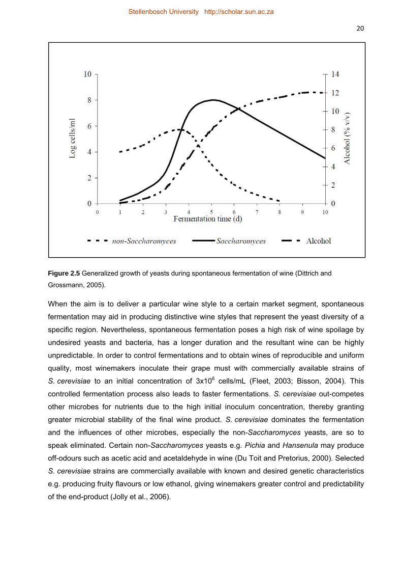
Dittrich, H.H., Grossmann, M., 2005. Mikrobiologie des Weines. Struttgart, Ulmer.
Dizy, M. and Bisson, L.F. 2000. Proteolytic activity of yeast stains during grape juice fermentation. American Journal of Enology and Viticulture 51, 155–167. Domizio, P., Romani, C., Comitini, F., Gobbi, M., Lencioni, L., Mannazzu, I., Ciani, M., 2011. Potential spoilage non-Saccharomyces yeasts in mixed cultures with Saccharomyces cerevisiae. Annals of Microbiology 61, 137–144. Dunn, B.M., 2002. Structure and mechanism of the pepsin-like family of aspartic peptidases. Chemical Reviews 102, 4431-4458. Du Toit, M., Pretorius, I.S., 2000. Microbial spoilage and preservation of wine: Using weapons from nature’s arsenal. A review. South African Journal of Enology and Viticulture 21, 74-96. Esteve-Zarzoso, B., Paloma Manzanares, P., Ramón, D., Querol, A., 1998. The role of non-Saccharomyces yeasts in industrial winemaking. International Microbiology 1, 143–148. Fernández, M., Ubeda, J.F., Briones, A.I., 2000. Typing of non-Saccharomyces yeasts with enzyme activities of interest in winemaking. International Journal of Food Microbiology 59, 29-36. Fernández-González, M., Di Stefano, R., Briones, A., 2003. Hydrolysis and transformation of terpene glycosides from muscat must by different yeast species. Food Microbiology 20, 35-41. Ferreira, R.B., Picarra-Pereira, M.A., Monteiro, S., Loureiro, V.B., Teixeira, A.R., 2001. The wine proteins. Trends in Food Science & Technology 12, 230–239. Feuillat, M., Charpentier, C., Maujean, A., 1998. Les composés azotés. In: Flanzy, C. ed. Oenologie: fondements scientifiques et techniques. Lavoisier, Paris, 94–116. Fleet, G.H., Lafon-Lafourcade, S., Ribéreau-Gayon, P., 1984. Evolution of yeasts and lactic acid bacteria during fermentation and storage of Bordeaux wines. Applied and Environmental Microbiology 48,1034-1038. Fleet, G.H., 2003. Yeast interactions and wine flavour. International Journal of Food Microbiology 86, 11-22 Friedman, R., Caflisch, A., 2010. On the orientation of the catalytic dyad in aspartic proteases. Proteins 78, 1575–1582.
Stellenbosch University http://scholar.sun.ac.za
31 Fujinaga, M., Cherney, M.M., Oyama, H., Oda, K., James, M.N.G., 2004. The molecular structure and catalytic mechanism of a novel carboxyl peptidase from Scytalidium lignicolum. The Proceedings of the National Academy of Sciences USA 101, 3364–3369. Goddard, M. R., 2008. Quantifying the complexities of Saccharomyces cerevisiae’s ecosystem engineering via fermentation. Ecology 89, 2077–2082. Gonzalez-Lopez, C.I., Szabo, R., Blanchin-Roland, S., Gaillardin, C., 2001. Genetic control of extracellular protease synthesis in the yeast Yarrowia lipolytica. Genetics 160, 417–427. Gonzalez-Ramos, D., Cebollero, E., Gonzalez, R., 2008. A recombinant Saccharomyces cerevisiae strain overproducing mannoproteins stabilizes wine against proteinhaze. Applied and Environmental Microbiology 74, 5533–5540. Heard, G.M., Fleet, G.H. 1985. Growth of natural yeast during the fermentation of inoculated wines. Applied and Environmental Microbiology 50, 727-728. Heard, G.M., Fleet, G.H., 1987. Occurrence and growth of yeast species during the fermentation of some Australian wines. Food Technology in Australia 38, 22-25. Henick-Kling, T., Ediger W., Daniel, P., Monk, P., 1998. Selective effects of sulfur dioxide and yeast starter culture addition on indigenous yeast populations and sensory characteristics of wine. Journal of Applied Microbiology 84, 865–876. Henschke, P. A,, Jiranek, V., 1993. Hydrogen sulfide formation during fermentation: effect of nitrogen composition in model grape must. International symposium on nitrogen in grapes and wine 172–184, Seattle, Davis: American Society for Enology and Viticulture. Hernandez-Orte, P., Bely, M., Cacho, J., Ferrreira, V., 2006. Impact of ammonium additions on volatile acidity,ethanol, and aromatic compound production by different Saccharomyces cerevisiae strains during fermentation incontrolled synthetic media. Australian Journal of Grape and Wine Research 12, 150–160. Hsu, J.C., Heatherbell, D. A., Flores, J.H., Watson B.T., 1987. Heat-unstable proteins in grape juice and wine. II. Characterization and removal by ultrafiltration. American Journal of Enology and Viticulture 38, 17-22. James M.N.G., Sielecki, A.R., 1986. Molecular structure of an aspartic proteinase zymogen, porcine pepsinogen, at 1.8 Å resolution. Nature 319, 33-38. Jolly, N.P., Augustyn, O.P.H. & Pretorius, I.S., 2003a. The occurrence of non-Saccharomyces yeast strains over three vintages in four vineyards and grape mustsfrom four production regions of the Western Cape, South Africa. South African Journal of Enology and Viticulture 24, 35-42. Jolly, N.P., Augustyn, O.P.H., Pretorius, I.S., 2003b. The use of Candida pulcherrima in combination with Saccharomyces cerevisiae for the production of Chenin blanc wine. South African Journal of Enology and Viticulture 24, 63-69. Jolly, N.P., Augustyn, O.P.H., Pretorius, I.S., 2006. The role and use of non-Saccharomyces yeasts in wine production. South African Journal of Enology and Viticulture 27, 15–38. Kim, D-H., Hong, Y-A., Park, H-D., 2008. Co-fermentation of grape must by Issatchenkia orientalis and Saccharomyces cerevisiae reduces the malic content in wine. Biotechnology Letters 30,1633–1638. Kurtzman, C.P. & Fell, J.W., 1998. The yeasts, a taxonomic study, fourth ed. Elsevier Science Publishers, Amsterdam. Lagace, L.S., Bisson, L.F., 1990. Survey of yeast acid proteases for effectiveness of wine haze reduction. American Journal of Enology and Viticulture 41, 147-155. Lambrechts, M.G., Pretorius I.S., 2000. Yeast and its importance to wine aroma. South African Journal of Enology and Viticulture 21, 97–129.
Stellenbosch University http://scholar.sun.ac.za
32 Le Bourse, D., Conreux A., Villaume, S., Lameiras, P., Nuzillard, J.-M., Jeandet, P., 2011. Quantification of chitinase and thaumatin-like proteins in grape juices and wines. Analytical and Bioanalytical Chemistry 401, 1541–1549. Lehle, L., Strahl, S., Tanner, W., 2006. Protein glycosylation, conserved from yeast to man: A model organism helps elucidate congenital human diseases. Angewandte Chemie International Edition 45, 6802–6818. Loureiro, V., Malfeito-Ferreira, M., 2003. Spoilage yeasts in the wine industry. International Journal of Food Microbiology 86, 23– 50. Madzak, C., Gaillardin, C., Beckerich, J-M., 2004. Heterologous protein expression and secretion in the non-conventional yeast Yarrowia lipolytica: a review. Journal of Biotechnology 109, 63–81. Manteau S., Lambert B., Jeandet P., Legendre L., 2003. Changes in chitinase and thaumatin-like pathogenesis-related proteins of grape berries during the Champagne winemaking process. Americam Journal of Enology and Viticulture 54, 267-272. Marangon, M., Van Sluyter, S.C., Neilson, K.A., Chan, C., Haynes, P.A., Waters, E.J., Falconer, R.J., 2011. Roles of grape thaumatin-like protein and chitinase in white wine haze formation. Journal Agricultural and Food Chemistry 59, 733–740. Martinand, V., Rietsch, M., 1891. Des microorganismes que l'onrencontre sur les raisins murs et de leur développement pendant la fermentation. Comptes Rendus de l' Académie des Sciences de Paris 112, 736-749.
McEwen, R.K., Young, T.W., 1998. Secretion and pH-dependent self-processing of the pro-form of the Yarrowia lipolytica acid extracellular protease. Yeast, 14, 1115-1125. Mendes Ferreira, A., Clίmaco, M.C., Mendes Faia, A., 2001. The role of non-Saccharomyces species in releasing glycosidic bound fraction of grape aroma components – a preliminary study. Journal of Applied Microbiology 91, 67-71. Mendoza, L.M., Manca de Nadra, M.C. Farías, M.E., 2007. Kinetics and metabolic behaviour of a composite culture of Kloeckera apiculata and Saccharomyces cerevisiae wine related strains. Biotechnology Letters 29, 1057–1063. Mendoza, L., Farías, M.E., 2010. Improvement of wine organoleptic characteristics by non-Saccharomyces yeasts. Current Research, Technology and Education Topics in Applied Microbiology and Microbial Biotechnology 2, 908-919. Mora, J., Barbas J.I., Mulet, A., 1990. Growth of yeast species during the fermentation of musts inoculated with Kluyveromyces thermotolerans and Saccharomyces cerevisiae. American Journal of Enology and Viticulture 41, 156–159. Moreno-Arribas, M.V., Pueyo, E., Polo, M.C., 2002. Analytical methods for the characterization of proteins and peptides in wines. Analytica Chimica Acta 458, 63–75. Müller-Thurgau, L. 1896. Das Zusammenwirken verschiedener Heferassen bei der Weingarung - Unsere bisherigen Erfahzungenueber die Anwendung der Reinhefen bei der Weingarung.Jahresberuft der deustchschweizerischen, Versuchstationin Wadesweil 1894-1895, 76-83. Naglik, J.R., Rodgers, C.A., Shirlaw, P.J., Dobbie, J.L. Fernandes-Naglik, L.L. Greenspan, D., Agabian, N., Challacombe, S.J., 2003a. Differential expression of Candida albicans secreted aspartyl proteinase and phospholipase B genes in humans correlates with active oral and vaginal infections. The Journal of Infectious Diseases 188, 469–79. Naglik,J.R. ,Challacombe, S.J., Hube, B., 2003b. Candida albicans secreted aspartyl proteinases in virulence and pathogenesis. Microbiology and Molecular Biology Reviews 67, 400–428. Naglik, J., Albrecht, A., Bader, O., Hube, B., 2004. Candida albicans proteinases and host/pathogen interactions. Cellular microbiology 6, 915-926.
Stellenbosch University http://scholar.sun.ac.za
33 Navia, M. A., Fitzgerald, P.M.D., McKeever, M., Leu, C-T., Heimbach, J.C., 1989. Three dimensional structure of aspartyl proteases from human immunodeficiency virus HIV-1. Nature 337, 615-620. Nguyen, H-V., Panon, G., 1998. The yeast Metschnikowia pulcherrima has an inhibitory effect against various yeast species. Sciences des Aliments 18, 515-526. Newport, G., Agabian, N., 1997. KEX2 Influences Candida albicans proteinase secretion and hyphal formation. The Journal of Biological Chemistry 272, 28954–28961. Northrop, D.B., 2001. Follow the protons: A low-barrier hydrogen bond unifies the mechanisms of the aspartic proteases. Accounts of Chemical Research 34, 790-797. Ogrydziak, D. M., 1993. Yeast extracellular proteases. Critical Reviews in Biotechnology 13, 1-55. Pardo, I., Garcia, M.I., Zuniga, M., Uruburu, F., 1989. Dynamics of microbial populations during fermentation of wines from the Utiel-Requena region of Spain. Applied and Environmental Microbiology 50, 539-541. Peters, I.I., Nelson, F.E., 1948. Preliminary characterization of the lipase of Mycotorula lipolytica. Journal Paper J-1481 of the Iowa Agricultural Experiment Station, Project 895. Piškur, J., Rozpędowska, E., Polakova, S., Merico, A., Compagno, C., 2006. How did Saccharomyces evolve to become a good brewer? Trends in Genetics 22, 183-186. Pocock, K.F., Hayasaka,Y., McCarthy, M.G., Waters, E.J., 2000. Thaumatin-like proteins and chitinases, the haze-forming proteins of wine, accumulate during ripening of grape (Vitis vinifera) berries and drought stress does not affect the final levels per berry at maturity. Journal of Agricultural Food Chemistry 48, 1637-1643. Pocock, K.F., Høj, P.B., Adams, K.S., Kwiatkowski, M.J., Waters, E.J., 2003. Combined heat and proteolytic enzyme treatment of white wines reduces haze forming protein content without detrimental effect. Australian Journal of Grape and Wine Research 9, 56-63. Pocock K.F., Waters, E.J., 2006. Protein haze in bottled white wines: How well do stability tests and bentonite fining trials predict haze formation during storage and transport? Australian Journal of Grape and Wine Research 12, 212–220. Pretorius, I.S., Van der Westhuizen, T.J., Augustyn, O.P.H., 1999. Yeast biodiversity in vineyards and wineries and its importance to the South African wine industry. South African Journal of Enology and Viticulture 20, 61-74. Punt, P.J., Drint-Kuijvenhoven A., Lokman B.C., Spencer, J.A., Jeenes D., Archer D.A. van den Hondel, C.A.M.J.J., 2003. The role of the Aspergillus niger furin-type protease gene in processing of fungal proproteins and fusion proteins. Evidence for alternative processing of recombinant (fusion-) proteins. Journal of Biotechnology 106, 23–32. Rao, M.B., Tanksale, A.M., Ghatge, M.S., Deshpande, V.V., 1998. Molecular and biotechnological aspects of microbial proteases. Microbiology and Molecular Biology Reviews 62, 597-635. Rawlings, N.D., Bateman, A., 2009. Pepsin homologues in bacteria. BMC Genomics 10, 437-448. Rawlings, N.D., Barrett, A.J., Bateman, A., 2009, MEROPS: the peptidase database. Nucleic Acids Research 38, Database issue D227–D233. Rawlings, N.D., Barrett, A.J., Bateman, A., 2011. Asparagine peptide lyases. A seventh catalytic type of proteolytic enzymes. The Journal of Biological Chemistry 286, 38321–38328. Ribereau-Gayon, P., D. Dubourdieu, B. Doneche, and A.Lonvaud. 2006. Handbook of Enology, second ed. John Wiley and Sons, Chichester, UK. Rodriguez, M.E., Lopez, C.A., Broock, M., Valles, S., Ramon, D., Caballero, A.C., 2004. Screening and typing of Patagonian wine yeasts for glycosidase activities. Journal of Applied Microbiology 96, 84-95.
Stellenbosch University http://scholar.sun.ac.za
34 Rodríguez, M.E., Lopes, C.A., Barbagelata, R.J., Barda, N.B., Caballero, A.C., 2010. Influence of Candida pulcherrima Patagonian strain on alcoholic fermentation behaviour and wine aroma. International Journal of Food Microbiology 138, 19-25. Romano, P., Suzzi, G., Comi, G. Zironi, R., 1993. Higher alcohol and acetic acid production by apiculate wine yeasts. Journal of Applied Bacteriology 73, 126-130. Romano, P., Fiore, C., Paraggio, M., Caruso, M., Capece, A., 2003. Function of yeast species and strains in wine flavour. International Journal of Food Microbiology 86,169-180. Rosi, I., Costamagna, L., 1987. Screening for extracellular acid protease(s) production by wine yeasts. Journalfor the Institute of Brewing, 93, 322-324. Salvadó, Z., Arroyo-López F.N., Barrio E., Querol A., Guillamón J.M, 2011a. Quantifying the individual effects of ethanol and temperature on the fitness advantage of Saccharomyces cerevisiae. Food Microbiology 28, 1155-1161. Salvadó, Z., Arroyo-López F.N., Guillamón J.M., Salazar, G., Querol, A., Barrio E., 2011b. Temperature adaptation markedly determines evolution within the genus Saccharomyces. Applied and Environmental Microbiology 77, 2292–2302. Sielecki, A.R., Fujinaga, M., Read, R.J., James, M.N.G., 1991. Refined structure of porcine pepsinogen at 1.8 Å resolution. Journal of Molecular Biology 219, 671-692. Somers, T. C., Ziemelis, G., 1973. Direct determination of wine proteins. American Journal of Enology and Viticulture, 24, 47–50. Swennen, D., Beckerich, J-M., 2007. Yarrowia lipolytica vesicle-mediated protein transport pathways. BMC Evolutionary Biology 7, 219-238. Swiegers, J.H., Bartowsky, E.J., Henschke, P.A. Pretorius, I.S., 2005. Yeast and bacterial modulation of wine aroma and flavour. Australian Journal Grape and Wine Research 11, 139-173.
Tanokura, M., Matsuzaki, H., Iwata, S., Nakagawa, A., Hamaya, T., Takizawa, T., Takahashi, K.,1992. Crystallization and preliminary X-ray investigation of proteinase A, a non-pepsin-type acid proteinase from Aspergillus niger var. macrosporus. Journal of Molecular Biology 223, 373-375.
Tattersall, D.B., Heeswijck, R., Hoj, P.B., 1997. Identification and characterization of a fruit-specific, thaumatin-like protein that accumulates at very high levels in conjunction with the onset of sugar accumulation and berry softening in grapes. Plant Physiology 114, 759–769. Tattersall, D.B., Pocock, K.F., Hayasaka, Y., Adams, K., van Heeswijck,R., Waters, E.J. and Høj, P.B., 2001. Pathogenesis related proteins –their accumulation in grapes during berry growth and their involvementin white wine heat instability. Current knowledge and futureperspectives in relation to winemaking practices. In: MolecularBiology and Biotechnology of the Grapevine. Ed. K.A. Roubelakis-Angelakis. Kluwer Academic Publishers: Dordrecht, Netherlands. pp. 183–201. Togni, G., Sanglard, D., Quadroni, M., Foundling, S.I., Monod, M., 1996. Acid proteinase secreted by Candida tropicalis: Functional analysis of preproregion cleavages in C. tropicalis and Saccharomyces cerevisiae. Microbiology 142, 493-503. Tsugawa, R., Nakase, T., Koyabashi, T., Yamashita, K., Okumura, S., 1969. Fermentation of n-paraffins by yeast. Part III. α-Ketoglutarate productivity of various yeasts. Agricultural Biolology and Chemistry (Tokyo) 33, 929–938. Tyndall, J.D.A., Nall, T., Fairlie, D.P., 2005. Proteases universally recognize β-strands in their active sites. Chemical Review 105, 973-1000. Valero, E., Mauricio J. C., Milán, M. C., Ortega, J. M., 1999. Changes in the urea content of wine under different fermentation and aging conditions by two Saccharomyces cerevisiae races. Biotechnology Letters, 21, 555-559.
Stellenbosch University http://scholar.sun.ac.za
35 Vaughan-Martini, A., Martini, A., 1995. Facts, myths and legends on the prime industrial microorganism. Journal of Industrial Microbiology 14, 514-522. Van Rensburg, P., Pretorius, I.S., 2000. Enzymes in winemaking: harnessing natural catalysts for efficient biotransformations: a review. South African Journal of Enology and Viticulture 21, 52-73. Van Sluyter, S.C., Marangon, M., Stranks, S.D., Neilson, K. A., Hayasaka, Y., Haynes, P. A., Menz, R. I., Waters, E.J., 2009. Two-step purification of pathogenesis-related proteins from grape juice and crystallization of thaumatin-like proteins. Journal of Agricultural and Food Chemistry 57, 11376-11382. Voilley, A., Lamer, C., Dubois, P., Feuillat, M., 1990. Influence ofmacromolecules and treatments on the behavior of aroma compounds in a model wine. Journal of Agricultural and Food Chemistry 38, 248–251. Von Heijne, G., 1985. Signal Sequences. The limits of variation. Journal of Molecular Biology 184, 99-105. Waters, E.J., Wallace, W., Williams, P.J., 1992. Identification of heat–unstable wine proteins and their resistance to peptidases. Journal of Agricultural and Food Chemistry 40, 1514-1519. Waters, E. J., Wallace, W., Tate, M. E., Williams, P. J., 1993. Isolation and partial characterization of a natural haze protectivefactor from wine. Journal of Agricultural and Food Chemistry 41, 724–730 Waters, E.J., Shirley, N.J., Williams, P.J., 1996. Nuisance proteins of wine are grape pathogenesis-related proteins. Journal of Agricultural and Food Chemistry 44, 3–5. Waters, E.J., Alexander, G., Muhlack, R., Pocock, K.F., Colby, C., O’Neill, B.K., Høj, P.B. Jones, P., 2005. Preventing protein haze in bottled white wine. Australian Journal of Grape and Wine Research 11, 215–225. White, T.C., Agabian, N., 1995. Candida albicans secreted aspartyl proteinases: Isoenzyme pattern is determined by cell type, and levels are determined by environmental factors. Journal of Bacteriology 177, 5215–5221. Williams, L.A., 1982. Heat release in alcoholic fermentation: a critical reappraisal. American Journal of Enology and Viticulture 33, 149-153. Younes, B., Cilindre, C., Villaume, S., Parmentier,M., Jeandet, P., Yann Vasserot, Y., 2011. Evidence for an extracellular acid proteolytic activity secreted by living cells of Saccharomyces cerevisiae PlR1. Impact on grape proteins. Journal of Agricultural and Food Chemistry 59, 6239–6246. Young, T.W., Wadeson, A., Glover, D.J., Quincey, R.V., Butlin, M.J., Kamei, E.A., 1996. The extracellular acid protease gene of Yarrowia lipolytica: sequence and pH-regulated transcription. Microbiology 142, 2913-2921. Yu, X., Li, H., Li, J., Chi, Z., 2010. Overexpression of acid Protease of Saccharomycopsis fibuligera in Yarrowia lipolytica and characterization of the recombinant acid protease for skimmed milk clotting. Biotechnology and Bioprocess Engineering 15, 467-475. Zironi, R., Romano, P., Suzzi, G., Battistutta, F., Comi, G., 1993. Volatile metabolites produced in wine by mixed and sequential cultures of Hanseniaspora guilliermondii or Kloeckera apiculata and Saccharomyces cerevisiae. Biotechnology Letters 15, 235–238. Zohre, D.E., Erten, H., 2002. The influence of Kloeckera apiculata and Candida pulcherrima yeasts on wine fermentation. Process Biochemistry 38, 319-324.
Stellenbosch University http://scholar.sun.ac.za
36
Chapter 3
Research results
Identification and partial characterization of
extracellular aspartic protease genes from
Metschnikowia pulcherrima IWBT Y1123 and
Candida apicola IWBT Y1384
Stellenbosch University http://scholar.sun.ac.za
37
Identification and partial characterization of extracellular aspartic protease genes from
Metschnikowia pulcherrima IWBT Y1123 and Candida apicola IWBT Y1384
Abstract
By using degenerate primers and Inverse-PCR, two extracellular aspartic protease encoding
genes were identified and sequenced from two yeast species of oenological origin:
Metschnikowia pulcherrima IWBT Y1123 named MpAPr1 and Candida apicola IWBT Y1384
named CaAPr1. MpAPr1 is 1137 bp long and the mature protein consists of 362 amino acids
with a molecular weight of 39.2 kDa. The prepeptide had a predicted pI of 4.22 and one
potential N-glycosylation site. The gene sequence of MpAPr1 shared significant homology to
only one nucleotide sequence, a hypothetical protein of Clavispora lusitaniae ATCC 42720 with
52% coverage and 65% identity scores. This is an indication of the novelty of the gene. The
putative CaAPr1 gene is 1101 bp long encoding a 367 amino acid long protein with a predicted
molecular weight of 39.1 kDa and a pI of 4.33. It is thought that the putative protein follows a
non-classical translocation process because no signal peptide could be predicted for the
protein. Three potential N-glycosylation sites were predicted for the putative protein. Both
MpAPr1 and CaAPr1 putative proteins showed homology to proteases of yeast genera.
Heterologous expression of MpAPr1 in S. cerevisiae YHUM272 confirmed that it encodes an
aspartic protease. MpAPr1 production and secretion was shown to be induced in the presence
of casein, grape juice proteins and to a lesser extent BSA. The MpAPr1 gene was found to be
present in 12 other M. pulcherrima strains; however plate assays revealed that the degree of
(XM_002004513.1), Yarrowia lipolytica (XM_500144.1) and even Homo sapiens (AC106053.6).
The hits showed coverage of the gene sequences that encode one of the active site regions of
the putative acid protease indicating the potential acid protease nature of MpAPr1.
Stellenbosch University http://scholar.sun.ac.za
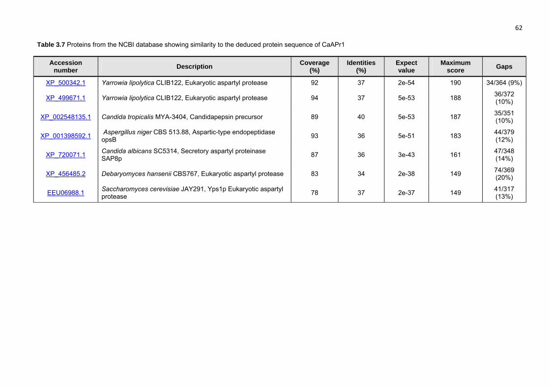
46 Seven of the proteins in the NCBI database that share similarities with the putative protein of
MpAPr1 are listed in Table 3.6. The best score was with the aspartyl protease of Clavispora
lusitaniae ATCC 42720, the same as with the nucleotide alignment. Unlike with the nucleotide
sequences, all the matches are to fungal proteins, particularly of the Candida genus with much
higher coverage and identity scores.
Similar results were obtained for the gene sequence of CaAPr1. Very low coverage scores
(between 2% and 7%) with relatively high identity scores (80% to 90%) were observed for the
region corresponding to the active site encoding gene sequences. The best score was given
with the aspartyl proteinase (PAPA) gene of the soil fungus Trichoderma asperellum
(AY611632.1). Although most of the matches were of fungal species, others included aspartic
peptidases and unidentified proteins from a variety of species such as Ajellomyces dermatitidis
mRNA prepropenicillopepsin XM_002625219.1, the house mouse Mus musculus (JN950245.1)
and the fish Dicentrarchus labrax (AM943112.1). Again, it is clear that the active site encoding
regions are highly conserved throughout different species. Table 3.7 shows seven of the
proteins that share similarities with the putative protein of CaAPr1. All the proteins are from
fungal species.
A phylogenetic tree of the two putative proteins with the proteins listed in Tables 3.6 and 3.7 is
shown in Figure 3.9. It is clear that the CaAPr1 protein is more closely related to the aspartic
proteases of Y. lipolytica than the MpAPr1 protein is. The MpAPr1 protein on the other hand, is
closely related to the Candida spp.
The role of M. pulcherrima strains has been investigated in fermentation studies (Jolly et al.,
2003 and Rodrίguez et al., 2010) and is known to secrete a number of enzymes (Charoenchai
et al., 1997; Strauss et al., 2001 and Jolly et al., 2006), and therefore holds great
biotechnological potential, especially for the wine industry. However, the proteolytic enzyme(s)
of M. pulcherrima has not been studied at a genetic level. It was subsequently decided to
continue studies only with the putative MpAPr1 gene.
3.3.5 Heterologous expression of the protease encoding gene of M. pulcherrima IWBT
Y1123 in S. cerevisiae YHUM272
The MpAPr1 putative gene sequence of M. pulcherrima IWBT Y1123 was cloned into the
shuttle vector pCEL13 for expression in the laboratory strain S. cerevisiae YHUM272. The yeast
was also transformed with the vector not containing the gene. Successful transformation was
confirmed by PCR with primers 5′-KPNPGK-631 and Mpulch_ IPCR_ R2 (nested). Colony PCR
was performed on the yeast transformed with MpAPr1, the yeast transformed with the empty
vector as well as with the untransformed yeast. The presence of the putative gene fragment
Stellenbosch University http://scholar.sun.ac.za
47 was detected only in the yeast transformed with MpAPr1 and not in the other strains (Figure
3.10A).
The extracellular acid protease activity of the transformed strains was investigated by plate
assays. Only the recombinant strain and the IWBT Y1123 strain showed extracellular protease
activity as indicated by the zone of clearance around the colonies (Figure 3.10B). No activity
was observed from the strain that contained the empty plasmid or from the untransformed
yeast. The results confirmed that the MpAPr1 gene from M. pulcherrima IWBT Y1123 indeed
encodes an extracellular acid protease enzyme.
3.3.6 Induction and substrate specificity investigation
The induction and substrate specificity of the MpAPr1 gene of M. pulcherrima IWBT Y1123
were studied upon exposure to different nitrogen sources by simultaneously investigating gene
expression and the presence of the protease in the extracellular medium. After pre-culturing
M. pulcherrima IWBT Y1123, cells were transferred to minimal medium and incubated for 1 day,
after which cultures were spiked with different nitrogen sources and incubated for 2 more days,
as explained in Material and Methods. The different nitrogen sources included ammonium
sulphate, BSA, casein and grape juice proteins. As a control, a fifth culture received no nitrogen
source addition for the final two-day incubation. The experiments were performed in triplicate.
The cells were harvested by centrifugation and the culture supernatants were concentrated by
ultra-filtration and used as the crude protease preparations. Total RNA was extracted from the
harvested cells. Reverse transcription was performed on mRNA and cDNA was used as
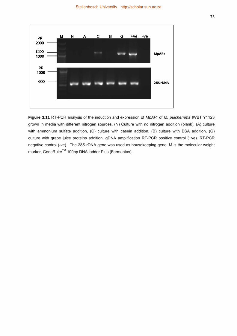
template for PCR. The 28S rDNA gene was used as constitutively expressed (housekeeping)
gene and was present in all the samples. The amplification showed a similar expression level of
the housekeeping gene in all the samples (Figure 3.11). Thus the amount of MpAPr1 transcript
could be compared between the cultures containing different nitrogen sources. The results
indicated that expression was induced in the presence of casein and grape juice proteins and
only slightly in the presence of BSA. No amplicon, and thus no expression, was observed either
in the presence of ammonium sulphate or in the absence of any nitrogen source.
In order to assess the presence of the protease and its activity in the extracellular medium, the
concentrated culture supernatants representing total extracellular proteins were analysed by
SDS-PAGE. The protease activity was visualized by zymography at pH 3.5 using gelatine as
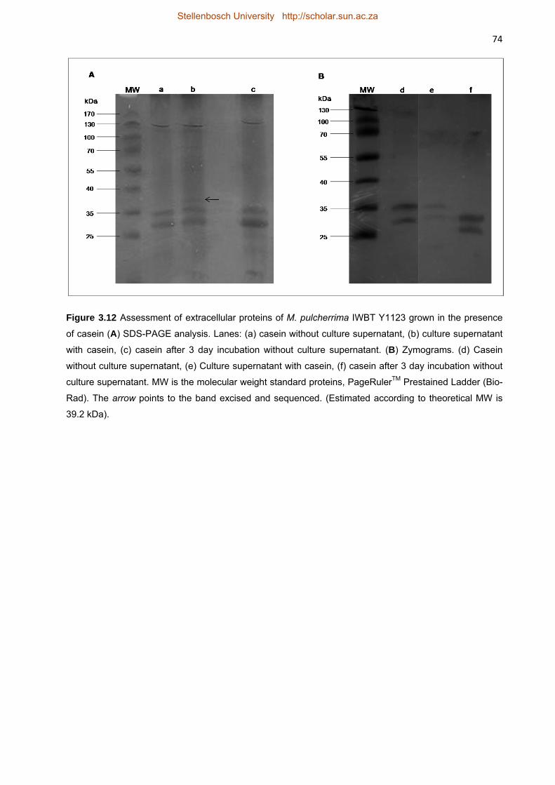
protease substrate. Figure 3.12 shows the extracellular proteins of M. pulcherrima IWBT Y1123
grown in the presence of casein. Lane (a) shows the profile of a commercially available casein
preparation alone before incubation with M. pulcherrima IWBT Y1123. The bands between
35 kDa and 25 kDa correspond to the molecular weights of two of the four subunits of casein.
Lane (b) shows the total extracellular proteins of the culture supernatant after incubation with
casein. The casein bands were still clearly visible but another band between 35 kDa and 40 kDa
(at approximately 39 kDa) was also visible. This band was excised from the gel and sequenced.
Stellenbosch University http://scholar.sun.ac.za
48 After 3 days of incubation in the medium without the presence of the yeast, no degradation was
observed of the casein (lane c). No protease activity was observed in the zymogram.
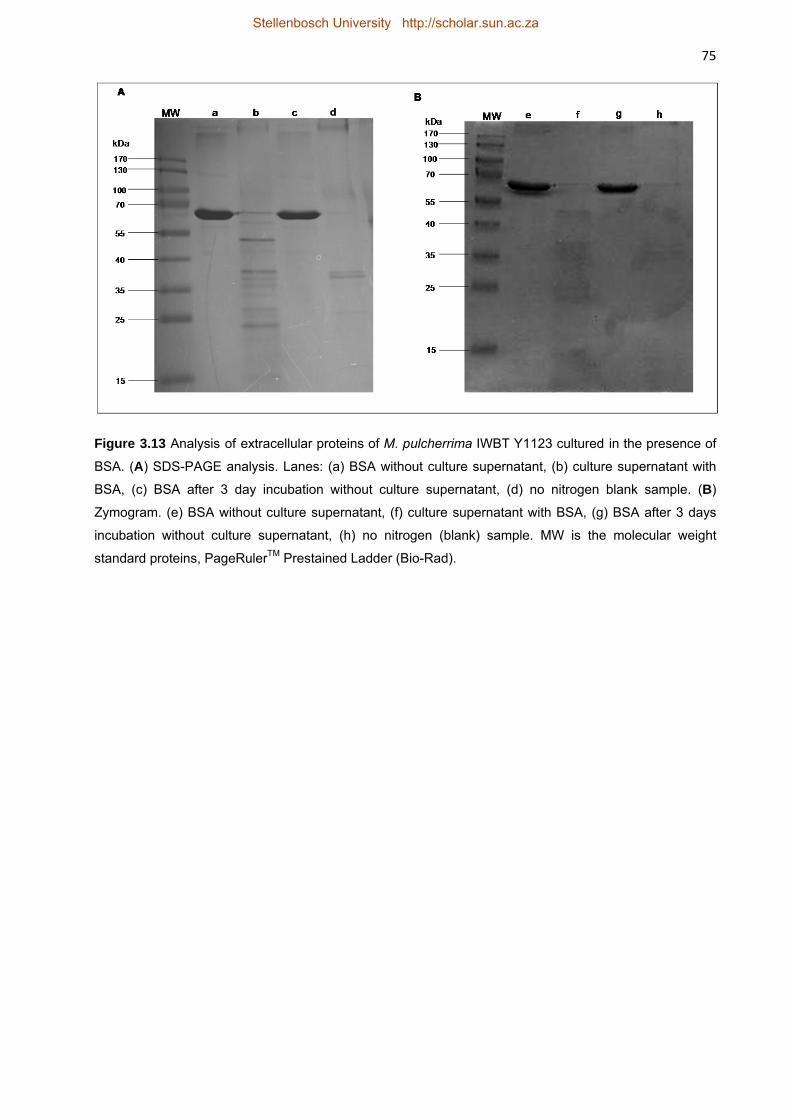
The analysis of the extracellular proteins of M. pulcherrima IWBT Y1123 after incubation with
BSA as nitrogen source is shown in Figure 3.13. The first blank sample, lane (a), shows the
profile of BSA alone before incubation with M. pulcherrima IWBT Y1123. The band at
approximately 68 kDa corresponds to the molecular weight of BSA. The profile in lane (b)
illustrates the proteins in the culture supernatant after incubation with M. pulcherrima IWBT
Y1123. An evident degradation of BSA is observed by a decrease in intensity of the band at
68 kDa. A number of low molecular weight bands are present which may be a combination of
hydrolysis artefacts of BSA and proteins secreted by the yeast. Lane (c) shows the profile of
BSA after 3 days incubation without the yeast. No degradation of BSA was observed indicating
that the protein is stable under the incubation conditions and hydrolysis must therefore be as a
result of proteolysis. No proteolytic activity could be observed by zymography either in the BSA
or the blank sample.
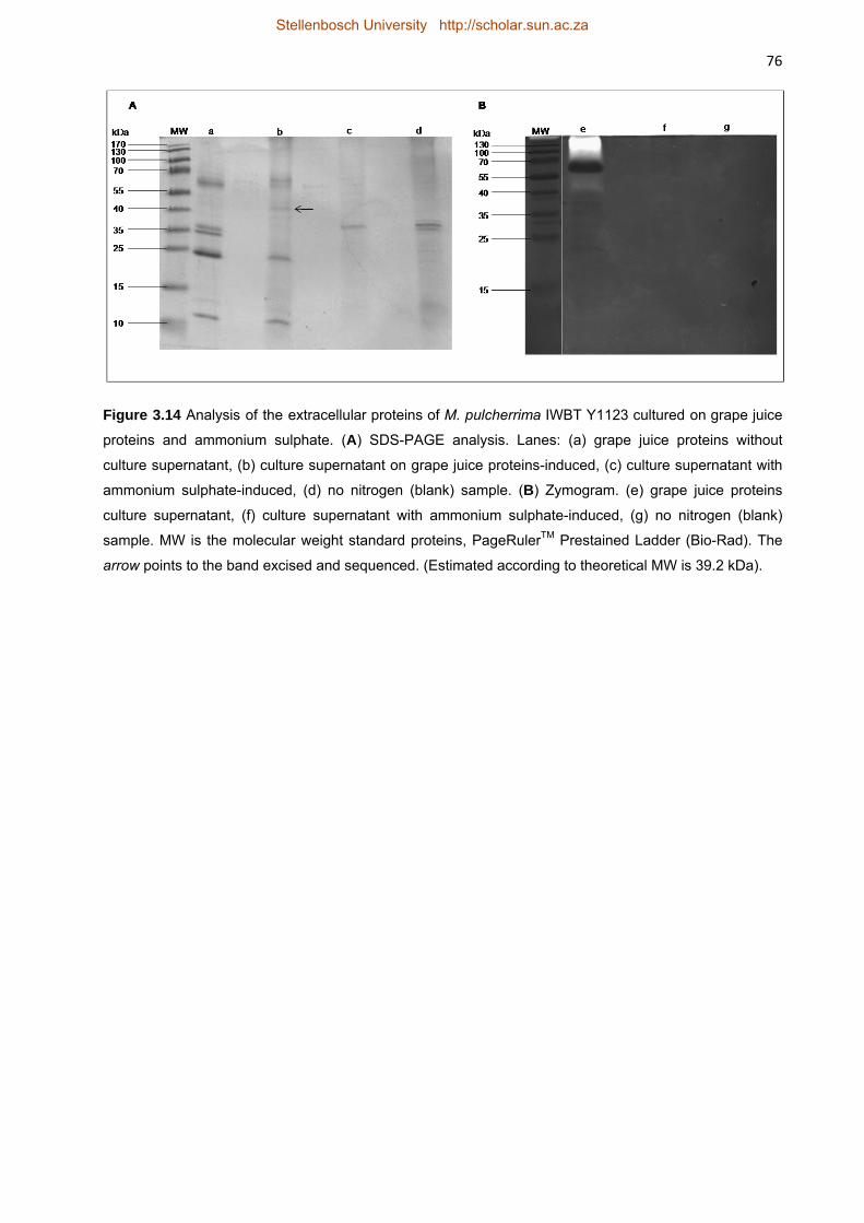
The analysis of the extracellular proteins of M. pulcherrima IWBT Y1123 grown on grape juice
proteins is illustrated in Figure 3.14. Lane (a) shows the profile of the grape proteins after
extraction from grape juice by acetone precipitation. The band at ~60 kDa may be grape
invertase (Marangon et al., 2009 and Le Bourse et al., 2011) and the intense band just beneath
25 kDa may be the common grape proteins, thaumatin-like (22 kDa) and/or chitinase (25 kDa)
(Waters et al., 1992 and Waters et al., 1996). These proteins are known to be very stable due to
their conformation thus degradation due to incubation conditions was not expected (Waters et
al., 2005). Lane (b) shows the extracellular protein profile of M. pulcherrima IWBT Y1123 grown
in the presence of grape juice proteins. The band at ~35 kDa disappeared and those at 10 kDa,
25 kDa and 60 kDa were fainter. New faint bands could be seen between 25 kDa and 40 kDa.
The arrow points to the band that was excised from the gel and sequenced. In lane (c) very faint
bands could be seen and a slightly more intense band was visible for the culture grown on
ammonium sulphate. Faint bands were also seen for the blank sample where no nitrogen
source was added. These could be proteins secreted by the yeast or released from early
autolysis due to starvation of the yeast. Activity could be visualized for the culture supernatant
grown with grape juice proteins but not with ammonium sulphate or the blank sample. This
indicated the presence of the protease. A clear zone was visible at the top of the lane and
another clear zone was observed at just above 40 kDa making it hard to estimate the molecular
weight of the protease from the zymogram activity.
In order to identify whether the protease was indeed present in the culture supernatants, protein
bands corresponding to the expected size as predicted by prediction software were manually
excised from lanes in SDS-PAGE gels from casein-induced and grape protein-induced protein
profiles. The bands were trypsin digested and Nano-LC-MS/MS analysis was performed. The
Stellenbosch University http://scholar.sun.ac.za
49 obtained peptide sequences were processed against the MASCOT database and the deduced
amino acid sequence of the MpAPr1 gene. The protein bands were positively identified to the
deduced protein sequence both from casein-induced and grape protein-induced cut-outs. For
the band from casein-induced culture identification of the protein resulted in 21.27% sequence
coverage of the protein with 6 peptides identified by the MASCOT search engine, and for the
band from the grape protein-induced, culture identification resulted in 19.9% sequence
coverage with 5 peptides identified.
A positive correlation was therefore found between the expression of the gene at transcription
level and protein production by the presence of the protease in the extracellular matrix of the
yeast grown in casein and grape juice proteins. However, activity could not be visualized in the
zymogram with casein. This could be because the concentration of the protease in the culture
supernatant was too low.
3.3.7 Genetic screening of 12 M. pulcherrima strains for the presence of MpAPr1
The identity of twelve M. pulcherrima strains previously isolated from grape juice of different
harvest seasons was confirmed by amplifying and sequencing the ITS-5.8S rDNA locus and
performing BLAST searches as well as by performing RFLP analysis on the amplicons. RFLP
analysis was performed with two restriction endonucleases, CfoI and HaeIII. The results in
Figure 3.15 confirm that the strains were all M. pulcherrima.
In order to evaluate the presence of MpAPr1, PCR was performed on the gDNA of the twelve
strains with the MpAPr1 amplifying primers (MpAPr1-F/MpAPr1-R). The gene appears to be
present in all the strains as seen in Figure 3.16. Extracellular acid protease activity of the twelve
M. pulcherrima strains was assessed using a plate assay. Yeasts, grown to stationary growth
phase in YPD broth, were spotted on skim milk plates at pH 3.5. Enzymatic activity was
visualised by a clear halo of at least 1 mm around the edges of the yeast colony. Assays were
performed in triplicate. Activity could be visualized in all the strains; however strain IWBT Y1123
had a greater activity than the rest of the strains indicated by a wider halo (Figure 3.16). VIN13,
used as negative control, displayed no activity at all.
The results showed a positive correlation between the presence of the gene, confirmed by PCR,
and protease activity screening on plates. However, activity was not equal amongst the strains
as illustrated by plate assays. The PCR fragments of seven of the putative genes were
sequenced and aligned with the sequence of IWBT Y1123 (data not shown). Only the sequence
of one strain, IWBT Y1065 showed distinguishable polymorphism with 1.2% nucleotide
differences compared to the other sequences. However, the deduced protein products revealed
only one amino acid difference, Val11 → Ile.
Stellenbosch University http://scholar.sun.ac.za
50 3.4 DISCUSSION
In this study, the putative gene and amino acid sequences of two aspartic proteases were
retrieved and characterized by in silico analysis. The first putative gene was from
M. pulcherrima IWBT Y1123 and the other from C. apicola IWBT Y1384. The putative proteins
were predicted to have molecular weights of 40.8 kDa and 39.1 kDa, respectively. The
molecular weight of the protein from strain IWBT Y1123 without the signal peptide (mature
protease) was predicted to be 39.2 kDa. These values are in line with what has been published
previously with the sizes of aspartic proteases ranging from 35 kDa to 50 kDa (Davies, 1990;
Rao et al., 1998). The putative genes were named MpAPr1 isolated from IWBT Y1123 and
CaAPr1 isolated from IWBT Y1384. The predicted pI values of the two putative proteins were
4.22 and 4.33 which are also in line with what has been reported in literature (Rao et al., 1998;
Tyndall et al., 2005).
By performing homology searches, it was found that both gene sequences shared homology
with genes from various species, including fish, insects and humans. The coverage scores,
although low, revealed high identity with the database sequences. These conserved regions
correspond to the active site-encoding regions on the putative protein sequences. It appears
that the active site-encoding regions, especially that which is located on the N-terminal end are
highly conserved throughout different species and indicates how the enzymes may have
evolved throughout different species but have most probably retained their function. This is
expected seeing that aspartic acid proteases are found in almost all living organisms: viruses,
bacteria, plants, mammals etc. (Davies 1990).
No signal peptide could be detected in the CaAPr1 protein and it was predicted that the putative
protein is also not a glycosylphosphatidylinositol(GPI)-anchored protein (data not shown).
Although a signal peptide was detected for the DNA deduced Axp aspartic protease of
Y. lipolytica 148, the prepro-region of the protein shared no homology with other extracellular
proteins and the secretory motif of the protein was distinct from the common motif for yeast
extracellular protease processing (Young et al.; 1996). McEwen and Young (1998) could not
confirm whether the Axp precursor contains the signal peptide but it was confirmed that Axp
translocation occurs co-translationally. Thus the signal peptide could have been cleaved co-
translationally. Nonetheless the translocation process of Axp is still unknown. Because of the
close homology of the CaAPr1 putative protein to the aspartic protease of Y. lipolytica, it may be
suggested that the two proteins could follow a similar secretion and maturation pathway which
is yet to be elucidated (Beckerich et al., 1998), or because of the lack of an identifiable signal
peptide, the putative CaAPr1 protein may present a novel translocation process.
Stellenbosch University http://scholar.sun.ac.za
51 The putative gene MpAPr1 was ligated into pCEL13, carrying the constitutive PGK1 promoter,
and expressed in S. cerevisiae YHUM272. Activity assays were performed on plates
supplemented with skim milk. A zone of clearance confirmed positive activity. The activity
assays confirmed that the MpAPr1 gene encodes an extracellular aspartic protease. The zone
of clearance was much wider in the IWBT Y1123 strain (native host) compared to the
transformed YHUM272 strain, indicating weaker activity in the recombinant strain. This could be
due to a number of factors: the differences in metabolic machinery of S. cerevisiae YHUM272
and M. pulcherrima IWBT Y1123. S. cerevisiae YHUM272 may not be systematically
recognizing the (unfamiliar) secretion signal peptide of the protein, or the signal peptide is
cleaved improperly. Expression may be lower under the PGK1 promoter of the plasmid. PGK1
gene expression is activated when yeast cells are grown on glucose, while PGK1 mRNA levels
are low when growth is on lactate (Chambers et al., 19898; Moore et al., 1991). The skim milk
media contained lactate and had a glucose concentration of 0.8%. Due to the presence of
lactate from the skim milk powder and the low glucose concentration, decreased activation of
the PGK1 promoter is possible. In 1987, Mellor et al. suggested that the absence of a
downstream activating sequence (DAS) in expression vectors can explain in part the low yield
of foreign proteins expressed under the control of the PGK1 promoter when compared to the
endogenous levels of 3-phosphoglycerate kinase (PGK). Further investigation is needed to
elucidate the decreased expression in the recombinant strain. Figure 3.10 shows that the
colonies of the recombinant S. cerevisiae YHUM272 carrying the MpAPr1 gene and that of
IWBT Y1123 was slightly bigger than the colonies of the strains not carrying the gene. In order
to assess the role of MpAPr1 expression on nitrogen source utilization, transformed and
untransformed YHUM272 strains were spotted on skim milk media not containing the essential
amino acids tryptophan, leucine and histidine. The cells not carrying the MpAPr1 gene did not
grow at all while the MpAPr1-transformed strain grew slightly and displayed minimal protease
activity (results not shown). This indicates the important role extracellular proteolytic activity can
play in the survival of yeasts under poor carbon and/or preferred nitrogen conditions.
M. pulcherrima IWBT Y1123 was grown in media containing different nitrogen sources (casein,
BSA, grape juice proteins and ammonium sulphate) in order to evaluate induced expression of
the MpAPr1 gene. RT-PCR established that the gene is strongly expressed in the presence of
casein and grape juice proteins, and only slightly in the presence of BSA. The strongest
expression of the gene appeared to be in the presence of grape proteins. Quantitative real-time
PCR could be performed in future in order to quantify MpAPr1 expression under different growth
conditions, particularly different nitrogen sources. The presence and activity of the MpAPr1
protease was assessed by performing SDS-PAGE analysis and zymography on cell-free
concentrated supernatants. Proteolysis was observed in the SDS-PAGE analysis of the BSA
induced culture and activity was observed in the grape protein-induced culture by zymography.
Sequencing of the protein bands corresponding to the expected size of the mature enzyme from
Stellenbosch University http://scholar.sun.ac.za
52 casein-induced and grape protein-induced cultures further confirmed the presence of the
MpAPr1 protein in the extracellular media. According to literature, protease secretion in yeasts
is induced by the presence of proteins as sole nitrogen source in the extracellular medium
(Dabas and Morschhäuser, 2008). It was not a surprise that the production and secretion of the
acid protease is induced by grape proteins seeing that these are present in the (natural)
environment from which the yeast was isolated, and may be a familiar source of nitrogen for the
yeast. Casein is a commonly used inducer for proteases from yeasts and fungi (Banerjee et al.,
1991; Gotoh et al., 1994), and BSA has also been used in a number of studies to induce the
production and secretion of proteases (Lagace and Bisson, 1990; Togni et al., 1996). Proteins
are considered alternative or secondary nitrogen sources for yeasts whereas amino acids,
ammonium, glutamine and urea are preferred nitrogen sources (Banerjee et al., 1991).
Therefore, the presence of the protease was expected in the cultures containing BSA, casein
and grape juice proteins and not with ammonium sulphate. The genes encoding proteases are
repressed when high concentrations of the preferred nitrogen sources are available (Dabas and
Morschhäuser, 2008). The GATA transcription factors, which control the use of alternative
nitrogen sources, have been studied extensively in S. cerevisiae and C. albicans (Marzluf et al.,
1997; Morschhäuser, 2011). Investigating the presence and role of these transcription factors in
M. pulcherrima would broaden our understanding of the expression of MpAPr1.
It appears that the MpAPr1 protease gene has no introns seeing that the predicted size of the
mature enzyme correspond to the size (~39 kDa) of the protein band excised from the SDS-
PAGE gel. This is also an indication of the low level of N-glycosylation of the protein hence the
ease of migration through the gel. Only one potential N-glycosylation site was predicted for the
deduced protein sequence.
In 1994, Gotoh and co-workers reported on the purification of an acid protease from
C. pulcherrima KSY 188-5 (teleomorph Metschnikowia) with a molecular weight of 36.5 kDa
estimated by SDS-PAGE and gel filtration, and a pI of 4.7 determined by isoelectric focusing,
which is similar to what has been found in this study for the protein of M. pulcherrima IWBT
Y1123, 39.2 kDa and pI 4.22, respectively. The enzyme also had a wide substrate specificity
hydrolysing casein, BSA, haemoglobin and collagen. Further characterization of MpAPr1 is
needed to confirm whether it is identical to the enzyme described by Gotoh et al. (1994).
Sequencing the gene encoding the protease from C. pulcherrima KSY 188-5 would also enable
one to better compare the two proteases. An acid protease gene of M. reukaufii W6b has also
been characterized (Li et al., 2009). This gene is 1527 bp long without any introns and encodes
a 508 amino acid long protein with an estimated molecular weight of 53.5 kDa and a predicted
pI of 4.2. The molecular weight of this protein is somewhat higher than the usual molecular
weight of yeast aspartic proteases, but had the same pI of the MpAPr1 protease.
Stellenbosch University http://scholar.sun.ac.za
53 The presence of MpAPr1 and extracellular protease activity were confirmed in 12
M. pulcherrima strains isolated from grape juice. As stated previously, activity was not equal
amongst the strains as illustrated by plate assays. An alignment of seven of the putative
protease encoding genes with that of IWBT Y1123 revealed that the sequence from strain IWBT
Y1065 had 1.2% nucleotide differences compared to the other sequences. The deduced amino
acid sequence of the putative gene had only one difference, Val11 → Ile, compared with the
other sequences. These two amino acids, valine and isoleucine, are both aliphatic and non-
polar and a substitution between the two is unlikely to affect protein activity. No correlation was
thus found between protein sequence and the degree of activity displayed on plates.
Sequencing and examining the nucleotide regions upstream and downstream of the gene-
encoding sequence may explain the varying degrees of activity.
Future work should include purification of the MpAPr1 enzyme and determination of its
biochemical properties such as optimum pH and temperature of the enzyme activity. Another
important aspect is to test the activity of the acid protease in wine, and whether it is able to
hydrolyse wine proteins and reduce protein haze formation under oenological conditions, as
potential inhibitors such as phenolic compounds and ethanol in wine may affect the activity of
the protease. This study showed that MpAPr1 can digest some grape juice proteins and thus
has potential to be tested for haze reduction abilities, its impact on aroma profile and whether
the protease activity will lead to a non-negligible increase in available assimilable nitrogen
content to be used by S. cerevisiae and lactic acid bacteria during alcoholic and malolactic
fermentations. An increase in assimilable nitrogen content will certainly have an impact on the
fermentation kinetics. Pollock et al. (2003) suggested heat treatment of the wine together with
protease treatment could effectively reduce white wine haze. Other potential biotechnological
applications of the enzyme include cheese manufacturing (Rao et al., 1998) and beer haze chill-
proofing (Ormrod et al., 1991).
Our work has provided insight into how some non-Saccharomyces yeasts may survive in wine
by extracellular proteolytic activity (Bossi et al., 2006). To our knowledge this the first report on
the extracellular aspartic protease encoding genes of M. pulcherrima and C. apicola.
3.5 Acknowledgements
The authors would like to thank Winetech, the THRIP funding programme of the National
Research Foundation, Stellenbosch University (Sub-Committee B) and the Harry Crossley
Foundation for financial support.
3.6 References
Albrecht, A., Felk, A., Pichova, I., Naglik, J.R., Schaller, M., de Groot, P., MacCallum, D., Odds, F.C., Schäfer, W., Klis, F., Monod, M., Hube, B., 2006. Glycosylphosphatidylinositol-anchored proteases of
Stellenbosch University http://scholar.sun.ac.za
54 Candida albicans target proteins necessary for both cellular processes and host-pathogen interactions. Journal of Biological Chemistry 281, 688-694. Banerjee, A., Ganesan, K., Datta, A., 1991. Induction of secretory acid proteinase in Candida albicans. Journal of General Microbiology 137, 2455-2461. Barrett, A.J., Rawlings, N.D., O'Brien, E.A., 2001. The MEROPS Database as a Protease Information System. Journal of Structural Biology 134, 95-102. Beckerich, J-M., Boisramé-Baudevin, A., Gaillardin, C., 1998. Yarrowia lipolytica: a model organism for protein secretion studies. International Microbiology 1, 123-130. Bossi, A., Bonizzato, L., Zapparoli, G., 2006. Acidic extracellular proteases from microrganisms of fairly acidic niche. Protein & Peptide Letters 13, 737-741. Charoenchai, C., Fleet, G.H., Henschke, P.A., Todd, B.E.N.T., 1997. Screening of non-Saccharomyces wine yeasts for the presence of extracellular hydrolytic enzymes. Australian Journal of Grape and Wine Research 3, 2-8. Chambers, A., Tsang, J.S.H., Stanway, C., Kingsman, A.J., Kingsman, S.M., 1989. Transcriptional control of the Saccharomyces cerevisiae PGK gene by RAPI. Molecular and Cellular Biology 9, 5516-5524. Ciani, M., Maccarelli, F., 1998. Oenological properties of non-Saccharomyces yeasts associated with wine-making. World Journal of Microbiology & Biotechnology 14, 199-203. Ciani, M., Comitini, F., 2011. Non-Saccharomyces wine yeasts have a promising role in biotechnological approaches to winemaking. Annals of Microbiology 61, 25-32. Collart, M.A., Oliviero, S., 1993. Preparation of yeast RNA. In: Ausubel, F.M., Brent, R., Kingston, R.E., Moore, D.D., Seidman J.G., Smith., J.A., Struhl, K. (Eds), Current protocols in molecular biology 5. John Wiley, New York, unit 13.12. Dabas, N., Morschhäuser, J., 2008. A transcription factor regulatory cascade controls secreted aspartic protease expression in Candida albicans. Molecular Microbiology 69, 586-602. Davies, D.R., 1990. The structure and function of aspartic proteases. Annual Reviews of Biophysics and Biophysical Chemistry 19, 189-215. Dujon, B., Sherman, D., Fischer, G., Durrens, P., Casaregola, S., Lafontaine, I., De Montigny, J., Marck, C., Neuveglise, C., Talla, E., Goffard, N., Frangeul, L., Aigle, M., Anthouard, V., Babour, A., Barbe, V., Barnay, S., Blanchin, S., Beckerich, J., Beyne, E., Bleykasten, C., Boisrame, A., Boyer, J., Cattolico, L., Confanioleri, F., De Daruvar, A., Despons, L., Fabre, E., Fairhead, C., Ferry-Dumazet, H., Groppi, A., Hantraye, F., Hennequin, C., Jauniaux, N., Joyet, P., Kachouri, R., Kerrest, A., Koszul, R., Lemaire, M., Lesur, I., Ma, L., Muller, H., Nicaud, J., Nikolski, M., Oztas, S., Ozier-Kalogeropoulos, O., Pellenz, S., Potier, S., Richard, G., Straub, M., Suleau, A., Swennen, D., Tekaia, F., Wesolowski-Louvel, M., Westhof, E., Wirth, B., Zeniou-Meyer, M., Zivanovic, I., Bolotin-Fukuhara, M., Thierry, A., Bouchier, C., Caudron, B., Scarpelli, C., Gaillardin, C., Weissenbach, J., Wincker, P., Souciet, J., 2004. Genome evolution in yeasts. Nature 430, 35-44. Dunn, B.M., 2002. Structure and mechanism of the pepsin-like family of aspartic peptidases. Chemical Reviews 102, 4431-4458. Feuillat, M., Charpentier, C., Maujean, A., 1998. Les composés azotés. In: Flanzy, C. (ed.),Oenologie: fondements scientifiques et techniques. Lavoisier, Paris, pp. 94–116. Folio, P., Ritt, J., Alexandre, H., Remize, F., 2008. Characterization of EprA, a major extracellular protein of Oenococcus oeni with protease activity. International journal of food microbiology 127, 26-31. Fleet, G.H., 2003. Yeast interactions and wine flavour. International Journal of Food Microbiology 86, 11-22. Gotoh, T., Kikuchi, K-I., Kodama, K., Konno, H., Kakuta, T., Koizumi, T., Nojiro, K., 1995. Purification and properties of extracellular carboxyl proteinase secreted by Candida pulcherrima. Bioscience, Biotechnology, and Biochemistry 59, 367-371.
Stellenbosch University http://scholar.sun.ac.za
55 Gundllapalli, S., Cordero Otero, R., Pretorius, I.S., 2006. Development of a screening method for the identification of a novel Saccharomyces cerevisiae mutant over-expressing Trichoderma reesei cellobiohydrolase II. Annals of Microbiology 56, 143–150. Hoffman, C.S., Winston, F., 1987. A ten-minute DNA preparation from yeast efficiently releases autonomous plasmids for transformation of Escherichia coli. Gene 57, 267-272. Jolly, N.P., Augustyn, O.P.H., Pretorius, I.S., 2003. The use of Candida pulcherrima in combination with Saccharomyces cerevisiae for the production of Chenin blanc wine. South African Journal of Enology and Viticulture 24, 63-69. Jolly, N.P., Augustyn, O.P.H., Pretorius, I.S., 2006. The role and use of non-Saccharomyces yeasts in wine production. South African Journal of Enology and Viticulture, 27, 15-39. Koszul, R., Malpertuy, A., Frangeul, L., Bouchier, C., Wincker, P., Thierry, A., Duthoy, S., Ferris, S., Hennequin, C., Dujon, B., 2003. The complete mitochondrial genome sequence of the pathogenic yeast Candida (Torulopsis) glabrata. FEBS letters 534, 39-48. Kurtzman, C.P., Robnett, C.J., 1998. Identification and phylogeny of ascomycetous yeasts from analysis of nuclear large subunit (26S) ribosomal DNA partial sequences. Antonie van Leeuwenhoek 73, 331–371. Laemmli, U.K., 1970. Cleavage of structural proteins during the assembly of the head of Bacteriophage T4. Nature, 277, 680-685. Lagace, L.S., Bisson, L.F., 1990. Survey of yeast acid proteases for effectiveness of wine haze reduction. American Journal of Enology and Viticulture 41, 147-155. Lambrechts, M.G., Pretorius I.S., 2000. Yeast and its importance to wine aroma. South African Journal of Enology and Viticulture 21, 97–129. Le Bourse, D., Conreux, A., Villaume, S., Lameiras, P., Nuzillard, J-M., Jeandet, P., 2011. Quantification of chitinase and thaumatin-like proteins in grape juices and wines. Analytical and Bioanalytical Chemistry, 401, 1541-1549. Li, J., Chi, Z., Liu, Z., Yue, L., Peng, Y., Wang, L.,2009. Cloning and characterization of a novel aspartic protease gene from marine-derived Metschnikowia reukaufii and its expression in E. coli. Applied Biochemistry and Biotechnology 159, 119-132. Lott, T.J., Burns, B.M., Zancope-Oliveira, R., Elie, C.M., Errol Reiss, E., 1998. Sequence analysis of the internal transcribed spacer 2 (ITS2) from yeast species within the genus Candida. Current Microbiology 36, 63–69. Marangon, M., Van Sluyter, S.C., Haynes, P.A., Waters, E.J., 2009. Grape and wine proteins: their fractionation by hydrophobic interaction chromatography and identification by chromatographic and proteomic analysis. Journal of Agricultural and Food Chemistry, 57, 4415–4425. Marzluf, G.A., 1997. Genetic regulation of nitrogen metabolism in the fungi. Microbiology and Molecular Biology Reviews 61, 17-32. McEwen, R.K., Young, T.W., 1998. Secretion and pH-dependent self-processing of the pro-form of the Yarrowia lipolytica acid extracellular protease. Yeast, 14, 1115-1125. Mendoza, L., Farías, M.E., 2010. Improvement of wine organoleptic characteristics by non-Saccharomyces yeasts. Current Research, Technology and Education Topics in Applied Microbiology and Microbial Biotechnology 2, 908-919. Moore, P.A., Sagliocco, F.A., Wood, R.M.C., Brown, A.J.P., 1991. Yeast glycolytic mRNAs are differentially regulated. Molecular and Cellular Biology 11, 5330-5337. Morschhäuser, J., 2011. Nitrogen regulation of morphogenesis and protease secretion in Candida albicans. International Journal of Medical Microbiology 301, 390-394. Naglik, J., Albrecht, A., Bader, O., Hube, B., 2004. Candida albicans proteinases and host/pathogen interactions. Cellular microbiology 6, 915-926.
Stellenbosch University http://scholar.sun.ac.za
56 Newport, G., Agabian, N., 1997. KEX2 influences Candida albicans proteinase secretion and hyphal formation. Journal of Biological Chemistry 272, 28954-28961. Ogrydziak, D.M., 1993. Yeast Extracellular Proteases. Critical Reviews in Biotechnology 13, 1-55. Ormrod, I. H. L., Lalor, E. F., Sharpe, F. R., 1991.The release of yeast proteolytic enzymes into beer. Journal of Institute of Brewing 97, 441-443. Pocock, K.F., Høj, P.B., Adams, K.S., Kwiatkowski, M.J., Waters, E.J., 2003. Combined heat and proteolytic enzyme treatment of white wines reduces haze forming protein content without detrimental effect. Australian Journal of Grape and Wine Research 9, 56-63. Rao, M.B., Tanksale, A.M., Ghatge, M.S., Deshpande, V.V., 1998. Molecular and biotechnological aspects of microbial proteases. Microbiology and Molecular Biology Reviews 62, 597-635. Rodríguez, M.E., Lopes, C.A., Barbagelata, R.J., Barda, N.B., Caballero, A.C., 2010. Influence of Candida pulcherrima Patagonian strain on alcoholic fermentation behaviour and wine aroma. International Journal of Food Microbiology 138, 19-25. Strauss, M.L.A., Jolly, N.P., Lambrechts, M.G., Van Rensburg, P., 2001. Screening for the production of extracellular hydrolytic enzymes by non-Saccharomyces wine yeasts. Journal of Applied Microbiology 91, 182-190. Togni, G., Sanglard, D., Quadroni, M., Foundling, S.I., Monod, M., 1996. Acid proteinase secreted by Candida tropicalis: Functional analysis of preproregion cleavages in C. tropicalis and Saccharomyces cerevisiae. Microbiology 142, 493-503. Tyndall, J.D.A., Nall, T., Fairlie, D.P., 2005. Proteases universally recognize β-strands in their active sites. Chemical Review 105, 973-1000. Van Dyk, D., Pretorius, I.S., Bauer, F.F., 2005. Mss11p is a central element of the regulatory network that controls FLO11 expression and invasive growth in Saccharomyces cerevisiae. Genetics 169, 91-106. Volschenk, H., Viljoen-Bloom, M., Van Staden, J., Husnik, J., Van Vuuren, H.J.J., 2004. Genetic engineering of an industrial strain of Saccharomyces cerevisiae for L-malic acid degradation via an efficient malo-ethanolic pathway. South African Journal of Enology and Viticulture 25, 63–73. Waters, E.J., Wallace, W., Williams, P.J., 1992. Identification of heat-unstable wine proteins and their resistance to peptidases. Journal of Agricultural and Food Chemistry 40, 1514-1519. Waters, E.J., Shirley, N.J., Williams P.J., 1996. Nuisance proteins of wine are grape pathogenesis-related proteins. Journal of Agricultural and Food Chemistry 44, 3-5. Waters, E.J., Alexander, G., Muhlack, R., Pocock, K.F., Colby, C., O’Neill, B.K., Høj, P.B., Jones, P., 2005. Preventing protein haze in bottled white wine. Australian Journal of Grape and Wine Research 11, 215–225. White, T.J., Bruns, T., Lee, S., Taylor, J., 1990. Amplification and direct sequencing of fungi ribosomal RNA genes for phylogenetics. In: Innis, M.A., Gelfand, D.H., Sninsky, J.J., White, T.J (Eds), PCR protocols. A guide to methods and applications. Academic Press, San Diego, pp 315-322. Younes, B., Cilindre, C., Villaume, S., Parmentier,M., Jeandet, P., Yann Vasserot, Y., 2011. Evidence for an extracellular acid proteolytic activity secreted by living cells of Saccharomyces cerevisiae PlR1. Impact on grape proteins. Journal of Agricultural and Food Chemistry 59, 6239–6246. Young, T.W., Wadeson, A., Glover, D.J., Quincey, R.V., Butlin, M.J., Kamei, E.A., 1996. The extracellular acid protease gene of Yarrowia lipolytica: sequence and pH-regulated transcription. Microbiology 142, 2913-2921.
Figure 3.5 The DNA and amino acid sequences of M. pulcherrima IWBT Y1123 putative secreted
aspartic protease. The putative secretion signal peptide is underlined. The cleavage site is indicated by
(‡). The active site amino acid sequences and active site flap amino acid sequence are shaded in yellow
and turquoise, respectively. The putative N-glycosylation site is boxed.
Stellenbosch University http://scholar.sun.ac.za
68
Figure 3.6 The conserved domains within the putative aspartic protease of M. pulcherrima IWBT Y1123
analysed in this study, indicating that the protein is a secreted aspartic protease-like (SAP-like) enzyme.
Stellenbosch University http://scholar.sun.ac.za
69
Figure 3.7 The DNA and amino acid sequence of C. apicola IWBT Y1384 secreted aspartic protease.
The putative secretion signal peptide is underlined. The cleavage site is indicated by (‡). The active site
amino acid sequences and active site flap amino acid sequence are shaded in yellow and turquoise,
respectively. The putative N-glycosylation sites are boxed.
1 ATGGTACTAGCTAAGAACTATGTTCATTTAGGCATTAAGCGTGCCTTACAAGGTGATCGTACTACCGCTAAC 72 1 M V L A K N Y V H L G I K R A L Q G D R T T A N 24
73 CGTGCACGTTCTAAGCGTGCATCCGTACCTGATCGATTGCAGAACAACATTTATCAGTACATTGCTACGTTA 144 25 R A R S K R A S V P D R L Q N N I Y Q Y I A T L 48
145 TCAATTGGAACACCTGGCCAAGCTACTGATGCTGCACTTGATACTGGCTCCTCAGATCTGTGGGTATTCACA 216 49 S I G T P G Q A T D A A L D T G S S D L W V F T 72
217 AATCAGACAAACTTCAAGGACAGTTTCAACCCTCAGAGTAGTTCTTCGTACAATCTGTTGAACAACGATTTT 288 73 N Q T N F K D S F N P Q S S S S Y N L L N N D F 96
289 TCAATTCAGTATGTGTCCGGGTCAGCCAGTGGAAGCTGGGTGACAGACACGGTAGATTATGGAGATGACAAG 360 97 S I Q Y V S G S A S G S W V T D T V D Y G D D K 120
361 GTCTCGTCGTTTCAGTTCGCAACTGTCAGCTCTCCAACTACTCAGGGCGAAACTGCCGGTGTGTTCGGTATC 432 121 V S S F Q F A T V S S P T T Q G E T A G V F G I 144
433 GGTCAAATTACCCAAGAATCATCTGCCGAGTACGGATCTACCTACCCGAATTTCCCAGTATCTCTTAAGAAT 504 145 G Q I T Q E S S A E Y G S T Y P N F P V S L K N 168
505 GAGTCGAAAATCCAATCTGTTGCTTATTCACTTTATTTAGACTCAATCAGTGCCCAGAATGGAAGCAGTGTG 576 169 E S K I Q S V A Y S L Y L D S I S A Q N G S S V 192
577 ACATTTGGTGCAGTGGATACCGCGAAATATAAGGGTGAGCTTTACAGTGTCCCTTTCACCTCCGATGTGAGT 648 193 T F G A V D T A K Y K G E L Y S V P F T S D V S 216
649 TTCAATGTTGACTTTGAAGTACTTGGTAACAAGTCGAATGGTGTGCTTGACTCTGGTACTTCATTGACCTAT 720 217 F N V D F E V L G N K S N G V L D S G T S L T Y 240
721 TTGGAACAATCCGTTGTGGATCAAATCGCCCAGCAGTACGGAGCAACTTTCGATTCAGAGCAGCAAACTTAC 792 241 L E Q S V V D Q I A Q Q Y G A T F D S E Q Q T Y 264
793 CTCATTCAGAGCAAGAGTGATTTAGCTAGCACTGATCCTCTGGTTTATACCATTGGAGGTGCAAAGATAGAA 864 265 L I Q S K S D L A S T D P L V Y T I G G A K I E 288
865 GTACCTGTCTCGGAACTGTTCATCGAGGACTCTTCTGATGGTACACTGGCACTCTCTATCCTCCCTAGTTCT 936 289 V P V S E L F I E D S S D G T L A L S I L P S S 312
937 ATGGCTCAAGATGTCATTTTACTTGGCGATAGCTTCTTGCGCTCAGCTTACGTTGTCTACAACTTACAAGGC 1008 313 M A Q D V I L L G D S F L R S A Y V V Y N L Q G 336
1009 AAAGTTGCTGGTATTGCACAAGCCAATTGGTCACCAGGCTCTCCCAACTTTGTCCCTATCACCGGAGATACT 1080 337 K V A G I A Q A N W S P G S P N F V P I T G D T 360
1081 ATTCCCGGATCTGTCAACTAA 1101 361 I P G S V N * 366
Stellenbosch University http://scholar.sun.ac.za
70
Figure 3.8 The conserved domains within the putative aspartic protease of C. apicola IWBT Y1384
analysed in this study, indicating that the protein is a secreted aspartic protease-like (SAP-like) enzyme.
Stellenbosch University http://scholar.sun.ac.za
71
Figure 3.9 Phylogenetic tree of the deduced protein sequences of MpAPr1 of M. pulcherrima IWBT Y1123 and CaAPr1 of C. apicola IWBT Y1384 with aspartic
proteases from other yeasts found on the NCBI website. The phylogenetic tree was constructed by using the neighbour joining method (PHYLIP 3.56). The scale bar
represents the number of base substitutions per site.
Stellenbosch University http://scholar.sun.ac.za
72
Figure 3.10 Confirmation of heterologous expression of MpAPr1 in S. cerevisiae YHUM272. (A) PCR
amplification with primers 5′-KPNPGK-631 and Mpulch_ IPCR_ R2. (a) colony PCR of YHUM272
transformed with MpAPr1, (b) amplification of pCEL13 plasmid without the insert, (c) colony PCR of
untransformed YHUM272, (d) amplification of pCEL13 ligated with MpAPr1 (PCR positive control), (e)
PCR negative control. M is the molecular weight marker, GeneRulerTM 100bp DNA ladder Plus
with MpAPr1, (2) YHUM272 transformed with empty vector, (3) untransformed YHUM272, (4)
M. pulcherrima IWBT Y1123 (positive control), (5) S. cerevisiae VIN13 (negative control).
Stellenbosch University http://scholar.sun.ac.za
73
Figure 3.11 RT-PCR analysis of the induction and expression of MpAPr of M. pulcherrima IWBT Y1123
grown in media with different nitrogen sources. (N) Culture with no nitrogen addition (blank), (A) culture
with ammonium sulfate addition, (C) culture with casein addition, (B) culture with BSA addition, (G)
culture with grape juice proteins addition. gDNA amplification RT-PCR positive control (+ve). RT-PCR
negative control (-ve). The 28S rDNA gene was used as housekeeping gene. M is the molecular weight
marker, GeneRulerTM 100bp DNA ladder Plus (Fermentas).
Stellenbosch University http://scholar.sun.ac.za
74
Figure 3.12 Assessment of extracellular proteins of M. pulcherrima IWBT Y1123 grown in the presence
of casein (A) SDS-PAGE analysis. Lanes: (a) casein without culture supernatant, (b) culture supernatant
with casein, (c) casein after 3 day incubation without culture supernatant. (B) Zymograms. (d) Casein
without culture supernatant, (e) Culture supernatant with casein, (f) casein after 3 day incubation without
culture supernatant. MW is the molecular weight standard proteins, PageRulerTM Prestained Ladder (Bio-
Rad). The arrow points to the band excised and sequenced. (Estimated according to theoretical MW is
39.2 kDa).
Stellenbosch University http://scholar.sun.ac.za
75
Figure 3.13 Analysis of extracellular proteins of M. pulcherrima IWBT Y1123 cultured in the presence of
BSA. (A) SDS-PAGE analysis. Lanes: (a) BSA without culture supernatant, (b) culture supernatant with
BSA, (c) BSA after 3 day incubation without culture supernatant, (d) no nitrogen blank sample. (B)
Zymogram. (e) BSA without culture supernatant, (f) culture supernatant with BSA, (g) BSA after 3 days
incubation without culture supernatant, (h) no nitrogen (blank) sample. MW is the molecular weight
standard proteins, PageRulerTM Prestained Ladder (Bio-Rad).
Stellenbosch University http://scholar.sun.ac.za
76
Figure 3.14 Analysis of the extracellular proteins of M. pulcherrima IWBT Y1123 cultured on grape juice
proteins and ammonium sulphate. (A) SDS-PAGE analysis. Lanes: (a) grape juice proteins without
culture supernatant, (b) culture supernatant on grape juice proteins-induced, (c) culture supernatant with
ammonium sulphate-induced, (d) no nitrogen (blank) sample. (B) Zymogram. (e) grape juice proteins
culture supernatant, (f) culture supernatant with ammonium sulphate-induced, (g) no nitrogen (blank)
sample. MW is the molecular weight standard proteins, PageRulerTM Prestained Ladder (Bio-Rad). The
arrow points to the band excised and sequenced. (Estimated according to theoretical MW is 39.2 kDa).
Stellenbosch University http://scholar.sun.ac.za
77
Figure 3.15 Identification of 12 M. pulcherrima strains by ITS and ITS-RFLP. (A) PCR amplification of the
ITS regions of M. pulcherrima strains. M, molecular weight marker, GeneRulerTM 100bp DNA ladder
(Fermentas). (B) Digestion of the ITS amplifications with restriction endonuclease CfoI. (C) Digestion of
the ITS amplifications with restriction endonuclease HaeIII. (+ve) PCR positive control in this case strain
M. pulcherrima IWBT Y1123, (-ve) PCR negative control. M is the molecular weight marker, FastRulerTM
Low Range DNA Ladder (Fermentas).
Stellenbosch University http://scholar.sun.ac.za
78
Figure 3.16 Protease activity screening of M. pulcherrima strains. (A) PCR amplifications with primers
MpAPr1-F/MpAPr1-R showing the presence of the MpAPr1 protease-encoding gene. M is the molecular
weight marker, GeneRulerTM 100bp DNA ladder Plus (Fermentas). (B) An example of a plate assay
showing the different degrees of activity with a positive result indicated by a clear halo around the colony;
(+) low activity, diameter of the halo from the edge of the colony 2mm, (++) medium activity, diameter of
the halo from the edge of the colony 3 mm, (+++) high activity, diameter of the halo from the edge of the
colony 5 mm, (-) no activity. Strain M. pulcherrima IWBT Y1123 was used as positive control (displaying
high activity) and S. cerevisiae VIN13 as negative control (displaying no activity). Strains IWBT Y1072
and IWBT Y1063 have low activity while strains IWBT Y1065 and IWBT Y1094 show medium activity.
Stellenbosch University http://scholar.sun.ac.za
79
Chapter 4
General discussion and conclusions
Stellenbosch University http://scholar.sun.ac.za
80
4. General discussion and conclusions
4.1 Results and general discussion
The enzymes of the aspartic endoprotease family are defined by the presence of two aspartic
acid residues in their catalytic sites that are critical for the functioning of the enzymes. These
enzymes function under acidic conditions. Literature reports that some non-Saccharomyces
yeasts of oenological origin have extracellular proteolytic activity under acidic conditions, e.g.
Candida stellata, Kloeckera apiculata and Candida pulcherrima (Charoenchai et al., 1997).
These poorly characterised enzymes could potentially fulfil a number of roles in wine production
such as the reduction of protein haze in white wines (Lagace and Bisson, 1990; Dizy and
Bisson, 2000). The amino acids and peptides liberated from the action of the proteases could
be used as sources of nitrogen by the fermentative microorganisms. Some amino acids can be
metabolised by yeasts to form aromatic compounds such as higher alcohols (Swiegers and
Pretorius, 2005), thus the proteolytic activity could have an indirect impact on the aroma profile
of the wine. The main aim of the study was to isolate and characterize extracellular aspartic
proteases at a genetic level from non-Saccharomyces yeasts that are of oenological origin.
The genes encoding extracellular aspartic proteases from two non-Saccharomyces yeasts were
isolated and characterized by in silico analysis. The yeasts were Metschnikowia pulcherrima
IWBT Y1123 and Candida apicola IWBT Y1384. These yeasts were isolated from grape must
and showed strong proteolytic activity during an enzymatic screening of 308 other yeasts.
The genes were retrieved by following a PCR approach using degenerate primers and inverse-
PCR. The gene from M. pulcherrima IWBT Y1123 was named MpAPr1. The gene is 1137 bp
long. The mature form of the protein encoded by the gene consists of 362 amino acids with a
predicted Mr of 39.2 kDa and one possible N-glycosylation site. The gene also encodes a
putative 16 amino acid signal peptide. The putative gene from C. apicola IWBT Y1384 was
named CaAPr1, is 1101 bp long and encodes a putative protein with Mr 39.1 kDa. This enzyme
had no predictable signal peptide and is thought to follow a non-classical secretion pathway and
had 3 possible N-glycosylation sites. The predicted pI values of the putative proteins were 4.22
for MpAPr1 and 4.33 for CaAPr1, respectively. These characteristics correspond to previously
published data on the extracellular proteases of yeasts. The AXP gene of Yarrowia lipolytica
148 encodes a mature acid protease of 353 amino acids with a predicted Mr of 39.2 kDa. The
gene also encodes a putative pre-peptide that is 17 amino acids long (Young et al., 1996).
Cryptococcus sp. S-2 possesses a CAP1 gene with an open reading frame of 1254 bp encoding
a protein of 417 amino acids long. The mature protein had an estimated Mr of 34 kDa on SDS-
PAGE (Rao et al., 2011).
Stellenbosch University http://scholar.sun.ac.za
81 From homology searches of the putative gene sequences, it was confirmed that the sequences
encoding the conserved active sites are indeed highly conserved throughout different species
(Davies, 1990; Dunn, 2002). Both putative protein sequences, MpAPr1 and CaAPr1, showed
homology to only fungal aspartic proteases with average identity scores of less than 40%,
indicating the novelty of the putative proteins. The CaAPr1 protein showed close homology to
the aspartic proteases of Y. lipolytica, which appears to follow a translation pathway other than
that described for Candida spp. (McEwen and Young, 1998; Naglik et al., 2004). CaAPr1 and
the acid proteases from Y. lipolytica may have similar processing pathways.
The putative MpAPr1 gene was cloned and expressed in Saccharomyces cerevisiae YHUM272.
Activity plate assays confirmed that MpAPr1 indeed codes for an extracellular aspartic protease.
The recombinant strain demonstrated weaker activity than the native host. A few explanations
may exist for this occurrence: differences in promoters could affect expression, metabolic
differences between the native and the recombinant strain and the signal peptide of the protein
may not be systematically recognized by the S. cerevisiae.
M. pulcherrima IWBT Y1123 was grown in media containing different nitrogen sources which
included casein, BSA, grape juice proteins and ammonium sulphate. Reverse transcription-PCR
(RT-PCR) confirmed that the expression of MpAPr1 in IWBT Y1123 was induced when casein,
grape juice proteins and BSA were used as sole nitrogen sources. The gene was not expressed
in the presence of ammonium sulphate. This result was expected since the secretion of
proteases in yeasts is induced when protein is the only available nitrogen source (Banerjee et
al., 1991; Dabas and Morschhäuser, 2008). Expression of the gene was the highest when grape
proteins were the sole nitrogen sources, as demonstrated by RT-PCR. The accurate profiling of
the MpAPr1 gene expression by qRT-PCR in the presence different nitrogen sources will shed
some light on the effect these nitrogen sources have on the growth of the yeast. Protease
activity in the presence of grape juice proteins was also confirmed by zymography, but not with
casein or BSA. A possible explanation for this could be that the concentration of the protease in
the gel was too low for activity to be detected, which correlates to the lower expression of the
gene in the presence of casein and BSA compared to the expression induced by the grape juice
proteins. Protein bands corresponding to the expected size of the putative protein was excised
from SDS-PAGE gels and analysed by nano-LC-MS/MS. The bands from the casein-induced
culture and the grape protein-induced culture had 21.3% and 20% identity with the putative
MpAPr1 protein sequence.
The presence of MpAPr1 was confirmed in 12 other M. pulcherrima strains by PCR. Plate
assays revealed that the strains did not have equal activity, as indicated by the size of the halo
surrounding the colonies. No correlation could be made between putative protein sequences
and the level of activity displayed on plates. Investigating the sequences upstream and
Stellenbosch University http://scholar.sun.ac.za
82 downstream of the gene-encoding sequences may provide some insight into the differences in
expression.
4.2 Conclusions and future prospects
The aim to isolate and characterize aspartic proteases encoding genes from non-
Saccharomyces yeasts isolated from grape must, as set out in Chapter 1, has been achieved.
The results obtained correspond well to existing data regarding aspartic proteases secreted by
yeasts (Rao et al., 1998). Some of the grape juice proteins have been hydrolysed when cultured
with M. pulcherrima IWBT Y1123. This illustrates the potential applicability of this yeast and /or
its secreted proteases. However, further investigations are needed to test whether the protease
is able to reduce haze formation and also whether the proteolytic activity will impact on the
assimilable nitrogen content and aroma profile of the wine. Some wine proteins are indeed
known to be insensitive to proteolytic attack due to their globular structure (Waters et al., 1992).
It has been suggested that partially ‘opening’ up the protein structure will expose peptide bonds
to be hydrolysed by proteases. One such proposal was suggested by Pocock and co-workers
(2006), who demonstrated that heat treatment together with proteolysis may reduce protein
haze.
This study has extended our understanding of how some yeasts may survive and even flourish
in wine which is characterized by low pH, low temperatures and the presence of a number of
inhibitors such as ethanol and polyphenols. Extracellular proteolytic activity that is adapted to
the acidic environment may aid in nutrient supply for the yeasts.
Future prospects should include purification of the enzymes, MpAPr1 and CaAPr1, e.g. by
anion-exchange chromatography and gel filtration, and biochemical characterization. This
should include determination of the activity range with regard to pH and temperature. The effect
of different potential inhibitors on the activity of the enzymes should also be tested, e.g. ethanol,
sulphur dioxide and polyphenols. The activity of the enzymes should also be tested in wine to
determine its applicability in wine biotechnology.
Mixed fermentations of M. pulcherrima IWBT Y1123 with S. cerevisiae may also be envisaged
to determine its influence on protein stability, wine aroma, and the interaction between the two
yeast species. By releasing proteases able to breakdown proteins present in grape must,
M. pulcherrima may advantageously affect the fermentation kinetics by increasing the available
assimilable nitrogen content for the growth of S. cerevisiae during alcoholic fermentation.
Sequential fermentations of M. pulcherrima species with S. cerevisiae have shown positive
results with regards to an increase in fruity and floral aroma in wine (Jolly et al., 2003;
Rodrίguez et al., 2010). The wines produced from sequential fermentations had higher
concentrations of aromatic compounds such as higher alcohols, esters and terpenols compared
Stellenbosch University http://scholar.sun.ac.za
83 to fermentations where only S. cerevisiae was inoculated in the grape must. Commercial
cultures of combined S. cerevisiae and non-Saccharomyces yeasts are already available on the
market. Such an example is of S. cerevisiae with Torulaspora delbrueckii and Kluyveromyces
thermotolerans from Viniflora® HARMONY.nsac. This combined culture is supposed to produce
a wine with a richer and rounder flavour with enhanced fruity aroma notes (Jolly et al., 2006).
Further investigations are required to determine the potential commercial value of
M. pulcherrima.
4.3 References
Banerjee, A., Ganesan, K., Datta, A., 1991. Induction of secretory acid proteinase in Candida albicans. Journal of General Microbiology 137, 2455-2461. Charoenchai, C., Fleet, G.H., Henschke, P.A., Todd, B.E.N.T., 1997. Screening of non-Saccharomyces wine yeasts for the presence of extracellular hydrolytic enzymes. Australian Journal of Grape and Wine Research 3, 2-8. Dabas, N., Morschhäuser, J., 2008. A transcription factor regulatory cascade controls secreted aspartic protease expression in Candida albicans. Molecular Microbiology 69, 586-602. Davies, D.R., 1990. The structure and function of aspartic proteases. Annual Reviews of Biophysics and Biophysical Chemistry 19, 189-215. Dizy, M. and Bisson, L.F. 2000. Proteolytic activity of yeast stains during grape juice fermentation. American Journal of Enology and Viticulture 51, 155–167. Dunn, B.M., 2002. Structure and mechanism of the pepsin-like family of aspartic peptidases. Chemical Reviews 102, 4431-4458. Jolly, N.P., Augustyn, O.P.H., Pretorius, I.S., 2003. The use of Candida pulcherrima in combination with Saccharomyces cerevisiae for the production of Chenin blanc wine. South African Journal of Enology and Viticulture 24, 63-69. Jolly, N.P., Augustyn, O.P.H., Pretorius, I.S., 2006. The role and use of non-Saccharomyces yeasts in wine production. South African Journal of Enology and Viticulture 27, 15–38. Lagace, L.S., Bisson, L.F., 1990. Survey of yeast acid proteases for effectiveness of wine haze reduction. American Journal of Enology and Viticulture 41, 147-155. McEwen, R.K., Young, T.W., 1998. Secretion and pH-dependent self-processing of the pro-form of the Yarrowia lipolytica acid extracellular protease. Yeast, 14, 1115-1125. Naglik, J., Albrecht, A., Bader, O., Hube, B., 2004. Candida albicans proteinases and host/pathogen interactions. Cellular microbiology 6, 915-926. Pocock, K.F., Høj, P.B., Adams, K.S., Kwiatkowski, M.J., Waters, E.J., 2003. Combined heat and proteolytic enzyme treatment of white wines reduces haze forming protein content without detrimental effect. Australian Journal of Grape and Wine Research 9, 56-63. Rao, M.B., Tanksale, A.M., Ghatge, M.S., Deshpande, V.V., 1998. Molecular and biotechnological aspects of microbial proteases. Microbiology and Molecular Biology Reviews 62, 597-635. Rao S, Mizutani O, Hirano T, Masaki K, Iefuji H., 2011. Purification and characterization of a novel aspartic protease from basidiomycetous yeast Cryptococcus sp. S-2. Journal of Bioscience and Bioengineering 112, 441-446.
Stellenbosch University http://scholar.sun.ac.za
84 Rodríguez, M.E., Lopes, C.A., Barbagelata, R.J., Barda, N.B., Caballero, A.C., 2010. Influence of Candida pulcherrima Patagonian strain on alcoholic fermentation behaviour and wine aroma. International Journal of Food Microbiology 138, 19-25. Swiegers, J.H., Pretorius, I.S., 2005. Yeast modulation of wine flavour. Advances in Applied Microbiology. 57, 131-175. Young, T.W., Wadeson, A., Glover, D.J., Quincey, R.V., Butlin, M.J., Kamei, E.A., 1996. The extracellular acid protease gene of Yarrowia lipolytica: sequence and pH-regulated transcription. Microbiology 142, 2913-2921. Waters, E.J., Wallace, W., Williams, P.J., 1992. Identification of heat–unstable wine proteins and their resistance to peptidases. Journal of Agricultural and Food Chemistry 40, 1514-1519.