www.pubs.acs.org/accounts Vol. XXX, No. XX ’ XXXX ’ 000–000 ’ ACCOUNTS OF CHEMICAL RESEARCH ’ A 10.1021/ar400051f & XXXX American Chemical Society Extramembrane Control of Ion Channel Peptide Assemblies, Using Alamethicin as an Example SHIROH FUTAKI,* , † DAISUKE NOSHIRO, † TATSUTO KIWADA, ‡ AND KOJI ASAMI † † Institute for Chemical Research, Kyoto University, Uji, Kyoto 611-0011, Japan, and ‡ Faculty of Pharmaceutical Sciences, Institute of Medical, Pharmaceutical, and Health Sciences, Kanazawa University, Kakuma-machi, Kanazawa 920-1192, Japan RECEIVED ON FEBRUARY 18, 2013 CONSPECTUS I on channels allow the influx and efflux of specific ions through a plasma membrane. Many ion channels can sense, for example, the membrane potential (the voltage gaps between the inside and the outside of the membrane), specific ligands such as neurotransmitters, and mechanical tension within the membrane. They modulate cell function in response to these stimuli. Researchers have focused on developing peptide- and non- peptide-based model systems to elucidate ion-channel protein functions and to create artificial sensing systems. In this Account, we employed a typical peptide that forms ion channels, alamethicin, as a model to evaluate our methodologies for controlling the assembly states of channel-forming molecules in membranes. As alamethicin self-assembles in membranes, it prompts channel formation, but number of peptide molecules in these channels is not constant. Using planar-lipid bilayer methods, we monitored the association states of alamethicin in real time. Many ligand-gated, natural-ion channel proteins have large extramembrane domains. As these proteins interact with specific ligands, those conformational alterations in the extramembrane domains are transmitted to the transmembrane, pore-forming domains to open and close the channels. We hypothesized that if we conjugated suitable extramembrane segments to alamethicin, ligand binding to the extramembrane segments could alter the structure of the extramembrane domains and influence the association states or association numbers of alamethicin in the membranes. We could then assess those changes by using single-channel current recording. We found that we could modulate channel assembly and eventual ion flux with attached leucine-zipper extramembrane peptide segments. Using conformationally switchable leucine-zipper extramembrane segments that respond to Fe 3þ , we fabricated an artificial Fe 3þ -sensitive ion channel; a decrease in the helical content of the extramembrane segment led to an increase in the channel current. When we added a calmodulin C-terminus segment, we formed a channel that was sensitive to Ca 2þ . This result demonstrated that we could prepare artificial channels that were sensitive to specific ligands by adding appropriate extramembrane segments from natural protein motifs that respond to external stimuli. In conclusion, our research points to the possibility of creating tailored sensor or signal transduction systems through the conjugation of a conformationally switchable extramembrane peptide/protein segment to a suitable transmembrane peptide segment. 1. Introduction Plasma membranes serve as a barrier between the inside and the outside of cells. Cell membrane proteins, including ion channels, transporters, and receptors, play important roles in transporting biological signals across membranes. 1,2 These proteins can contain several subunits. Given the difficulty of analyzing the three-dimensional (3D) structures of these proteins, for example by crystallography, nuclear mag- netic resonance (NMR) spectroscopy, or electron microscopy, substantial uncertainty remains regarding the mechanisms for selective transmission of stimuli from the exterior of membranes into cells. Ion channels transmit signals through
Transcript
www.pubs.acs.org/accounts Vol. XXX, No. XX ’ XXXX ’ 000–000 ’ ACCOUNTS OF CHEMICAL RESEARCH ’ A10.1021/ar400051f & XXXX American Chemical Society
Extramembrane Control of Ion Channel PeptideAssemblies, Using Alamethicin as an Example
SHIROH FUTAKI,*, † DAISUKE NOSHIRO,†
TATSUTO KIWADA,‡ AND KOJI ASAMI††Institute for Chemical Research, Kyoto University, Uji, Kyoto 611-0011, Japan,and ‡Faculty of Pharmaceutical Sciences, Institute of Medical, Pharmaceutical,
and Health Sciences, Kanazawa University, Kakuma-machi, Kanazawa920-1192, Japan
RECEIVED ON FEBRUARY 18, 2013
CONS P EC TU S
I on channels allow the influx and efflux of specific ions through a plasmamembrane. Many ion channels can sense, for example, the membrane
potential (the voltage gaps between the inside and the outside of themembrane), specific ligands such as neurotransmitters, and mechanicaltension within the membrane. They modulate cell function in response tothese stimuli. Researchers have focused on developing peptide- and non-peptide-based model systems to elucidate ion-channel protein functions andto create artificial sensing systems.
In this Account, we employed a typical peptide that forms ion channels,alamethicin, as a model to evaluate our methodologies for controlling theassembly states of channel-forming molecules in membranes. As alamethicinself-assembles in membranes, it prompts channel formation, but number ofpeptide molecules in these channels is not constant. Using planar-lipidbilayer methods, we monitored the association states of alamethicin in realtime.
Many ligand-gated, natural-ion channel proteins have large extramembranedomains. As these proteins interact with specific ligands, those conformationalalterations in the extramembrane domains are transmitted to the transmembrane, pore-forming domains to open and close thechannels. We hypothesized that if we conjugated suitable extramembrane segments to alamethicin, ligand binding to theextramembrane segments could alter the structure of the extramembrane domains and influence the association states or associationnumbers of alamethicin in the membranes. We could then assess those changes by using single-channel current recording. We foundthatwe couldmodulate channel assembly and eventual ion fluxwith attached leucine-zipper extramembrane peptide segments. Usingconformationally switchable leucine-zipper extramembrane segments that respond to Fe3þ, we fabricated an artificial Fe3þ-sensitiveion channel; a decrease in the helical content of the extramembrane segment led to an increase in the channel current.
When we added a calmodulin C-terminus segment, we formed a channel that was sensitive to Ca2þ. This result demonstratedthat we could prepare artificial channels that were sensitive to specific ligands by adding appropriate extramembrane segmentsfrom natural protein motifs that respond to external stimuli.
In conclusion, our research points to the possibility of creating tailored sensor or signal transduction systems through the conjugationof a conformationally switchable extramembrane peptide/protein segment to a suitable transmembrane peptide segment.
1. IntroductionPlasma membranes serve as a barrier between the inside
and the outside of cells. Cell membrane proteins, including
ion channels, transporters, and receptors, play important
roles in transporting biological signals across membranes.1,2
These proteins can contain several subunits. Given the
difficulty of analyzing the three-dimensional (3D) structures of
these proteins, for example by crystallography, nuclear mag-
netic resonance (NMR) spectroscopy, or electron microscopy,
substantial uncertainty remains regarding the mechanisms
for selective transmission of stimuli from the exterior of
membranes into cells. Ion channels transmit signals through
B ’ ACCOUNTS OF CHEMICAL RESEARCH ’ 000–000 ’ XXXX ’ Vol. XXX, No. XX
Control of Ion Channel Peptide Assemblies Futaki et al.
ion flux. The channel pores are paths that allow ions to pass
through a membrane; in most cases, they also have filter
functions that allow the penetration of specific inorganic ions
(typically, Naþ, Kþ, Ca2þ, and Cl�). Many ion-channel proteins
are equipped with gating machinery that controls ion influx
and efflux in accordancewith changes inmembrane potential,
ligand binding, and mechanical tension. For example, the
nicotinic acetylcholine receptor (nAChR) channel protein com-
prises an assembly of five subunits that span membranes to
form a pore in the center of the assembly.3 The interaction of
acetylcholine with the alpha subunit of the channel protein
leads to a structural alteration in the extramembrane domain
of the channel protein, opening of the channel pore, and the
eventual influx of sodium ions into cells.4 Thus, stimuli from
nerve termini are transmitted to postsynaptic cells, and neuro-
transmission is accomplished.
Modeling the functions of ion channels with simplified
systems is a challenge. Tomodel ion-channel functions using
synthetic molecules and to create novel signal-transmitting
and sensing devices, a variety of artificial ion-channel types
has been reported.5�10 Channels and pores with gating
controllable by external stimuli such as temperature, light,
pH, smallmolecularweight ligands ormetals have also been
reported.11�17 While nonpeptide/protein-based artificial
ion channels may provide unique frameworks for ion chan-
nels, peptide/protein-based artificial ion channels enable
the utilization of natural channel protein structural motifs.
The availability of solid-phase peptide synthesis (SPPS),18
gene manipulation, and single-channel recording using the
planar lipid bilayer method19 or the patch-clamp
technique20 may also facilitate the design, synthesis, and
evaluation of these channels. We used the natural antimi-
crobial peptide alamethicin as a framework for artificial ion
channels. Through the attachment of extramembrane seg-
ments, we endowed the original channel peptide with
unique characteristics. In this Account, we describe the
motivation for our interest in alamethicin channels and
introduce our approach.
2. Historical Surveys of Peptide/Protein-BasedArtificial Ion Channels and Their ApplicationStudies of the behavior of ion channels formed by antimi-
crobial peptides (e.g., alamethicin21 and gramidicin22) were
reported, along with development of an electrophysiologi-
cal technique using planar lipid bilayers (black lipid
membranes), as early as the 1970s.23 In 1988, Oiki et al.
reported the self-assembly of a simple, 23-residue peptide
corresponding to a putative transmembrane domain of a
nAChR and observed that the synthetic channel exhibited
features characteristic of an authentic nAChR channel, in-
cluding single-channel conductance, discrimination of ca-
tions over anions, and channel lifetimes for open and closed
states in the millisecond time range.24 At that time, only the
primary structure of the channelwas available, and littlewas
known regarding its 3D structure. The study byOiki et al. not
only suggested the amino acid sequences responsible for
channel pore formation but also opened avenues for con-
structing artificial channels with native channel-like beha-
viors through self-assembly of appropriate peptides. This
possibility was exemplified by Lear et al., who designed a de
novo amphiphilic 21-residue peptide comprising only serine
(Ser) and leucine (Leu), H2N-(Leu-Ser-Ser-Leu-Leu-Ser-Leu)3-
CONH2, which formed channels that behaved like those
observed in nAChR.25 As alternatives to approaches that
simulate channel pores by using helical peptides, methods
based on forming stacks of cyclic peptides in membranes to
yield nanotubes have also been developed.26 In addition,
Bayley and co-workers developed approaches that utilize
and frequent interchange are usually observed during anal-
ysis of Alm channels. Each conductance level corresponds to
an associated state of different numbers or different con-
formations of Alm molecules.
We have been interested in developing control methods
for assembly structure formation and gating of channel-
forming peptides, and these methodologies may elucidate
the mechanisms of the structural formation and functional
exertion of natural membrane proteins, including ligand-
gated ion channels.29 A better understanding of the self-
assembly process should also benefit the development of
artificial ion-channel-based receptors and sensors. The
synthesis of Alm is labor intensive; the steric hindrance of
the Aib residue at the R-carbon in Alm necessitates that
special care be taken during solid-phase synthesis, such as
the employment of amino acid fluoride30 for peptide chain
elongation. Despite the labor-intensive synthesis of Alm, we
have employed Alm as a framework in our research for the
following reasons. (i) The characteristics of Alm channels
have been studied in detail, allowing us to use the extensive
information available during the design and evaluation of
channel systems. (ii) Differences in the numbers of Alm
molecules forming a single channel should yield different
pore sizes, thereby yielding a corresponding channel current
(or channel conductance) that is detectable in real time by
using the planar lipid bilayermethod. The assembly number
and eventual channel conductance of Alm in membranes
are subject to change. The association states of the channel
peptide forming a single channel are detectable in real time
using channel current levels in the planar-lipid-bilayermeth-
od. Therefore, the fidelity of our approach can be analyzed
by studying changes in the association states of the chan-
nels. (iii) The channel conductance of Alm (typically between
0.1 and a few nanosiemens) is significantly higher than that
of natural ion-channel proteins (typically∼40 pS in the case
of nAChR). Because analysis of a single channel at lower
conductance levels requires careful removal of electrical
noise, the higher conductance of Alm facilitates easier
analysis of channel characteristics. (iv) Alm molecules are
easily incorporated into lipid bilayers by the addition of Alm
to electrolytes, which further facilitates the analysis of Alm
channel characteristics. Difficulty in membrane insertion
and the lowprobability of channel formation has often been
an obstacle for channel current measurement using the
planar-lipid-bilayermethod. (v) Alm usually inserts intomem-
ranes from its N-terminus in a voltage-dependent manner
when a negative voltage is applied to the trans side of the
membrane (compared with the peptide addition, or cis, side),
thereby enabling one-directional insertion of Alm molecules
intomembranes. Characteristics (i)�(v) of Almare reviewed in
detail in references 21, 22, and 28.
4. Assembly Control of Alm UsingExtramembrane SegmentsAs described above, assembly of a 23-residue peptide corre-
sponding to a transmembrane segment of a nAChR channel
protein exhibited conductance and kinetics characteristics
very similar to those of a nAChR channel.24 In a nAChR
channel, conformational alteration of the extramembrane
segments caused by interaction with its ligand acetylcholine
is transmitted into the transmembrane segments to stabilize
the open state of the channel.4 We hypothesized that if
we attached an appropriate extramembrane segment to a
channel-forming peptide and if the extramembrane segment
could induce conformational alteration by some stimuli, the
conformational alteration in the extramembrane segment
would affect the assembly state of the channel-forming
peptide and thus its channel current. To evaluate thisworking
hypothesis, we prepared hybrid peptides of Alm and leucine-
zipper peptides as a model.31
The leucine-zipper motif is one of the simplest protein
motifs formed by the association of two amphiphilic helical
segments of about 30�40 residues.32 In this motif, hydro-
phobic leucine residues occur every seven residues. The
leucine residues form hydrophobic cores in the presence
of additional hydrophobic amino acids. Hydrophobic inter-
actions between two helices stabilize the coiled-coil dimer
structure. One of the best-studied leucine-zipper peptides
that forms a stable homodimer is derived from the yeast
transcription factor GCN4.33 We employed this segment as
the extramembrane segment of Alm and evaluated the
influence of its presence on peptide assembly. Various
approaches have been reported to create novel channel
systems based on Alm.34,35 Most of these approaches were
designed to modify the pore linings or orifices. In addition,
several approaches have been reported that utilize cross-
linking of ion-channel-forming peptides and other mole-
cules to stabilize membrane assembly.36�42 Before our
study, few reports had evaluated the influence of extramem-
brane segments on channel assembly and whether they
effectively utilize the conformational alteration of extra-
membrane segments in channel assembly.
A hybrid peptide composed of Alm and a GCN4 leucine
zipper (Alm-LeuZ)29,31 was prepared using Fmoc-SPPS (Fmoc =
fluorenylmethyloxycarbonyl). While peptide segments
D ’ ACCOUNTS OF CHEMICAL RESEARCH ’ 000–000 ’ XXXX ’ Vol. XXX, No. XX
Control of Ion Channel Peptide Assemblies Futaki et al.
corresponding to the GCN4 leucine-zipper segment were
prepared using a standard Fmoc-SPPS protocol, introduction
of the Aib residues and any amino acids next to Aib residues
was accomplished using a Fmoc-amino-acid fluoride (e.g.,
Fmoc-Aib-F).30 The circular dichroism (CD) spectrum of Alm-
LeuZ in the presence of liposomes suggests that this peptide
forms a helical structure in the membrane and that Alm and
LeuZ segments independently form helical structures with-
out interacting with each other. For the channel-current
analysis of Alm-LeuZ, the planar-lipid-bilayer method was
employed, which allows analysis of the ion-flux through a
single channel pore in real time (i.e., single-channel current
measurement) with sensitivity comparable to that of the
patch-clamp technique. As is typical in Alm channels, Alm
without an extramembrane segment formed channels with
several levels of channel conductance (Figure 1A). On the
other hand, introduction of the LeuZ segment into Alm as an
extramembrane yielded a single conductance level corre-
sponding to that of the Alm-LeuZ tetramer assembled on a
peptide template,29 suggesting that the introduction of a
helical peptide segment significantly affected the assembly
state (Figure 1B). We designed two analogues of Alm-LeuZ
FIGURE 1. Schematic representation of a possible association state and the single channel records of alamethicin amide (Ac-UPUAUAQUVUGL-UPVUUEQF-amide, without an extramembrane segment) (A), Alm-LeuZ (Ac-UPUAUAQUVUGLUPVUUEQF-GGGG-RXKQLEDKVEELLSKNYHLENEVA-RLKKLVGE-amide, bearing an extramembrane segment having an R-helical structure) (B), Alm-[Gly]LeuZ (Ac-UPUAUAQUVUGLUPVUUEQF-GGGG-RXKQGEDKVEEGLSKNYHGENEVARGKKLVGE-amide, bearing an extramembrane segment having a random-coil structure) (C). Ac = acetyl; U = Aib;X = norleucine (Nle). Voltage, þ100 mV; electrolyte, unbuffered 1 M KCl. Reprinted with permission from ref 31. Copyright 2001 AmericanChemical Society.
Vol. XXX, No. XX ’ XXXX ’ 000–000 ’ ACCOUNTS OF CHEMICAL RESEARCH ’ E
Control of Ion Channel Peptide Assemblies Futaki et al.
with leucine-zipper segments with reduced helical contents.
The decrease in the helical content in the extramembrane
segments led to a higher peptide aggregation number (or a
different conformation) that produced higher channel con-
ductance (Figure 1C). The assemblymodulation in Alm-LeuZ
may be attributable to formation of metastable pseudo-
tetramer assemblies facilitated by frequent exchange of
the pairs of GCN4 dimers that are proximately located on
the membranes after assembly of the transmembrane Alm
segments. However, even when extramembrane segments
do not show strong affinity, steric hindrance among the
extramembrane segments prevents excess assembly of Alm
molecules in the membrane, leading to a reduced associa-
tion number.
The above results suggest that employment of confor-
mationally switchable extramembrane segments that are
sensitive to external stimuli might influence the assembly
states of Almmolecules in themembranes and the eventual
channel current. In another words, these systems may be
useful as artificial sensor systems, in which stimuli are
transmitted as alterations in channel current levels.
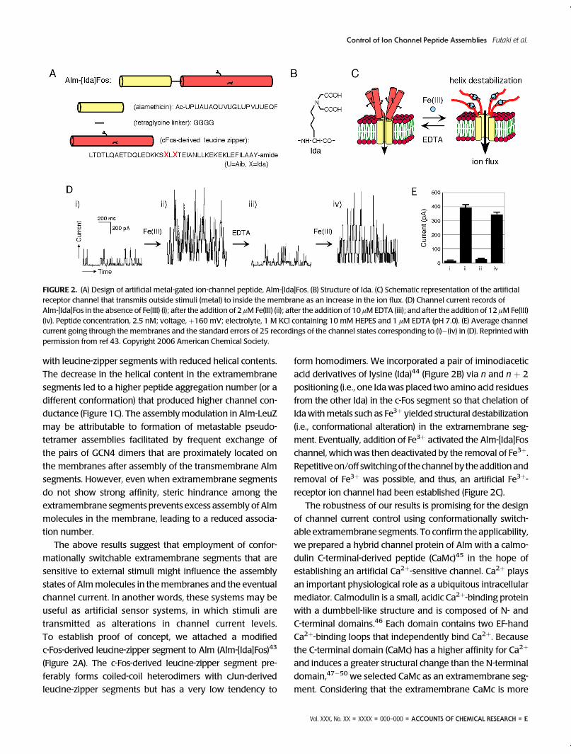
To establish proof of concept, we attached a modified
c-Fos-derived leucine-zipper segment to Alm (Alm-[Ida]Fos)43
(Figure 2A). The c-Fos-derived leucine-zipper segment pre-
ferably forms coiled-coil heterodimers with cJun-derived
leucine-zipper segments but has a very low tendency to
form homodimers. We incorporated a pair of iminodiacetic
acid derivatives of lysine (Ida)44 (Figure 2B) via n and n þ 2
positioning (i.e., one Idawas placed two amino acid residues
from the other Ida) in the c-Fos segment so that chelation of
Idawithmetals such as Fe3þ yielded structural destabilization
(i.e., conformational alteration) in the extramembrane seg-
ment. Eventually, addition of Fe3þ activated the Alm-[Ida]Fos
channel, which was then deactivated by the removal of Fe3þ.
Repetitiveon/off switchingof the channelby theadditionand
removal of Fe3þ was possible, and thus, an artificial Fe3þ-
receptor ion channel had been established (Figure 2C).
The robustness of our results is promising for the design
of channel current control using conformationally switch-
able extramembrane segments. To confirm the applicability,
we prepared a hybrid channel protein of Alm with a calmo-
dulin C-terminal-derived peptide (CaMc)45 in the hope of
establishing an artificial Ca2þ-sensitive channel. Ca2þ plays
an important physiological role as a ubiquitous intracellular
mediator. Calmodulin is a small, acidic Ca2þ-binding protein
with a dumbbell-like structure and is composed of N- and
C-terminal domains.46 Each domain contains two EF-hand
Ca2þ-binding loops that independently bind Ca2þ. Because
the C-terminal domain (CaMc) has a higher affinity for Ca2þ
and induces a greater structural change than the N-terminal
domain,47�50 we selected CaMc as an extramembrane seg-
ment. Considering that the extramembrane CaMc is more
FIGURE 2. (A) Design of artificial metal-gated ion-channel peptide, Alm-[Ida]Fos. (B) Structure of Ida. (C) Schematic representation of the artificialreceptor channel that transmits outside stimuli (metal) to inside the membrane as an increase in the ion flux. (D) Channel current records ofAlm-[Ida]Fos in the absence of Fe(III) (i); after the addition of 2 μMFe(III) (ii); after the addition of 10 μMEDTA (iii); and after the addition of 12 μMFe(III)(iv). Peptide concentration, 2.5 nM; voltage, þ160 mV; electrolyte, 1 M KCl containing 10 mM HEPES and 1 μM EDTA (pH 7.0). (E) Average channelcurrent going through the membranes and the standard errors of 25 recordings of the channel states corresponding to (i)�(iv) in (D). Reprinted withpermission from ref 43. Copyright 2006 American Chemical Society.
F ’ ACCOUNTS OF CHEMICAL RESEARCH ’ 000–000 ’ XXXX ’ Vol. XXX, No. XX
Control of Ion Channel Peptide Assemblies Futaki et al.
hydrophilic than a LeuZ segment, we employed the more
hydrophobic Alm (Rf50) as a transmembrane segment to
stably retain the hybrid protein in the membranes. Alm
(Rf50) has a glutamine (Gln) at position 18 (Figure 3A),
whereas Alm has a glutamic acid (Glu) in that location.21,22
Preparation of Rf50-CaMc was accomplished by using
native chemical ligation, since the CaMc segment has more
than 70 amino acid residues and is thus difficult to synthe-
size chemically. Therefore, a CaMc segment bearing an extra
cysteine on the N-terminus (Cys-CaMc) was prepared by
recombinant expression using intein-mediated purification
with an affinity chitin-binding tag (IMPACT) system for liga-
tion with the Rf50 thioester.51,52 Because the N-terminal
region of the CaMc segment (which is proximal to the
transmembrane Rf50 segment) is rich in the acidic amino
acids aspartic acid (Asp) and Glu, we performed the channel
current recording of Rf50-CaMc under acidic conditions (pH
5.4) to reduce electrostatic repulsion, which would have
prevented Rf50-CaMc from self-assembling in membranes
to form channels.
In the absence of Ca2þ, Rf50-CaMc yielded burstlike chan-
nel currents with no discrete channel conductance levels.45
The addition of Ca2þ significantly stabilized the channel open
state (corresponding to a 6-mer assembly) and increased the
mean channel current by 6-fold (Figure 3B). Conversely,Mg2þ
produced no significant changes in the channel current. Thus,
successful employment of a natural protein motif as the
extramembrane gating segment for Almwas achieved,while
maintaining a simple structure and the intrinsic functions of
the extramembrane and channel-forming segments. The
addition of Ca2þ led to an increase in the surface hydropho-
bicity of the extramembrane segment, which may also have
influenced assembly modulation together with steric hin-
drance between extramembrane segments in the Rf50-CaMc
channel. In summary, useof anatural ligand-bindingpeptide/
protein segment in an Alm extramembrane segment may
lead to a tailored ligand-gated channel.
5. Assembly Control of Alm by MetalChelationAs described above, formation of Alm channels is voltage-
dependent, and a helix dipole interaction between Alm and
membranes' electric fields leads to insertion of the N-termi-
nus into membranes.21�23 Therefore, the absence of a
membrane potential should lead to cancellation of both
themembrane insertion state of Alm and channel assembly.
Many natural membrane proteins have hydrophilic regions
that are exposed to the aqueous environment on either side
of transmembrane segments, which prevent the liberation
of the transmembrane segments from the membranes.
Disposition of hydrophilic amino acids on both sides of
Alm may contribute to the stabilization of its insertion state
and assembly in membranes. Alm has a negatively charged
Glu at position 18, which has been reported to hamper
the membrane insertion of the C-terminus.22 Disposition
of charged residues on the N-termini would prevent the
FIGURE 3. (A) Structure of Rf50 and Rf50-CaMc. U = Aib; Pheol = phenylalaninol; Ac = acetyl. Two Ca2þ-binding loops are highlighted in bold.(B) Schematic representation of Ca2þ-stimulated gating of the Rf50-CaMc channel, and channel current records of Rf50-CaMc in the absence (left) orpresence (right) of 5mMCaCl2. Voltage,þ180mV; electrolyte, 1 M KCl containing 20mM2-(N-morpholino)ethanesulfonic acid (MES), 0.2mM EDTA,and 10 mM dithiothreitol (DTT) (pH 5.4). Reprinted with permission from ref 45. Copyright 2013 American Chemical Society.
Vol. XXX, No. XX ’ XXXX ’ 000–000 ’ ACCOUNTS OF CHEMICAL RESEARCH ’ G
Control of Ion Channel Peptide Assemblies Futaki et al.
inserted Alm segment from turning back to the C-terminus
side, therefore extending the lifespan of the channel. There-
fore, we designed an Alm analogue with a histidine-glycine
(His-Gly) extension on its N-terminus (HG-Alm).53 HG-Alm
formed a channel with multiple conductance states, as
observed in Alm, in an electrolyte containing ethylenedia-
minetetraacetic acid (EDTA) as a metal chelating agent. A
higher voltage (250�300mV) was necessary to activate the
HG-Alm channel compared with Alm (100�150 mV). How-
ever, once channels were formed, longer channel lifetimes
were obtained, even at lower voltages than the voltage
necessary for Alm channel activation.
Unexpectedly, we also found that the channel open life-
time of HG-Alm was considerably longer in the presence of
metals, such as Zn2þ, Ni2þ, and Co2þ, with stabilization of
specific conductance states corresponding to the 8-, 10-, and
12-mer assemblies (Figure 4).53 Althoughwe prepared other
analogues of Alm bearing a lysine (Lys) or Glu at the
N-terminus instead of a His, no similar stabilization effect
by metals was observed. These results suggest that metal
chelationwith theHis forms ametastable dimer assembly of
HG-Alm molecules. Similarly, blocking the N-terminus ami-
no group of His or protonation of the imidazole nitrogen at
low pH also inhibited stabilized assembly. Utilizing the
significant difference in the conductance levels and lifetimes
of HG-Alm channels in the absence and presence of metal
ions, we showed the feasibility of reversible metal switching
of the HG-Alm assembly and the channel current. Addition-
ally, we found that substitution of Aib in HG-Alm with other
6. Scope and Limitations of Using Extra-membrane Segments for Channel GatingAs exemplified by the use of leucine-zipper and CaMc
segments, the strategy of utilizing appropriate extramem-
brane segments is promising for channel assembly control
or gating. Although approaches using modification of chan-
nel pores of natural proteins and those formed with as-
sembled peptides have been applied for practical sensing
(e.g., for DNA sequencing or small molecules),55�57 such
techniques are successful only when the channel pores are
appropriately sized for proper sensing. The analytes must
penetrate the pores, and the interaction between the pores
and the analytesmust accelerate or inhibit the ion flux in the
membrane. Sensing cannot be accomplished if the pores are
too small or too large for the analytes, and adapters, including
cyclodextrins, are often employed to adjust pore size.55,58
When the pore sizes are suitable, these approaches may
attain very sensitive analyte detection. These detectionmeth-
ods are often based on stochastic sensing, and very sensitive
analysis of the channel current is required. Alternatively,
approaches that use extramembrane segments as gating
machinery are less limited with respect to channel pore size.
Depending on the availability of appropriate membrane
segments that specifically interact with ligands (or analytes)
and lead to structural alteration in extramembrane segments
and eventually in channel pores, there ismuchmore room for
channel design. Alternatively, in Alm-based channels, since
the probability of channel formation is voltage-dependent,
the numbers of peptides that form a pore, and the eventual
channel conductance can vary. These characteristics may be
disadvantages of Alm-based approaches in terms of reprodu-
cibility. Thus, employment of suitable extramembrane seg-
ments that yield fixed numbers of segments and the
embedding of relatively large numbers of channel peptides
in themembrane to average the currentmaybenecessary for
future application of this approach to practical sensors.
To analyze the channel current of artificial channel
peptides/proteins, the planar lipid bilayer19 and the tip-dip59
methods have been used. The former approach employs
two chambers connected by a pinhole (typically, having
a diameter of a few hundred micrometers), where a lipid
bilayer is formed. The latter approachuses glass pipettes, as in
the patch-clamp technique. With repeated dipping of the
pipet tips into buffers with surface monolayer membranes,
single bilayers are formed on the ends of the glass pipettes.
These procedures have sensitivities equivalent to that of the
patch-clamp technique and allow single-channel analysis.
FIGURE 4. Schematic representation and typical channel currentrecords of HG-Alm in the absence and presence of ZnCl2. Voltage,þ80 mV; electrolyte, 1 M KCl containing 10 mM HEPES (pH 7.0).Reprint from ref 53.
H ’ ACCOUNTS OF CHEMICAL RESEARCH ’ 000–000 ’ XXXX ’ Vol. XXX, No. XX
Control of Ion Channel Peptide Assemblies Futaki et al.
However, because difficulties often arise in the formation of
membranes, further development is needed. Given that the
chambers used for analysis by these methods are usually in
the milliliter range, downsizing of the system is desired to
facilitate easier and less-sample-consuming analysis. Re-
cently, methods using droplet interface bilayers have been
introduced to allow easier formation of lipid bilayers. In these
methods, aqueous droplets are submerged in an oil/lipid
mixture.60 When the droplets join together, the lipid mono-
layers surrounding them combine at the interface to form
a lipid bilayer. This approach also allows analysis using
much smaller volumes than with the traditional planar-
lipid-bilayer methods and has potential for high through-
put screening of channel peptides/proteins using micro- to
nanoliter-range buffer volumes.61 This system is also bene-
ficial for reducing the electrical noise that typically perturbs
the analysis of very fine currents in the nano- to picoampere
range. Current studies of artificial-ligand-gated ion channels
introduced using this method are in the proof-of-concept
stage, but practical application of these techniques in artificial
sensors may be achieved in the future.
7. ConclusionUsing our Alm-based channels as examples, we showed that
the simple disposition of a structurally switchable extra-
membrane segment effectively influenced the assembly
conditions of the transmembrane Alm segment. This system
can serve as an artificial ligand-sensitive receptor channel
and has potential uses in the development of artificial
sensors, especially in combination with small-scale sensing
systems, including the droplet interface bilayer method.
tures in membranes even without functional moieties that
interact with each other. We have demonstrated the trans-
mittance of alterations in extramembrane structures into the
assembled states of Alm in membranes; this concept may
also be used to control the assembly of other membrane
proteins. Assuming that transmembrane segments have a
tendency to assemble in membranes, membrane proteins
composed of more than one protein unit may form higher-
ordered structures in membranes even in the absence of
strong interactions among extramembrane segments, but
extramembrane segments can prevent excess assembly of
the subunits in the membrane, as shown in the case of Alm-
LeuZ channels. The method involved in the structural forma-
tion of membrane proteins is likely to be more complicated
than that of soluble proteins because correct folding and
packing of the transmembrane segments in the membranes
without interfering with each other or the extramembrane
segments in aqueous environments are necessary. However,
the movement of membrane proteins is two-dimensionally
restricted, and their structures can become stabilized, present-
ing their extramembrane segments on both sides. Therefore,
it might be easier to control the structure and assembly of
membrane proteins than proteins in solution.
We have reported control of the assembly of transmem-
brane segment Alm by introducing appropriate extramem-
brane segments. In addition, we recently developed an
alternative approach to the assembly control of the epider-
mal growth factor receptor using dimerization linkers based
on leucine zipper recognition.62 Extramembrane engineer-
ing is a promising area for assembly control of membrane
proteins and the eventual control of cell functions.
This work was supported by Grants-in-Aid for Scientific Researchfrom the Ministry of Education, Culture, Sports, Science andTechnology of Japan. D.N. received a JSPS Research Fellowshipfor Young Scientists.
BIOGRAPHICAL INFORMATION
Shiroh Futaki obtained his Ph.D. in 1989 from Kyoto University.Following his appointment as a Research Associate and an Associ-ate Professor at the University of Tokushima, he moved to KyotoUniversity in 1997.Meanwhile, he spent 16months (1989�1991)in the United States as a Postdoctoral Associate in the Departmentof Biochemistry, Rockefeller University. He has been a Professor ofBiochemistry at the Institute of Chemical Research, Kyoto Univer-sity, since 2005. His research interests include design of bioactivepeptides having unique functions (ion-channel formation, cellpenetration, DNA-binding, and so on).
Daisuke Noshiro graduated from Faculty of PharmaceuticalSciences, Kyoto University in 2007. He obtained his Ph.D. in 2012fromKyotoUniversityunder the supervisionofProfessor ShirohFutaki.After obtaining his Ph.D., he joined the research group of ProfessorHagan Bayley, Department of Chemistry, theUniversity of Oxford as apostdoctoral fellow. His main research focus is in the field of mem-brane protein engineering for sensing applications. He is a recipientof the Naito Foundation Subsidy for Inter-Institute Researches.
Tatsuto Kiwada was involved in the development ofAlm-[Ida]Fos channels and obtained his Ph.D. in 2005 from KyotoUniversity. After four years' postdoctoral training at the laboratoryof Professor Shigetada Nakanishi, Osaka Bioscience Institute,
Vol. XXX, No. XX ’ XXXX ’ 000–000 ’ ACCOUNTS OF CHEMICAL RESEARCH ’ I
Control of Ion Channel Peptide Assemblies Futaki et al.
he was appointed as an Assistant Professor at Clinical and Analy-tical Sciences, Kanazawa University in 2009. His current researchinterest is development of novel platinum anticancer drug.
Koji Asami is an Associate Professor at the Institute for ChemicalResearch, Kyoto University since 1986. After he obtained his Ph.D.in 1978 from Kyoto University, he was a Research Associate inPhysiology at Kochi Medical School from 1979 to 1986 and was avisiting Associate Professor of theDepartment of Bioengineering atthe University of Pennsylvania from 1987 to 1989. His researchinterests focus on the electric and dielectric properties of artificialand biological membranes.
FOOTNOTES
*To whom correspondence should be addressed. Phone: þ81-774-38-3210. Fax:þ81-774-32-3038. E-mail: [email protected] authors declare no competing financial interest.
REFERENCES1 Alberts, B.; Johnson, A.; Lewis, J.; Raff, M.; Roberts, K.; Walter, P. Molecular Biology of the
Cell, 5th ed.; Garland Science: New York, 2007.2 Hille, B. Ion Channels of ExcitableMembranes, 3rd ed.; Sinauer Associates Inc.: Sunderland,
MA, 2001.3 Numa, S. Molecular basis for the function of ionic channels. Biochem. Soc. Symp. 1986,
52, 119–143.4 Miyazawa, A.; Fujiyoshi, Y.; Unwin, N. Structure and gating mechanism of the acetylcholine
receptor pore. Nature 2003, 423, 949–955.5 Matile, S.; Vargas Jentzsch, A.; Montenegro, J.; Fin, A. Recent synthetic transport systems.
Chem. Soc. Rev. 2011, 40, 2453–2474.6 Sakai, N.; Mareda, J.; Matile, S. Rigid-rod molecules in biomembrane models: from
7 Bayley, H. Designed membrane channels and pores. Curr. Opin. Biotechnol. 1999, 10,94–103.
8 Astier, Y.; Bayley, H.; Howorka, S. Protein components for nanodevices. Curr. Opin. Chem.Biol. 2005, 9, 576–584.
9 Fyles, T.M. Synthetic ion channels in bilayermembranes. Chem. Soc. Rev. 2007, 36, 335–347.10 Futaki, S. Peptide ion channels: design and creation of function. Biopolymers 1998, 47,
75–81.11 Jung, Y.; Bayley, H.; Movileanu, L. Temperature-responsive protein pores. J. Am. Chem.
Soc. 2006, 128, 15332–15340.12 Gorteau, V.; Bollot, G.; Mareda, J.; Pasini, D.; Tran, D. H.; Lazar, A. N.; Coleman, A. W.;
Sakai, N.; Matile, S. Synthetic multifunctional pores that open and close in response tochemical stimulation. Bioorg. Med. Chem. 2005, 13, 5171–5180.
13 Sakai, N.; Sord�e, N.; Das, G.; Perrottet, P.; Gerard, D.; Matile, S. Synthetic multifunctionalpores: deletion and inversion of anion/cation selectivity using pM and pH. Org. Biomol.Chem. 2003, 1, 1226–1231.
14 Jog, P. V.; Gin, M. S. A light-gated synthetic ion channel. Org. Lett. 2008, 10, 3693–3696.15 Tian, Y.; Hou, X.; Wen, L.; Guo, W.; Song, Y.; Sun, H.; Wang, Y.; Jiang, L.; Zhu, D. A
Wang, Y.; Jiang, L. Gating of single synthetic nanopores by proton-driven DNA molecularmotors. J. Am. Chem. Soc. 2008, 130, 8345–8350.
17 Hou, X.; Guo, W.; Xia, F.; Nie, F. Q.; Dong, H.; Tian, Y.; Wen, L.; Wang, L.; Cao, L.; Yang, Y.;Xue, J.; Song, Y.; Wang, Y.; Liu, D.; Jiang, L. A biomimetic potassium responsivenanochannel: G-quadruplex DNA conformational switching in a synthetic nanopore. J. Am.Chem. Soc. 2009, 131, 7800–7805.
18 Fmoc Solid Phase Peptide Synthesis: A Practical Approach; Chan, W. C., White, P. D., Eds.;Oxford University Press: Oxford, 2000.
19 Montal, M. Reconstitution of channel proteins from excitable cells in planar lipid bilayermembranes. J. Membr. Biol. 1987, 98, 101–115.
20 Neher, E.; Sakmann, B. Single-channel currents recorded from membrane of denervatedfrog muscle fibres. Nature 1976, 260, 799–802.
21 Sansom, M. S. The biophysics of peptide models of ion channels. Prog. Biophys. Mol. Biol.1991, 55, 139–235.
22 Woolley, G. A.; Wallace, B. A. Model ion channels: gramicidin and alamethicin. J. Membr.Biol. 1992, 129, 109–136.
23 Gordon, L. G.; Haydon, D. A. The unit conductance channel of alamethicin. Biochim.Biophys. Acta 1972, 255, 1014–1018.
24 Oiki, S.; Danho, W.; Madison, V.; Montal, M. M2 delta, a candidate for the structure liningthe ionic channel of the nicotinic cholinergic receptor. Proc. Natl. Acad. Sci. U.S.A. 1988,85, 8703–8707.
25 Lear, J. D.; Wasserman, Z. R.; DeGrado, W. F. Synthetic amphiphilic peptide models forprotein ion channels. Science 1988, 240, 1177–1181.
26 Ghadiri, M. R.; Granja, J. R.; Buehler, L. K. Artificial transmembrane ion channels fromself-assembling peptide nanotubes. Nature 1994, 369, 301–304.
27 Bayley, H. Triggers and switches in a self-assembling pore-forming protein. J. Cell Biochem.1994, 56, 177–182.
28 Cafiso, D. S. Alamethicin: a peptide model for voltage gating and protein-membraneinteractions. Annu. Rev. Biophys. Biomol. Struct. 1994, 23, 141–165.
29 Futaki, S.; Asami, K. Ligand-induced extramembrane conformation switch controllingalamethicin assembly and the channel current. Chem. Biodiversity 2007, 4, 1313–1322.
30 Kaduk, C.; Wenschuh, H.; Beyermann, M.; Forner, K.; Carpino, L. A.; Bienert, M. Synthesisof Fmoc-amino acid fluorides via DAST, an alternative fluorinating agent. Lett. Pept. Sci.1995, 2, 285–288.
31 Futaki, S.; Fukuda, M.; Omote, M.; Yamauchi, K.; Yagami, T.; Niwa, M.; Sugiura, Y.Alamethicin-leucine zipper hybrid peptide: a prototype for the design of artificial receptorsand ion channels. J. Am. Chem. Soc. 2001, 123, 12127–12134.
32 Landschulz, W. H.; Johnson, P. F.; McKnight, S. L. The leucine zipper: a hypotheticalstructure common to a new class of DNA binding proteins. Science 1988, 240,1759–1764.
33 O'Shea, E. K.; Rutkowski, R.; Kim, P. S. Evidence that the leucine zipper is a coiled coil.Science 1989, 243, 538–542.
34 Chugh, J. K.; Wallace, B. A. Peptaibols: models for ion channels. Biochem. Soc. Trans.2001, 29, 565–570.
35 Woolley, G. A. Channel-forming activity of alamethicin: effects of covalent tethering. Chem.Biodiversity 2007, 4, 1323–1337.
36 Matsubara, A.; Asami, K.; Akagi, A.; Nishino, N. Ion-channels of cyclic template-assembledalamethicins that emulate the pore structure predicted by barrel-stave model. J. Chem.Soc., Chem. Commun. 1996, 2069–2070.
37 Duclohier, H.; Kociolek, K.; Stasiak, M.; Leplawy, M. T.; Marshall, G. R. C-terminallyshortened alamethicin on templates: influence of the linkers on conductances. Biochim.Biophys. Acta 1999, 1420, 14–22.
38 Sakoh, M.; Okazaki, T.; Nagaoka, Y.; Asami, K. N-terminal insertion of alamethicin inchannel formation studied using its covalent dimer N-terminally linked by disulfide bond.Biochim. Biophys. Acta 2003, 1612, 117–121.
39 Okazaki, T.; Sakoh, M.; Nagaoka, Y.; Asami, K. Ion channels of alamethicin dimerN-terminally linked by disulfide bond. Biophys. J. 2003, 85, 267–273.
40 Futaki, S.; Aoki, M.; Fukuda, M.; Kondo, F.; Niwa, M.; Kitagawa, K.; Nakaya, Y. Assemblingof the four individual helices corresponding to the transmembrane segments (S4 in repeatI-IV) of the sodium channel. Tetrahedron Lett. 1997, 38, 7071–7074.
41 Futaki, S.; Aoki, M.; Ishikawa, T.; Kondo, F.; Asahara, T.; Niwa, M.; Nakaya, Y.; Yagami, T.;Kitagawa, K. Chemical ligation to obtain proteins comprising helices with individual aminoacid sequences. Bioorg. Med. Chem. 1999, 7, 187–192.
42 Cornell, B. A.; Braach-Maksvytis, V. L. B.; King, L. G.; Osman, P. D. J.; Raguse, B.;Wieczorek, L.; Pace, R. J. A biosensor that uses ion-channel switches. Nature 1997, 387,580–583.
43 Kiwada, T.; Sonomura, K.; Sugiura, Y.; Asami, K.; Futaki, S. Transmission ofextramembrane conformational change into current: construction of metal-gated ionchannel. J. Am. Chem. Soc. 2006, 128, 6010–6011.
44 Futaki, S.; Kiwada, T.; Sugiura, Y. Control of peptide structure and recognition byFe(III)-induced helix destabilization. J. Am. Chem. Soc. 2004, 126, 15762–15769.
45 Noshiro, D.; Sonomura, K.; Yu, H. H.; Imanishi, M.; Asami, K.; Futaki, S. Construction of aCa2þ-gated artificial channel by fusing alamethicin with a calmodulin-derivedextramembrane segment. Bioconjugate Chem. 2013, 24, 188–195.
46 Babu, Y. S.; Sack, J. S.; Greenhough, T. J.; Bugg, C. E.; Means, A. R.; Cook, W. J.Three-dimensional structure of calmodulin. Nature 1985, 315, 37–40.
47 Finn, B. E.; Even€as, J.; Drakenberg, T.; Waltho, J. P.; Thulin, E.; Fors�en, S. Calcium-inducedstructural changes and domain autonomy in calmodulin. Nat. Struct. Biol. 1995, 2,777–783.
48 Barth, A.; Martin, S. R.; Bayley, P. M. Specificity and symmetry in the interaction ofcalmodulin domains with the skeletal muscle myosin light chain kinase target sequence.J. Biol. Chem. 1998, 273, 2174–2183.
49 Martin, S. R.; Bayley, P. M. The effects of Ca2þ and Cd2þ on the secondary and tertiarystructure of bovine testis calmodulin. A circular-dichroism study. Biochem. J. 1986, 238,485–490.
50 Linse, S.; Helmersson, A.; Fors�en, S. Calcium binding to calmodulin and its globulardomains. J. Biol. Chem. 1991, 266, 8050–8059.
J ’ ACCOUNTS OF CHEMICAL RESEARCH ’ 000–000 ’ XXXX ’ Vol. XXX, No. XX
Control of Ion Channel Peptide Assemblies Futaki et al.
51 Xu, M. Q.; Evans, T. C., Jr. Intein-mediated ligation and cyclization of expressed proteins.Methods 2001, 24, 257–77.
52 Futaki, S.; Sogawa, K.; Maruyama, J.; Asahara, T.; Niwa, M.; Hojo, H. Preparation of peptidethioesters using Fmoc-solid-phase peptide synthesis and its application to the construction ofa template-assembled synthetic protein (TASP). Tetrahedron Lett. 1997, 38, 6237–6240.
53 Noshiro, D.; Asami, K.; Futaki, S. Metal-assisted channel stabilization: disposition of a singlehistidine on the N-terminus of alamethicin yields channels with extraordinarily long lifetimes.Biophys. J. 2010, 98, 1801–1808.
54 Noshiro, D.; Asami, K.; Futaki, S. Control of leakage activities of alamethicin analogs bymetals: side chain-dependent adverse gating response to Zn2þ. Bioorg. Med. Chem. 2012,20, 6870–6876.
55 Maglia, G.; Heron, A. J.; Stoddart, D.; Japrung, D.; Bayley, H. Analysis of single nucleic acidmolecules with protein nanopores. Methods Enzymol. 2010, 475, 591–623.
56 Ma, L.; Cockroft, S. L. Biological nanopores for single-molecule biophysics. ChemBioChem2010, 11, 25–34.
57 Kawano, R.; Osaki, T.; Sasaki, H.; Takinoue, M.; Yoshizawa, S.; Takeuchi, S. Rapiddetection of a cocaine-binding aptamer using biological nanopores on a chip. J. Am. Chem.Soc. 2011, 133, 8474–8477.
58 Li, W.W.; Claridge, T. D.; Li, Q.;Wormald, M. R.; Davis, B. G.; Bayley, H. Tuning the cavity ofcyclodextrins: altered sugar adaptors in protein pores. J. Am. Chem. Soc. 2011, 133,1987–2001.
59 Zweifach, A.; Lewis, S. A. Characterization of a partially degraded Naþ channel from urinarytract epithelium. J. Membr. Biol. 1988, 101, 49–56.
60 Bayley, H.; Cronin, B.; Heron, A.; Holden, M. A.; Hwang, W. L.; Syeda, R.; Thompson, J.;Wallace, M. Droplet interface bilayers. Mol. BioSyst. 2008, 4, 1191–1208.
61 Funakoshi, K.; Suzuki, H.; Takeuchi, S. Lipid bilayer formation by contactingmonolayers in amicrofluidic device for membrane protein analysis. Anal. Chem. 2006, 78, 8169–8174.
62 Nakase, I.; Okumura, S.; Tanaka, G.; Osaki, K.; Imanishi, M.; Futaki, S. Signal transductionusing an artificial receptor system that undergoes dimerization upon addition of a bivalentleucine-zipper ligand. Angew. Chem., Int. Ed. 2012, 51, 7464–7467.