50
Eye Care for Critically Ill Adults
Eye Care for Critically Ill Adults

i Eye Care for Critically Ill Adults 2014
Full title Eye Care for Critically Ill Adults SHPN (ACI) 140005
Guideline owner NSW Agency for Clinical Innovation Intensive Care Coordination and Monitoring Unit
ISBN 978-1-74187-951-3
Executive authorisation Dr Nigel Lyons CE ACI
Author Kay Johnson CNS – KM Kaye Rolls CNC - KM
Guideline development network members (Expert intensive care clinical expert nurses)
Intensive CareAntony Altea CNEKelvin Smith CNCMartin Boyle CNCRand Butcher CNC
Project manager Kaye Rolls
Project officer Janet Masters
ICCMU Director Dr Sean Kelly
ICCMU Manager Di Kowal
Version number 2.3
Year published/implementation 2014
Year for review 2017
Funding • ACI-ICCMU is the primary project funder• NaMO provided funding for the Project Officer’s salary• Baxter Healthcare provided an un-conditional education grant which was used to fund the
2012 project startup meeting. Baxter took no part in the process of developing any of the guidelines
Related NSW health policies and guidelines
• Infection Control Policy (PD2007_036) • Hand Hygiene Policy (PD2010_058)• Infection Control Policy: Prevention & Management of Multi-Resistant Organisms
(PD2007_084)• NSW Work Health & Safety Act (2011)
Disclaimer• This clinical practice guideline (CPG) is aimed at providing clinicians working in NSW hospitals’ intensive care units (ICU) with
recommendations to frame the development of policies and procedures related to the eye care practices in adult ICUs.• This CPG is a revision of 2007 eye care guideline and includes: 1) an update of the evidence base; 2) an evaluation of how this
literature applies to the NSW intensive care context; 3) the extensive clinical knowledge of the guideline development network members (GDN); and 4) a consensus development process.
• The CPG is not intended to replace the critical evaluation processes that underpin the development of local policy and procedure nor does it replace a clinician’s judgment in an individual case.
• Users of this CPG must critically evaluate this CPG as it relates to local circumstances and any changes in the literature that may have occurred since the dates of the literature review conducted. In addition, NSW Health clinicians must review NSW State Government policy documents to identify any directives that may relate to this clinical practice.
• These guidelines are intended for use in NSW acute care facilities.• Content within this publication was accurate at the time of publication. This work is copyright. It may be reproduced in whole or part
for study or training purposes subject to the inclusion of an acknowledgment of the source. • It may not be reproduced for commercial usage or sale. Reproduction for purposes other than those indicated above, requires written
permission from the Agency for Clinical Innovation.
Suggested citation: Johnson K and Rolls K (2014) Eye Care for Critically Ill Adults, Agency for Clinical Innovation NSW Government Version 2 Chatswood, NSW, Australia. ISBN: 878-1-74187-951-3
AGENCY FOR CLINICAL INNOVATION Agency for Clinical InnovationLevel 4, Sage Building PO Box 699 Chatswood NSW 205767 Albert Avenue, Chatswood NSW 2067 T +61 2 9464 4666 | F +61 2 9464 4728
E [email protected] | www.aci.health.nsw.gov.auFurther copies of this publication can be obtained from the Agency for Clinical Innovation website at: www.aci.health.nsw.gov.au Disclaimer: Content within this publication was accurate at the time of publication. This work is copyright. It may be reproduced in whole or part for study or training purposes subject to the inclusion of an acknowledgment of the source.
It may not be reproduced for commercial usage or sale. Reproduction for purposes other than those indicated above, requires written permission from the Agency for Clinical Innovation.© Agency for Clinical Innovation 2014
GUIDELINE PROVENANCE
OpthamologyBev LathamJill GrassoDr Peter McCluskeyDr Michael Hennessey

ii Eye Care for Critically Ill Adults 2014
ABOUT THE ACIThe Agency for Clinical Innovation (ACI) works with clinicians, consumers and managers to design and promote better healthcare for NSW. It does this by:
• Service redesign and evaluation – applying redesign methodology to assist healthcare providers and consumers to review and improve the quality, effectiveness and efficiency of services.
• Specialist advice on healthcare innovation – advising on the development, evaluation and adoption of healthcare innovations from optimal use through to disinvestment.
• Initiatives including Guidelines and Models of Care – developing a range of evidence-based healthcare improvement initiatives to benefit the NSW health system.
• Implementation support – working with ACI Networks, consumers and healthcare providers to assist delivery of healthcare innovations into practice across metropolitan and rural NSW.
• Knowledge sharing – partnering with healthcare providers to support collaboration, learning capability and knowledge sharing on healthcare innovation and improvement.
• Continuous capability building – working with healthcare providers to build capability in redesign, project management and change management through the Centre for Healthcare Redesign.
ACI Clinical Networks, Taskforces and Institutes provide a unique forum for people to collaborate across clinical specialties and regional and service boundaries to develop successful healthcare innovations.
A priority for the ACI is identifying unwarranted variation in clinical practice and working in partnership with healthcare providers to develop mechanisms to improve clinical practice and patient care.
Eye care is an essential component of nursing care for critically ill patients who are particularly vulnerable to eye injury because their illness and treatment can compromise ocular protective mechanisms.
The purpose of this guideline is to provide intensive care clinicians with best practice recommendations so that the evidence-based treatment and care can be delivered and patients can receive the therapy they need.
Developed under the auspices of the Intensive Care Best Practice Manual Project, this guideline highlights the ability of the Agency for Clinical Innovation (ACI) to facilitate strong working relationships with clinicians as well other executive branches of the Ministry.
On behalf of the ACI, I would like to thank Susan Pearce, Chief Nursing and Midwifery Officer for providing state executive sponsorship for the project and funds for the Project Officer. I would also like to extend my appreciation to the LHD executives for facilitating the participation of LHD staff in developing these guidelines, which I commend to you the clinicians of NSW.
Dr Nigel LyonsChief Executive, Agency for Clinical Innovation
FOREWORD

iii Eye Care for Critically Ill Adults 2014
Table 1: Guideline development network members
GUIDELINE MANAGEMENT TEAM
Kay Johnson Chair GDN ACI-ICCMU (past) RN St Vincents Private
Kaye Rolls Project Manager ACI-ICCMU
Antony Altea CNE Intensive care Port Macquarie
Kelvin Smith CNC Intensive care JHH
Martin Boyle CNC Intensive care POW
Rand Butcher CNC Intensive care Tweed Heads
Sarah Jones CNC Intensive care St George
Bev Latham Ophthalmology Sydney Eye Hospital
Jill Grasso Ophthalmology Sydney Eye Hospital
Dr Peter McCluskey Ophthalmology ACI Opthamology Network
Dr Michael Hennessy Ophthalmology ACI Opthamology Network
iv Eye Care for Critically Ill Adults 2014
GUIDELINE PROVENANCE .................................................................................................... iGuideline development network ................................................................................................................................... iii
FOREWORD .........................................................................................................................ii
ABOUT THE ACI ...................................................................................................................ii
1. EXECUTIVE SUMMARY .................................................................................................... 1
2. INTRODUCTION .............................................................................................................. 4Purpose ........................................................................................................................................................................4
Projected outcomes for this guideline .......................................................................................................................4
Scope .... ........................................................................................................................................................................4
Target clinicians ..............................................................................................................................................................4
Guideline development .................................................................................................................................................4
How to use the guideline ...............................................................................................................................................4
Format of guideline .......................................................................................................................................................5
Rating of the evidence for recommendations .................................................................................................................5
Glossary ........................................................................................................................................................................6
3. BACKGROUND ................................................................................................................ 8Summary of normal anatomy and physiology of the anterior ocular surface ...................................................................8
Function of tears and blinking mechanism .....................................................................................................................8
Abnormal physiology ....................................................................................................................................................9
Epidemiology of ocular complications in the critically ill adult .........................................................................................9
Why critically ill patients are at increased risk of ocular surface disorders .......................................................................9
Eye care for the critically ill patient ...............................................................................................................................10
4. RECOMMENDATIONS FOR PRACTICE ............................................................................ 12Patient assessment ......................................................................................................................................................12
Interventions ...............................................................................................................................................................17
Infection prevention ...............................................................................................................................................18
Workplace health and safety ..................................................................................................................................19
Governance ...........................................................................................................................................................20
5. GUIDELINE DEVELOPMENT PROCESS ............................................................................ 21
6. APPENDICES ................................................................................................................. 22
7. REFERENCES ................................................................................................................. 41
TABLE OF CONTENTS

v Eye Care for Critically Ill Adults 2014
TABLESTable 1: Guideline development network members ...................................................................................................... iii
Table 2: NHMRC grading of recommendations ..............................................................................................................5
Table 3: Eye care methods used by authors ................................................................................................................. 11
Table 4: Risk factors for ocular surface disorders ..........................................................................................................13
Table 5: Opthalmology abnormalities ..........................................................................................................................15
Table 6: GDN and EVP members .................................................................................................................................21
Table 7: Consensus results ...........................................................................................................................................21
FIGURESFigure 1: Horizontal section of a schematic eye .............................................................................................................8
Figure 2: Dry eye ...........................................................................................................................................................9
Figure 3: Dry eye ...........................................................................................................................................................9
Figure 5: Method of eyelid taping ...............................................................................................................................14
Figure 6: Grading lagaopthalmos ................................................................................................................................14
Figure 7: Chemosis ......................................................................................................................................................15
Figure 8: Corneal abrasion ..........................................................................................................................................15
Figure 9: Allergic conjunctivitis ....................................................................................................................................15
Figure 10: Marginal keratitis ........................................................................................................................................15
Figure 11: Viral conjunctivitis .......................................................................................................................................15
Figure 12: Bacterial ulcer .............................................................................................................................................15
Figure 13: Bacterial conjunctivitis .................................................................................................................................15
Figure 14: Red eye in septic patient .............................................................................................................................16
Figure 16: Moisture chamber ......................................................................................................................................17
APPENDICESAppendix 1: 2012 literature review ..............................................................................................................................22
Appendix 2: NHMRC levels of evidence .......................................................................................................................23
Appendix 3: NHMRC grading of recommendations .....................................................................................................24
Appendix 4: Summary of 2012 eye studies ..................................................................................................................25
Appendix 5: Incidence summary for iatrogenic opthalmological complications .............................................................28
Appendix 6: Risk factors for iatrogenic opthalmologic complications ...........................................................................30
Appendix 7: Clinical practices effective in preventing iatrogenic opthalmological complications ...................................33
Appendix 8: Clinical practice guidelines (AGREE tool used for assessment) ..................................................................36
Appendix 9: Research papers not included in 2012 literature review ............................................................................39
Appendix 10: Updated literature review 2013 ..............................................................................................................40

1 Eye Care for Critically Ill Adults 2014
1. EXECUTIVE SUMMARY
Ocular surface disease (OSD), due to superficial corneal exposure, has been reported to occur in up to 60% of critically ill patients (1-3). Lagophthalmos or incomplete eyelid closure is thought to be the primary mechanism underlying the development of this condition (4-7).
The purpose of this guideline is to inform intensive care practice related to the provision of eye care for critically ill patients. The underlying aim of the guideline is to minimise the prevalence of ocular surface disorders in this group of patients.
Projected outcomes for this guideline include:
• Facilitation of the diffusion of evidence-based eye care recommendations into clinical eye care practice
• To support the early detection of eye disease, timely referral for conditions, and systematic delivery of eye toilet and treatment
• Improvement of patient quality of care by routinely
addressing iatrogenic ophthalmologic issues, ensuring that on discharge from the unit, visual compromise is not added to existing co morbidities (8).
This guideline has been developed from a limited research base supporting the provision of eye care for the critically ill adult. It provides an update to the 2007 Eye Care Clinical Practice Guideline (CPG), and literature reviewed has been sourced from studies published between 2007 and July 2013. The methodological quality of relevant studies found on iatrogenic ocular surface disorders among intensive care patients has been variable. This has been influenced by differences in definitions used, assessment techniques and study design. To date, published studies have largely been descriptive, yielding limited evidence to support specific nursing eye care practices. The available research was evaluated against the designations of levels of evidence stipulated by the National Health and Medical Research Council (NHMRC) (9).
SECTION RECOMMENDATION GOR
Assessment
1
Eye health assessment should be part of routine patient physical assessment practice and be performed on admission and then routinely at the beginning of the new nursing shift. The initial assessment should include input from the patients’ family to identify pre-admission ocular conditions and treatment and to identify the need for ophthalmology review.
D
2.
Admission and ongoing assessment should include, but is not limited to the following:
• risk factors for OSD
• ability for patient to maintain complete eyelid closure
• evaluation of eye and eyelid cleanliness
• corneal dryness or discolouration
• eye care interventions
• effectiveness of eye care interventions.
C
Table continues on page 2
2 Eye Care for Critically Ill Adults 2014
SECTION RECOMMENDATION GOR
3.
An assessment by intensive care medical staff should be undertaken when the following are found:
• signs of infection
• patients with red eyes and/or general sepsis
• cornea that is dull and cloudy, or with white lines or spots visible.
C
4.Where red eyes are identified, with or without exudate, bilateral swabs for culture should be taken.
C
5.Eyelid closure should be maintained to protect the eyes of intensive care patients who are unable to independently maintain complete lid closure.
B
6.All patients should receive regular eye cleaning to remove debris, secretions, dried ointment and/or other ocular medications.
D
7.
For all patients with, or at risk of lagopthalmos, second hourly eye care must be undertaken to prevent drying of ocular epithelial surfaces, and reduce the risk of infection. Interventions include:
• cleaning of the eye (with saline soaked gauze)
• closure of the eyelid by use of either
• ocular lubricant, or
• creation of a moisture chamber by use of polyethylene wrap
The frequency of eye cleansing should vary with the frequency of eye intervention required.
C
Consensus
8.If eyelid closure cannot be maintained passively then mechanical taping methods should be used to close the eye.
C
Consensus
9.If eye infection is suspected, consideration should be given to commencing broad-spectrum topical antibiotic treatment until the result of swabs are available.
D
10.Clinicians should take care to ensure that patient eyes are not exposed to aspirates during tracheal or oropharyngeal suction procedures.
D
11.
Medical Officers should assess the patient for iatrogenic ophthalmologic complications (at the micro epithelial level) at least weekly in intensive care patients with a length of stay greater than seven days using readily available practical methods.
D
12.
Patients should be referred for specialist ophthalmological consultation where
• clinical practices fail to achieve sustained eyelid closure within 24 hours and/or
• when iatrogenic ophthalmologic complications are identified, or
• patient response to treatment is limited.
C
Consensus
13.
Clinicians are to undertake a risk assessment to identity the risk of contamination and mucosal or conjunctival splash injuries when caring for patients PPE (including goggles/face shield/gloves and gown/apron) as per NSW 2007 Infection Prevention control policy should be worn according to the risk assessment.
PD2010_058
Table continued from page 1
Table continues on page 3
3 Eye Care for Critically Ill Adults 2014
Table continued from page 2
SECTION RECOMMENDATION GOR
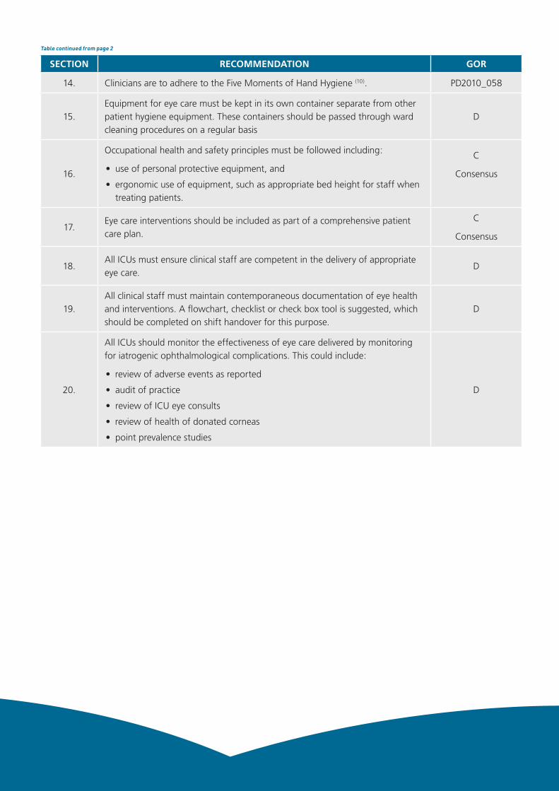
14. Clinicians are to adhere to the Five Moments of Hand Hygiene (10). PD2010_058
15.Equipment for eye care must be kept in its own container separate from other patient hygiene equipment. These containers should be passed through ward cleaning procedures on a regular basis
D
16.
Occupational health and safety principles must be followed including:
• use of personal protective equipment, and
• ergonomic use of equipment, such as appropriate bed height for staff when treating patients.
C
Consensus
17.Eye care interventions should be included as part of a comprehensive patient care plan.
C
Consensus
18.All ICUs must ensure clinical staff are competent in the delivery of appropriate eye care.
D
19.All clinical staff must maintain contemporaneous documentation of eye health and interventions. A flowchart, checklist or check box tool is suggested, which should be completed on shift handover for this purpose.
D
20.
All ICUs should monitor the effectiveness of eye care delivered by monitoring for iatrogenic ophthalmological complications. This could include:
• review of adverse events as reported
• audit of practice
• review of ICU eye consults
• review of health of donated corneas
• point prevalence studies
D

4 Eye Care for Critically Ill Adults 2014
Eye care is an important aspect of the nursing management of critically ill patients, especially for those patients whose ocular protective mechanisms may be compromised (11). Dryness of the cornea and disruption to corneal epithelial surface lining may result in sequelae of corneal abrasion, erosion, infection, ulceration, scarring, rupture or blindness (8). The intensive care patient is at increased risk for any of these events due to having a co-existing compromised immune response and being exposed to environmental factors and pathogens (12). Additionally, for the critically ill patient, lagopthalmos, or incomplete eyelid closure, is an important clinical sign contributing to the development of ocular surface disease (OSD) (13-15).
PurposeThis guideline has been developed to provide intensive care clinicians with recommendations to guide eye care practice for critically ill patients.
Projected outcomes for this guideline:• Facilitation of the diffusion of evidence-based eye care
recommendations into clinical eye care practice.
• To support the early detection of eye disease, timely referral for conditions and systematic delivery of eye toilet and treatment.
• Improvement of patient quality of care by routinely addressing iatrogenic ophthalmologic issues, ensuring that on discharge from the unit, visual compromise is not added to existing co morbidities (8).
ScopeGuideline development addresses clinical practices aimed at maintaining/optimising the eye health of critically ill adults nursed in intensive care units (ICUs) in NSW. In particular, practice recommendations are most relevant for patients at increased risk for iatrogenic ophthalmological complications due to a compromise in
level of consciousness and/or impaired ability to control eye opening and closure. Guideline development has been based on the assumption that readers possess a working knowledge of anatomy and physiology of the eye.
Target cliniciansThis guideline is for the use of all intensive care clinicians, especially for clinicians responsible for the care of any patient in whom protection of the ocular surface cannot be achieved by independent complete eyelid closure. Clinicians who use this guideline must ensure they have a working knowledge of anatomy and physiology of the eye, as well as of ocular protective mechanisms that may become compromised during episodes of critical illness and treatment.
Guideline developmentThis guideline is a revision of 2007 Eye Care Clinical Practice Guidelines (16). A guideline development network (GDN) group was formed in November 2011 to review the original guideline, and the primary authors undertook an updated literature review (Appendix 1). Provisional recommendations based on the available evidence were developed and revised by GDN members. Subsequent to this, the revised guideline was written and the revised clinical practice guideline (CPG) sent to the GDN members who assigned their level of agreement with recommendation statements. The guideline narrative was also revised based on group feedback. Due to the delay in publishing the guideline another search was undertaken covering literature published between 1/1/2012-8/7/2013 (Appendix 10). Because no controlled studies were identified no changes were made to the guideline.
How to use the guidelineClinical judgement should be exercised when applying the principles described in this guideline. Where ophthalmic complications have occurred, the directions of the ophthalmologist should take precedence over the recommendations outlined in this document.
2. INTRODUCTION

5 Eye Care for Critically Ill Adults 2014
Format of guidelineRecommendations and relevant explanatory literature are presented. Detailed evidence used to support statements may be found in the integrative literature review (Appendix 1).
Rating of the evidence for recommendationsThe Australian NHMRC taxonomy 2009(17) was used for grading the level of evidence of a study and grading a recommendation for practice. The assignment of a level of evidence for an individual paper, and the grading of a recommendation was done by the principle authors. If a recommendation did not have an evidence base, the clinical opinion of the guideline network members has been used to inform guideline recommendations.
GRADE OF RECOMMENDATION
DESCRIPTION
A Body of evidence can be trusted to guide evidence
B Body of evidence can be trusted to guide practice in most situations
CBody of evidence provides some support for recommendation/s but care should be taken in its application
D Body of evidence is weak and recommendation must be applied with caution
Consensus Consensus was set as a median of ≥ 7
NHMRC (18)
Table 2: NHMRC grading of recommendations

6 Eye Care for Critically Ill Adults 2014
GlossaryBacterial keratitis ..........................Inflammation of the cornea secondary to bacterial infection
Chemosis ......................................Swelling of the conjunctiva, often preventing eyelid closure.
CI .................................................Confidence interval
CNC .............................................Clinical nurse consultant
CNS ..............................................Clinical nurse specialist
CONSORT .....................................Consolidated Standards on Reporting Trials http://www.consort-statement.org/?o=1001
Corneal abrasions ........................ Superficial disruption to corneal epithelial lining. Common conditions may be secondary to foreign body or contact lens use.
Corneal erosion ............................ Small/punctate or changes/break in the corneal epithelium creating a breach in the defence mechanisms of the cornea, leaving it vulnerable to pathogenic organisms. Left untreated, corneal erosion may result in ulceration and scarring and compromised vision.
CPG ..............................................Clinical practice guideline
Dry Eye ......................................... Lack of normal eye tear film and lubrication. Corneal defences are compromised due to lack of IgA and other immune mediators.
ETT ...............................................Endotracheal tube
EVP ...............................................External validation panel
Exposure keratitis/ ......................... Inflammation of the cornea, either sterile or microbial, may result in epithelial breakdown.
Filamentary keratitis ..................... A condition caused by the formation of epithelial filaments of varying size and length, attached at one or both ends of the cornea. Patients often experience a foreign body sensation, grittiness, discomfort, photophobia, eyelid twitching, increased blinking or pain.
GCS ..............................................Glasgow Coma Scale
GDN .............................................Guideline development network
GOR .............................................Grading of recommendations
HDU .............................................High dependency unit
Hypopyon .....................................An accumulation of pus in the anterior chamber of the eye
ICC ...............................................Intensive care collaborative
ICC-CDC .......................................Intensive care collaborative – consensus development conference
ICCMU ..........................................NSW Intensive Care Coordination and Monitoring Unit
ICU ............................................... Intensive care unit includes all types of units designated as such in NSW. May include units currently designated as ICU, HDU, critical care units
Injection .......................................Conjunctival redness
Keratopathy .................................. Ocular surface breach predisposing to corneal infection, inclusive of any corneal disease, dysfunction or abnormality.
Lagophthalmos ............................. The inability to close or poor closure of the eyelids.
Microbial keratitis .......................... Inflammation of the cornea secondary to bacterial, viral or fungal infection. May result in corneal ulceration and perforation.

7 Eye Care for Critically Ill Adults 2014
Neurotrophic keratopathy ............. A degenerative disease characterised by decreased corneal sensitivity and poor corneal healing. This disease leaves the cornea susceptible to injury and decreases reflex tearing. Epithelial breakdown can lead to ulceration, infection, and perforation secondary to poor healing.
NHMRC ........................................National Health and Medical Research Council
Ocular SurfaceDisease (OSD) ......... General term covering conditions of superficial corneal exposure. These may range from micro/punctuate lesions to larger geographical defects de-epitheliazing the cornea.
OR ................................................Odds ratio
PICO .............................................Population intervention comparison outcome
Punctate epithelial keratopathy .....Micro epithelial defects to the corneal surface
RCT ..............................................Randomised control trial
SR .................................................Systematic review

8 Eye Care for Critically Ill Adults 2014
3. BACKGROUND
Summary of normal anatomy and physiology of the anterior ocular surfaceThe ocular surface is protected from injury and infection by a number of structures including: (refer Figure 1)
1) retractable eyelids, which have a mucous membrane covering that is continuous with the eyeball, and epithelium of the sclera, cornea and conjunctiva. Eyelids mechanically protect the eyes from dehydration and injury (19).
2) an opaque sclera, which ensures that light transmitted to the globe enters only through the transparent corneal covering of the pupil (19).
3) an avascular cornea, which functions to admit and refract light. If injured, it may be slow to heal. Five layers of corneal tissue (superficially epithelium changing to deeper endothelial tissue) provide a protective barrier against abrasion and erosion, and also provide a permeability barrier against eye pathogens (20), and
4) conjunctival epithelium, which extends from the eyelid margins anteriorly, sharply turning on itself to cover the sclera, creating a moist sac. This sac is continuous with the epithelium lining the ducts of tear producing glands, and plays a central role in the defence of ocular surface microbial injury (20). The conjunctiva has a rich blood supply. If damaged, redness and swelling may be present. Tissues may protrude between the eyelids, exacerbating the effects of lagopthalmos, and resulting in corneal opacity and vision loss (20).
Proper functioning of the above structures, and transparency of the cornea are therefore essential requirements for eye surface protection and vital for vision (20, 21). Under normal physiology, closure of the eyelids occurs, and is protective of the ocular surface, blink reflex and tear production are present, and the sclera and cornea appear bright and clear (19).
Figure 1: Horizontal section of a schematic eye
Images from Eye Care Emergency Manual, used by permission of ACI-Ophthalmology Network (22)
Function of tears and blinking mechanismComplex physiology underlies the action of eyelid closure and blinking. These two actions provide a mechanical barrier to ocular injury, and prevent drying out and desiccation of the corneal epithelium by distributing tear film across the exposed surface of the eye (21, 23,
24). Lacrimal gland production of tear film is inherent to healthy eye function (20). Tear film contains bactericidal enzymes (lysozyme, lactoferrin), and proteins (IgA). Tears help to provide a defence against microbial colonisation by providing a medium for transport of leucocytes in the event of eye injury or infection (19). Any increase in irritation from the cornea or conjunctiva will trigger a lacrimal reflex, resulting in an increased tear volume for the eye (20). Blinking and tear production also aids in smoothing out corneal irregularities, protects the air-corneal interface and refractive surface of the cornea. It also supports the clearance of metabolic waste via nasolacrimal drainage mechanisms and enables oxygen delivery to the cells of an avascular cornea (19).

9 Eye Care for Critically Ill Adults 2014
Abnormal physiologyAny disruption to ocular epithelial tissue may compromise vision and predispose the cornea to infection and OSD (21, 25). Mechanisms underlying the development of conditions such as corneal abrasion, erosion, or pathogenic invasion primarily relate to lack of eyelid closure and interruption to blinking reflex and blinking frequency (20, 21, 23). In the ICU population the use of muscle relaxants and sedation have been identified as contributing to lagopthalmos and of placing patients at increased risk (1, 3, 23).
Epidemiology of ocular complications in the critically ill adultIatrogenic eye complications cover a range of OSD involving structures such as the cornea, sclera and conjunctiva. Pathologies may range from microepithelial corneal punctures (often associated with dry eye syndrome), to corneal abrasion, erosion, ulceration, infection and scarring (24). Superficial keratopathy, that is, any breach of the ocular surface (1, 15, 21), in the ICU population, has been found to predispose to infection of the corneal epithelium (keratitis) (1, 19, 26). This infection may present as microbial, bacterial or fungal in origin (14, 21, 23). Keratitis in the presence of corneal exposure has been found to be a key factor in the development of ocular surface disease (1, 2, 14, 19, 27) and has resulted in serious complications such as vision loss, corneal rupture, and the need for corneal transplantation (1, 12, 21, 28, 29).
A high incidence of OSD among ICU patients has been reported with a range from 23%-60% of patients affected. Of these, exposure keratopathy has been found
in 23%-40% (2, 26). Superficial keratopathy has been found in 60% of patients sedated or on neuromuscular blockade (1). Microbial keratitis has been found to be more prevalent than the non-ulcerative sterile form of keratitis (77% vs. 10%) (3). (See Table 5.)
Why critically ill patients are at increased risk of ocular surface disordersThere are a number of causes of impaired ocular defence mechanisms in critically ill patients including:
• an alteration in level of consciousness, impacting on the blink reflex and lagopthalmos
• metabolic derangements
• immunosuppression
• mechanical ventilation
• medications such as sedatives, muscle relaxants and paralysis
• open suction technique
• systemic disease (2, 5, 13, 24).
The ICU environment is also a pathogen-rich environment. This may contribute to the increased exposure of the ocular surface to microorganisms (28). Multi-resistant organisms associated with microbial keratitis include: pseudomonas aeruginosa, acinetobacter, staph epidermis, enterococcus, enterobacter, proteus mirabilis and klebsiella pneumonae (6, 21, 28). Regular eye care has been found to reduce the developmnet of corneal abnormalities and infections in ICU populations (4, 14, 29). Meticulous nursing care is therefore essential to prevent iatrogenic ophthalmological complications and potentially serious visual impairment (11, 24).
Figure 2: Dry eye Figure 3: Dry eye
Images from Eye Care Emergency Manual, used by permission of ACI-Ophthalmology Network (22)

10 Eye Care for Critically Ill Adults 2014
Eye care for the critically ill patient Regular eye care for intubated and ventilated patients is considered routine nursing practice. Anecdotally however, it has been shown that practice varies greatly between intensive care units regarding the frequency and method of eye care undertaken. Historically, specific eye care practice has included regimens of cleaning the eyes with sterile water or normal saline every two to four hours (4, 15, 29, 30), twice daily (7) or daily (31). Installation of a lubricating liquid, such as methylcellulose eye drops, has also been commonly used (7, 15, 29, 30, 32). Eye ointment has been applied for high risk patients, or where evidence of eye injury may be apparent, such as when conjunctival oedema is present (2, 4, 5, 12, 30, 32-34). For conditions of conjunctival or corneal exposure, methods such as passive eye closure (33), eye taping (2, 5, 15, 30), padding with gel membranes (2, 15, 29, 34), and creation of moisture closed chambers using polyethylene film (2, 4, 7, 30,
32) or goggles (5) have been described (see Table 3). From the literature review it is unclear if any of thee methods identified contributed to ocular surface protection, or to the maintenance of eyelid closure (33), as there has been a limited number of quality studies, and significant variability in the methods of eye care used in studies.

11 Eye Care for Critically Ill Adults 2014
Table 3: Eye care methods used by authors
Author Eye care method
Bates J et al. Clinical Intensive Care 2004
Routine eye care to all patients daily: cleaning lids with saline and sterile gauze daily, plus ocular lubricant at least twice daily. Corneal Care with adhesive to tape the eyelids closed. Geliperm/Polyacrylamide Gel Membrane Changed at regular intervals to prevent drying.
Cortese D et al. American Journal of Critical Care 1995
All patients eye toilet with n/s 2/24 Polyethylene Cover (PC) over the eyes to create a moisture chamber. Changed daily. Methylcellulose (hypromellose) lubricating drops 2/24.
Ezra D et al. Intensive Care Medicine 2009
• Lacrilube applied to inferior conjunctival fornix 6/24.
• Geliperm dressing cut to completely cover the top and lower lid and applied onto the closed eye 4/24 or sooner if signs of drying.
• Staff trained in eye care, particularly in early recognition of drying Geliperm.
Guler E et al. Journal of Clinical Nursing 2011
For all subjects, standard eye care with sterile n/s soaked gauze conducted twice daily. Then Polyethylene cover applied to one eye every 12/24, and Carbomer Methylcellulose drops 6/24 to the other eye.
Joyce N Joanna Briggs Institute (Systematic Review) 2002
Polyethylene Cover used. Hypromellose eye drops two drops 2/24 combined with 1–1.27cm Duratears ointment 4/24.
Koroloff N et al. Intensive Care Medicine 2004
• Standard care for both groups: 2/24 eye cleaning with n/s.
• Lacri-lube ointment 2/24 plus 2/24 Hypromellose drops combination.
• Polyethylene Cover/cling wrap placed over the eyes to create a moisture chamber. Micropore used to seal the edge. Changed every shift, or when necessary.
Laight S et al. Intensive and Critical Care Nursing 1996
Sterile water used for eye cleaning 2/24 for all patients. Hypromellose drops 1/24 – 6/24 with corneal dryness. Micropore used for mechanical eye closure. Gel membrane/Geliperm covers applied on clean eye only, and assessed 2/24 for dryness and change.
Lenart S, Garrity J American Journal of Critical Care 2000
1.27cm Duratears artificial tear ointment 4/24.passive eyelid closure.
Rosenberg J et al. Critical Care Medicine 2008
Moisture chamber (MC); lubricating ointment.
Sivasankar S et al. Indian Journal of Critical Care Medicine 2006
• Closed moisture chamber created using swimming goggles and sterile water moistened gauze 12/24.
• Open chamber method (ocular lubricant and mechanical eye closure using securing tape 12/24).
So H International Journal of Nursing Studies 2008
• All subjects received standard eye care: cleansing of the eyelids and surrounding skin 4/24 with n/s.
• Lanolin/Durotears ointment: 1cm applied into the “V” pocket between the eyeball and lower lid of each eye 4/24.
• Polyethylene cover/Gladwrap, tailored to cover the eyes from the eyebrow to the cheekbone, snuggly adhering to form a closed moisture chamber. Micropore adhesive tape use to secure edges of the wrap if the seal was not adequate. PC wrap changed daily, or when visibly soiled.

12 Eye Care for Critically Ill Adults 2014
4. RECOMMENDATIONS FOR PRACTICE
SECTION RECOMMENDATION GOR
Assessment
1
Eye health assessment should be part of routine patient physical assessment practice and be performed on admission and then routinely at the beginning of the new nursing shift. The initial assessment should include input from the patients’ family to identify pre-admission ocular conditions and treatment and to identify the need for ophthalmology review.
D
2.
Admission and ongoing assessment should include, but is not limited to the following:
• risk factors for OSD
• ability for patient to maintain complete eyelid closure
• evaluation of eye and eyelid cleanliness
• corneal dryness or discolouration
• eye care interventions
• effectiveness of eye care interventions.
C
3.
An assessment by intensive care medical staff should be undertaken when the following are found:
• signs of infection
• patients with red eyes and/or general sepsis
• cornea that is dull and cloudy, or with white lines or spots visible.
C
4.Where red eyes are identified, with or without exudate, bilateral swabs for culture should be taken.
C
Patient assessment
A limited numbers of studies have focussed on eye care assessment for the adult intensive care patient. Research areas have ranged from identifying risk factors for lagopthalmos (13), risk factors for OSD (13, 15) and studies on eyelid cleanliness and corneal dryness (15, 24). Consensus support is given to a comprehensive patient history and assessment on admission and at regular intervals (11, 24) such as at shift handover as an essential component of clinical care. These recommendations are based on existing findings that critical illness, pre-existing conditions and intensive care treatment all contribute to an increased risk of iatrogenic eye complications for the critically ill adult.
To ensure that all-important information is obtained, and
in keeping with good clinical practice, clinicians should
approach family members for information regarding the
patient’s medical and surgical history. This history should
include ocular conditions and treatment on admission, in
order to assess the risks of and early recognition of OSD (11). Highly effective eye regimes may be compromised
by interruption to treatment. On admission, previous
eye injury or surgery, the presence of an artificial lens, a
history of cataracts, glaucoma and any other pre-existing
eye treatment and medications, such as anticholinergic
drops, should be elicited (35, 36).

13 Eye Care for Critically Ill Adults 2014
A number of studies have focussed on the role of critical illness, pre-existing conditions and the treatment environment of intensive care as factors contributing to iatrogenic risk for eye complications. Critical illness commonly presents with a range of conditions potentially affecting ocular defence mechanisms. The following medical conditions have been investigated: immunosuppression (13), sepsis and trauma (6, 21), multi-organ failure (1, 6), burns (35), Guillian Barre Syndrome (37), myasthenia gravis (38), collagen disease and diabetes (36), neurological presentations (23, 39), and ocular conditions arising due to various complications of systemic disease (3,
39). Pre-existing eye conditions also place this population of adult intensive care patients at greater risk, especially if interruption to existing treatment regimes were to occur by virtue of admission to the intensive care unit (8).
The patient treatment process within the intensive care environment may also create barriers to ocular
health and integrity of eye function. The use of sedation and neuromuscular blockade has been identified as a precursor to lagopthalmos (5, 7), and this relationship has been shown to strengthen with an increased length of ICU stay (4, 7, 21) and an increased length in ventilation time (6, 12, 21). Unconscious patients are vulnerable to eye injury and infection due to inadequate lid closure and epithelial exposure (13, 36). This may lead to drying of the conjunctiva and corneal epithelium, infection, permanent corneal scarring and visual loss (15, 24). In the ICU environment, other risks for eye infection and corneal disruption arise from respiratory pathogens (6, 14, 19), high gas flow, CPAP, the use of tracheal or oropharyngeal open suction (12), copious secretions, patient positioning (for example proning) (36) and cross infection from other body infective surface wounds. (12, 14)
Risk factor Level of risk Overview of evidence References
Lagopthalmos Probably highly significant
2 x RCT 1 x Prospective cohort1 x Observational 1 x Retrospective case control 1 x Narrative review
(5, 7)(40)(6)(41)(36)
Length of sedation/use of neuromuscular blockade
Probably significant 1 x RCT2 x Observational 2 x Clinical practice guideline1 x Narrative review
(36)(6, 21)(11, 24)(36)
Length of stay Probably a function of critical illness
2 x RCT1 x Observational
(4, 7)(21)
Length of ventilation Probably a function of critical illness
2 x Observational1 x Narrative review and
meta-analysis
(6, 21)(12)
Medical conditions Possibly a risk 1 x RCT2 x Observational1 x Narrative review
(5)(6, 21)(36)
Respiratory pathogens Possibly a risk 1 x Narrative review and Meta-analysis
1 x Narrative review
(12)
(36)
Table 4: Risk factors for ocular surface disorders
14 Eye Care for Critically Ill Adults 2014
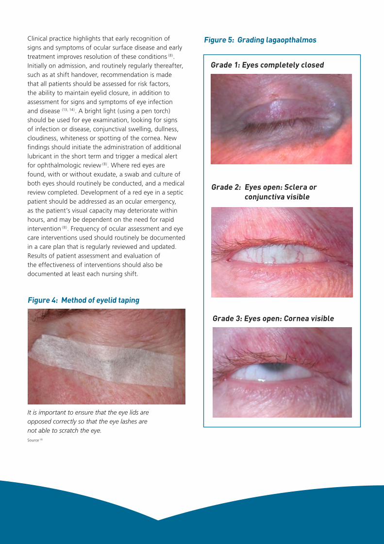
Clinical practice highlights that early recognition of signs and symptoms of ocular surface disease and early treatment improves resolution of these conditions (8). Initially on admission, and routinely regularly thereafter, such as at shift handover, recommendation is made that all patients should be assessed for risk factors, the ability to maintain eyelid closure, in addition to assessment for signs and symptoms of eye infection and disease (13, 14). A bright light (using a pen torch) should be used for eye examination, looking for signs of infection or disease, conjunctival swelling, dullness, cloudiness, whiteness or spotting of the cornea. New findings should initiate the administration of additional lubricant in the short term and trigger a medical alert for ophthalmologic review (8). Where red eyes are found, with or without exudate, a swab and culture of both eyes should routinely be conducted, and a medical review completed. Development of a red eye in a septic patient should be addressed as an ocular emergency, as the patient’s visual capacity may deteriorate within hours, and may be dependent on the need for rapid intervention (8). Frequency of ocular assessment and eye care interventions used should routinely be documented in a care plan that is regularly reviewed and updated. Results of patient assessment and evaluation of the effectiveness of interventions should also be documented at least each nursing shift.
Figure 4: Method of eyelid taping
It is important to ensure that the eye lids are opposed correctly so that the eye lashes are not able to scratch the eye.Source (8)
Figure 5: Grading lagaopthalmos
Grade 1: Eyes completely closed
Grade 2: Eyes open: Sclera or conjunctiva visible
Grade 3: Eyes open: Cornea visible

15 Eye Care for Critically Ill Adults 2014
Table 5: Opthalmology abnormalities
Figure 6: Chemosis
Figure 8: Allergic conjunctivitis
Figure 10: Viral conjunctivitis
Figure 12: Bacterial conjunctivitis
Figure 7: Corneal abrasion
Figure 9: Marginal keratitis
Figure 11: Bacterial ulcer
Figure 13: Red eye in septic patient
Figures 6-13 sourced from Eye Emergency Manual (22).

16 Eye Care for Critically Ill Adults 2014
SECTION RECOMMENDATION GOR
5.Eyelid closure should be maintained to protect the eyes of intensive care patients who are unable to independently maintain complete lid closure.
B
6.All patients should receive regular eye cleaning to remove debris, secretions, dried ointment and/or other ocular medications.
D
7.
For all patients with, or at risk of lagopthalmos, second hourly eye care must be undertaken to prevent drying of ocular epithelial surfaces, and reduce the risk of infection. Interventions include:
• cleaning of the eye (with saline soaked gauze)
• closure of the eyelid by use of either
• ocular lubricant, or
• creation of a moisture chamber by use of polyethylene wrap
The frequency of eye cleansing should vary with the frequency of eye intervention required.
C
Consensus
8.If eyelid closure cannot be maintained passively then mechanical taping methods should be used to close the eye.
C
Consensus
9.If eye infection is suspected, consideration should be given to commencing broad-spectrum topical antibiotic treatment until the result of swabs are available.
D
10.Clinicians should take care to ensure that patient eyes are not exposed to aspirates during tracheal or oropharyngeal suction procedures.
D
11.
Medical Officers should assess the patient for iatrogenic ophthalmologic complications (at the micro epithelial level) at least weekly in intensive care patients with a length of stay greater than seven days using readily available practical methods.
D
12.
Patients should be referred for specialist ophthalmological consultation where
• clinical practices fail to achieve sustained eyelid closure within 24 hours and/or
• when iatrogenic ophthalmologic complications are identified, or
• patient response to treatment is limited.
C
Consensus
Interventions
Incomplete eye closure (lagopthalmos) has been identified as strongly contributing to the development of iatrogenic ocular surface disorders (OSD) (5-7, 13, 24, 36, 41). The vulnerability of ICU patients to lagopthalmos has been attributed to a number of factors including reduced level of consciousness, tracheal intubation, prolonged sedation, paralysis, prolonged mechanical ventilation and PEEP. Medical conditions with significant metabolic derangement and positive fluid balances also contribute (1, 5-7, 12, 14, 19,
21, 26, 27, 42). Exposure of the eye due to inadequate lid closure may lead to drying of the conjunctival and corneal epithelium, and trigger a cascade of infection and corneal
erosion resulting in permanent corneal scarring and visual loss(8). Early identification of incomplete eyelid closure by regular assessment of eyelid position (Figure 5), provides a strategy for early intervention to close and protect the eyes. However, while the underlying principle of the eye care CPG is to ensure eye lid closure, this strategy is based on consensus opinion that treatable causes for lagopthalmos have first been identified and addressed.
Various methods have been used to provide protective barriers and moisture to the corneal surface. Evidence supporting practice however has been inconsistent, due to variations in definitions and methodologies used.

17 Eye Care for Critically Ill Adults 2014
Study outcomes on the effectiveness of interventions used should therefore be viewed with caution. Regardless, support exists for the use of lubricants in all unconscious or heavily sedated patients (13) as lubricants have been found to decrease the risk of corneal dehydration and infection (26). The literature also supports the use of lubricants over eye drops, as ointment has been shown to provide longer lasting eye moisture, and require less frequent installation (33). Lubricants have been found to be better than passive eyelid closure in reducing the incidence of corneal erosion (32, 33), less effective than mechanical eye covers (except Geliperm) to reduce corneal breakdown (2, 5, 15), and less effective than polyethylene cover moisture chamber to reduce the incidence of exposure keratopathy (4, 30, 32). Other studies have found efficiency with the use of either polyethylene covers or lubricants to decrease the incidence of corneal breakdown
(4). Combination use of 1.27cm Duratears ointment with polyethylene covers has been shown to result in a low incidence of OSD (5.3% - 6.8%)(4, 33), and Micropore edging has additionally been used with polyethylene covers in order to create a better seal (4, 30). Research using swimming goggles as a moisture chamber and changed 12/24 has proved inconclusive in reducing the incidence of OSD (5). While a meta-analysis (12) supports the use of moisture chambers over the use of lubricants, these findings have been based on studies with a moderate to high risk of bias.
For patients unable to maintain eyelid closure independently, interventions to cover the eye and to maintain corneal moisture (Appendix 7 Clinical practice effective in preventing iatrogenic opthalmological complications) appear to reduce the incidence of eye complications (2, 4, 5, 12, 30, 33, 34, 43). These interventions include the use of either passive or mechanical means to obtain complete lid closure (13, 32, 33). Mechanical eye covers have been advocated as a strategy to minimise the risk of eye infection in cases of respiratory infection and wherein open tracheal suction techniques may be in use (26-
28). These covers have been advocated for use in combination with eye ointment (4, 29, 30, 32), paraffin gauze, dressing and tape (15). All interventions include the use of regular eye hygiene. Eye cleaning with saline soaked gauze 2/24 – 4/24 to remove exudate, debris or dried ocular medications (13, 15,
29, 34) has evidence-based support. However, while the use of normal saline over sterile water remains debatable (4, 13, 15, 30), agreement exists on the need to promote patient comfort and healing by frequently cleaning the eyes with eye care interventions utilised.
Given the limited success at protecting and supporting ocular epithelial integrity associated with moisture chambers, mechanical covers, and passive eye closure, additional mechanical means of eye closure by taping with Micropore has also been suggested (5, 15). The proviso with
this recommendation is that extreme care should be taken to prevent injury because the tissues surrounding the eyes are delicate and inadvertent application of tape to the cornea may cause damage (20, 21).
To summarise, available evidence lends support to routine eye hygiene for all patients, and eyelid cleansing if lids are unclean (34, 44). Eye lubricants, eye covers and eye taping have been found to either decrease the incidence or the severity of OSD once apparent (5, 34, 44). Furthermore, that incomplete eyelid closure is indicative of a need for eye hygiene, eye lubricant and eye covers, with the exception of the use of Geliperm (15, 24, 34).
Eliminating lagopthalmos and ocular surface exposure has been shown to be essential for the prevention of microbial colonisation and infection (23). Signs of infection may include redness, pain or discharge (15), lid and conjunctival swelling with hyperaemia, lid margin crusting or corneal clouding (14,
24). Suspicion of infection, medical review and subsequent to obtaining bilateral eye swabs for culture (15), and medical consideration for ophthalmologic referral, consideration should also be given to the use of a broad-spectrum antibiotic until the result of eye swabs become available. Two antibiotics have been cited in the literature for interim use in this situation: gentamycin, for use when respiratory pathogen involvement is suspected (14), or otherwise, a chloramphenicol prescription (6).
The frequency for medical assessment of iatrogenic ocular surface disease in ICU patients cited in the literature varies. This has ranged from weekly (13) to more frequent examination especially with symptomatic patients (15, 24). Regardless, timely specialist referral is recommended for symptomatic patients, or for patients in whom treatment response is limited, or the adopted interventions do not achieve the goal of eyelid closure (13-15).
Figure 14: Moisture chamber
Image courtesy RNSH ICU Eye Care Guideline

18 Eye Care for Critically Ill Adults 2014
Infection preventionSECTION RECOMMENDATION GOR
13.
Clinicians are to undertake a risk assessment to identity the risk of contamination and mucosal or conjunctival splash injuries when caring for patients PPE (including goggles/face shield/gloves and gown/apron) as per NSW 2007 Infection Prevention control policy should be worn according to the risk assessment.
PD2007_036
Australian Guidelines for Prevention &
Control of Infection in Healthcare.
14 Clinicians are to adhere to the Five Moments of Hand Hygiene (10). PD2010_058
15.Equipment for eye care must be kept in its own container separate from other patient hygiene equipment. These containers should be passed through ward cleaning procedures on a regular basis.
D
Hand hygiene
The NSW Health Hand Hygiene Policy (PD2010_058) states that all staff must perform hand hygiene as per the Five Moments for Hand Hygiene (http://www.hha.org.au/). Hand hygiene must occur before touching the patient; prior to a procedure; after a procedure or body fluid exposure risk; after touching a patient; after touching a patient’s surroundings. Hand hygiene can be performed using appropriate soap solutions and water or ABHR (alcohol-based hand rub). Soap and water must be used when hands are visibly soiled.
NSW Ministry of Health policies
Prevention of infection is an important aspect of any clinical practice guideline. Users are directed to the following policy directives covering infection control. Local policy must also be consulted.
1. Infection Control Policy - http://www0.health.nsw.gov.au/policies/pd/2007/PD2007_036.html
2. Infection Control Policy: Prevention & Management of Multi-Resistant Organisms (MRO) http://www0.health.nsw.gov.au/policies/pd/2007/PD2007_084.html
3. Hand Hygiene Policy http://www0.health.nsw.gov.au/policies/pd/2010/pdf/PD2010_058.pdf
4. Australian Guidelines for the Prevention and Control of Infection in Health Care http://www.nhmrc.gov.au/_files_nhmrc/publications/attachments/cd33_complete.pdf
Other relevant policies and standards
1. Australian Guidelines for the Prevention and Control of Infection in Health Care http://www.nhmrc.gov.au/_files_nhmrc/publications/attachments/cd33_complete.pdf
Personal protective equipment
The Australian Guidelines for the Prevention and Control of Infection in Health Care and the NSW Infection Control Policy (PD2007_036) state that all procedures that generate or have the potential to generate secretions or excretions require that either a face shield or a mask with protective goggles be worn.
Therefore, the use of personal protective equipment (PPE) to prevent mucosal or conjunctival splash injury is
Based on the 'My 5 moments for Hand Hygiene', URL: http://www.who.int/gpsc/5may/background/5moments/en/index.html © World Health Organization 2009. All rights reserved.

19 Eye Care for Critically Ill Adults 2014
as far as reasonably practicable. Organisations must provide appropriate PPE for use by staff. Staff have a responsibility to use that PPE according to policy.
The worker has an obligation under the NSW Work Health and Safety Act 2011 to;
i) take all reasonable care for their own safety
ii) take care that their acts or omissions do not adversely affect the health and safety of other persons
iii) comply with any reasonable instruction they are given.
mandatory while suctioning the patient (both open and closed suction). This must include mask and goggles or face shield; gloves and gown/apron.
Critically ill patients are at increased risk of eye infections due to impaired mechanisms such as eyelid closure and reduced tear film (13, 20, 23, 24). Regular eye hygiene is an integral component of eye care interventions provided for critically ill patients and should routinely pre-empt eye treatment.
Eye care equipment should be kept in containers separate from other hygiene equipment. Additionally, medications including eye lubricants must be for single
patient use only, and must be kept in locations and disposed of as indicated by the manufacturer. Critically ill patients are also at risk of ocular infections due to exposure to respiratory pathogens during suction procedures (12, 24, 27, 36). Accordingly, clinicians should consider interventions to limit this exposure including:
• use of eye covers
• methods of limiting aerolisation of secretions (such as closed tracheal suction systems)
• ensuring suction catheters are not passed over or near patient’s eyes.
Prevention of work injury is an important aspect of any clinical practice guideline. Users are directed to the following policy directives covering work health and safety. Local policy must also be consulted.
NSW Work Health and Safety Act 2011 www.legislation.nsw.gov.au/maintop/view/inforce/act+10+2011+cd+0+N
The NSW Work Health and Safety Act 2011 states that organisations must eliminate risks to the health and safety of workers where at all possible. When it is not possible to eliminate risks, the risk must be minimised
Workplace health and safetySECTION RECOMMENDATION GOR
16.
Occupational health and safety principles must be followed including:
• use of personal protective equipment, and
• ergonomic use of equipment, such as appropriate bed height for staff when treating patients.
Consensus

20 Eye Care for Critically Ill Adults 2014
SECTION RECOMMENDATION GOR
17.Eye care interventions should be included as part of a comprehensive patient care plan.
Consensus
18.All ICUs must ensure clinical staff are competent in the delivery of appropriate eye care.
Consensus
19.All clinical staff must maintain contemporaneous documentation of eye health and interventions. A flowchart, checklist or check box tool is suggested, which should be completed on shift handover for this purpose.
D
20.
All ICUs should monitor the effectiveness of eye care delivered by monitoring for iatrogenic ophthalmological complications. This could include:
• review of adverse events as reported
• audit of practice
• review of ICU eye consults
• review of health of donated corneas
• point prevalence studies
D
Governance
Governance mechanisms are essential if the eye health of critically ill patients is to be maintained and incidence of iatrogenic ophthalmological complications minimised. These mechanisms include:
• contemporaneous documentation
• inclusion of ophthalmological problems of critical illness in clinician education
• evaluation of practices and patient outcomes.
Eye care interventions should be included in a comprehensive patient care plan, which is regularly reviewed and updated. This approach facilitates awareness of changes to the patient’s condition, eye care treatment requirements and a record of treatment outcomes (13). Standing orders may be useful in ensuring timely intervention such as the initiation of ocular antibiotics where infection is suspected. Contemporaneous documentation of patient eye status (and treatment), recorded each nursing shift as a minimum, may be aided by the use of a tick box checklist tool for attachment to either paper flow chart, or CIS entry.
Staff training in eye care practice has been identified as being essential to addressing the incidence of OSD in ICU (34). For this reason, staff education on the essentials of eye care practice, including hand hygiene and infection control for eye care management, has been
recommended. A comprehensive education program is also suggested, including content covering ocular physiology and pathophysiology, treatment options, eye care guidelines and care plan development.
Currently, Australasian data on the epidemiology of iatrogenic ophthalmological complications in ICU is limited. To date, limited data has been obtained in NSW through the use of the IIMS incidence reporting mechanism. Ongoing monitoring OSD rates and the effectiveness of eye care practices does have support in the literature (13). The use of existing auditing tools and outcome assessment measures, such as IIMS and practice audit reporting, should be considered in order to identify both individual and system issues negatively affecting patient quality of care. Consensus among GDN members also supports the usefulness of auditing processes to track the rate and need for ophthalmologic intervention, to review the health of donated corneal tissue, and for use as a point prevalence study to identify the incidence of OSD for patients in the ICU unit at any time point.. In conclusion, it has also been recommended that the use of data and information gathered through auditing processes, iteratively inform eye care practice and policy development at a local ICU level.
21 Eye Care for Critically Ill Adults 2014
5. GUIDELINE DEVELOPMENT HISTORY
1. November 2011 – New guideline development network formed; new systematic review and guideline scope development
2. November-February 2012 – Literature review (Table 7)
3. March 2012 – Consensus development meeting –
recommendation development
4. April 2012 – GDN consensus (Table 7)
5. August 2013 – Network consultation
NAME ROLE HOSPITAL
EXTERNAL VALIDATION
Jeff Breeding, CNC Intensive care SVH – Sydney
Leanne Schubert, CNE Intensive care Manning
Mark McLennan, CNC Intensive care Lismore
Philip Marshall, NUM Intensive care Sutherland
Skye Vagg, CNE Intensive care Griffith
Sue Lamb, CNC Intensive care Gosford
Table 6: External validation members
RECOMMENDATION 1 2 3 4 5 6 7 8 9 10
GDN9
(8.25-9)9
(8-9)8.5
(8-9)8
(8-8.75)9
(8-9)9
(8-9)9
(7.25-9)7.5
(7-8)9
(8-9)9
(8.25-9)
EVP8
(7-9)8
(7-8)8.5
(8-9)8
(8-8.75)9
(8-9)9
(8-9)9
(7.25-9)7.5
(7-8)9
(8-9)9
(8.25-9)
RECOMMENDATION 11 12 13 14 15 16 17 18 19
GDN7.5
(7-8.75)8
(7.25-8.75)9
(8.5-9)9
(8.5-9)9
(8-9)9
(8.25-9)8.5
(8-9)9
(8-9)8
(8-8.75)
EVP7.5
(7-8.75)8
(7.25-8.75)9
(8.5-9)9
(8.5-9)9
(8-9)9
(8.25-9)8.5
(8-9)9
(8-9)8
(8-8.75)
Median (IQR)
Table 7: Consensus results

22 Eye Care for Critically Ill Adults 2014
COMMENT
6. APPENDICES
Appendix 1: 2012 literature review
IntroductionThe search for literature to inform this guideline update and review was undertaken within the context of the 2007 Eye Care CPG. Initially a bibliography citation search was conducted using keywords: eyes and adult intensive care patients. Animal, paediatric, burns and trauma studies were excluded. Following this, a structured search of databases was conducted and outlined below.
Results of search strategiesStructured research questions:
1. What is the incidence of iatrogenic ophthalmological complications in the adult ICU population?
2. What risk factors have been identified for iatrogenic ophthalmological complications in adult ICU patients?
3. What clinical practices are effective in preventing ophthalmological complications?
P Population (of interest) All Adult ICU patients with subgroup of patients at most risk
I Intervention Any intervention
C Control (group) N/A ✓
O Outcome (measured) OSD
Search strategy
Databases: Pubmed, OvidSP
Key words: Eye + intensive care/critical care (+ guidelines/clinical practice/eye assessment/eye exam/eye risk factors/iatrogenic ophthalmic complications/prone positioning/nursing/corneal/epithelial damage/infection)
Publication years: 2006 - 2012
Other search filters: Meshing of terms, and combined searches included in strategy. Adults (plus 13 -18 yrs and older), humans: male and female.
English language only
Adult 45 3 paediatric
How many articles first hit? 19 bibliography citation search, 20 PubMed, 9 OVID SP.
Studies reviewed for the 2012 Eye Care Clinical Practice Guidelines development have been organised according to the above three research questions.

23 Eye Care for Critically Ill Adults 2014
Literature review processThe primary authors (KJ and KR) reviewed each article independently using the data extraction tool. Disagreements were resolved through discussion
Description of literature identifiedOnly 17 papers were found with only seven suitable for grading according to NHMRC guidelines (see Table 8 for details). Appendix 4 are the summary tables for the literature used to inform the development of recommendations. Appendix 10 contains papers that were reviewed but not used to develop practice recommendations.
Literature synthesis processThe primary authors developed four summary tables using the data extraction tools.
Strengths and limitations of the reviewFor this systematic review of ocular surface disorders in intensive care patients most studies identified as relevant had a moderate to high risk of bias. Substantial variability in definitions used within studies made it difficult to compare study outcomes, and to assess relevance for clinical practice. As a result of this heterogeneity, outcome findings should be critically interpreted.
Process of guideline developmentGDN members received the literature review. A single day meeting was held where the recommendations were developed by discussion. Following this meeting, a draft guideline was developed by the primary authors.. Infection prevention clinicians were consulted to address these issues. Recommendation agreement was achieved by sending the draft guideline document to GDN members with a recommendation agreement form. They were then asked to assign their level of agreement (Likert 1-9) with the recommendation statement. A median score of 7 was set for consensus to be reached. Table 7 Consensus results sets out the results of the EVP process for this guideline.
Appendix 2: NHMRC levels of evidence
LEVEL INTERVENTION
I A systematic review of level II studies
II A randomised controlled trial
III-1 A pseudo-randomised controlled trial
III-2
A comparative study with concurrent controls:
• non-randomised, experimental trial
• cohort study
• case-control study
• interrupted time series with a control group
III-3
A comparative study without concurrent controls:
• historical control study
• two or more single arm study
• interrupted time series without a parallel control group
IV Case series with either post-test or pre-test/post-test outcomes
GPG Guidelines from international organisation
NHMRC grades (45)

24 Eye Care for Critically Ill Adults 2014
ComponentA
ExcellentB
GoodC
SatisfactoryD
Poor
Evidence base 2 One or more level I studies with low risk of bias or several level II studies with a low risk of bias
One or two level II studies with a low risk of bias or an SR/several level III studies with a low risk of bias
One or two level III studies with a low risk of bias, or level I or II studies with a moderate risk of bias
Level IV studies, or level I to III studies/SRs with a high risk of bias
Consistency 3 All studies consistent Most studies consistent and inconsistency may be explained
Some inconsistency reflecting genuine uncertainty around clinical question
Evidence is inconsistent
Clinical impact Very large Substantial Moderate Slight or restricted
Generalisability Population/s studied in body of evidence are the same as the target population for the guideline
Population/s studied in the body of evidence are similar to the target population for the guideline
Population/s studied in body of evidence differ to target population for guideline but it is clinically sensible to apply this evidence to target population 3
Population/s studied in body of evidence differ to target population and hard to judge whether it is sensible to generalise to target population
Applicability Directly applicable to Australian healthcare context
Applicable to Australian healthcare context with few caveats
Probably applicable to Australian healthcare context with some caveats
Not applicable to Australian healthcare context
NHMRC grades (45)
Appendix 3: NHMRC grading of recommendations
2 Level of evidence determined from the NHMRC evidence hierarchy – Table 3, Part B.
3 If there is only one study, rank this component as ‘not applicable’.
4 For example, results in adults that are clinically sensible to apply to children or psychosocial outcomes for one cancer that may be applicable to patients with another cancer.

25 Eye Care for Critically Ill Adults 2014
STU
DY
MET
HO
DLO
ESA
MPL
E SI
ZEIN
TER
VEN
TIO
NO
UTC
OM
ES/
REC
OM
MEN
DA
TIO
NS
CO
UN
TRY
BIA
S R
ISK
Des
alu
I 20
08 (6
)O
bser
vatio
nal
IV56
Dai
ly e
ye e
xam
55.4
% d
evel
oped
OSD
Sub
Saha
ran
Hig
h
Meh
ta S
20
07 (3
)Re
tros
pect
ive
Cas
e Se
ries
IV -
D12
Med
ical
rec
ord
revi
ew f
or
evid
ence
of
eye
exam
on
adm
issi
on.
50%
ocu
lar
lesi
ons
seco
ndar
y to
can
dida
Indi
aH
igh
Mel
a E
2010
(21)
Coh
ort
III -
270
Dai
ly e
ye e
xam
, wee
kly
cultu
res
until
sed
atio
n ce
ased
.
77%
mic
robi
al k
erat
itis,
10%
non
-ulc
erat
ive
kera
titis
sec
onda
ry t
o co
rnea
l exp
osur
eG
reec
eH
igh
Oh
E 20
08 (4
1)Re
tros
pect
ive
Cas
e C
ontr
olIv
-D
216
Retr
ospe
ctiv
e ch
art
revi
ewLo
w in
cide
nce
of O
SD =
8.6
%. O
f w
hich
: co
rnea
l dry
ness
(72.
2%),
redn
ess
(41.
2%),
lago
ptha
lmos
(13.
4%),
eye
disc
harg
e (1
3%) a
nd
Incr
ease
d in
trao
cula
r pr
essu
re (1
.9%
).
Kor
eaM
oder
ate
Rose
nber
g J
2008
(12)
Nar
rativ
e re
view
and
m
eta
-Ana
lysi
s
III-3
264
Use
of
open
and
clo
sed
cham
bers
: lub
ricat
ing
oint
men
t vs
. moi
stur
e ch
ambe
r (M
C).
MC
mor
e ef
fect
ive
than
lubr
icat
ing
oint
men
t in
pr
even
ting
expo
sure
ker
atop
athy
.U
SM
oder
ate
So H
20
08 (4
)RC
TII
- C-
D11
6La
nolin
vs.
pol
yeth
ylen
e co
ver
(PC
) moi
stur
e ch
ambe
r.
Low
inci
denc
e of
cor
neal
abr
asio
ns f
rom
stu
dy.
No
diff
eren
ce b
etw
een
grou
ps, b
oth
equa
lly
effe
ctiv
e.
Hon
g K
ong
(Chi
na)
Mod
erat
e
Ezra
D 2
009
(34
)RC
TIII
-240
Lacr
ilube
vs.
Gel
iper
mN
o di
ffer
ence
bet
wee
n gr
oups
for
exp
osur
e ke
ratit
is p
rovi
ded
that
sta
ff a
re t
rain
ed in
eye
ca
re.
UK
Mod
erat
e
Gul
er E
201
1(7)
RCT
III-1
18PC
vs.
car
bom
er d
rops
PC b
ette
r th
an c
arbo
mer
dro
ps f
or d
ry e
ye
synd
rom
e an
d co
rnea
l abr
asio
n.Tu
rkey
Hig
h
Ram
irez
R 20
08
(36
)
Nar
rativ
e re
view
IV
Revi
ew o
f ey
e co
nditi
ons
ofte
n en
coun
tere
d in
ICU
Lago
ptha
lmos
mos
t im
port
ant
risk
fact
or f
or
eye
com
plic
atio
ns in
ICU
. Sed
ated
pat
ient
s at
gr
eate
r ris
k th
an t
hose
abl
e to
blin
k. A
dequ
ate
eye
care
sho
uld
incl
ude
freq
uent
ass
essm
ent
from
bot
h ph
ysic
ians
and
nur
sing
per
sonn
el,
and
shou
ld b
e co
nsid
ered
the
sta
ndar
d of
car
e in
ICU
.
Mex
ico
Mod
erat
e to
hi
gh:
App
endi
x 4:
Sum
mar
y of
201
2 ey
e st
udie
s
Tabl
e co
ntin
ues
on p
age
26

26 Eye Care for Critically Ill Adults 2014
STU
DY
MET
HO
DLO
ESA
MPL
E SI
ZEIN
TER
VEN
TIO
NO
UTC
OM
ES/
REC
OM
MEN
DA
TIO
NS
CO
UN
TRY
BIA
S R
ISK
McH
ugh
J 20
08
(46
)
Pros
pect
ive
coho
rtIII
-2-D
18
Whe
ther
juni
or IC
U
doct
ors
are
com
para
ble
with
opt
halm
olog
ists
fo
r de
tect
ing
expo
sure
ke
rato
path
y.
No
diff
eren
ce b
etw
een
grou
ps f
or e
xpos
ure
kera
titis
. Sig
nific
ant
diff
eren
ce in
det
ectin
g m
icro
scop
ic p
unct
uate
lesi
ons
attr
ibut
able
to
varia
tion
of e
xam
inat
ion
tool
s us
ed.
UK
Mod
erat
e to
hi
gh
Kon
no R
201
1 (1
1)C
linic
al
prac
tice
guid
elin
e (C
PG)
Gui
delin
eSR
x 5
RC
T,
M-A
of x
3
RCT,
Qua
si
expe
rimen
tal
&
Obs
erva
tiona
l st
udie
s,
Pros
pect
ive
RCT
Que
stio
n: W
hat
is t
he
best
ava
ilabl
e ev
iden
ce
for
the
effe
ctiv
enes
s of
ey
e ca
re in
ICU
?
Eye
care
is v
ery
impo
rtan
t to
mai
ntai
n th
e in
tegr
ity o
f the
ocu
lar s
urfa
ce th
roug
hout
illn
ess
and
to p
reve
nt e
ye c
ompl
icat
ions
. Fin
ding
s: P
C
cove
r bet
ter t
han
drop
s to
dec
reas
e co
rnea
l ab
rasio
n (II
), D
urat
ears
bet
ter t
han
pass
ive
eyel
id
clos
ure,
Moi
stur
e ch
ambe
r bet
ter t
han
lubr
ican
t fo
r exp
osur
e ke
rato
path
y (I)
, No
diff
eren
ce
betw
een
Lacr
ilube
oin
tmen
t and
pol
yeth
ylen
e fil
m (g
ladw
rap)
(II),
Ass
essm
ent s
houl
d in
clud
e ris
k fa
ctor
s fo
r iat
roge
nic
eye
com
plic
atio
ns (I
II),
rate
s of
iatr
ogen
ic e
ye c
ompl
icat
ions
sho
uld
be
mon
itore
d, R
egul
ar a
sses
smen
t of e
ye c
are
shou
ld
be p
erfo
rmed
, If e
yelid
s ca
n't b
e cl
osed
- us
e of
m
echa
nica
l met
hods
suc
h as
tape
+/-
Lac
rilub
e sh
ould
be
cons
ider
ed (I
V),
Prom
pt re
ferr
al fo
r su
spec
ted
iatr
ogen
ic e
ye c
ompl
icat
ions
, esp
ecia
lly
if w
hite
/yel
low
spo
ts fo
und
on s
urfa
ce o
f cor
nea,
or
con
junc
tival
disc
harg
e or
redn
ess
(IV),
Gel
iper
m
as e
ffec
tive
as L
acril
ube,
in p
reve
ntin
g ex
posu
re
kera
topa
thy
(EK
) in
the
criti
cally
ill (
II), O
intm
ent
as e
ffec
tive
as g
ladw
rap
to p
reve
nt E
K, T
imel
y re
ferr
al a
nd m
onito
ring
of ra
tes
of ia
trog
enic
eye
co
mpl
icat
ions
sho
uld
be d
one.
Aus
tral
iaM
oder
ate
Tabl
e co
ntin
ued
from
pag
e 25
Tabl
e co
ntin
ues
on p
age
27

27 Eye Care for Critically Ill Adults 2014
STU
DY
MET
HO
DLO
ESA
MPL
E SI
ZEIN
TER
VEN
TIO
NO
UTC
OM
ES/
REC
OM
MEN
DA
TIO
NS
CO
UN
TRY
BIA
S R
ISK
Mar
shal
l A 2
008
(1
3)
CPG
RC
T x
5,
Retr
ospe
ctiv
e C
hart
revi
ew
x 2,
pre
-pos
t O
bser
vatio
nal
stud
y x
1
Que
stio
n: 1
) Risk
fact
ors
for d
evel
opin
g O
SD
in IC
U. 2
) Inc
iden
ce o
f ia
trog
enic
opt
halm
olog
ical
co
mpl
icat
ions
. 3)
Wha
t cl
inic
al p
ract
ices
are
ef
fect
ive
in p
reve
ntin
g ia
trog
enic
opt
halm
olog
ical
co
mpl
icat
ions
?
Pt a
sses
smen
t fo
r ab
ility
to
mai
ntai
n ey
elid
cl
osur
e fo
r ia
trog
enic
opt
halm
ic c
ompl
icat
ions
. Ra
tes
of O
SD s
houl
d be
mon
itore
d. T
imel
y re
ferr
al s
houl
d be
mad
e fo
r su
spec
t ia
trog
enic
co
mpl
icat
ions
. Eye
lid c
losu
re s
houl
d be
m
aint
aine
d if
clos
ure
not
obta
ined
pas
sive
ly.
All
patie
nts
who
can
not
achi
eve
eyel
id c
losu
re
inde
pend
ently
sho
uld
rece
ive
eye
care
2/2
4.
Aus
tral
iaLo
w
Yi Y
20
09 (2
4)
Mas
ters
Th
esis
: SR
and
Eye
Car
e C
PG f
or IC
U
patie
nts
with
al
tere
d le
vel o
f co
nsci
ousn
ess.
Met
a -a
naly
sis
x 1,
SR
Que
stio
n: is
evi
denc
e-ba
sed
eye
care
pro
toco
l m
ore
effe
ctiv
e th
an
rout
ine
care
in d
ecre
ase
the
inci
denc
e +
/or
seve
rity
of O
SD in
ICU
pa
tient
s w
ith a
ltere
d le
vel
of c
onsc
ious
ness
?
Reco
mm
enda
tions
mad
e w
ith: A
sses
smen
t,
Inte
rven
tion,
use
of
eye
cove
rs, u
se o
f ey
e lu
bric
ant,
eye
cov
ers
for
suct
ion,
and
the
pr
even
tion
and
man
agem
ent
of c
onju
nctiv
al
oede
ma.
Hon
g K
ong
(Chi
na)
Low
Tabl
e co
ntin
ued
from
pag
e 26

28 Eye Care for Critically Ill Adults 2014
SHO
RT
REF
EREN
CE
DES
IGN
/MET
HO
DSA
MPL
E D
ESC
RIP
TIO
NO
UTC
OM
ES/F
IND
ING
S LE
VEL
OF
BIA
S
Des
alu
, I. e
t al
200
8.
Inte
rnet
J of
Em
erg
&
Inte
nsiv
e C
are
Med
(6)
• Pr
ospe
ctiv
e ob
serv
atio
nal a
dult
and
paed
iatr
ic s
tudy
ove
r 4
/12.
• D
aily
eye
exa
m b
y nu
rse.
Flu
ores
cein
st
aini
ng p
ost
+ve
res
ult.
• Pt
’s d
ivid
ed in
to 2
gps
for
ana
lysi
s: O
SD
=+
ve:-
ve.
• N
o ro
utin
e ey
e ca
re g
iven
• N
=56
.
• Va
riabi
lity
of a
ge, (
mea
n 36
.55,
SD
16.
68, R
a 5
-78
) se
x. (m
ale:
fem
ale
3:1)
an
d di
seas
e: (t
raum
a 32
%
n=
18, p
ost-
op 2
1.4%
n
=12
)
N=
31/5
6 (5
5.4%
) dev
elop
ed O
SD: (
conj
unct
ival
ch
ange
s/oe
dem
a, e
xuda
te,c
orne
al d
isor
der
/ha
zine
ss/u
lcer
, dry
ness
iden
tified
.).
Hig
h
Meh
ta, S
et
al 2
007
JAPI
(3)
• Re
tros
pect
ive
Cas
e Se
ries/
cha
rt R
v of
pt
reco
rds.
.
• Ey
e ex
am a
s pa
rt o
f ad
mis
sion
rec
ord.
• In
dire
ct o
phth
alm
osco
py.
• N
= 1
2Adu
lt pt
s D
x w
ith
cand
ida
seps
is.
• M
ale:
fem
= 9
:3
• M
ean
age
= 52
.7 (R
a 26
-97)
• 8
/12
> x
2 sit
es c
andi
da
• 7/
12 N
IDD
M, 7
/12
Surv
ival
• N
= 6
/12
(50%
) with
ocu
lar
lesi
ons,
at
trib
utab
le t
o ca
ndid
emia
. Les
ion
U/L
in 5
/6,
& B
/L in
1/6
.
• Fo
llow
up
mad
e fo
r 2
case
s, w
hich
res
olve
d in
48
/48
with
ant
ifung
al.
HIg
h
Mel
a E.
et
al. 2
010
Ana
esth
etic
s an
d In
tens
ive
Car
e (2
1)
(See
als
o S.
3 of
Sum
mar
y)
• Pr
ospe
ctiv
e ob
serv
atio
nal 1
2/1
2 st
udy.
• St
anda
rdis
ed e
ye c
are
prot
ocol
in u
se.
• C
orne
al e
xam
O/a
dm, a
nd d
aily
.
• C
onju
nctiv
al c
ultu
res
BE t
aken
O/a
dm a
nd
ever
y 7/
365
till s
edn
ceas
ed.
• St
anda
rdla
b an
alys
is o
f cu
lture
s
• A
b R
x fo
r pr
ophy
laxi
s ad
min
iste
red
acco
rdin
g to
lab
resu
lts
N=
134
Adu
lt ge
n IC
U p
ts
in a
larg
e un
iv h
ospi
tal i
n S
Gre
ece.
N=
64 E
xclu
ded
(n=
59<7
/365
, n=
5 du
r to
pr
e-ex
istin
g co
nditi
on).
N=
70 a
naly
sed.
• N
=70
Ana
lyse
d
• N
=54
(77%
) col
onis
ed b
y @
leas
t x
1 ex
ogen
ous
bact
eria
l spe
cies
with
in R
a: 7
-42
days
: 26
/54
(48%
x 1
spe
cies
), 28
/54
(51%
x
2 or
>sp
ec)
• N
=16
(N
o ch
ange
fro
m a
dmis
sion
of
norm
al
flora
). 7/
16 n
on-u
lcer
ativ
e st
erile
ker
atiti
s du
e to
cor
neal
exp
osur
e (1
0%)
• Ti
me
to +
ve c
ultu
re: n
=46
@ 7
d(8
5%),
n=
2 @
14d
, n=
2 @
21d
, n=
2 @
28d
, n=
2 @
42d
.
Hig
h
Oh
E e
t al
200
8 J
of C
lin N
urs
(41)
Retr
ospe
ctiv
e ca
se c
ontr
ol s
tudy
.•
Adu
lt IC
U p
ts ,>
17
yrs
age.
• N
= 2
500
• N
= 2
16 e
ye d
isor
ders
do
cum
ente
d.
• N
= 5
22 c
ontr
ols.
Mat
ched
fo
r ag
e an
d ge
nder
.
Inci
denc
e fo
r ey
e di
sord
ers
= 8
.6%
Type
s:
Mod
erat
e
App
endi
x 5:
Inc
iden
ce s
umm
ary
for
iatr
ogen
ic o
ptha
lmol
ogic
al c
ompl
icat
ions
Tabl
e co
ntin
ues
on p
age
29

29 Eye Care for Critically Ill Adults 2014
SHO
RT
REF
EREN
CE
DES
IGN
/MET
HO
DSA
MPL
E D
ESC
RIP
TIO
NO
UTC
OM
ES/F
IND
ING
S LE
VEL
OF
BIA
S
Oh
E e
t al
200
8 J
of C
lin N
urs
(41)
Retr
ospe
ctiv
e ca
se c
ontr
ol s
tudy
.•
Adu
lt IC
U p
ts ,>
17
yrs
age.
• N
= 2
500
• N
= 2
16 e
ye d
isor
ders
do
cum
ente
d.
• N
= 5
22 c
ontr
ols.
Mat
ched
fo
r ag
e an
d ge
nder
.
Inci
denc
e fo
r ey
e di
sord
ers
= 8
.6%
Type
s:
Cor
neal
dr
ynes
s15
6/2
1672
.2%
Redn
ess
89/2
1641
.2%
Lago
ptha
lmos
29/2
1613
.4%
Dis
char
ge28
/216
13.0
%
Incr
IOP
4/2
161.
9%
Mod
erat
e
Ro
sen
ber
g J
et a
l 200
8 C
rit C
are
Med
(See
als
o S.
3 of
Sum
mar
y) (1
2)
• N
arra
tive
revi
ew a
nd m
eta-
anal
ysis
. M-A
• O
n th
e us
e of
ope
n an
d cl
osed
cha
mbe
rs.
lubr
icat
ing
oint
men
t (L
O) v
s. m
oist
ure
cham
ber
(MC
)
• Q
UO
RUM
gui
delin
e, C
ochr
ane
Q s
tatis
tic
for
hete
roge
neity
and
ran
dom
eff
ects
m
odel
use
d.
• Ex
posu
re k
erat
opat
hy in
cide
nce:
• M
C8
/113
(7.1
%)
• LO
32/1
51(2
1.2%
)
• O
R 0.
208
(95%
CI 0
.090
-0.4
79, p
<0.
001
)
• H
eter
ogen
eity
p 0
.666
• C
oncl
usio
n: M
C m
ore
effe
ctiv
e
Mod
erat
e
So H
et
al 2
008
Int
J of
N
ursi
ng S
tud
(4)
(See
als
o S.
3 of
Sum
mar
y)
• RC
T ov
er 2
0/1
2 In
20
04-2
005
.
• Le
gitim
ate
eye
care
tea
m u
sed.
• In
itial
bed
side
eye
exa
m t
o as
sess
: blin
k re
flex,
eye
lid
posi
tion,
con
junc
tival
sta
tus,
an
d co
rnea
l cha
nges
.
• -
ve F
luor
esce
in t
este
d Pt
wer
e ra
ndom
ised
in
to e
ither
pol
yeth
ylen
e co
vers
or
lano
lin
eye
oint
men
t.
• Fl
uore
scei
n te
st d
one
1/7
for
1 w
eek
by e
ye c
are
team
, the
n 1/
52 u
ntil
stud
y co
mpl
etio
n/e
nd p
oint
.
• N
= 1
16 a
dult
patie
nts.
• N
= 5
9 m
oist
ure
cham
ber
• N
= 5
7 la
nolin
• Fa
irly
hom
ogen
ous
grou
ps.
• Lo
w in
cide
nce
of c
orne
al a
bras
ions
in t
his
stud
y= 7
/116
. (6%
)
• 4
/59
(6.8
%) =
PC
• 3
/57
(5.3
%) =
Lan
olin
oin
tmen
t:
• N
o di
ffer
ence
bet
wee
n gr
oups
.
• N
B: 1
5/3
14 (4
.7%
) Pat
ient
s te
sted
+ve
Fl
uore
scei
n on
adm
issi
on.
Mod
erat
e
Tabl
e co
ntin
ued
from
pag
e 28

30 Eye Care for Critically Ill Adults 2014
SHO
RT
REF
EREN
CE
DES
IGN
/MET
HO
DSA
MPL
E D
ESC
RIP
TIO
NO
UTC
OM
ES/F
IND
ING
S LE
VEL
OF
BIA
S
Des
alu
, I.e
t al
200
8.
Inte
rnet
J o
f Em
erg
&
Inte
nsiv
e C
are
Med
(6)
(See
als
o S.
2 of
Sum
mar
y)
• Pr
ospe
ctiv
e ob
serv
atio
nal.
• A
dult
and
paed
iatr
ic p
opul
atio
n, 4
/12.
St
udy.
• D
aily
eye
exa
m b
y? n
urse
. Flu
ores
cein
st
aini
ng p
ost
+ve
res
ult.
• Pt
’s d
ivid
ed in
to 2
gps
for
ana
lysi
s: O
SD
=+
ve:-
ve.
• N
o ro
utin
e ey
e ca
re g
iven
• N
=56
.
• Va
riabi
lity
of a
ge, (
mea
n 36
.55,
SD
16.
68, R
a 5
-78
) se
x. (m
ale:
fem
ale
3:1)
&
Dis
ease
: (tr
aum
a 32
%
n=
18, p
ost
op 2
1.4%
n
=12
)
Ris
k fa
cto
rs:
• D
urat
ion
of s
edat
ion
(p 0
.008
) Ven
tilat
ion
leng
th (p
0.0
01),
MO
F.(0
.043
),
• Sa
line
irrig
n (p
0.02
0).
Lago
ptha
lmos
(p 0
.07)
.
Hig
h
Ezra
D e
t al
200
8 In
tens
ive
Car
e M
ed
(20
09) (4
7)
(See
als
o S.
3 of
Sum
mar
y)
• Pr
ospe
ctiv
e RC
T C
ontr
alat
eral
eye
stu
dy,
over
6/1
2.
• St
udy
end
pts:
A c
orne
al e
xpos
ure
scor
e >
3,
dis
char
ged
from
ITU
, ext
ubat
ed, b
link
refle
x re
cove
ry, i
f nu
rsed
pro
ne, d
ied.
• Pt
ran
dom
isat
ion
and
staf
f tr
aini
ng.
• N
= 4
0 ad
ult p
ts =
80
eyes
.
• 16
fem
ale/
24 m
ale.
• M
ean
age
= 53
.5 (S
D 2
0.5)
• M
edia
n le
ngth
of e
ye c
are
=
7 (2
-15
days
)
• M
ean
Apa
che
= 16
(Ra
5-40
)
Prov
ided
tha
t st
aff
are
give
n tr
aini
ng, N
o si
gnif
diff
Bw
eith
er L
acril
ube
or G
elip
erm
gps
on
outc
ome
mea
sure
s of
: eye
ope
ning
, pal
pabl
e ap
ertu
re, c
onju
nctiv
al o
edem
a (M
cNam
us
p=
0.69
), or
cor
neal
exp
osur
e (W
ilcox
on
mat
ched
pai
rs r
ank
test
p=
0.38
)
Gu
ler,
E e
t al
201
1
JCN
(7)
• Pr
ospe
ctiv
e RC
T. /
Subj
ects
use
d as
ow
n co
ntro
l: C
/Lat
eye
stu
dy.
• 1
/24
eye
exam
for
dry
ness
and
ocu
lar
lesi
on. r
ando
mis
atio
n in
to In
terv
entio
n an
d C
ontr
ol.
• St
udy
end
pt =
5/7
or
if co
rnea
l les
ion
dete
cted
.
• N
= 1
8 pt
s =
36
eyes
. Ove
r 5
/7.
• M
ixed
adu
lt an
d pa
edia
tric
po
pula
tion.
• Po
lyet
hyle
ne C
over
(PC
) bet
ter
than
C
arbo
mer
Dro
ps(C
D) f
or d
ry e
yes
synd
rom
e (D
ES) a
nd c
orne
al a
bras
ion.
• Pr
edic
tive
fact
ors
incl
uded
inco
mpl
ete
lid
clos
ure.
Li
d c
losu
re &
+ve
flu
oro
sta
in
Gd
11/
3 po
sLi
ds c
lose
d
21/
1 po
sC
onju
nctiv
a on
ly
310
/11
1/3
Cor
nea
43
/3 p
os½
or
>
expo
sed
Mod
erat
e
App
endi
x 6:
Ris
k fa
ctor
s fo
r ia
trog
enic
opt
halm
olog
ic c
ompl
icat
ions
Tabl
e co
ntin
ues
on p
age
)31

31 Eye Care for Critically Ill Adults 2014
SHO
RT
REF
EREN
CE
DES
IGN
/MET
HO
DSA
MPL
E D
ESC
RIP
TIO
NO
UTC
OM
ES/F
IND
ING
S LE
VEL
OF
BIA
S
Meh
ta, S
et
al 2
007
JAPI
(3)
(See
als
o S.
2 of
Sum
mar
y
• Re
tros
pect
ive
Cas
e Se
ries/
cha
rt R
v of
pt
reco
rds.
• Ey
e ex
am a
s pa
rt o
f ad
mis
sion
rec
ord.
• In
dire
ct o
phth
alm
osco
py.
• N
= 1
2Adu
lt pt
s D
x w
ith
cand
ida
seps
is.
• M
ale:
fem
= 9
:3
• M
ean
age
= 52
.7 (R
a 26
-97)
• 8
/12
> x
2 s
ites
cand
ida
• 7/
12 N
IDD
M, 7
/12
Surv
ival
• N
= 6
/12
(50%
) with
ocu
lar
lesi
ons,
at
trib
utab
le t
o ca
ndid
emia
. Dia
gnos
tic
cond
ition
s in
crea
sing
ris
k :3
/6 p
ts
imm
unos
uppr
esse
d
• 3
/6 p
ts w
ere
NID
DM
• 1
/12
Dia
b re
tinop
athy
.
Mod
erat
e to
hig
h
Mel
a E.
et
al. 2
010
Ana
esth
etic
s an
d In
tens
ive
Car
e (2
1)
(See
als
o S.
2 of
Sum
mar
y)
• Pr
ospe
ctiv
e ob
serv
atio
nal 1
2/1
2 st
udy.
• St
anda
rdis
ed e
ye c
are
prot
ocol
in u
se.
• C
orne
al e
xam
O/a
dm, a
nd d
aily
.
• C
onju
nctiv
al c
ultu
res
BE t
aken
O/a
dm a
nd
ever
y 7/
365
till s
edn
ceas
ed.
• St
d la
b an
alys
is o
f cu
lture
s
• A
b R
x fo
r pr
ophy
laxi
s ad
min
iste
red
acco
rdin
g to
lab
resu
lts
• N
=13
4 A
dult
gen
ICU
pts
in
a la
rge
univ
hos
pita
l in
S G
reec
e.
• N
=64
Exc
lude
d
• N
=70
ana
lyse
d.
• N
=70
Ana
lyse
d
• N
=54
(77%
) col
onise
d by
@ le
ast x
1 e
xoge
nous
ba
cter
ial s
peci
es w
ithin
Ra:
7-4
2 da
ys:
• Th
is s
tudy
obs
erve
d th
at m
icro
bial
co
loni
satio
n w
as t
ime
depe
nden
t. T
he
long
er t
he t
ime
in IC
U, s
edat
ed a
nd M
V, t
he
grea
ter
the
colo
nisa
tion.
85%
of
pts
show
ed
mic
robi
al c
hang
es w
ithin
the
ist
7 da
ys.
100%
col
onis
atio
n by
day
42.
Hig
h
Oh
E e
t al
200
8
J of
Clin
Nur
s (4
1)
(See
als
o S.
2 of
Sum
mar
y)
• Re
tros
pect
ive
Cas
e co
ntro
l stu
dy•
Adu
lt IC
U p
ts ,>
17
yrs
age.
• N
= 2
500
• N
= 2
16 e
ye d
isor
ders
do
cum
ente
d.
• N
= 5
22 c
ontr
ols.
Mat
ched
fo
r ag
e an
d ge
nder
.
Mod
erat
e
So H
et
al 2
008
Int
J of
Nur
sing
Stu
d. (4
) (S
ee a
lso
S.2&
3 of
Su
mm
ary)
• RC
T ov
er 2
0/1
2 In
20
04-2
005
.
• Le
gitim
ate
Eye
care
tea
m u
sed.
• In
itial
bed
side
eye
exa
m t
o as
sess
: blin
k re
flex,
eye
lid
posn
, con
junc
tival
sta
tus,
and
co
rnea
l cha
nges
.
• -
ve F
luor
esce
in t
este
d Pt
wer
e ra
ndom
ized
in
to e
ither
pol
yeth
ylen
e co
vers
or
Lano
lin
eye
oint
men
t.
• Fl
uore
scei
n te
st d
one
1/7
for
1 w
eek
by e
ye
care
tea
m, t
hen
1/52
till
stud
y co
mpl
etio
n/
end
poin
t.
• N
= 1
16 a
dult
pts.
• N
= 5
9 m
oist
ure
cham
ber
• N
= 5
7 la
nolin
• Fa
irly
hom
ogen
ous
grou
ps.
Mod
erat
e
Tabl
e co
ntin
ued
from
pag
e 30
Tabl
e co
ntin
ues
on p
age
32

32 Eye Care for Critically Ill Adults 2014
SHO
RT
REF
EREN
CE
DES
IGN
/MET
HO
DSA
MPL
E D
ESC
RIP
TIO
NO
UTC
OM
ES/F
IND
ING
S LE
VEL
OF
BIA
S
Ram
irez
R e
t al
200
8 C
rit C
are
& S
hock
(36
)
• N
arra
tive
revi
ew (
Not
SR
)•
Eye
com
plic
atio
ns a
re c
omm
on in
ICU
• La
gopt
halm
os is
the
mos
t im
port
ant
risk
fact
or.
• Pa
tient
s se
date
d an
d un
cons
ciou
s ar
e at
gre
ater
ris
k fo
r co
rnea
l ins
ults
tha
n th
ose
able
to
blin
k. O
ther
ris
k fa
ctor
s in
clud
e: o
pen
resp
irato
ry a
nd o
ro-
phar
ynge
al s
uctio
n, m
edic
atio
ns u
sed,
dis
ease
d st
ates
(dia
bete
s, a
rter
ial
hype
rten
sion
), se
dativ
es, n
euro
mus
cula
r bl
ocka
ge, d
ec L
OC
, lag
opth
alm
os,
mec
hani
cal a
nd p
rone
ven
tilat
ion.
mod
erat
e
Ro
sen
ber
g. J
et
al 2
008
Crit
Car
e M
ed.(1
2)
• N
arra
tive
revi
ew a
nd m
eta-
anal
ysis
.M-A
)
• O
n th
e us
e of
ope
n an
d cl
osed
cha
mbe
rs,
lubr
icat
ing
oint
men
t (L
O) v
s. m
oist
ure
cham
ber
(MC
). Q
UO
RUM
gui
delin
e,
Coc
hran
e Q
sta
tistic
for
het
erog
enei
ty a
nd
rand
om e
ffec
ts m
odel
used
.
• Ri
sks
for
expo
sure
ker
atop
athy
:
• M
ech
Vent
ilatio
n, P
aral
ysis
and
Sed
n, O
2 th
erap
y, o
pen
suct
ion
and
dec
Rapi
d Ey
e M
vt.
mod
erat
e
Jam
mal
(40
)•
Pros
pect
ive
coho
rt
• pa
tient
s w
ithou
t bl
ink
refle
x an
d ve
ntila
ted
> 2
4 hr
s
• Te
rtia
ry IC
U
• 6
mon
ths
• 74
• 31
% la
goth
amos
• La
gopt
halm
os s
core
• 2-
OR
14.4
(C
I 2.6
7-77
.2; o
=.0
02)
• 3
- O
R 13
6 (C
I14.
97-1
242;
p<
0.0
00
)
• 54
% c
hem
osis
low
Tabl
e co
ntin
ued
from
pag
e 31
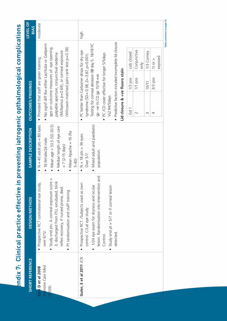
33 Eye Care for Critically Ill Adults 2014
SHO
RT
REF
EREN
CE
DES
IGN
/MET
HO
DSA
MPL
E D
ESC
RIP
TIO
NO
UTC
OM
ES/F
IND
ING
S LE
VEL
OF
BIA
S
Ezra
D e
t al
200
8 In
tens
ive
Car
e M
ed
(20
09)
• Pr
ospe
ctiv
e RC
T co
ntra
late
ral e
ye s
tudy
, ov
er 6
/12.
• St
udy
end
pts:
A c
orne
al e
xpos
ure
scor
e >
3,
dis
char
ged
from
ITU
, ext
ubat
ed, b
link
refle
x re
cove
ry, i
f nu
rsed
pro
ne, d
ied.
• Pt
ran
dom
isat
ion
and
staf
f tr
aini
ng
• N
= 4
0 ad
ult
pts
= 8
0 ey
es.
• 16
fem
ale/
24 m
ale.
• M
ean
age
= 5
3.5
(SD
20.
5)
• M
edia
n le
ngth
of
eye
care
=
7 (2
-15
days
)
• M
ean
Apa
che
= 1
6 (R
a 5-
40)
• Pr
ovid
ed t
hat
staf
f ar
e gi
ven
trai
ning
,
• N
o si
gnif
diff
Bw
eith
er L
acril
ube
or G
elip
erm
gp
s on
out
com
e m
easu
res
of: e
ye o
peni
ng,
palp
able
ape
rtur
e, c
onju
nctiv
al o
edem
a (M
cNam
us p
=0.
69),
or c
orne
al e
xpos
ure
(Wilc
oxon
mat
ched
pai
rs r
ank
test
p=
0.38
)
mod
erat
e
Gu
ler,
E e
t al
201
1 JC
N•
Pros
pect
ive
RCT.
/Su
bjec
ts u
sed
as o
wn
cont
rol:
C/L
at e
ye s
tudy
.
• 1
/24
eye
exam
for
dry
ness
and
ocu
lar
lesi
on. R
ando
mis
atio
n in
to In
terv
entio
n an
d C
ontr
ol.
• St
udy
end
pt =
5/7
or
if co
rnea
l les
ion
dete
cted
.
• N
= 1
8 pt
s =
36
eyes
. O
ver
5/7
.
• M
ixed
adu
lt an
d pa
edia
tric
po
pula
tion.
• PC
bet
ter t
han
Car
bom
er d
rops
for d
ry e
ye
synd
rom
e (S
D=
0.38
, Z=
-3.8
7, p
<0.
001)
. Te
stin
g fo
r cor
neal
abr
asio
n (@
day
5: 1
8/1
8 PC
gp
–ve
Vs
CD
gp
15/1
8 +
ve.
• PC
/CD
cov
er e
ffec
tive
for l
onge
r 5/5
days
Vs
2.94
/5da
ys.
• Pr
edic
tive
fact
ors
incl
uded
inco
mpl
ete
lid c
losu
re
Lid
clo
sure
& +
ve fl
uo
ro s
tain
Gd
11/
3 po
sLi
ds c
lose
d
21/
1 po
sC
onju
nctiv
a on
ly
310
/11
1/3
Cor
nea
43
/3 p
os½
or
>
expo
sed
high
App
endi
x 7:
Clin
ical
pra
ctic
e ef
fect
ive
in p
reve
ntin
g ia
trog
enic
opt
halm
olog
ical
com
plic
atio
ns
Tabl
e co
ntin
ues
on p
age
34
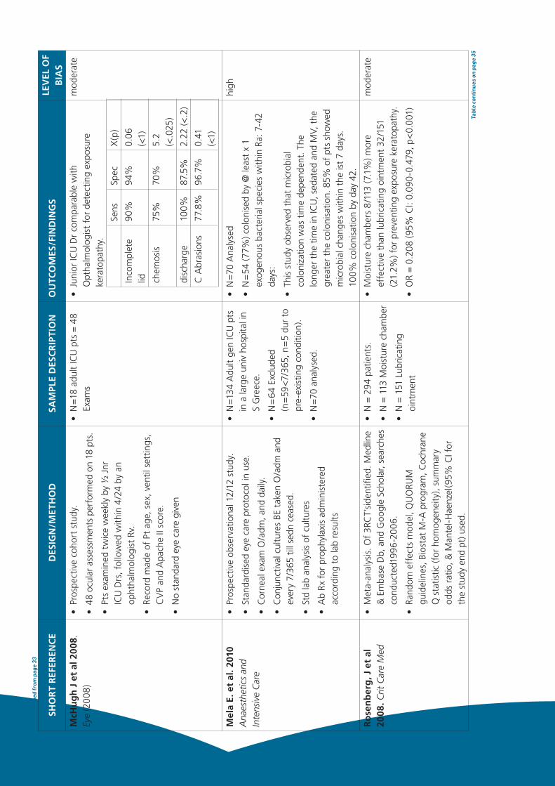
34 Eye Care for Critically Ill Adults 2014
SHO
RT
REF
EREN
CE
DES
IGN
/MET
HO
DSA
MPL
E D
ESC
RIP
TIO
NO
UTC
OM
ES/F
IND
ING
S LE
VEL
OF
BIA
S
McH
ug
h J
et
al 2
008.
Ey
e (2
008
)•
Pros
pect
ive
coho
rt s
tudy
.
• 48
ocu
lar
asse
ssm
ents
per
form
ed o
n 18
pts
.
• Pt
s ex
amin
ed t
wic
e w
eekl
y by
½ J
nr
ICU
Drs
, fol
low
ed w
ithin
4/2
4 by
an
opht
halm
olog
ist
Rv.
• Re
cord
mad
e of
Pt
age,
sex
, ven
til s
ettin
gs,
CV
P an
d A
pach
e II
scor
e.
• N
o st
anda
rd e
ye c
are
give
n
• N
=18
adu
lt IC
U p
ts =
48
Exam
s•
Juni
or IC
U D
r co
mpa
rabl
e w
ith
Opt
halm
olog
ist
for
dete
ctin
g ex
posu
re
kera
topa
thy.
Sens
Spec
X(p
)
Inco
mpl
ete
lid90
%94
%0.
06
(<1)
chem
osis
75%
70%
5.2
(<.0
25)
disc
harg
e10
0%87
.5%
2.22
(<.2
)
C A
bras
ions
77.8
%96
.7%
0.41
(<
1)
mod
erat
e
Mel
a E.
et
al. 2
010
Ana
esth
etic
s an
d In
tens
ive
Car
e
• Pr
ospe
ctiv
e ob
serv
atio
nal 1
2/1
2 st
udy.
• St
anda
rdis
ed e
ye c
are
prot
ocol
in u
se.
• C
orne
al e
xam
O/a
dm, a
nd d
aily
.
• C
onju
nctiv
al c
ultu
res
BE t
aken
O/a
dm a
nd
ever
y 7/
365
till s
edn
ceas
ed.
• St
d la
b an
alys
is o
f cu
lture
s
• A
b R
x fo
r pr
ophy
laxi
s ad
min
iste
red
acco
rdin
g to
lab
resu
lts
• N
=13
4 A
dult
gen
ICU
pts
in
a la
rge
univ
hos
pita
l in
S G
reec
e.
• N
=64
Exc
lude
d (n
=59
<7/3
65, n
=5
dur
to
pre-
exis
ting
cond
ition
).
• N
=70
ana
lyse
d.
• N
=70
Ana
lyse
d
• N
=54
(77%
) col
onis
ed b
y @
leas
t x
1 ex
ogen
ous
bact
eria
l spe
cies
with
in R
a: 7
-42
days
:
• Th
is s
tudy
obs
erve
d th
at m
icro
bial
co
loni
zatio
n w
as t
ime
depe
nden
t. T
he
long
er t
he t
ime
in IC
U, s
edat
ed a
nd M
V, t
he
grea
ter
the
colo
nisa
tion.
85%
of
pts
show
ed
mic
robi
al c
hang
es w
ithin
the
ist
7 da
ys.
100%
col
onis
atio
n by
day
42.
high
Ro
sen
ber
g, J
et
al
2008
. Crit
Car
e M
ed•
Met
a-an
alys
is. O
f 3R
CT’
side
ntifi
ed. M
edlin
e &
Em
base
Db,
and
Goo
gle
Scho
lar,
sear
ches
co
nduc
ted1
996
-20
06.
• Ra
ndom
eff
ects
mod
el, Q
UO
RUM
gu
idel
ines
, Bio
stat
M-A
pro
gram
, Coc
hran
e Q
sta
tistic
(for
hom
ogen
eity
), su
mm
ary
odds
rat
io, &
Man
tel-
Hae
nzel
(95%
CI f
or
the
stud
y en
d pt
) use
d.
• N
= 2
94 p
atie
nts.
• N
= 1
13 M
oist
ure
cham
ber
• N
= 1
51 L
ubric
atin
g oi
ntm
ent
• M
oist
ure
cham
bers
8/1
13 (7
.1%
) mor
e ef
fect
ive
than
lubr
icat
ing
oint
men
t 32
/151
(2
1.2%
) for
pre
vent
ing
expo
sure
ker
atop
athy
.
• O
R =
0.2
08 (
95%
CI:
0.09
0-0
.479
, p<
0.0
01)
mod
erat
e
Tabl
e co
ntin
ued
from
pag
e 33
Tabl
e co
ntin
ues
on p
age
35

35 Eye Care for Critically Ill Adults 2014
SHO
RT
REF
EREN
CE
DES
IGN
/MET
HO
DSA
MPL
E D
ESC
RIP
TIO
NO
UTC
OM
ES/F
IND
ING
S LE
VEL
OF
BIA
S
So H
et
al 2
008
Int
J of
N
ursi
ng S
tud.
• RC
T ov
er 2
0/1
2 In
20
04-2
005
.
• Le
gitim
ate
eye
care
tea
m u
sed.
• In
itial
bed
side
eye
exa
m t
o as
sess
: blin
k re
flex,
eye
lid
posn
, con
junc
tival
sta
tus
and
corn
eal c
hang
es.
• -
ve fl
uore
scei
n te
sted
Pt
wer
e ra
ndom
ised
in
to e
ither
pol
yeth
ylen
e co
vers
or
lano
lin
eye
oint
men
t.
• Fl
uore
scei
n te
st d
one
1/7
for
1 w
eek
by e
ye c
are
team
, the
n 1/
52 u
ntil
stud
y co
mpl
etio
n/e
nd p
oint
.
• N
= 1
16 a
dult
pts.
• N
= 5
9 m
oist
ure
cham
ber
• N
= 5
7 la
nolin
• Fa
irly
hom
ogen
ous
grou
p.
• N
o st
at s
igni
f: p
=.5
19 B
w t
he u
se o
f po
lyet
hyle
ne c
over
s 4
/59
(6.8
%) v
s.
lano
lin o
intm
ent
3/5
7 (5
.3%
) for
exp
osur
e ke
rato
path
y.
mod
erat
e
Tabl
e co
ntin
ued
from
pag
e 34

36 Eye Care for Critically Ill Adults 2014
SHO
RT
REF
EREN
CE
DES
IGN
/M
ETH
OD
REC
OM
MEN
DA
TIO
NS
FOR
PR
AC
TIC
E M
ETH
OD
OLO
GIC
AL
QU
ALI
TY
Ko
nn
o R
et
al 2
011
JBI
• Ey
e C
areC
PG
Follo
ws
the
AG
REE
App
rais
al T
ool.
1.
Regu
lar
asse
ssm
ent
of e
yes
and
care
nee
ded.
2.
Ass
essm
ent
to b
e in
clus
ive
of r
isk
fact
ors
for
iatr
ogen
ic e
ye c
ompl
icat
ions
: pat
ient
s’ a
bilit
y to
m
aint
ain
eyel
ids
clos
ed, a
nd s
igns
of
susp
ecte
d ey
e co
mpl
icat
ions
.
3.
Use
of
tape
or
/ and
Lac
rilub
e is
rec
omm
ende
d if
eyel
ids
cann
ot b
e cl
osed
.
4.
Use
of
poly
ethy
lene
film
s is
rec
omm
ende
d ov
er r
egul
ar e
ye in
stal
latio
ns a
nd/o
r dr
ops
for
redu
cing
the
inci
denc
e of
cor
neal
abr
asio
ns.
5.
App
licat
ion
of a
rtifi
cial
tea
r oi
ntm
ent
(Dur
atea
rs) i
s re
com
men
ded
over
pas
sive
eye
lid c
losu
re
alon
e, f
or r
educ
ing
the
inci
denc
e of
cor
neal
abr
asio
ns.
6.
Hyd
rom
ello
se lu
bric
atin
g oi
ntm
ent
(Lac
rilub
e) a
nd P
olye
thyl
ene
film
(Gla
dwra
p) a
re e
qual
ly
effe
ctiv
e in
pre
vent
ing
corn
eal d
amag
e.
7.
Refe
rral
for
any
sus
pect
ed ia
trog
enic
eye
com
plic
atio
ns s
uch
as w
hite
or
yello
w s
pots
on
the
surf
ace
of t
he c
orne
a, o
r co
njun
ctiv
al d
isch
arge
and
red
ness
.
8. T
o m
onito
r th
e ra
te o
f ia
trog
enic
eye
com
plic
atio
ns in
all
ICU
s.
9.
Ocu
lar
lubr
ican
t (L
acril
ube)
and
pol
yacr
ylam
ide
hydr
ogel
dre
ssin
gs (G
elip
erm
) may
be
used
fo
r th
e pr
even
tion
of e
xpos
ure
kera
topa
thy
in t
he c
ritic
ally
ill.
high
Mar
shal
l A e
t al
200
8 A
ust
Crit
Car
e•
Eye
Car
e C
PG
• A
dult
patie
nts
1.
For I
CU
nur
ses
to a
sses
s ea
ch p
atie
nt fo
r the
risk
fact
ors
of ia
trog
enic
oph
thal
mol
ogic
com
plic
atio
ns.
2.
Ass
essm
ent o
f the
abi
lity
of th
e pa
tient
to m
aint
ain
eyel
id c
losu
re s
houl
d be
per
form
ed d
aily
in
inte
nsiv
e ca
re p
atie
nts.
3.
Obs
erva
tion
for i
atro
geni
c op
htha
lmol
ogic
com
plic
atio
ns (a
t the
mic
ro e
pith
elia
l lev
el) s
houl
d be
pe
rfor
med
at l
east
wee
kly
in in
tens
ive
care
pat
ient
s us
ing
prac
tical
met
hods
read
ily a
vaila
ble
to b
usy
clin
icia
ns (f
or e
xam
ple,
the
inst
illatio
n of
fluo
resc
ein
and
use
of a
cob
alt b
lue
pen
torc
h).
4.
It is
reco
mm
ende
d th
at a
ll in
tens
ive
care
uni
ts m
onito
r the
rate
s of
iatr
ogen
ic o
phth
alm
olog
ic
com
plic
atio
ns.
5.
Refe
rral
mus
t be
mad
e in
a ti
mel
y m
anne
r for
any
sus
pect
ed ia
trog
enic
oph
thal
mol
ogic
co
mpl
icat
ions
in in
tens
ive
care
pat
ient
s.
6.
Eyel
id c
losu
re s
houl
d be
mai
ntai
ned
in in
tens
ive
care
pat
ient
s w
ho c
anno
t mai
ntai
n co
mpl
ete
eyel
id
clos
ure.
7.
If ey
elid
clo
sure
can
not b
e m
aint
aine
d pa
ssiv
ely
then
mec
hani
cal m
etho
ds s
houl
d be
use
d.
8.
All
patie
nts
who
can
not a
chie
ve e
yelid
clo
sure
inde
pend
ently
and
unc
onsc
ious
or h
eavi
ly s
edat
ed
patie
nts
shou
ld re
ceiv
e ey
e ca
re e
very
2 h
ours
(cle
anin
g w
ith s
alin
e so
aked
gau
ze a
nd th
e ad
min
istra
tion
of a
n ey
e sp
ecifi
c lu
bric
ant).
Hig
h
App
endi
x 8:
Clin
ical
pra
ctic
e gu
idel
ines
(AG
REE
tool
use
d fo
r as
sess
men
t)
Tabl
e co
ntin
ues
on p
age
37

37 Eye Care for Critically Ill Adults 2014
SHO
RT
REF
EREN
CE
DES
IGN
/M
ETH
OD
REC
OM
MEN
DA
TIO
NS
FOR
PR
AC
TIC
E M
ETH
OD
OLO
GIC
AL
QU
ALI
TY
Yi Y
200
9 U
niv
of H
ong
Kon
g M
aste
rs T
hesi
s
http
://h
ub.h
ku.h
k/bi
tstr
eam
/107
22/5
6966
/1/
FullT
ext.p
df?a
ccep
t=1
• SR
and
Eye
Car
e C
PG
• IC
U P
ts w
ith
dec
LOC
• U
ses
SIG
N
met
hod
Ass
essm
ent
1.
Ass
ess
risk
fact
ors
for
OSD
reg
ular
ly o
n al
l new
ly a
dmitt
ed IC
U p
atie
nts
rega
rdle
ss o
f th
eir
leve
ls o
f co
nsci
ousn
ess.
Pat
ient
s w
ho a
re a
t ris
k w
ill r
ecei
ve t
he c
orre
spon
ding
eye
car
e in
terv
entio
ns (G
OR
D)
a.
Ass
ess
the
risk
fact
ors
for
inco
mpl
ete
lid c
losu
re a
t le
ast
daily
. Pat
ient
s w
ho a
re a
t ris
k w
ill
rece
ive
the
follo
win
g ey
e as
sess
men
ts. T
he r
isk
fact
ors
incl
ude:
red
uced
con
scio
us le
vel,
prot
ectiv
e ey
e re
flexe
s, u
se o
f se
dativ
es o
r ne
urom
uscu
lar
rela
xant
s, t
rach
eal i
ntub
atio
n,
use
of P
EEP
of 5
or
abov
e, v
entil
atio
n in
pro
ne p
ositi
on, c
onju
nctiv
al o
edem
a, a
nd
sign
ifica
nt m
etab
olic
der
ange
men
t (c
ardi
ac o
r re
nal f
ailu
re) (
GO
R B
)
2.
Ass
ess
the
inco
mpl
ete
lid c
losu
re a
t le
ast
ever
y 8
hour
s, u
sing
a b
right
han
d-he
ld t
orch
in li
ne
with
eye
lash
es (G
OR
B)
a.
Patie
nts
who
are
una
ble
to m
aint
ain
com
plet
e lid
clo
sure
will
rec
eive
eye
hyg
iene
, eye
co
ver
or e
ye lu
bric
ant
(GO
R A
)
3.
Ass
ess
the
ocul
ar s
urfa
ce d
ryne
ss (d
ulln
ess
and
abse
nce
of s
park
les
at le
ast
ever
y 4
hour
s us
ing
a ha
nd-h
eld
torc
h (G
OR
D)
a.
Dry
ocu
lar
surf
ace
indi
cate
s a
need
of
havi
ng e
ye c
over
or
eye
lubr
ican
t (S
ee 7
.0-9
.1)
(GO
R D
)
4.
Ass
ess
the
lid c
lean
lines
s at
leas
t ev
ery
4 ho
urs.
Mor
e fr
eque
nt a
sses
smen
t is
req
uire
d fo
r pa
tient
s w
ith s
igns
of
eye
infe
ctio
n or
cop
ious
eye
dis
char
ge, o
r re
spira
tory
infe
ctio
n w
ith
copi
ous
sput
um p
rodu
ctio
n (t
hat
requ
ires
freq
uent
suc
tioni
ng a
t le
ast
2-ho
urly
), es
peci
ally
PA
ER in
fect
ion.
Unc
lean
lids
indi
cate
a n
eed
for
eye
hygi
ene
(see
6.0
and
6.1
) (G
OR
D)
5.
Ass
ess
the
sign
s of
OSD
s at
leas
t da
ily, u
sing
rea
dily
ava
ilabl
e to
ols
such
as
fluor
esce
in
stai
n an
d co
balt
blue
han
d-he
ld t
orch
. Oth
er s
igns
of
OSD
incl
ude
lid s
wel
ling,
con
junc
tival
sw
ellin
g w
ith h
yper
aem
ia, l
id m
argi
n cr
ustin
g, c
orne
al c
loud
ing,
epi
thel
ial l
oss,
red
ness
or
disc
harg
e (G
OR
C)
a.
Ass
ess
the
sign
s of
OSD
mor
e fr
eque
ntly
for
pat
ient
s w
ith r
espi
rato
ry in
fect
ion
espe
cial
ly
thos
e w
ith P
AER
infe
ctio
n or
cop
ious
spu
tum
pro
duct
ion
(tha
t re
quire
s at
leas
t 2h
ourly
su
ctio
ning
(GO
R D
)
b.
Sign
s of
OSD
s in
dica
te a
pro
mpt
med
ical
and
oph
thal
mic
con
sulta
tion
for
early
tre
atm
ent
and
com
plic
atio
ns p
reve
ntio
n (G
OR
C)
c.
Sign
s of
eye
infe
ctio
n in
dica
te a
n ey
e sw
ab f
or c
ultu
re a
nd m
ore
freq
uent
eye
hyg
iene
(G
OR
D)
Mod
erat
e
Tabl
e co
ntin
ued
from
pag
e 36
Tabl
e co
ntin
ues
on p
age
38

38 Eye Care for Critically Ill Adults 2014
SHO
RT
REF
EREN
CE
DES
IGN
/M
ETH
OD
REC
OM
MEN
DA
TIO
NS
FOR
PR
AC
TIC
E M
ETH
OD
OLO
GIC
AL
QU
ALI
TY
Yi Y
200
9 U
niv
of H
ong
Kon
g M
aste
rs T
hesi
s
http
://h
ub.h
ku.h
k/bi
tstr
eam
/107
22/5
6966
/1/
FullT
ext.p
df?a
ccep
t=1
• SR
and
Eye
Car
e C
PG
• IC
U P
ts w
ith
dec
LOC
• U
ses
SIG
N
met
hod
Inte
rven
tio
ns
6.
Perf
orm
lid
clea
nsin
g at
leas
t 4
-hou
rly f
or p
atie
nts
with
inco
mpl
ete
lid c
losu
re a
nd u
ncle
an
lids.
Mor
e fr
eque
nt li
d cl
eans
ing
is in
dica
ted
for
eye
infe
ctio
n or
cop
ious
eye
dis
char
ge, o
r re
spira
tory
infe
ctio
n w
ith c
opio
us s
putu
m (t
hat
requ
ires
freq
uent
suc
tioni
ng a
t le
ast
2-ho
urly
), es
peci
ally
PA
ER in
fect
ion.
(GO
R C
)
7.
Lid
clea
nsin
g w
ith s
teril
e w
ater
or
norm
al s
alin
e so
aked
ste
rile
gauz
e, in
onc
e-sw
ab-o
nce
man
ner,
is r
ecom
men
ded
over
eye
. How
ever
, the
use
of
norm
al s
alin
e is
stil
l con
trov
ersi
al u
ntil
furt
her
evid
ence
is a
vaila
ble.
Nur
ses’
han
d hy
gien
e is
em
phas
ised
. (G
OR
C)
Eye
cove
rs8.
For
pat
ient
s w
ith in
com
plet
e lid
clo
sure
and
dry
ocu
lar
surf
ace,
eye
s sh
ould
be
kept
clo
sed
by
mec
hani
cal e
ye c
over
s. M
echa
nica
l eye
cov
er is
pre
ferr
ed o
ver
eye
lubr
ican
t. (G
OR
A)
a.
Tran
spar
ent
poly
ethy
lene
cov
ers
(Gla
dwra
p) is
sug
gest
ed t
o ap
ply
on c
lean
eye
s fr
om
eyeb
row
s to
che
ekbo
nes,
with
Mic
ropo
re s
ealin
g ed
ge if
nec
essa
ry. C
hang
e th
e po
lyet
hyle
ne c
over
s da
ily o
r w
hene
ver
nece
ssar
y (s
uch
as s
oile
d or
tor
n). (
GO
R A
)
b.
Mic
ropo
re t
apin
g is
NO
T re
com
men
ded
until
fur
ther
evi
denc
e is
ava
ilabl
e. (G
OR
D)
c.
Gel
iper
m a
nd C
orne
a C
are
cove
rs a
re N
OT
reco
mm
ende
d. (G
OR
B)
Eye
lub
rica
nt
9.
Eye
lubr
ican
t is
rec
omm
ende
d w
hen
eye
cove
r is
not
app
licab
le, s
uch
as t
he p
atie
nts
with
eye
in
fect
ion
or c
opio
us e
ye s
ecre
tion,
or
occa
sion
al s
pont
aneo
us b
link
refle
x.
a. D
urat
ears
is s
ugge
sted
to
appl
y to
the
“V
” po
cket
bet
wee
n ey
ebal
l and
low
er li
d ev
ery
4 ho
urs
(GO
R A
)
b H
ypro
mel
lose
, Lac
rilub
e an
d H
L co
mbi
natio
n ar
e N
OT
reco
mm
ende
d. (G
OR
A)
Eyec
ove
rs d
uri
ng
su
ctio
nin
g10
. A
pply
eye
cov
ers
durin
g op
en t
rach
eal o
roro
phar
ynge
al s
uctio
ning
(for
pat
ient
s w
ith
resp
irato
ry in
fect
ion)
and
cop
ious
spu
tum
pro
duct
ion
(tha
t re
quire
s su
ctio
ning
at
leas
t 2-
hour
ly).
(GO
R C
)
a.
Shou
ld n
ot w
ithdr
aw t
he s
uctio
n ca
thet
er a
cros
s pa
tient
’s f
ace
afte
r su
ctio
ning
. (G
OR
D)
Prev
enti
on
or
man
agem
ent
of
con
jun
ctiv
al o
edem
a11
. To
red
uce
or p
reve
nt c
onju
nctiv
al o
edem
a, e
leva
te t
he h
ead
of b
ed, a
nd c
heck
for
ap
prop
riate
tig
htne
ss o
f ai
rway
sec
urin
g ta
ping
. (G
OR
D)
12.
Prev
entio
n of
Ven
tilat
or a
ssoc
iate
d pn
eum
onia
red
uces
the
ris
k of
eye
infe
ctio
n. F
or
exam
ple,
use
ase
ptic
tec
hniq
ue d
urin
g op
en t
rach
eal s
uctio
ning
, and
fol
low
the
VA
P bu
ndle
ca
re p
roto
col a
s im
plem
ente
d in
the
tar
get
ICU
.
Mod
erat
e
Tabl
e co
ntin
ued
from
pag
e 37

39 Eye Care for Critically Ill Adults 2014
FULL NAME OF PAPER REASONS FOR
NON-INCLUSION
Carstens, J.(2009)Eye Care: (ICU): Clinical InformationEvidence Summaries (July 30 2009) The Joanna Briggs Institute for Evidence Based Nursing and Midwifery, Systematic Review. Adelaide, SA.
• Reference source only
• X 4 recommendations based on Joyce N 2002 and Marshall A 2008
Coyer, F. Wheeler, M. Wetzig, S. Couchman, B. (2007)Nursing Care of the mechanically ventilated patient: What does the evidence say? Part Two
Intensive and Critical Care Nursing Vol 23, Issue 2, April 2007. pp 71-80
• SR but only ltd cover of eye care in ICU popn
• (x 3 references previously cited in other studies)
• Eye care not central to this study
Dawson D(2005)Development of a new eye care guideline for critically ill patients. Intensive and Critical Care Nursing. Vol 21, Issue 2, April 2005, pp 119-122
• Reference source only
• Incidence only useful +/- algorithm
Dingwall, L. (2010)Care of the eyes (Chpt3) in Personal Hygiene Care, Wiley-Blackwell, Oxford, UK.
• Reference source only
• Non ICU population
Ervin, AM.Wojciechowski, R. Schein, O. (2010) Punctal occlusion for dry eye syndrome. A Cochrane Review in the Cochrane Library 2010, Issue 9. John Wiley & Sons Publication.
• Reference source only
• Treatment review for eye condition
• Non ICU population
Ewald, M. Hammersmith, K.(2009) Review and diagnosis and management of recurrent erosion syndrome. Current Opinion in Opthalmology, 2009, 20:287-291
• Reference source only
• Not acute iatrogenic ophthalmic complication
• Non ICU population
Federici, L. proust, H. Conrath, J. Ridings, B (2006) Bilateral corneal endothelial decompensation after post septicaemia coma. J Fr Opthalmology, 2006 May; 29(5):556-8
• Abstract English, text in French
Fullbrook, P. (2006) Eye Care in ACCCN: Crit Care Nursing. P.190-191 • Reference source only
• Book pages on Eye Care for Critical Care Patients
• Nil author reference
Germano, E. Mello, M. Sena, D. Correia, J. Amorim, M. (2009)Incidence and risk factors of corneal epithelial defects in mechanically ventilated children. Crit Care Med 2009 Vol.37, No.3
• Brazilian paediatric study in 2001
Jackson W (2008) Blepharitis: current strategies for diagnosis and management. Can J Opthalmology. Vol 43, No.2, 2008
• Reference source only
• Non ICU population
Kam, R. Hayes, M. Joshi, N.(2011) Ocular care and complications in the critically ill. Trends in Anaesthesia and critical Care 1 (2011) 257-262
• Narrative review only
• Reference source only
Kocacal, E. Eser, I. (2008) Significant problems in ICU patients eye complications, medical education. Turkiye Klinikleri J med Sci 2008, 28:193-7
• Abstract in English, text in Turkish
Konno, R. (2011) Eye Care: (ICU) Clinicain Information. The Joanna Briggs Institute for Evidence Based Nursing and Midwifery, Systematic Review. Adelaide, SA. 03/03/2011
• Reference source only.
• Annotated CPG for ICU pts
• Insufficient information to assess quality
Maxeiner, H. Jekat, R. (2010) Resuscitation and conjunctival petechial hemorrhages. J Forensic Leg Med. 2010 Feb; 17(2):87-91. Epub 2009 Oct 6.
• Non ICU population: out of hospital cardiac arrest
• Majority of pts examined were deceased
Appendix 9: Research papers not included in 2012 Eye Care CPG
Table continues on page 40

40 Eye Care for Critically Ill Adults 2014
FULL NAME OF PAPER REASONS FOR
NON-INCLUSION
Oh, E. Lee, W. Yoo, J. Kim, S. Ko, I. Chu, S. Song, E. Kang, S. (2009)Factors related to the incidence of eye disorders in Korean patients at intensive care units. J. of Clinical Nursing, 18, 29-35.
• Ref source for incidence data only. Cases studied, not matched for confounding variables.
Pazzaolini, A. Pantaleo, F. (2006) Hygiene & eye care in critically ill patient. Prof Inferm. 2006 Oct-Dec; 59(4):246-8
• Article in Italian
Schutz, J. Mavrakanas, N.(2009)The value of opthalmic independent medical examination: analysis of 344 cases. Br J Opthalmology. 2009 Oct;93(10): 1371-5. Epub 2009 Jun 30.
• Non ICU population
Sorce, L. (2009)Preventing corneal abrasions in critically ill children receiving neuromuscular blockade: a RCT. Pediatr Crit Care Med. 2009 Vol. 10, No. 2
• Paediatric population
Sundling, V. Gulbrandsen, P. jervekk,J. Straand, J. (2008) Care of vision and ocular health in diabetic members of a national diabetes organization: a cross sectional study. BMC Health Services Research 2008, 8:159
• Non ICU sample
Suwan-apichon, O. Reyes, J. herretes, S. Vedula, S. chuck, R. (2009) Topical corticosteroids as adjunctive therapy for bacterial keratitis. A Cochrane Review in the Cochrane Library 2009, Issue 1. John Wiley & Sons Publication.
• Reference source only
Watson, S.Barker, N. (2009) Interventions for recurrent corneal erosions. A Cochrane Review in the Cochrane Library 2009, Issue 1. John Wiley & Sons Publication.
• Reference source only
• Community population
Xue, Y. (2011)Eye Care: ICU. The Joanna Briggs Institute for Evidence Based Nursing and Midwifery, Systematic Review. Adelaide, SA. 03/03/2011
• Reference use only for eye care algorithm
• Narrative review
STUDY NAME OUTCOME
Azfar et al Protocolized eye care prevents corneal complications in ventilated patients in a medical intensive care unit
al Saudi J Anaesth. 2013 Jan-Mar; 7(1): 33–36.
doi: 10.4103/1658-354X.109805
• Not included
• Limited quality improvement study –significant measurement bias
Ahmadi-Nejad Comparing the Effectiveness of Two Methods of Eye Care in the Prevention of Ocular Surface Disorders in Patients Hospitalized in Intensive Care Unit- journals.ajaums.ac.ir
• Not available
Werli-Avargenga Nursing Interventions for Adult Intensive care patients with risk for corneal injury: a systematic review. International journal of Nursing knowledge, 24(1): 25-29
• Not included because SR identified same studies as this review
• Quality limited due to sample bias
Grixti Uncommon ophthalmologic disorders in intensive care unit patients Journal of Critical Care 27(746.e9-e22
• Not included as SR on disorders not treatment
Jammal Exposure keratopathy in sedated and ventilated patients Journal of Critical Care 27: 537-541
• Included
Appendix 10: Updated literature review 2013A PUBMED and CINAHL search was undertaken using the same criteria as the 2012 search except years covered were 1/1/2012-8/7/2013. No controlled studies were found. No changes were made to the guideline
Table continued from page 39
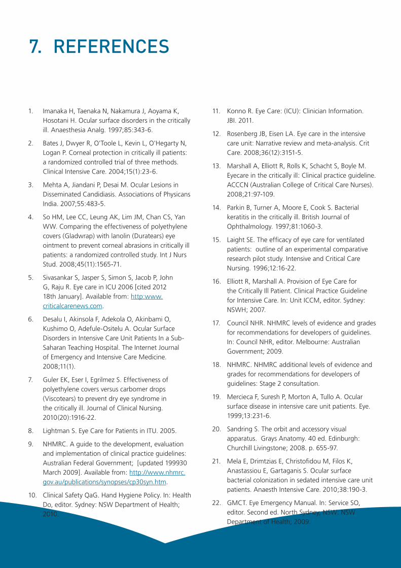
41 Eye Care for Critically Ill Adults 2014
1. Imanaka H, Taenaka N, Nakamura J, Aoyama K, Hosotani H. Ocular surface disorders in the critically ill. Anaesthesia Analg. 1997;85:343-6.
2. Bates J, Dwyer R, O'Toole L, Kevin L, O'Hegarty N, Logan P. Corneal protection in critically ill patients: a randomized controlled trial of three methods. Clinical Intensive Care. 2004;15(1):23-6.
3. Mehta A, Jiandani P, Desai M. Ocular Lesions in Disseminated Candidiasis. Associations of Physicans India. 2007;55:483-5.
4. So HM, Lee CC, Leung AK, Lim JM, Chan CS, Yan WW. Comparing the effectiveness of polyethylene covers (Gladwrap) with lanolin (Duratears) eye ointment to prevent corneal abrasions in critically ill patients: a randomized controlled study. Int J Nurs Stud. 2008;45(11):1565-71.
5. Sivasankar S, Jasper S, Simon S, Jacob P, John G, Raju R. Eye care in ICU 2006 [cited 2012 18th January]. Available from: http:www.criticalcarenews.com.
6. Desalu I, Akinsola F, Adekola O, Akinbami O, Kushimo O, Adefule-Ositelu A. Ocular Surface Disorders in Intensive Care Unit Patients In a Sub-Saharan Teaching Hospital. The Internet Journal of Emergency and Intensive Care Medicine. 2008;11(1).
7. Guler EK, Eser I, Egrilmez S. Effectiveness of polyethylene covers versus carbomer drops (Viscotears) to prevent dry eye syndrome in the critically ill. Journal of Clinical Nursing. 2010(20):1916-22.
8. Lightman S. Eye Care for Patients in ITU. 2005.
9. NHMRC. A guide to the development, evaluation and implementation of clinical practice guidelines: Australian Federal Government; [updated 199930 March 2009]. Available from: http://www.nhmrc.gov.au/publications/synopses/cp30syn.htm.
10. Clinical Safety QaG. Hand Hygiene Policy. In: Health Do, editor. Sydney: NSW Department of Health; 2010.
11. Konno R. Eye Care: (ICU): Clinician Information. JBI. 2011.
12. Rosenberg JB, Eisen LA. Eye care in the intensive care unit: Narrative review and meta-analysis. Crit Care. 2008;36(12):3151-5.
13. Marshall A, Elliott R, Rolls K, Schacht S, Boyle M. Eyecare in the critically ill: Clinical practice guideline. ACCCN (Australian College of Critical Care Nurses). 2008;21:97-109.
14. Parkin B, Turner A, Moore E, Cook S. Bacterial keratitis in the critically ill. British Journal of Ophthalmology. 1997;81:1060-3.
15. Laight SE. The efficacy of eye care for ventilated patients: outline of an experimental comparative research pilot study. Intensive and Critical Care Nursing. 1996;12:16-22.
16. Elliott R, Marshall A. Provision of Eye Care for the Critically Ill Patient. Clinical Practice Guideline for Intensive Care. In: Unit ICCM, editor. Sydney: NSWH; 2007.
17. Council NHR. NHMRC levels of evidence and grades for recommendations for developers of guidelines. In: Council NHR, editor. Melbourne: Australian Government; 2009.
18. NHMRC. NHMRC additional levels of evidence and grades for recommendations for developers of guidelines: Stage 2 consultation.
19. Mercieca F, Suresh P, Morton A, Tullo A. Ocular surface disease in intensive care unit patients. Eye. 1999;13:231-6.
20. Sandring S. The orbit and accessory visual apparatus. Grays Anatomy. 40 ed. Edinburgh: Churchill Livingstone; 2008. p. 655-97.
21. Mela E, Drimtzias E, Christofidou M, Filos K, Anastassiou E, Gartaganis S. Ocular surface bacterial colonization in sedated intensive care unit patients. Anaesth Intensive Care. 2010;38:190-3.
22. GMCT. Eye Emergency Manual. In: Service SO, editor. Second ed. North Sydney, NSW: NSW Department of Health; 2009.
7. REFERENCES

42 Eye Care for Critically Ill Adults 2014
23. Kam K, Hayes M, Joshi N. Ocular Care and complications in the critically ill. Trends in Anaesthesia and Critical Care. 2011(1):257-62.
24. Yi WY. Evidence-based Eye Protocol for ICU Patients with Altered Level of Consciousness: University of Hong Kong; 2009.
25. Black MD, Schorr C, Levy MM. Knowledge translation and the multifaceted intervention in the intensive care unit. Critical Care Medicine. 2012;40(4):1324-8.
26. Hernandez EV, Mannis MJ. Superficial keratopathy in Intensive Care unit patients. American Journal of Opthalmology. 1997;124:212-6.
27. Hilton E, Adams AA, Uliss A, Lesser ML, Samuels S, Lowy FD. Nosocomial bacterial eye infections in intensive care units. The Lancet. 1983(June 11,1983).
28. Ommeslag D, Colardyn F, De Laey J-J. Eye infections caused by respiratory pathogens in mechanically ventilated patients. Crit Care Med. 1987;15(1):80-1.
29. Cortese D, Capp L, McKinley S. Moisture chamber versus lubrication for the prevention of corneal epithelial breakdown. American Journal of Critical Care. 1995;4:425-8.
30. Koroloff N, Boots R, Lipman J, Thomas P, Rickard C, Coyer F. A randomised controlled study of the efficacy of hypromellose and Lacri-Lub combination versus polyethylene/Cling wrap to prevent corneal epithelial breakdown in the semiconscious intensive care patient. Intensive Care Medicine. 2004;30:1122-6.
31. Bates DW, Gawande AA. Error in medication: What have we learned? Annals of Internal Medicine. 2000;132(9):763-7.
32. Joyce N. Eye care for the intensive care patient. Adelaide: The Joanna Briggs Institute for Evidence Based Nursing and Midwifery, 2001.
33. Lenart SB, Garrity JA. Eye care for patients receiving neuromuscular blocking agents or propofol during mechanical ventilation. American Journal of Critical Care. 2000;9(3):188-91.
34. Ezra D, Chan M, Solebo L, Malik A, Crane E, Coombes A, et al. Randomized trial comparing ocular lubricants and polyacrylamide hydrogel dressings in the prevention of exposure keratopathy in the critically ill. Intensive Care Medicine. 2009;35:455-61.
35. Smith SB, Coffee T, Yowler C, Steinemann TI. Risk Factors for Ophthalmic Complications in Patients with burns. J Burn Care Res 2010;31:911-917. 2010;31(6):911-7.
36. Ramirez F, Ibarra S, Varon J, Tang R. The Neglected Eye: Ophthalmological Issues in the Intensive Care Unit. Crit Care & Shock. 2008;11:72-82.
37. Vriesendorp F. Clinical features and diagnosis of Guillain-Barre syndrome in adults. Up to Date Online. 2012;2012.
38. Pelak V, Quan D. Ocular myasthenia gravis. UpToDate Online. 2011;2012.
39. Klig J. Opthalmologic Complications of Systemic Disease. Emerg Med Clin N Am. 2008;26(2008):217-31.
40. Jammal H, Khader Y, Shihadeh W, Ababneh L, AlJizawi G, AlQasem A. Exposure keratopathy in sedated and ventilated patients. Journal of Critical Care. 2012;27(6):537-41.
41. Oh EG, Lee WH, Yoo JS, Kim SS, Ko IS, Chu SH, et al. Factors related to incidence of eye disorders in Korean patients at intensive care units. Journal of Clinical Nursing. 2008;18:29-35.
42. Sorce LH, S. Gauvreau, K. Mets, M. Hunter, D. Rahmani, B. Wu, C. Curley, M. Preventing corneal abrasions in critically ill children receiving neuromuscular blockade: A randomized, controlled trial. Pediatr Crit Care Med 2009;10(2):171-5.
43. Cotese D, Capp L, McKinley S. Moisture chamber versus lubrication for the prevention of corneal epithelial breakdown. American Journal of Critical Care. 1995;4:425-8.
44. Suresh P, Mercieca F, Morton A, Tullo AB. Eye care for the critically ill. Intensive Care Medicine. 2000;26:162-6.
45. Hillier S, Grimmer-Somers K, Merlin T, Middleton P, Salisbury J, Tooher R, et al. FORM: an Australian method for formulating and grading recommendations in evidence-based clinical guidelines. BMC Medical Research Methodology. 2011;11:23.
46. McHugh J, Alexander P, Kalhoro A, Lonides.A. Screening for ocular surface disease in the intensive care unit. Eye. 2007,22(12): 1465-1468.
47. Ezra.D.G, M H, Coombes A. Assessment of corneal epitheliopathy in the critically ill. Intensive Care Medicine. 2004.

43 Eye Care for Critically Ill Adults 2014


