36
EYE STRUCTURE and VISION
| Date post: | 24-Dec-2015 |
| Category: |
Documents |
| Upload: | jason-webster |
| View: | 213 times |
| Download: | 0 times |
EYE STRUCTURE and VISION

GENERAL FACTS ABOUT THE EYE
Is a sphere about 1 inch in diameter (about 2.5 cm.)
Is protected by the orbital socket of the skull and by the eyebrows,eyelids, and eyelashes.
Continuously bathed in fluid secreted by the lacrimal glands(tears empty into nasal cavity.)
Secretions have some antibiotic properties.
Thin membrane which lines the eyelids and covers part of the eyeis called the conjunctiva. It secretes mucous to help lubricate theeye.
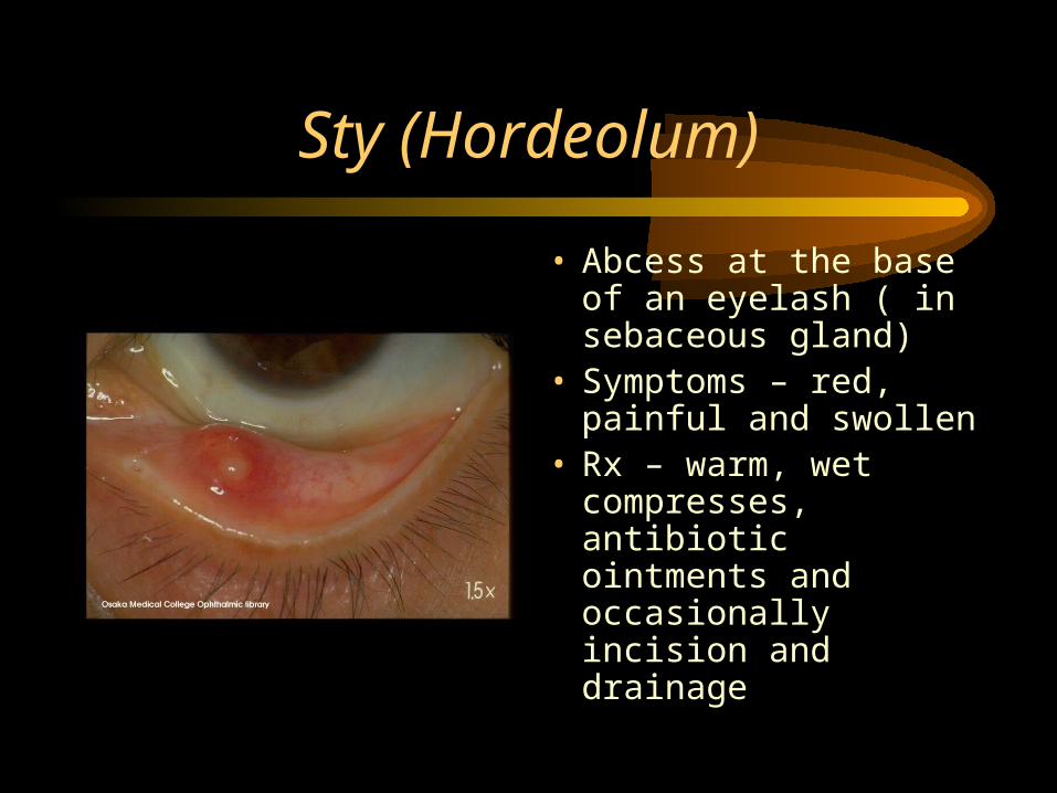
Sty (Hordeolum)
• Abcess at the base of an eyelash ( in sebaceous gland)
• Symptoms – red, painful and swollen
• Rx – warm, wet compresses, antibiotic ointments and occasionally incision and drainage

Conjunctivitis• Commonly known as pink eye• Inflammation of conjunctival
membranes in front of the eye• Caused by a variety of
pathogens including the bacterium Staphylococcus and Chlaymdia
• Symptoms are redness, pain, swelling, and discharge
• Highly contagious

Wall of the eye is made up of 3 layers or coats.
• Sclera - – Outer layer
– “white of the eye”
– tough, fibrous capsule which maintains the eye’s shape
– Extrinsic Muscles - muscles that are attached to the sclera and responsible for moving the eye.

Cornea - “Window of the Eye”• Located in very front center
of the sclera
• Transparent - no blood vessels
• Allows outside light to pass through the eye.
• Gets oxygen and nutrients through lymph fluid.
• Has pain and touch receptors
• Injury = scarring and impaired vision.

Choroid Coat - Middle Layer of the Eye
• Contains many blood vessels that provides nourishment to the eye.
• Pupil - dark circular opening in the center
• Iris - Colored muscular layer surrounding pupil
• Intrinsic Muscles in Iris- contraction of muscle dilates or constricts pupil. The larger the pupil - the more light that can enter the eye.
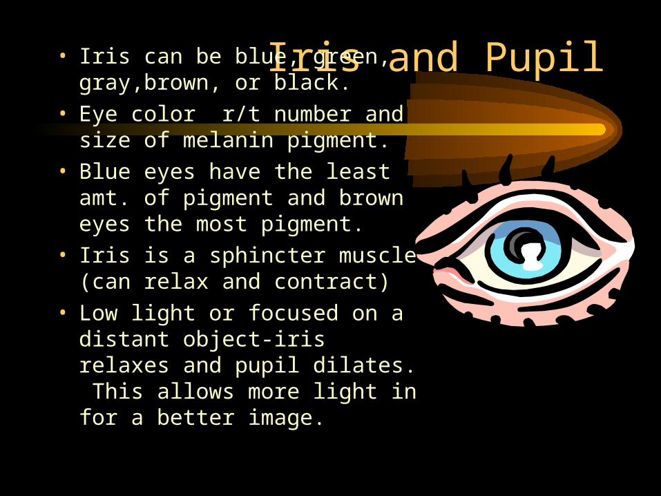
Iris and Pupil• Iris can be blue, green, gray,brown, or black.
• Eye color r/t number and size of melanin pigment.
• Blue eyes have the least amt. of pigment and brown eyes the most pigment.
• Iris is a sphincter muscle (can relax and contract)
• Low light or focused on a distant object-iris relaxes and pupil dilates. This allows more light in for a better image.
PUPILS• The word “pupil” comes
from the Latin word “pupa” that means doll. The use of the word pupil for the center of the eye may have come from the observation that if you look into the eye of another person, a small version of yourself (a doll) is reflected back.
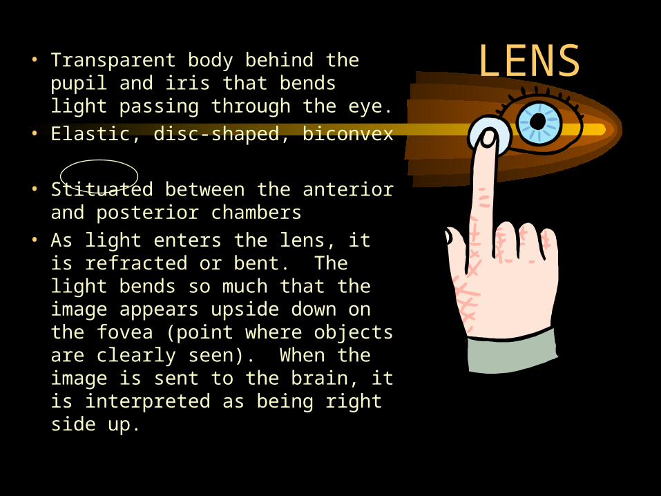
LENS• Transparent body behind the pupil and iris that bends light passing through the eye.
• Elastic, disc-shaped, biconvex
• Stituated between the anterior and posterior chambers
• As light enters the lens, it is refracted or bent. The light bends so much that the image appears upside down on the fovea (point where objects are clearly seen). When the image is sent to the brain, it is interpreted as being right side up.

Cataracts
• Lens of eyes gradually becomes cloudy
• Frequently occurs in people over 70
• Causes a painful, gradual blurring and loss of vision
• Pupil turns from black to milky white
• Rx – surgical removal of the lens
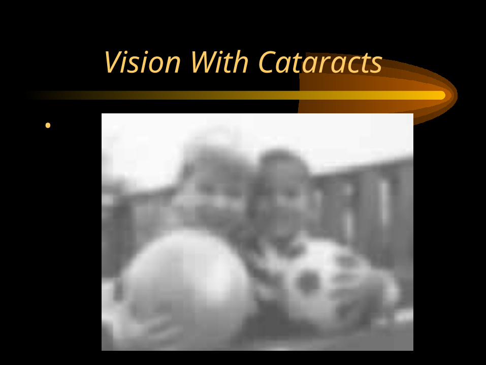
Vision With Cataracts
•

Chambers of Fluid Within the Eye
(help to maintain the eyeball’s spherical shape
• Anterior Chamber– Filled with a clear
watery fluid called aqueous humor.
– Found in front of the lens.
– Constantly replenished by blood vessels behind the iris.
• Posterior Chamber– Filled with a clear,
jelly-like fluid called vitreous humor.
– Found in back of lens.
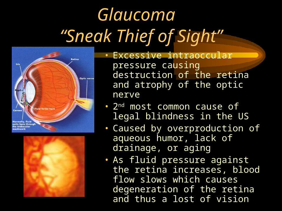
Glaucoma “Sneak Thief of Sight”
•
• Excessive intraoccular pressure causing destruction of the retina and atrophy of the optic nerve
• 2nd most common cause of legal blindness in the US
• Caused by overproduction of aqueous humor, lack of drainage, or aging
• As fluid pressure against the retina increases, blood flow slows which causes degeneration of the retina and thus a lost of vision
Glaucoma (continued)
Damage appears first at the edge of the retina, causing a gradual lost of peripheral vision called “tunnel vision.”
Symptoms develop gradually – mild aching, loss of peripheral vision, halo around the light
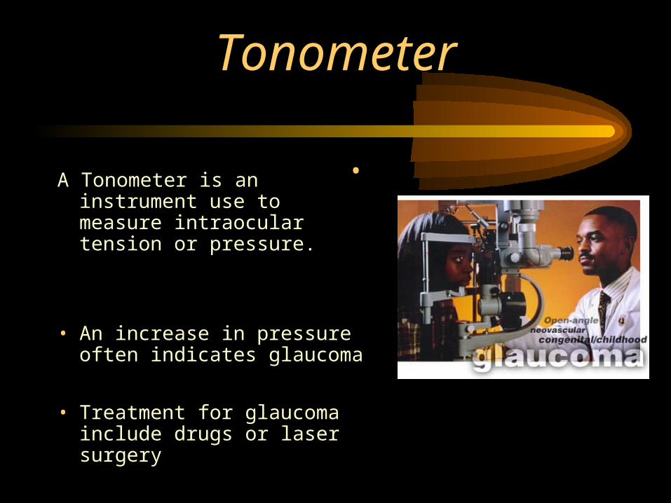
Tonometer
A Tonometer is an instrument use to measure intraocular tension or pressure.
• An increase in pressure often indicates glaucoma
• Treatment for glaucoma include drugs or laser surgery
•
Retina - Innermost Layer• Located between the posterior chamber and the choroid
coat.
• Light rays focus an image on the retina
• It is upon this light-sensitive layer that light rays from an object form an image.
• The image travels by electrical impulses via the optic nerve to the visual part of the cerebral cortex (occipital lobe) where it is interpreted.
• If light rays do not focus correctly on the retina, can correct condition with glasses or contact lenses which will bend the light rays as required.
• Contains rods and cones.
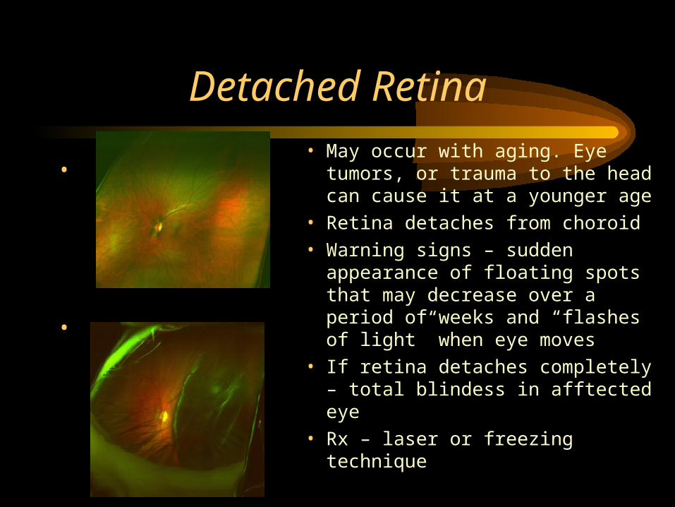
Detached Retina
•
•
• May occur with aging. Eye tumors, or trauma to the head can cause it at a younger age
• Retina detaches from choroid
• Warning signs – sudden appearance of floating spots that may decrease over a period of weeks and “flashes of light” when eye moves
• If retina detaches completely – total blindess in afftected eye
• Rx – laser or freezing technique
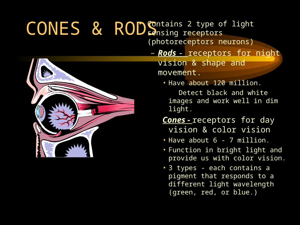
CONES & RODS• Contains 2 type of light sensing receptors (photoreceptors neurons)
– Rods - receptors for night vision & shape and movement.
• Have about 120 million.
Detect black and white images and work well in dim light.
Cones - receptors for day vision & color vision
• Have about 6 - 7 million.
• Function in bright light and provide us with color vision.
• 3 types - each contains a pigment that responds to a different light wavelength (green, red, or blue.)
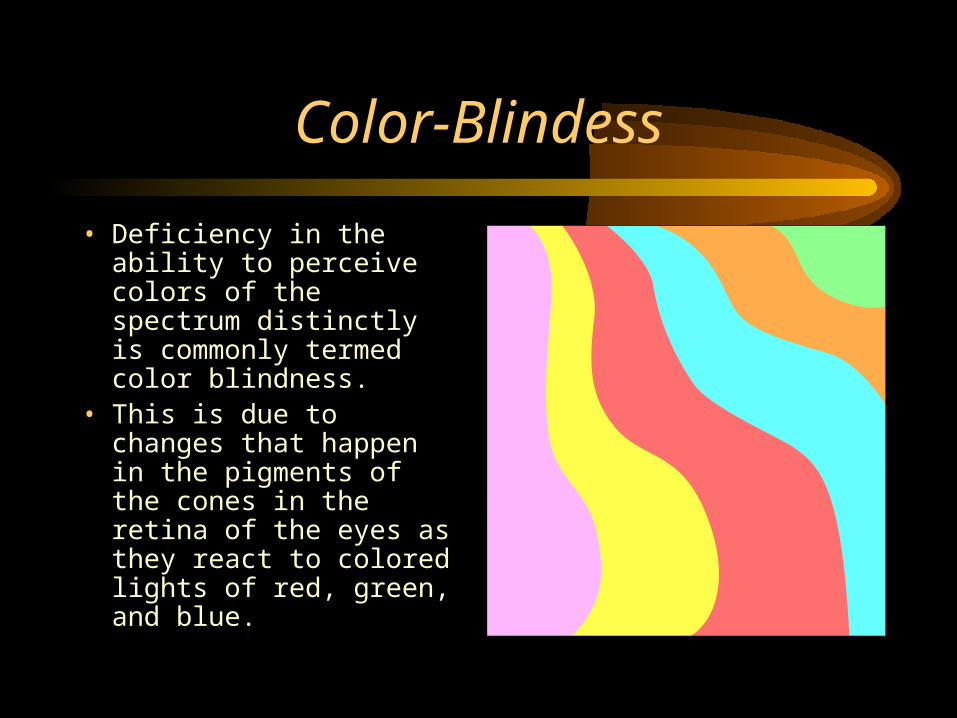
Color-Blindess
• Deficiency in the ability to perceive colors of the spectrum distinctly is commonly termed color blindness.
• This is due to changes that happen in the pigments of the cones in the retina of the eyes as they react to colored lights of red, green, and blue.
Tyeps of Color Blindness
• Achromatic Vision
• Total color blindness and very rare.
• Person cannot recognize any color at all. ( These people see everything in white, gray, or black.)
• Cause is the cones in the retina are defective or there may be none at all.
Daltonism
Most common.
•Person cannot tell the difference between red and green.
•It is an hereditary disorder.

Colorblindness• Ishihara color plates are used
to test for color blindness.• Patients are asked to trace the
pattern of color with their fingers as you observe them. There are letters and numbers that are one color within another.
• Make sure the room is well lit with natural daylight if possible so the patient will not have to squint.
• Make sure you have normal vision before you give the test.
OPTIC DISC AND FOVEA• If you look at the retina with an
ophthalmoscope you will see a yellow disc (macula lutea.)
• Within this disc is the fovea (contains cones for color vision.)
• Slightly to the side of the fovea is a pale disc called the optic disc or blind spot. (Contain no rods or cones and is insensitive to light.)
• Never fibers gather here to form the optic disc
• Each eye compensates for the blind spot of the other eye.
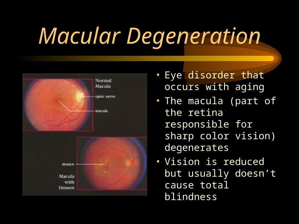
Macular Degeneration
• Eye disorder that occurs with aging
• The macula (part of the retina responsible for sharp color vision) degenerates
• Vision is reduced but usually doesn’t cause total blindness
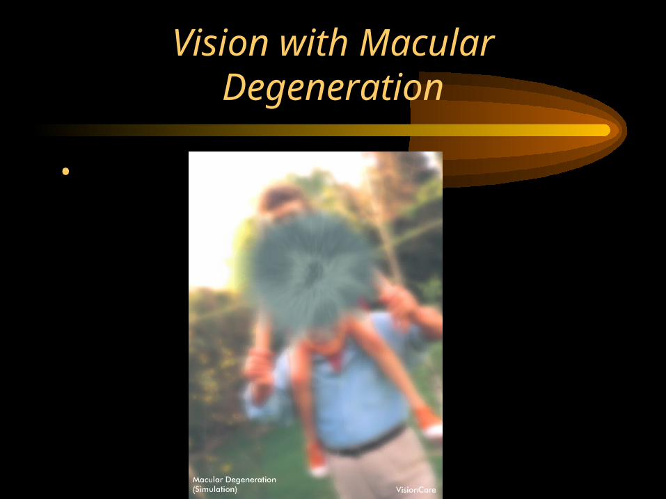
Vision with Macular Degeneration
•
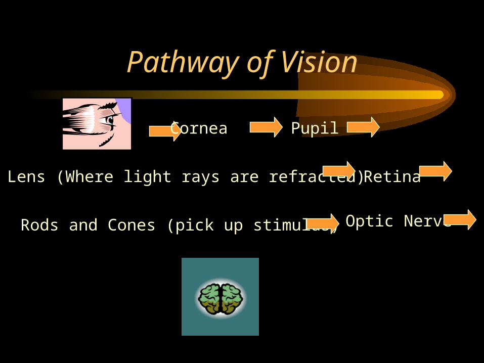
Pathway of Vision
Cornea Pupil
Lens (Where light rays are refracted) Retina
Rods and Cones (pick up stimulus) Optic Nerve

Myopia• Nearsighted• Eyeball too long• Light focuses in front of the
retina (in normal eye light focuses on the retina)
• Concave lenses help• Surgical Tx – Photo
Refractive Keratectomy – laser therapy used to reshape the anterior cornea of the eye.
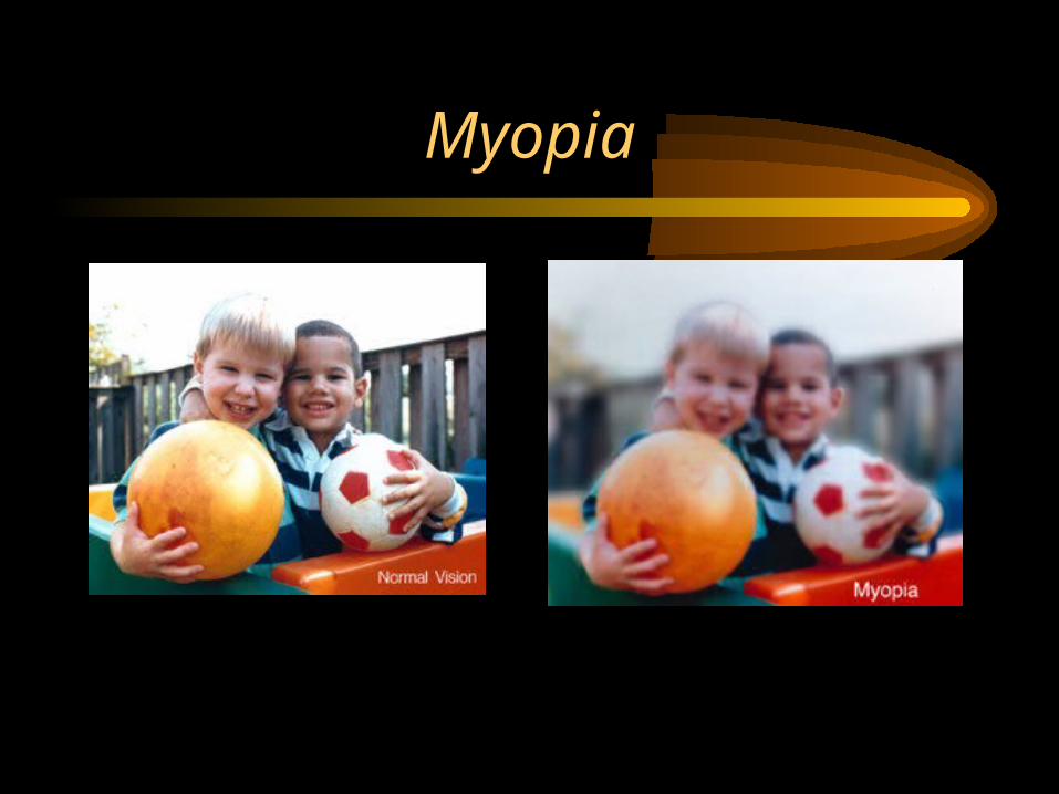
Myopia

Hyperopia
• Farsighted• Light focuses behind
the retina because the eyeball is too short
• Objects must be moved farther away from the eye to be seen clearer
• Convex lenses help
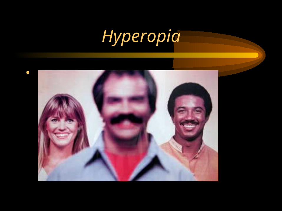
Hyperopia
•
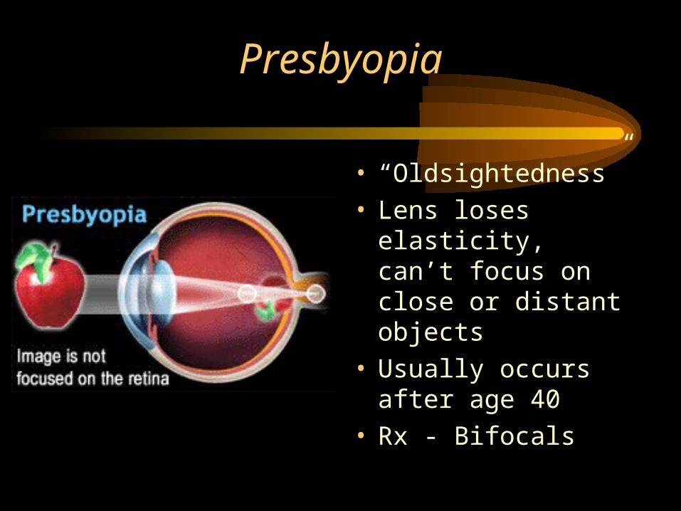
Presbyopia
• “Oldsightedness”• Lens loses elasticity,
can’t focus on close or distant objects
• Usually occurs after age 40
• Rx - Bifocals
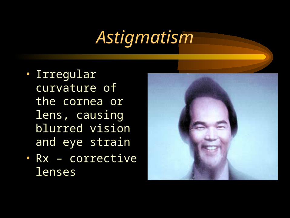
Astigmatism
• Irregular curvature of the cornea or lens, causing blurred vision and eye strain
• Rx – corrective lenses

Stabismus
• Strabismus (cross-eye)
• Eye muscles do not coordinate their actions
• If not corrected early, visual centers in the brain will learn to ignore information from one eye causing permanent blindness in affected eye
• Usually in children
• Rx – eye exercises or surgery

Additional Visual Disorders
• Amblyopia – reduction or dimness of vision
• Diplopia – double vision
Lights out folks!


