Fabrication, Characterization andCytotoxicity of Spherical-ShapedConjugated Gold-Cockle Shell DerivedCalcium Carbonate Nanoparticles forBiomedical ApplicationsHanan Karimah Kiranda1, Rozi Mahmud2, Danmaigoro Abubakar3 and Zuki Abubakar Zakaria1,3*
Abstract
The evolution of nanomaterial in science has brought about a growing increase in nanotechnology, biomedicine,and engineering fields. This study was aimed at fabrication and characterization of conjugated gold-cockleshell-derived calcium carbonate nanoparticles (Au-CSCaCO3NPs) for biomedical application. The synthetic techniqueemployed used gold nanoparticle citrate reduction method and a simple precipitation method coupled with mechanicaluse of a Programmable roller-ball mill. The synthesized conjugated nanomaterial was characterized for its physicochemicalproperties using transmission electron microscope (TEM), field emission scanning electron microscope (FESEM) equippedwith energy dispersive X-ray (EDX) and Fourier transform infrared spectroscopy (FTIR). However, the intricacy of cellularmechanisms can prove challenging for nanomaterial like Au-CSCaCO3NPs and thus, the need for cytotoxicity assessment.The obtained spherical-shaped nanoparticles (light-green purplish) have an average diameter size of 35 ± 16 nm, highcarbon and oxygen composition. The conjugated nanomaterial, also possesses a unique spectra for aragonite polymorphand carboxylic bond significantly supporting interactions between conjugated nanoparticles. The negative surface chargeand spectra absorbance highlighted their stability. The resultant spherical shaped conjugated Au-CSCaCO3NPs could be agreat nanomaterial for biomedical applications.
BackgroundThe production of monodisperse nanoparticles hasemerged significant in electronic, optical, biomedical,and magnetic applications [1–4]. Their evolution andthat of biomaterials has favorably enhanced pharmaceu-ticals [5], biomedical systems [6], drug delivery systems[7], cosmetics, and water treatment [7–9]. In the sameregard, the development of conjugated materials that arebiocompatible, biogenic, and nontoxic could have
valuable contributions to the fields of bioscience andbiomedicine [10]. Additionally, biocompatible metallicconjugated bio and nanomaterial could contribute tomore scientific advancements for biomedical applica-tions such as tissue engineering [5], therapeutics [11],and drug delivery [12]. This has been shown in recentworks elaborately, like the use of injectable self-assembling collagen-gold hybrid hydrogel [13], colloidalgold-collagen core-shell nanoconjugates [14], and co-assembled carrier-free nano drugs for antitumor therapy[15]. A number of studies have also documented thatmetallic nanoparticles can produce enzyme electrodes inelectrochemical biosensors with inorganic non-silicaporous materials [16]. Furthermore, the synthesized gra-phene oxide-albumin nano-hybrids have also displayed
* Correspondence: [email protected] of Molecular Biomedicine, Institute of Bioscience, Universiti PutraMalaysia, 43400 UPM, Serdang, Malaysia3Department of Veterinary Preclinical Sciences, Faculty of VeterinaryMedicine, Universiti Putra Malaysia, 43400 UPM, Serdang, MalaysiaFull list of author information is available at the end of the article
their potential benefit towards enhanced photodynamictherapy [17]. Altogether, this has only sparked moreinterest with other possible applications such as biomed-ical imaging and bio-sensory systems [16, 18].Calcium carbonate as a raw, natural mineral has been
used in a wide range of applications including biomed-ical, industrial, and nanotechnology [10, 19–21]. Aragon-ite as a calcium carbonate polymorph richly exists incockle shell (Anadara granosa), a molluscs popularly,also found in Malaysia [22]. Aragonite is biogenic unlikethe other calcium carbonate polymorphs of calcite andvaterite, making up to 95–98% of cockle shell. Calciumcarbonate, an inorganic material of aragonite polymorph,naturally and commonly exists within the cockle shells[23]. Aragonite polymorph has increasingly attractedattention in research field due to its biocompatibilityproperties and promising potential in the developmentof anticancer drug delivery systems [24] and biomedicalimaging [25, 26]. Currently, most of prior researchstudies have revealed mainly two methods of productionof calcium carbonate [26]. They include the co-precipitation or double decomposition and carbonationof CO2 gas through calcium hydroxide under controlledsettings, which regrettably none produces biogenic cal-cium carbonate [26–28]. Therefore, the products containa mixture of calcite and vaterite in high quantities whichare unsuitable for biomedical use because of their non-biocompatibility and high toxicity reports [26].However, with the increasing use of nanotechnology in
biomedical applications, the present study is focused onthe synthesis of controlled cockle shell-derived calciumcarbonate nanoparticles (CSCaCO3NPs) with unique sizeand shape using dodecyl dimethyl betaine (BS-12) [29].This is inspired by prior works that utilize BS-12 as biomineralization catalyst in the synthesis of CSCaCO3NPsthat can easily be manipulated for bio-applications, costefficient, and relatively pure nanoparticles [30]. Themorphology and size of synthesized nanoparticles arecrucial in determining their physicochemical properties,with focus on metal nanoparticles given their vast poten-tial biomedical applications [31]. Gold nanoparticles(AuNPs) have continuously been used due to their op-tical properties, different size range, and color which aredependent on absorption maxima variations or the syn-thesis method employed [32]. AuNPs’ size and shapeaffect their absorption and emission characteristics inthe light visible spectrum, making them vary from visibleto near infrared regions. Therefore, due to their synthe-sis [33], physicochemical properties [34], biocompatibil-ity [35], and surface functionalization [36], they can bemanipulated for different and particular applications[37]. In addition, it also has been stated that in medicaldiagnostics, they are not completely used and their valuepossibly obscure [37].
So perhaps upon appropriate functionalization, theycould be redesigned for cancer imaging [38], cancertreatment [39], drug delivery [40], and sensory gadgets[41]. A coating is essential to fabricate nano-hybridbiomaterial with functionalized properties like goldnanoparticles (AuNPs) conjugated with porous calciumcarbonate nano-spheres [16, 42]. The resultant conju-gated gold-calcium carbonate nanomaterial or nano-composite hybrid, which could retain the advantageousparental traits such as biocompatibility, good solubility,and dispersibility in solution [16]. Conjugated gold nano-particles that exhibit strong color change and localizedsurface plasmon resonance (LSPR) could be excellentcandidates for potential multiple receptor systems such asaptamers, peptides, and antibodies [35, 43–45]. The fabri-cation of water-soluble conjugated polymers and its appli-cations in biosensors, fluorescence imaging, and drugdelivery have been successfully realized [46–48]. However,the conjugated nanoparticles or nanomaterial has progres-sively improved advantages such as photo stability [48, 49]and low cytotoxicity [50] over the years except for friend-lier preparation [51] and separation features [48].Herewith, the AuNPs and CSCaCO3NPs are control-
lably synthesized and used to fabricate and characterizebiogenic conjugated gold-cockle shell-derived calciumcarbonate nanoparticles (Au-CSCaCO3NPs) whosediameter size ranges from 19–51 nm. Initially, theAuNPs preparation is inspired by the classic Turkevichmethod [52] and the cockle shell derived nanoparticlesusing the dodecyl dimethyl betaine synthetic approach[26]. The modifications in the synthetic parameters suchas concentration could proficiently decrease or increasetheir size. Consequently, the synthesized nanomaterialwas characterized and investigated for cytotoxicity. TheAu-CSCaCO3NPs preparation added advantages are;easy synthesis and cost efficiency.
Methods/ExperimentalMaterials and Chemical ReagentThe gold salt (tetra chloroauric acid containing 49% goldsolution) and the tri-sodium citrate were purchasedfrom prima nexus Sdn Bhd (Malaysia). Fresh cockle shellwas obtained from local market (Pasar borong, SeriKembangan, Selangor, Malaysia). Dodecyl dimethyl beta-ine (BS-12) and indocyanine green dye (ICG) were pur-chased from Sigma-Aldrich (Steinheim, Germany).Dulbecco’s modified Eagle’s medium (DMEM), fetal bo-vine serum (FBS), antibiotics combination (glutamine100 mmol/L, penicillin 100 U/mL, and streptomycin100 μg/mL), phosphate-buffered saline (PBS), dimethylsulfoxide (DMSO), and MTT (3-Dimethylthiazo-2, 5-diphynyltetrazolium Bromide dye) were purchased fromNaclai tesque, Inc., Kyoto, Japan. All other reagents usedwere of analytical grade.
Kiranda et al. Nanoscale Research Letters (2018) 13:1 Page 2 of 10
Synthesis of Gold NanoparticlesThe synthesis was achieved using a method earlier de-scribed by Verma et al. [53] with slight modifications inconcentrations, 1% tetra chloroauric acid containing49% gold solution. Approximately, 0.1% of the gold solu-tion was prepared and diluted in a series of concentra-tions of 15, 25, and 20 mM in different conical flasks,respectively. The solutions were then heated at 100 °Con a hot plate coupled with the magnetic stirring (6positioned, WiseStir ® Korea). Then, about 1% tri-sodium citrate was added to the boiling solution withcontinuous magnetic stirring until color transition(yellowish gold solution turned colorless then to blackthen finally turned brilliant red) was observed. The heatwas turned off after 15 min and allowed to cool at roomtemperature. The synthesized gold nanoparticles werethen stored at − 4 °C for further use. The reaction wasshown in the equation below:
Preparation and Synthesis of Cockle Shell-DerivedCalcium Carbonate Nanoparticles (CSCaCO3NPs)Three kilograms of freshly obtained cockle shells werethoroughly cleaned, scrubbed, and washed. The cockleshell powder was produced according to the method de-scribed by Islam et al. [54]. The cleaned cockle shell wasdried in an oven (Memmert UM500, GmbH Co,Germany) at 50 °C for 7 days. The cockle shells wereground into powder using a blender (Blender HCB, 550,USA) and sieved with a stainless laboratory test sieve(Endecott Ltd., made in London, England) with the aper-ture of 90 μm to obtain micron-size powder. The pow-der was dried for 7 days at 74 °C in the oven. Thepowder was further packed in airtight polythene plasticbag for later use. The cockle shell-derived calcium car-bonate nanoparticles were synthesized according to theapproach described by Islam et al. [55], with slight modi-fications to the method and synthesis parameters. Twograms of cockle shell powder were taken into 250 mlconical flask followed by 50 ml of double deionizedwater, and a concentration of 0.5 ml of BS-12 was addedinto the conical flask. The mixture in the conical flaskwas vigorously stirred at 1000 rpm, with a temperatureof 50 °C for 135 min using a systematic multi-hotplateand magnetic stirrer with small magnetic bar. The pre-pared sample was separated from the mother liquidusing double ring filter paper of size 125 mm (FiltresFioroni, China). The residue was then washed thor-oughly to remove the excess BS-12. The final products,
CSCaCO3NP powder, were packed in dry-clean con-tainer and dried for 3 days (Oven Memmert UM500,GmbH Co, Germany) at 74 °C. The container was prop-erly wrapped and sealed with Para film after addition ofmultiple small marble balls inside. The container wasplaced on a Programmable roller-ball mill (BML-6,Wisemix ® Korea) at speed of 200 rpm for 5 days. Thesample was stored in airtight polythene in oven forfurther use.
Synthesis of Conjugated Gold-Cockle Shell-DerivedCalcium Carbonate Nanoparticles (Au-CSCaCO3NPs) andInco-operation of Near Infrared (NIR) DyeIn this procedure, 0.2 g of CSCaCO3NPs and 5 mg ofnear infrared (NIR) Indocyanine green dye (ICG) weredispersed in 20 ml of gold colloid solution (pH 7)(AuNPs-solution), as similarly described by Cai et al.[16], in a clean empty conical flask. Further synthesismodifications were made, where the sample was soni-cated for 20 min and incubated on magnetic stirrer witha small magnetic bar at 200 rpm for 3 days. The samplewas ultra-centrifuged at a speed of 10,000 rpm for10 min to obtain light-green-purplish, Au-CSCaCO3NPcomposite. The supernatant was decanted and pelletwashed with a series of deionized water. The preparedcomposite material was dried in the oven for 4 days andstored in airtight polythene in oven for further analysis.
Characterization of Conjugated Gold-Cockle Shell-DerivedCalcium Carbonate Nanoparticles (Au-CSCaCO3NPs)The particle size and morphology of the nanomaterialwas analyzed using transmission electron microscope(TEM). The nanomaterial was dispersed in absolute al-cohol and sonicated for 40 min. Approximately, 5 μl ofthe suspended sample solution was pipetted out on tocopper grip specimen mount. The sample was viewedunder TEM (Hitachi H-7100). The field emission scan-ning electron microscope (FESEM) (Model JOEL 7600F)operated at voltage of 5 KV and equipped with energydispersive X-ray spectroscopy unit (EDX). This was usedto characterize the surface features of the Au-CSCaCO3NPs. The material was dispersed in absolutealcohol and sonicated for 1 h. About 50 μl of the sus-pended sample solution was pipetted out on to coppergrip specimen mount, dried overnight, and scannedusing the electron beams. In addition, the Fourier trans-form infrared spectrometer (FTIR) was also used forfunctional analysis of the synthesized conjugated nano-material; the nano material was calibrated in 1 wt% inKer (FTIR Model 100, Perkin Elmer) in the range of400–4000 cm−1. Furthermore, analysis for the synthe-sized nano conjugate size and zeta potential was doneusing zetasizer (Nano ZS, Malvern Instruments). Thematerial was suspended in deionized water and
Kiranda et al. Nanoscale Research Letters (2018) 13:1 Page 3 of 10
sonicated for 50 min; the homogenous suspension wasdeposited in the zetasizer cuvette and examined for par-ticle size and zeta potential. The presence of differentanalytes of the conjugated nano composite was moni-tored using Uv-Vis spectrophotometer (UV - 2600) atdifferent wavelength ranging from 300 to 800 nm.
Cell Culture and Cytotoxicity StudiesHuman breast adenocarcinoma cell line (JCRB: MCF-7)and the mouse fibroblast cell line (JCRB: NIH3T3) werecultured in DMEM (high glucose) supplemented with10% FBS and antibiotics combination (glutamine100 mmol/L, penicillin 100 U/mL, and streptomycin100 μg/mL). The culture flasks (Eppendorf culture T-25and T-75) were incubated in 5% carbondioxide at 37 °C,and cells at 80–90% confluence were used for seedingand treatment process.
Cells Seeding and TreatmentThe cells were seeded into 96-well sterile plates at adensity of 5 × 10³ cells per well and incubated for 24 hovernight. The media in each well were removed, andthe cells were treated and co-cultured in replicates withconjugated nano composite suspension (Au-CSCa-CO3NP) for a period of 24, 48, and 72 h. After treatmentexposure was completed, the media in the wells were as-pirated and washed with PBS before they were replacedwith another fresh media prior to experimentaltreatments.
Preparation of Au-CSCaCO3NPs for TreatmentStock solution of Au-CSCaCO3NPs at a concentrationof 1 mg/ml in 10% serum free DMEM media was pre-pared. After cell seeding of MCF-7 cells and NIH3T3cells in 96-well plates, the plates were treated andincubated with different concentrations in microgram(100–1.56) of the Au-CSCaCO3NPs solutions.
(MTT) 3-Dimethylthiazo-2, 5-diphynyltetrazolium BromideReagent Preparation and ProtocolTypically, 5 mg of MTT reagent powder was dissolvedin 1 ml of PBS facilitated by sonicator vortex for uni-form mixture. After cell seeding and treatment, the wellplates were cleared and 20 μl of MTT reagent was addedto each well. Immediately after, the plates were allowedto incubate for 3–4 h to allow binding of the MTT tothe mitochondria of the cells. After incubation, 1 ml ofDMSO was added to each of the wells which releasedthe color product into the solution. The plates were keptin a dark room for 30 min, and optical density (OD) ofthe solution was measured with a micro plate reader atwavelength of 570 nm [56]. The experiments were con-ducted in triplicates for each cell line, and the mean
values were recorded. The percentage of cell viabilitywas determined using the formula below.
Percentage of cell viability ¼ A Sample=A Controlð Þ � 100
where ASample was average OD reading of different in-cubated treated cells of both cell lines and AControl wasaverage OD reading of the different incubated cells incomplete culture media only. The cytotoxicity of thecells was then assessed from the average triplicate valuesand exhibited as mean ± standard deviation (SD).
Statistical AnalysisStatistical data analysis were done using SPSS software(Version 10, Chicago, USA). The experiments were donein triplicates and expressed as mean ± standard deviation(M ± SD). The significance threshold was p < 0.01.
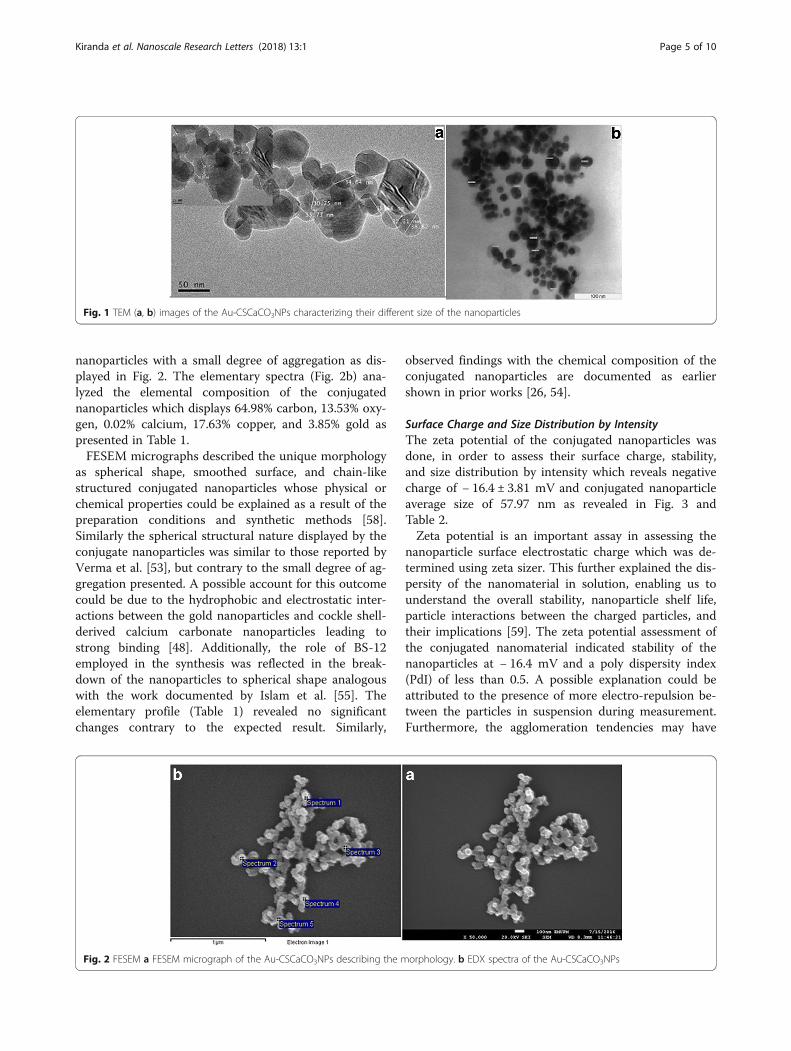
Results and DiscussionPhysicochemical Properties of the Conjugated Au-CSCaCO3NPsTransmission Electron MicroscopeThe purpose of the TEM micrographs was to assess thesize of the synthesized conjugated Au-CSCaCO3NPs whichshow well dispersed nanoparticles with average diametersize of 35 ± 16 nm within the range of (19–51 nm). Thedifferences in size attributed to the synthesis conditionswere as shown in Fig. 1.TEM micrographs of the nano conjugate showed ran-
ging diameter of 19–51 nm and dispersed nanoparticles.The uniquely obtained nano-size could be attributed tothe controlled synthetic conditions employed. Anotherpossible explanation for the nanoparticle dispersity couldbe due to the negatively charged layer of citrate ionswhich aided in the repulsions of nanoparticles from eachother and also, due to electrostatic repulsion and theconjugate hydration surface layer preventing aggregationand increasing conjugate stability as similarly reportedby Jazayeri et al. [56]. Furthermore, the citrate cappingreagent plays a role in the synthesis, which allowed formore dispersity and stability of the nanoparticle conju-gate as reported by Rawat et al. [57]. The unique particlesize showed the different absorbed gold nanoparticles in-side calcium carbonate nano-sphere matrix similar towork done by Cai et al. [16], contributing to the ob-served resulting particle size shown. However, this resultalso confirms reports that calcite has poor ability toaccommodate gold nanoparticles [16].
Field Emission Scanning Electron Microscopy (FESEM) andEnergy Dispersive X-ray Spectroscopy (EDX)The FESEM micrograph assessed the morphology andshape of the synthesized nanoparticles which showsspherically shaped and chain-like Au-CSCaCO3NPs
Kiranda et al. Nanoscale Research Letters (2018) 13:1 Page 4 of 10
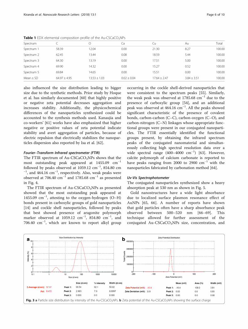
nanoparticles with a small degree of aggregation as dis-played in Fig. 2. The elementary spectra (Fig. 2b) ana-lyzed the elemental composition of the conjugatednanoparticles which displays 64.98% carbon, 13.53% oxy-gen, 0.02% calcium, 17.63% copper, and 3.85% gold aspresented in Table 1.FESEM micrographs described the unique morphology
as spherical shape, smoothed surface, and chain-likestructured conjugated nanoparticles whose physical orchemical properties could be explained as a result of thepreparation conditions and synthetic methods [58].Similarly the spherical structural nature displayed by theconjugate nanoparticles was similar to those reported byVerma et al. [53], but contrary to the small degree of ag-gregation presented. A possible account for this outcomecould be due to the hydrophobic and electrostatic inter-actions between the gold nanoparticles and cockle shell-derived calcium carbonate nanoparticles leading tostrong binding [48]. Additionally, the role of BS-12employed in the synthesis was reflected in the break-down of the nanoparticles to spherical shape analogouswith the work documented by Islam et al. [55]. Theelementary profile (Table 1) revealed no significantchanges contrary to the expected result. Similarly,
observed findings with the chemical composition of theconjugated nanoparticles are documented as earliershown in prior works [26, 54].
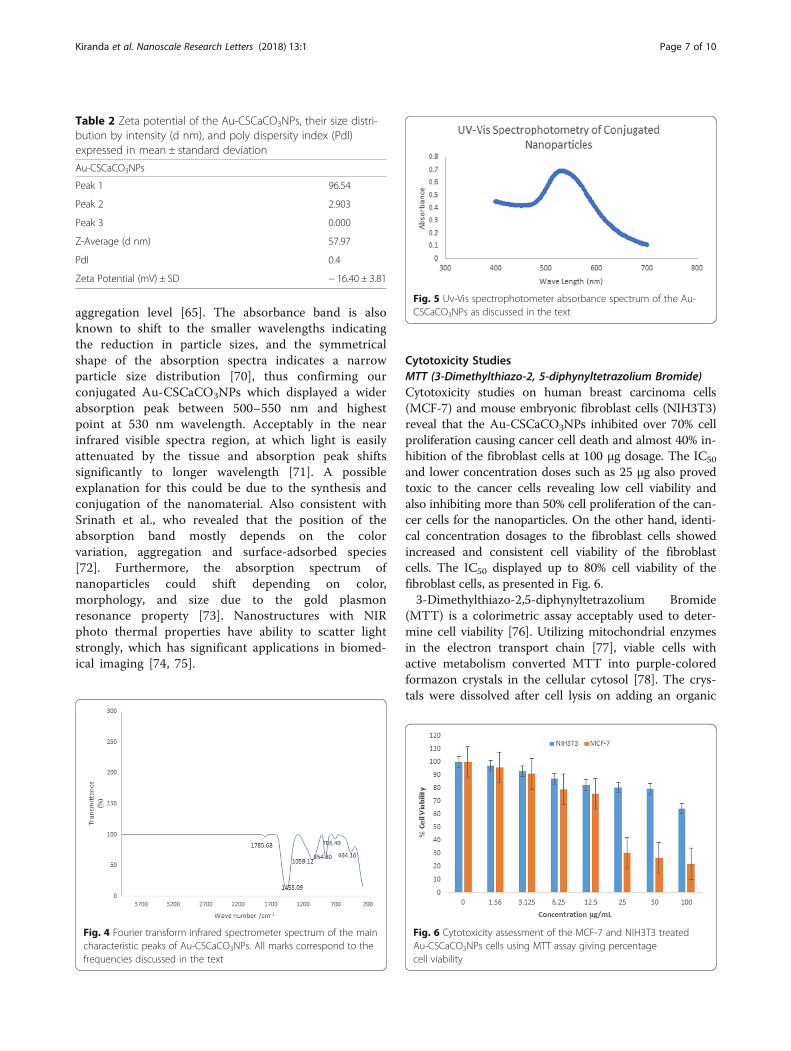
Surface Charge and Size Distribution by IntensityThe zeta potential of the conjugated nanoparticles wasdone, in order to assess their surface charge, stability,and size distribution by intensity which reveals negativecharge of − 16.4 ± 3.81 mV and conjugated nanoparticleaverage size of 57.97 nm as revealed in Fig. 3 andTable 2.Zeta potential is an important assay in assessing the
nanoparticle surface electrostatic charge which was de-termined using zeta sizer. This further explained the dis-persity of the nanomaterial in solution, enabling us tounderstand the overall stability, nanoparticle shelf life,particle interactions between the charged particles, andtheir implications [59]. The zeta potential assessment ofthe conjugated nanomaterial indicated stability of thenanoparticles at − 16.4 mV and a poly dispersity index(PdI) of less than 0.5. A possible explanation could beattributed to the presence of more electro-repulsion be-tween the particles in suspension during measurement.Furthermore, the agglomeration tendencies may have
Fig. 1 TEM (a, b) images of the Au-CSCaCO3NPs characterizing their different size of the nanoparticles
Fig. 2 FESEM a FESEM micrograph of the Au-CSCaCO3NPs describing the morphology. b EDX spectra of the Au-CSCaCO3NPs
Kiranda et al. Nanoscale Research Letters (2018) 13:1 Page 5 of 10
also influenced the size distribution leading to biggersize due to the synthetic methods. Prior study by Hoqueet al. has similarly documented [60] that highly positiveor negative zeta potential decreases aggregation andincreases stability. Additionally, the physicochemicaldifferences of the nanoparticles synthesized could beaccounted to the synthesis methods used. Kanaujia andco-workers’ [61] works have also emphasized that highernegative or positive values of zeta potential indicatestability and avert aggregation of particles, because ofelectric repulsion that electrically stabilizes the nanopar-ticles dispersion also reported by Isa et al. [62].
Fourier–Transform Infrared spectrometer (FTIR)The FTIR spectrum of Au-CSCaCO3NPs shows that themost outstanding peak appeared at 1455.09 cm−1
followed by peaks observed at 1059.12 cm−1, 854.80 cm−1, and 464.16 cm−1, respectively. Also, weak peaks wereobserved at 706.40 cm−1 and 1785.68 cm−1 as presentedin Fig. 4.The FTIR spectrum of Au-CSCaCO3NPs as presented
showed that the most outstanding peak appeared at1455.09 cm−1, attesting to the oxygen-hydrogen (O–H)bonds present in carboxylic groups of gold nanoparticles[14] and cockle shell nanoparticles, followed by peaksthat best showed presence of aragonite polymorphmarker observed at 1059.12 cm−1, 854.80 cm−1, and706.40 cm−1, which are known to report alkyl group
occurring in the cockle shell-derived nanoparticles thatwere consistent to the spectrum peaks [55]. Similarly,the weak peak was observed at 1785.68 cm−1 due to thepresence of carboxylic group [54], and an additionalpeak was observed at 464.16 cm−1. All the peaks showedsignificant characteristic of the presence of covalentbonds, carbon-carbon (C–C), carbon-oxygen (C–O), andcarbon-nitrogen (C–N) linkages whose appropriate func-tional groups were present in our conjugated nanoparti-cles. The FTIR essentially identified the functionalgroups present, by obtaining the infrared spectrumpeaks of the conjugated nanomaterial and simultan-eously collecting high spectral resolution data over awide spectral range (400–4000 cm−1) [63]. However,calcite polymorph of calcium carbonate is reported tohave peaks ranging from 2000 to 2900 cm−1 with thenanoparticles fabricated by carbonation method [64].
Uv-Vis SpectrophotometerThe conjugated nanoparticles synthesized show a heavyabsorption peak at 530 nm as shown in Fig. 5.Gold nanostructures have a wide light absorbance
due to localized surface plasmon resonance effect ofAuNPs [65, 66]. A number of reports have shownthat gold particles often have a sharp absorbance peakobserved between 500–520 nm [66–69]. Thistechnique allowed for further assessment of theconjugated Au-CSCaCO3NPs size, concentration, and
Table 1 EDX elemental composition profile of the Au-CSCaCO3NPs
Fig. 3 a Particle size distribution by intensity of the Au-CSCaCO3NPs. b Zeta potential of the Au-CSCaCO3NPs showing the surface charge
Kiranda et al. Nanoscale Research Letters (2018) 13:1 Page 6 of 10
aggregation level [65]. The absorbance band is alsoknown to shift to the smaller wavelengths indicatingthe reduction in particle sizes, and the symmetricalshape of the absorption spectra indicates a narrowparticle size distribution [70], thus confirming ourconjugated Au-CSCaCO3NPs which displayed a widerabsorption peak between 500–550 nm and highestpoint at 530 nm wavelength. Acceptably in the nearinfrared visible spectra region, at which light is easilyattenuated by the tissue and absorption peak shiftssignificantly to longer wavelength [71]. A possibleexplanation for this could be due to the synthesis andconjugation of the nanomaterial. Also consistent withSrinath et al., who revealed that the position of theabsorption band mostly depends on the colorvariation, aggregation and surface-adsorbed species[72]. Furthermore, the absorption spectrum ofnanoparticles could shift depending on color,morphology, and size due to the gold plasmonresonance property [73]. Nanostructures with NIRphoto thermal properties have ability to scatter lightstrongly, which has significant applications in biomed-ical imaging [74, 75].
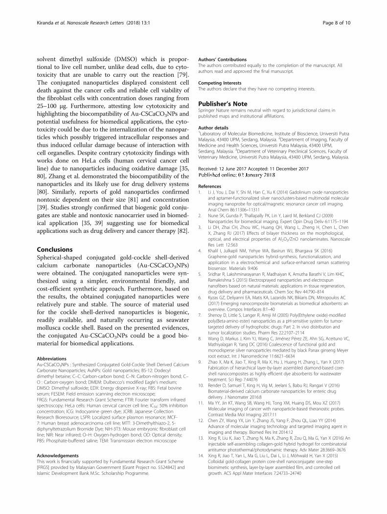
Cytotoxicity StudiesMTT (3-Dimethylthiazo-2, 5-diphynyltetrazolium Bromide)Cytotoxicity studies on human breast carcinoma cells(MCF-7) and mouse embryonic fibroblast cells (NIH3T3)reveal that the Au-CSCaCO3NPs inhibited over 70% cellproliferation causing cancer cell death and almost 40% in-hibition of the fibroblast cells at 100 μg dosage. The IC50
and lower concentration doses such as 25 μg also provedtoxic to the cancer cells revealing low cell viability andalso inhibiting more than 50% cell proliferation of the can-cer cells for the nanoparticles. On the other hand, identi-cal concentration dosages to the fibroblast cells showedincreased and consistent cell viability of the fibroblastcells. The IC50 displayed up to 80% cell viability of thefibroblast cells, as presented in Fig. 6.3-Dimethylthiazo-2,5-diphynyltetrazolium Bromide
(MTT) is a colorimetric assay acceptably used to deter-mine cell viability [76]. Utilizing mitochondrial enzymesin the electron transport chain [77], viable cells withactive metabolism converted MTT into purple-coloredformazon crystals in the cellular cytosol [78]. The crys-tals were dissolved after cell lysis on adding an organic
Table 2 Zeta potential of the Au-CSCaCO3NPs, their size distri-bution by intensity (d nm), and poly dispersity index (PdI)expressed in mean ± standard deviation
Au-CSCaCO3NPs
Peak 1 96.54
Peak 2 2.903
Peak 3 0.000
Z-Average (d nm) 57.97
PdI 0.4
Zeta Potential (mV) ± SD − 16.40 ± 3.81
Fig. 4 Fourier transform infrared spectrometer spectrum of the maincharacteristic peaks of Au-CSCaCO3NPs. All marks correspond to thefrequencies discussed in the text
Fig. 5 Uv-Vis spectrophotometer absorbance spectrum of the Au-CSCaCO3NPs as discussed in the text
Fig. 6 Cytotoxicity assessment of the MCF-7 and NIH3T3 treatedAu-CSCaCO3NPs cells using MTT assay giving percentagecell viability
Kiranda et al. Nanoscale Research Letters (2018) 13:1 Page 7 of 10
solvent dimethyl sulfoxide (DMSO) which is propor-tional to live cell number, unlike dead cells, due to cyto-toxicity that are unable to carry out the reaction [79].The conjugated nanoparticles displayed consistent celldeath against the cancer cells and reliable cell viability ofthe fibroblast cells with concentration doses ranging from25–100 μg. Furthermore, attesting low cytotoxicity andhighlighting the biocompatibility of Au-CSCaCO3NPs andpotential usefulness for biomedical applications, the cyto-toxicity could be due to the internalization of the nanopar-ticles which possibly triggered intracellular responses andthus induced cellular damage because of interaction withcell organelles. Despite contrary cytotoxicity findings withworks done on HeLa cells (human cervical cancer cellline) due to nanoparticles inducing oxidative damage [35,80], Zhang et al. demonstrated the biocompatibility of thenanoparticles and its likely use for drug delivery systems[80]. Similarly, reports of gold nanoparticles confirmednontoxic dependent on their size [81] and concentration[39]. Studies strongly confirmed that biogenic gold conju-gates are stable and nontoxic nanocarrier used in biomed-ical application [35, 39] suggesting use for biomedicalapplications such as drug delivery and cancer therapy [82].
ConclusionsSpherical-shaped conjugated gold-cockle shell-derivedcalcium carbonate nanoparticles (Au-CSCaCO3NPs)were obtained. The conjugated nanoparticles were syn-thesized using a simpler, environmental friendly, andcost-efficient synthetic approach. Furthermore, based onthe results, the obtained conjugated nanoparticles wererelatively pure and stable. The source of material usedfor the cockle shell-derived nanoparticles is biogenic,readily available, and naturally occurring as seawatermollusca cockle shell. Based on the presented evidences,the conjugated Au-CSCaCO3NPs could be a good bio-material for biomedical applications.
AbbreviationsAu-CSCaCO3NPs : Synthesized Conjugated Gold-Cockle Shell Derived CalciumCarbonate Nanoparticles; AuNPs: Gold nanoparticles; BS-12: Dodecyldimethyl betaine; C–C: Carbon-carbon bond; C–N: Carbon-nitrogen bond; C–O : Carbon-oxygen bond; DMEM: Dulbecco’s modified Eagle’s medium;DMSO: Dimethyl sulfoxide; EDX: Energy dispersive X-ray; FBS: Fetal bovineserum; FESEM: Field emission scanning electron microscope;FRGS: Fundamental Research Grant Scheme; FTIR: Fourier transform infraredspectroscopy; HeLa cells: Human cervical cancer cell line; IC50: 50% inhibitionconcentration; ICG: Indocyanine green dye; JCRB: Japanese CollectionResearch Bioresource; LSPR: Localized surface plasmon resonance; MCF-7: Human breast adenocarcinoma cell line; MTT: 3-Dimethylthiazo-2, 5-diphynyltetrazolium Bromide Dye; NIH-3T3: Mouse embryonic fibroblast cellline; NIR: Near infrared; O–H: Oxygen-hydrogen bond; OD: Optical density;PBS: Phosphate-buffered saline; TEM: Transmission electron microscope
AcknowledgementsThis work is financially supported by Fundamental Research Grant Scheme[FRGS] provided by Malaysian Government [Grant Project no. 5524842] andIslamic Development Bank M.Sc. Scholarship Programme.
Authors’ ContributionsThe authors contributed equally to the completion of the manuscript. Allauthors read and approved the final manuscript.
Competing InterestsThe authors declare that they have no competing interests.
Publisher’s NoteSpringer Nature remains neutral with regard to jurisdictional claims inpublished maps and institutional affiliations.
Author details1Laboratory of Molecular Biomedicine, Institute of Bioscience, Universiti PutraMalaysia, 43400 UPM, Serdang, Malaysia. 2Department of Imaging, Faculty ofMedicine and Health Sciences, Universiti Putra Malaysia, 43400 UPM,Serdang, Malaysia. 3Department of Veterinary Preclinical Sciences, Faculty ofVeterinary Medicine, Universiti Putra Malaysia, 43400 UPM, Serdang, Malaysia.
Received: 12 June 2017 Accepted: 11 December 2017
References1. Li J, You J, Dai Y, Shi M, Han C, Xu K (2014) Gadolinium oxide nanoparticles
and aptamer-functionalized silver nanoclusters-based multimodal molecularimaging nanoprobe for optical/magnetic resonance cancer cell imaging.Anal Chem 86:11306–11311
2. Nune SK, Gunda P, Thallapally PK, Lin Y, Laird M, Berkland CJ (2009)Nanoparticles for biomedical imaging. Expert Opin Drug Deliv 6:1175–1194
3. Li DH, Zhai CH, Zhou WC, Huang QH, Wang L, Zheng H, Chen L, ChenX, Zhang RJ (2017) Effects of bilayer thickness on the morphological,optical, and electrical properties of Al2O3/ZnO nanolaminates. NanoscaleRes Lett 12:563
4. Khalil I, Julkapli NM, Yehye WA, Basirun WJ, Bhargava SK (2016)Graphene-gold nanoparticles hybrid-synthesis, functionalization, andapplication in a electrochemical and surface-enhanced raman scatteringbiosensor. Materials 9:406
5. Sridhar R, Lakshminarayanan R, Madhaiyan K, Amutha Barathi V, Lim KHC,Ramakrishna S (2015) Electrosprayed nanoparticles and electrospunnanofibers based on natural materials: applications in tissue regeneration,drug delivery and pharmaceuticals. Chem Soc Rev 44:790–814
7. Shenoy D, Little S, Langer R, Amiji M (2005) Poly(Ethylene oxide)-modifiedpoly(Beta-amino ester) nanoparticles as a pH-sensitive system for tumor-targeted delivery of hydrophobic drugs: Part 2. In vivo distribution andtumor localization studies. Pharm Res 22:2107–2114
8. Wang D, Markus J, Kim YJ, Wang C, Jiménez Pérez ZE, Ahn SG, Aceituno VC,Mathiyalagan R, Yang DC (2016) Coalescence of functional gold andmonodisperse silver nanoparticles mediated by black Panax ginseng Meyerroot extract. Int J Nanomedicine 11:6621–6634
9. Zhao X, Ma K, Jiao T, Xing R, Ma X, Hu J, Huang H, Zhang L, Yan X (2017)Fabrication of hierarchical layer-by-layer assembled diamond-based core-shell nanocomposites as highly efficient dye absorbents for wastewatertreatment. Sci Rep 7:44076
10. Render D, Samuel T, King H, Vig M, Jeelani S, Babu RJ, Rangari V (2016)Biomaterial-derived calcium carbonate nanoparticles for enteric drugdelivery. J Nanomater 2016:8
11. Ma YY, Jin KT, Wang SB, Wang HJ, Tong XM, Huang DS, Mou XZ (2017)Molecular imaging of cancer with nanoparticle-based theranostic probes.Contrast Media Mol Imaging 2017:11
12. Chen ZY, Wang YX, Lin Y, Zhang JS, Yang F, Zhou QL, Liao YY (2014)Advance of molecular imaging technology and targeted imaging agent inimaging and therapy. Biomed Res Int 2014:12
13. Xing R, Liu K, Jiao T, Zhang N, Ma K, Zhang R, Zou Q, Ma G, Yan X (2016) Aninjectable self-assembling collagen-gold hybrid hydrogel for combinatorialantitumor photothermal/photodynamic therapy. Adv Mater 28:3669–3676
14. Xing R, Jiao T, Yan L, Ma G, Liu L, Dai L, Li J, Möhwald H, Yan X (2015)Colloidal gold-collagen protein core-shell nanoconjugate: one-stepbiomimetic synthesis, layer-by-layer assembled film, and controlled cellgrowth. ACS Appl Mater Interfaces 7:24733–24740
Kiranda et al. Nanoscale Research Letters (2018) 13:1 Page 8 of 10
15. Zhang R, Xing R, Jiao T, Ma K, Chen C, Ma G, Yan X (2016) Carrier-free,Chemophotodynamic dual Nanodrugs via self-assembly for synergisticantitumor therapy. ACS Appl Mater Interfaces 8:13262–13269
16. Cai WY, Xu Q, Zhao XN, Zhu JJ, Chen HY (2006) Porous gold-Nanoparticle−CaCO3 hybrid material: preparation, characterization, andapplication for horseradish Peroxidase assembly and directelectrochemistry. Chem Mater 18:279–284
17. Xing R, Jiao T, Liu Y, Ma K, Zou Q, Ma G, Yan X (2016) Co-assembly ofgraphene oxide and albumin/photosensitizer nanohybrids towardsenhanced photodynamic therapy. Polymers (Basel) 8:181
18. Tang D, Gao W, Yuan Y, Guo L, Mei X (2017) Novel biocompatible Aunanostars@PEG Nanoparticles for in vivo CT imaging and renal clearanceproperties. Nanoscale Res Lett 12:565
19. Zhang J, Li Y, Xie H, Su BL, Yao B, Yin Y, Li S, Chen F, Fu Z (2015) Calciumcarbonate nanoplate assemblies with directed high-energy facets: additive-free synthesis, high drug loading, and sustainable releasing. ACS Appl MaterInterfaces 7:15686–15691
20. Roy K, Alam N, Debnath SC (2014) Role of surface modified nano calciumcarbonate as filler and linseed oil as an extender in the vulcanization ofacrylonitrile butadiene rubber (NBR) nanocomposites. Int J Innov Res SciEng 2:69-75
21. Brecevic L, Kralj D (2007) On calcium Carbonates : from fundamentalresearch to application. Croat Chem Acta 80:467–484
22. Hariharan M, Varghese N, Cherian AB, Sreenivasan PV, Paul J, Antony AKA(2014) Synthesis and characterisation of CaCO3 (Calcite) nano particles fromcockle shells using chitosan as precursor. Int J Sci Res Publ 4:1–5
23. Shafiu Kamba A, Zakaria ZAB (2014) Osteoblasts growth behaviour on bio-based calcium carbonate aragonite nanocrystal. Biomed Res Int 2014:9
24. Svenskaya Y, Parakhonskiy B, Haase A, Atkin V, Lukyanets E, Gorin D, AntoliniR (2013) Anticancer drug delivery system based on calcium carbonateparticles loaded with a photosensitizer. Biophys Chem 182:11–15
25. Parakhonskiy BV, Svenskaya YI, Yashchenok AM, Fattah HA, Inozemtseva OA,Tessarolo F, Antolini R, Gorin DA (2014) Size controlled hydroxyapatite andcalcium carbonate particles: synthesis and their application as templates forSERS platform. Colloids Surf B: Biointerfaces 118:243–248
26. Islam KN, Zuki ABZ, Ali ME, Bin Hussein MZ, Noordin MM, Loqman MY,Wahid H, Hakim MA, Abd Hamid SB (2012) Facile synthesis of calciumcarbonate nanoparticles from cockle shells. J Nanomater 2012:1–5
27. Kamba AS, Ismail M, Azmi T, Ibrahim T, Abu Z, Zakaria B (2014)Biocompatibility of bio-based calcium carbonate nanocrystals aragonitepolymorph on NIH3T3 fibroblast cell line. African J Tradit ComplementAltern Med 11:31–38
28. Shafiu KA, Zakaria ZAB (2014) Osteoblasts growth behaviour on bio-basedcalcium carbonate aragonite nanocrystal. Biomed Res Int 2014:9
29. Shafiu KA, Ismail M, Tengku-Ibrahim TA, Zakaria ZAB (2013) Synthesis andcharacterisation of calcium carbonate aragonite nanocrystals from cockleshell powder (Anadara granosa). J Nanomater 2013:9
30. Islam KN, Bakar MZBA, Ali ME, Hussein MZB, Noordin MM, Loqman MY,Islam A, Islam MS, Rahman MM, Ullah M (2013) A novel method for thesynthesis of calcium carbonate (aragonite) nanoparticles from cockle shells.Powder Technol 235:70–75
31. Patra JK, Baek KH (2015) Novel green synthesis of gold nanoparticles usingCitrullus lanatus rind and investigation of proteasome inhibitory activity,antibacterial, and antioxidant potential. Int J Nanomedicine 10:7253–7264
32. Zhang J, Wang L, Pan D, Song S, Boey FYC, Zhang H, Fan C (2008) Visualcocaine detection with gold nanoparticles and rationally engineeredaptamer structures. Small 4:1196–1200
33. Hanžić N, Jurkin T, Maksimović A, Gotić M (2015) The synthesis of goldnanoparticles by a citrate-radiolytical method. Radiat Phys Chem 106:77–82
34. Jain PK, Lee KS, El-Sayed IH, El-Sayed MA (2006) Calculated absorption andscattering properties of gold nanoparticles of different size, shape, andcomposition: applications in biological imaging and biomedicine. J PhysChem B 110:7238–7248
35. Marisca O, Kantner K, Pfeiffer C, Zhang Q, Pelaz B, Leopold N, Parak W,Rejman J (2015) Comparison of the in vitro uptake and toxicity ofcollagen- and synthetic polymer-coated gold nanoparticles. Nano 5:1418–1430
36. Cheng K, Kothapalli SR, Liu H, Koh AL, Jokerst JV, Jiang H, Yang M, Li J,Levi J, Wu JC, Gambhir SS, Cheng Z (2014) Construction and validationof nano gold tripods for molecular imaging of living subjects. J AmChem Soc 136:3560–3571
37. Yasun E, Kang H, Erdal H, Cansiz S, Ocsoy I, Huang Y, Tan W (2013) Cancercell sensing and therapy using affinity tag-conjugated gold nanorods.Interface Focus 3:1–9
38. Popovtzer R (2011) Targeted gold nanoparticles enable molecular CTimaging of cancer: an in vivo study. Int J Nanomedicine 6:2859
39. Wójcik M, Lewandowski W, Król M, Pawłowski K, Mieczkowski J, Lechowski R,Zabielska K (2015) Enhancing anti-tumor efficacy of doxorubicin by non-covalent conjugation to gold nanoparticles—in vitro studies on felinefibrosarcoma cell lines. PLoS One 10:1–15
40. Ruan S, Yuan M, Zhang L, Hu G, Chen J, Cun X, Zhang Q, Yang Y, HeQ (2015) Tumor microenvironment sensitive doxorubicin delivery andrelease to glioma using angiopep-2 decorated gold nanoparticles.Biomaterials 37:425–435
41. Wang H, Liu Y, Li M, Huang H, Xu HM, Hong RJ, Shen H (2010) MultifunctionalTiO2 nanowires-modified nanoparticles bilayer film for 3D dye-sensitized solarcells. Optoelectron Adv Mater Rapid Commun 4:1166–1169
42. Dobrowolska P, Krajewska A, Gajda-Rączka M, Bartosewicz B, Nyga P,Jankiewicz B (2015) Application of Turkevich method for gold Nanoparticlessynthesis to fabrication of SiO2@Au and TiO2@au Core-Shell nanostructures.Materials (Basel) 8:2849–2862
44. Di Pasqua AJ, Ii REM, Ship Y, Dabrowiak JC, Asefa T (2009) Preparation ofantibody-conjugated gold nanoparticles. Mater Lett 63:1876–1879
45. Tan YN, Su X, Zhu Y, Lee JY (2010) Sensing of transcription factor throughcontrolled-assembly of metal nanoparticles modified with segmented DNAelements. ACS Nano 4:5101–5110
46. Hartnett ME, Tinkham N, Paynter L, Geisen P, Rosenberg P, Koch G, CohenKL (2009) Aqueous vascular endothelial growth factor as a predictor ofmacular thickening following cataract surgery in patients with diabetesmellitus. Am J Ophthalmol 148:895–901
47. Gao B, Li H, Xia D, Sun S, Ba X (2011) Amphiphilic dendritic peptides:synthesis and behavior as an organogelator and liquid crystal. Beilstein JOrg Chem 7:198–203
48. Xing C, Yang G, Liu L, Yang Q, Lv F, Wang S (2012) Conjugated polymers forlight-activated antifungal activity. Small 8:524–529
49. Li K, Ding D, Huo D, Pu KY, Thao NNP, Hu Y, Li Z, Liu B (2012)Conjugated polymer based nanoparticles as dual-modal probes fortargeted in vivo fluorescence and magnetic resonance imaging. AdvFunct Mater 22(15):3107–3115
50. Geng J, Li K, Pu KY, Ding D, Liu B (2012) Conjugated polymer and goldnanoparticle Co-loaded PLGA nanocomposites with eccentric internalnanostructure for dual-modal targeted cellular imaging. Small 8:2421–2429
51. Zhang J, Badugu R, Lakowicz JJR (2008) Fluorescence quenching ofCdTe nanocrystals by bound gold nanoparticles in aqueous solution.Plasmonics 3:3–11
53. Verma HN, Singh P, Chavan RM (2014) Gold nanoparticle: synthesis andcharacterization. Vet World 7:72–77
54. Islam KN, Bakar MZBA, Noordin MM, Bin HMZ, Rahman NSBA, Ali ME (2011)Characterisation of calcium carbonate and its polymorphs from cockle shells(Anadara granosa). Powder Technol 213:188–191
55. Islam KN, Ali ME, Bakar MZBA, Loqman MY, Islam A, Islam MS, Rahman MM,Ullah M (2013) A novel catalytic method for the synthesis of sphericalaragonite nanoparticles from cockle shells. Powder Technol 246:434–440
56. Isa T, Zakaria ZAB, Rukayadi Y, Hezmee MNM, Jaji AZ, Imam MU, HammadiNI, Mahmood SK (2016) Antibacterial activity of ciprofloxacin-encapsulatedcockle shells calcium carbonate (Aragonite) nanoparticles and itsbiocompatability in macrophage J774A.1. Int J Mol Sci 17:713
57. Jazayeri MH, Amani H, Pourfatollah AA, Pazoki-Toroudi H,Sedighimoghaddam B (2016) Various methods of gold nanoparticles (GNPs)conjugation to antibodies. Sens Bio-Sensing Res 9:17–22
58. Rawat P, Rajput YS, Bharti MK, Sharma R (2016) A method for synthesis ofgold nanoparticles using 1-amino-2- naphthol-4-sulphonic acid as reducingagent. Curr Sci 110:2–5
59. Schmidt H (2001) Nanoparticles by chemical synthesis, processing tomaterials and innovative applications. Appl Organomet Chem 15:331–343
60. Johnston BD, Kreyling WG, Pfeiffer C, Schäffler M, Sarioglu H, Ristig S,Hirn S, Haberl N, Thalhammer S, Hauck SM, Semmler‐Behnke M (2017)Colloidal Stability and Surface Chemistry Are Key Factors for the
Kiranda et al. Nanoscale Research Letters (2018) 13:1 Page 9 of 10
Composition of the Protein Corona of Inorganic Gold Nanoparticles.Adv. Funct. Mater 27:1-9.
61. Hoque E, Shehryar M, Islam KN (2013) Material Science & EngineeringProcessing and characterization of cockle shell calcium carbonate (CaCO3)bioceramic for potential application in bone tissue engineering. J Mater SciEng 2:2–6
62. Das S, Ng WK, Kanaujia P, Kim S, Tan RBH (2011) Formulation design,preparation and physicochemical characterizations of solid lipidnanoparticles containing a hydrophobic drug: effects of process variables.Colloids Surf B: Biointerfaces 88:483–489
63. Kurien S (2005) Analysis of FTIR spectra of nanoparticles of MgAl2O4,SrAl2O4, and NiAl2O4, 1595:64–78
64. Wang C, Liu Y, Bala H, Pan Y, Zhao J, Zhao X, Wang Z (2007) Facilepreparation of CaCO3 nanoparticles with self-dispersing properties in thepresence of dodecyl dimethyl betaine. Colloids Surfaces A PhysicochemEng Asp 297:179–182
65. Amendola V, Meneghetti M (2009) Size evaluation of gold nanoparticles byUV-vis spectroscopy. J Phys Chem C 113:4277–4285
66. Sadrolhosseini AR, Rashid SA, Zakaria A (2017) Synthesis of goldnanoparticles dispersed in palm oil using laser ablation technique. JNanomater 2017:5
67. Verma MS, Chen PZ, Jones L, Gu FX (2015) Controlling “chemical nose”biosensor characteristics by modulating gold nanoparticle shape andconcentration. Sens Bio-Sensing Res 5:13–18
68. Haiss W, Thanh NTK, Aveyard J, Fernig DG (2007) Determination of sizeand concentration of gold nanoparticles from UV-Vis spectra. AnalChem 79:4215–4221
69. Rahman S (2016) Size and concentration analysis of gold nanoparticles withultraviolet-visible spectroscopy. Undergrad J Math Model One + Two 7:13
70. Norfazila SM, Mohd JR (2014) Synthesis and ultraviolet visible spectroscopystudies of Chitosan capped gold nanoparticles and their reactions withanalytes. Sci World J 2014:7
71. Luo S, Zhang E, Su Y, Cheng T, Shi C (2011) A review of NIR dyes in cancertargeting and imaging. Biomaterials 32:7127–7138
72. Srinath BS, Ravishankar Rai V (2015) Biosynthesis of highly monodispersed,spherical gold nanoparticles of size 4–10 nm from spent cultures ofKlebsiella pneumoniae. 3. Biotech 5:671–676
73. Bao G, Mitragotri S, Tong S (2013) Multifunctional nanoparticles for drugdelivery and molecular imaging. Annu Rev Biomed Eng 4:253–282
74. Vats M, Mishra SK, Baghini MS, Chauhan DS, Srivastava R, De A (2017) Nearinfrared fluorescence imaging in nano-therapeutics and photo-thermalevaluation. Int J Mol Sci 18:924
75. Abadeer NS, Murphy CJ (2016) Recent progress in cancer thermal therapyusing gold nanoparticles. J Phys Chem C 120:4691–4716
76. Mosmann T (1983) Rapid colorimetric assay for cellular growth andsurvival: application to proliferation and cytotoxicity assays. J ImmunolMethods 65:55–63
77. Fotakis G, Timbrell JA (2006) In vitro cytotoxicity assays: comparison of LDH,neutral red, MTT and protein assay in hepatoma cell lines followingexposure to cadmium chloride. Toxicol Lett 160:171–177
78. Silva AC, Kumar A, Wild W, Ferreira D, Santos D, Forbes B (2012) Long-termstability, biocompatibility and oral delivery potential of risperidone-loadedsolid lipid nanoparticles. Int J Pharm 436:798–805
79. Senthilraja P, Kathiresan K (2015) In vitro cytotoxicity MTT assay in Vero,HepG2 and MCF -7 cell lines study of Marine Yeast. J Appl Pharm Sci5:080–084
80. Zhang Y, Ma P, Wang Y, Du J, Zhou Q, Zhu Z, Yang X, Yuan J (2012)Biocompatibility of porous spherical calcium carbonate microparticles onHela cells. World J Nano Sci Eng 2:25–31
81. Pan Y, Neuss S, Leifert A, Fischler M, Wen F, Simon U, Schmid G, Brandau W,Jahnen-Dechent W (2007) Size-dependent cytotoxicity of goldnanoparticles. Small 3:1941–1949
82. Mukherjee S, Dasari M, Priyamvada S, Kotcherlakota R, Bollu VS, Patra CR(2015) A green chemistry approach for the synthesis of goldnanoconjugates that induce the inhibition of cancer cell proliferationthrough induction of oxidative stress and their in vivo toxicity study. JMater Chem B 3:3820–3830
Kiranda et al. Nanoscale Research Letters (2018) 13:1 Page 10 of 10