Accepted Manuscript Title: Fabrication of controlled-release budesonide tablets via desktop (FDM) 3D printing Author: Alvaro Goyanes Hanah Chang Daniel Sedough Grace B. Hatton Jie Wang Asma Buanz Simon Gaisford Abdul W. Basit PII: S0378-5173(15)30308-2 DOI: http://dx.doi.org/doi:10.1016/j.ijpharm.2015.10.039 Reference: IJP 15294 To appear in: International Journal of Pharmaceutics Received date: 13-8-2015 Revised date: 9-10-2015 Accepted date: 11-10-2015 Please cite this article as: Goyanes, Alvaro, Chang, Hanah, Sedough, Daniel, Hatton, Grace B., Wang, Jie, Buanz, Asma, Gaisford, Simon, Basit, Abdul W., Fabrication of controlled-release budesonide tablets via desktop (FDM) 3D printing.International Journal of Pharmaceutics http://dx.doi.org/10.1016/j.ijpharm.2015.10.039 This is a PDF file of an unedited manuscript that has been accepted for publication. As a service to our customers we are providing this early version of the manuscript. The manuscript will undergo copyediting, typesetting, and review of the resulting proof before it is published in its final form. Please note that during the production process errors may be discovered which could affect the content, and all legal disclaimers that apply to the journal pertain.
Transcript
Accepted Manuscript
Title: Fabrication of controlled-release budesonide tablets viadesktop (FDM) 3D printing
Author: Alvaro Goyanes Hanah Chang Daniel SedoughGrace B. Hatton Jie Wang Asma Buanz Simon GaisfordAbdul W. Basit
To appear in: International Journal of Pharmaceutics
Received date: 13-8-2015Revised date: 9-10-2015Accepted date: 11-10-2015
Please cite this article as: Goyanes, Alvaro, Chang, Hanah, Sedough, Daniel, Hatton,Grace B., Wang, Jie, Buanz, Asma, Gaisford, Simon, Basit, Abdul W., Fabricationof controlled-release budesonide tablets via desktop (FDM) 3D printing.InternationalJournal of Pharmaceutics http://dx.doi.org/10.1016/j.ijpharm.2015.10.039
This is a PDF file of an unedited manuscript that has been accepted for publication.As a service to our customers we are providing this early version of the manuscript.The manuscript will undergo copyediting, typesetting, and review of the resulting proofbefore it is published in its final form. Please note that during the production processerrors may be discovered which could affect the content, and all legal disclaimers thatapply to the journal pertain.
Fabrication of controlled-release budesonide tablets via desktop (FDM) 3D printing Alvaro Goyanes1, Hanah Chang1, Daniel Sedough1, Grace B. Hatton1, Jie Wang1, Asma Buanz1, Simon Gaisford1,2, Abdul W. Basit1,2* [email protected] 1UCL School of Pharmacy, University College London, 29-39 Brunswick Square, London, WC1N 1AX, UK 2FabRx Ltd., 3 Romney Road, Ashford, Kent TN24 0RW, UK *Corresponding author. Tel: 020 7753 5865.
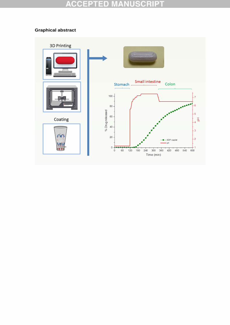
Graphical abstract
Abstract The aim of this work was to explore the feasibility of using fused deposition modelling (FDM) 3D printing (3DP) technology with hot melt extrusion (HME) and fluid bed coating to fabricate modified-release budesonide dosage forms. Budesonide was sucessfully loaded into polyvinyl alcohol filaments using HME. The filaments were transfomed into capsule-shaped tablets (caplets) containing 9 mg budesonide using a FDM 3D printer; the caplets were then overcoated with a layer of enteric polymer. The final printed formulation was tested in a dynamic dissolution bicarbonate buffer system, and two commercial budesonide products, Cortiment® (Uceris®) and Entocort®, were also investigated for comparison. Budesonide release from the Entocort® formulation was rapid in conditions of the upper small intestine while release from the Cortiment® product was more delayed and prolonged. In contrast, the new 3D printed caplet formulation started to release in the mid-small intestine but release then continued in a sustained manner throughout the distal intestine and colon. This work has demonstrated the potential of combining FDM 3DP with established pharmaceutical processes, including HME and film coating, to fabricate modified release oral dosage forms. Keywords: Three dimensional printing; modified release; fused filament fabrication; budesonide; inflammatory bowel disease; colonic delivery
1. Introduction 3-dimensional printing (3DP) technology is being explored as a viable method of personalizing medicines at the point of use (Sanderson, 2015). Indeed, 3DP has the potential to circumvent formulation challenges associated with myriad drugs, including those with narrow therapeutic indices to those whose metabolism is influenced by genetic polymorphisms. In addition it facilitates the development of formulations incorporating more than one drug (Goyanes et al., 2015e). Such approaches could arguably revolutionize clinical management in practice, helping to improve medicine compliance and to achieve better therapeutic outcomes. The first equipment used to prepare 3D printed medicines was based on powder bed - liquid 3D printing technology (Katstra et al., 2000; Rowe et al., 2000; Yu et al., 2009). This technology uses liquids to bind multiple layers of powder to create the desired geometry. The limited choice of binder solutions together with the need for high powder flow and moisture content control are some of the drawbacks that limit the use of these 3D printer types. However, in 2015, the first 3D printed formulation (Spritam®), based on powder bed - liquid 3D printing technology (ZipDose®), was approved by the FDA (Aprecia_Pharmaceuticals, 2015). Spritam® (levetiracetam) is a fast dissolving tablet formulation indicated for the treatment of epileptic seizures. This new product exemplifies the opportunities for the use of 3DP technologies to manufacture medicines at industrial scale, in addition to personalised therapy at the point of use. Fused-deposition modeling (FDM) is a more recent 3DP approach, in which an extruded polymer filament is passed through a heated tip. The heat softens the polymer, which is then deposited onto a build plate to harden. Layers of the deposited and hardened polymer are then built up to create an object in three dimensions. This printing method can fabricate hollow objects as well as dosage forms with different drug release profiles; the latter is achieved by altering either the infill percentage (Goyanes et al., 2014) or surface area/volume ratio of the formulations (Goyanes et al., 2015d). FDM 3DP has higher resolution in comparison with previous printing methods, and can, therefore, achieve better dosing accuracy that is also easily adjusted by changing parameters in the computer software. The method of loading polyvinyl alcohol (PVA) polymer filaments with drug prior to printing has relied on the need for alcoholic solutions containing the active drug (Goyanes et al., 2014; Goyanes et al., 2015a; Skowyra et al., 2015), in which filaments are incubated overnight, but because of the passive nature of the process, it is only possible to achieve low drug loadings. One means of overcoming this problem lies in the use of hot-melt extrusion (HME) to obtain drug-loaded PVA filaments (Goyanes et al., 2015d). In the pharmaceutical industry, HME is the process by which a rotating screw is used to pump drugs and/or excipients at elevated temperatures through a die to generate a product of uniform shape. HME is used to incorporate drugs within a matrix at a molecular level to form solid solutions/dispersions for drug delivery systems such as pellets and granules (Breitenbach, 2002; Mehuys et al., 2005; Schilling et al., 2010). Moreover, HME can reduce the number of processing steps in dosage form manufacturing, and can be automated as a continuous process to give better drug homogeneity.
In this study, the synthetic corticosteroid budesonide was selected as the model drug. Budesonide possesses strong affinity for corticosteroid receptors and features both potent topical anti-inflammatory effects (Gionchetti et al., 2014) and low systemic bioavailability. It is often used in the treatment of inflammatory bowel disease (IBD) (McConnell et al., 2009). Specifically in IBD, modifying budesonide release is a particularly desirable goal for the purposes of increasing its duration of action, achieving optimal therapeutic levels and reducing associated systemic side effects through targeting specific regions of disease activity in the gut. This is achieved mainly by the use of pH-dependent coatings applied to tablets or pellets, though there are significant differences in drug release and distribution between the commercial formulations (Goyanes et al., 2015b). Here, we aimed to create a new budesonide dosage form, combining FDM 3DP with HME and fluid bed coating, to achieve appropriate dissolution kinetics. The suitability of the HME process to produce budesonide 3D-printable filaments was assessed, along with the potential of this method as a valid manufacturing process. The performance of the 3D printed formulation was evaluated and compared with two commercial pH dependent budesonide formulations (Cortiment® 9mg and Entocort® CR 3mg) in a dynamic physiological in vitro dissolution model of the human gastrointestinal tract. 2. Materials and Methods 2.1 Materials Polyvinyl alcohol (PVA), a water-soluble synthetic polymer with a molecular formula of (C2H4O)n), was purchased as an extruded commercial filament from Makerbot Inc., USA (1.75mm diameter, print temperature 190-220°C). Budesonide powder of micronized grade was obtained from Sigma Aldrich, UK (molecular weight: 430.53g/mol (Figure 1); BCS class II, aqueous solubility 0.0215mg/mL (Yalkowsky and He, 2003)). Eudragit® L100 (pH threshold of 6) was acquired from Evonik, Darmstadt, Germany, Triethyl citrate (TEC) and talc were purchased from Sigma Aldrich, UK. Isopropanol supplied were of analytical grade. Salts for preparing buffer dissolution were purchased from VWR International Ltd., Poole, UK. Commercial medicines tested in this study were as follows: - Cortiment® 9mg, prolonged release tablets (Ferring Pharmaceuticals, UK) (commercialized as Uceris® in the US) is tablet formulation with sustained release hydrophilic/lipophilic matrix core known as Multi Matrix System (MMX®) and an outer coating comprising methacrylic acid – methyl methacrylate copolymer (1:1) (Eudragit® L) and methacrylic acid – methyl methacrylate copolymer (1:2) (Eudragit® S) (eMC-Cortiment, 2015). - Entocort® CR 3 mg capsules (AstraZeneca UK Limited, UK). Hard gelatine capsules for oral administration containing budesonide 3 mg as gastro-resistant prolonged-release granules. Granules coated with ethylcellulose and an outer layer of methacrylic acid-
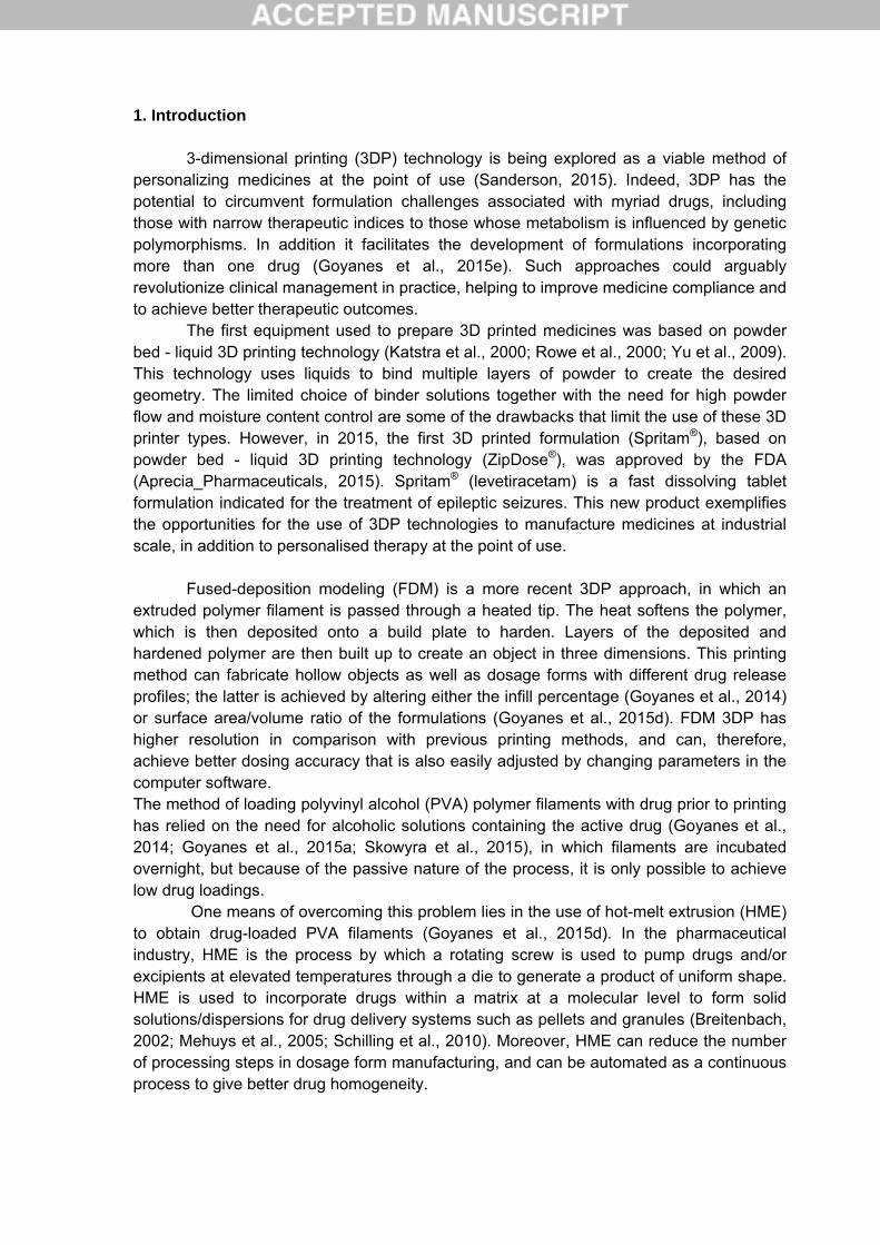
methyl methacrylate copolymer (1:1) (Eudragit® L); the enteric polymer has a dissolution pH threshold of 5.5 (Edsbacker and Andersson, 2004). 2.2 Methods 2.2.1 Preparation of PVA filament loaded with budesonide The commercial PVA filament was cut into small cylindrical pellets (~2mm) using a Pharma 11 Varicut Pelletizer (Thermo Fisher Scientific, UK). A commercial grinder (Wahl ZX789, Wahl store, UK) was used to produce a fine PVA powder with a small particle size which was then sifted through a sieve with a mesh size of 1000 µm. Budesonide (2g, 5% drug w/w) was manually mixed in together with the PVA powder (38g) using a mortar and pestle. The mixture was then extruded using a single-screw extruder (Noztek Pro filament extruder, NozteK, UK) at 170 °C through a nozzle with diameter 1.75mm at screw speed of 15 rpm. The resulting filament was stored in a vacuum desiccator before printing. Budesonide loading in the filaments was determined by HPLC analysis. 2.2.2 3D printing of budesonide dosage forms Dosage forms were fabricated with the drug-loaded filament using a standard fused-deposition modelling 3D printer, MakerBot Replicator 2X Desktop (MakerBot Inc., USA). The template used to print the dosage form was designed with AutoCAD 2014® (Autodesk Inc., USA) and exported as a stereolithography (.stl) file into MakerWare v. 2.4.1 (MakerBot Inc., USA). The selected geometry of the dosage forms was a rounded hard capsule-shaped tablet (caplet) (Figure 2). The dimensions of the caplet were altered such that the axis measurements were: X= 12.49mm; Y= 4.64mm; and Z= 4.64mm using the scale function. The printer settings used were as follows: standard resolution with the raft option deactivated, support option activated and extrusion temperature of 190 °C; speed while extruding: 90mm/s; speed while traveling: 150mm/s; number of shells 2 and layer height of 0.20mm. The infill percentage was set to 100% to produce high-density caplets. 2.2.3 Coating of 3D printed caplets The coating solution was prepared by adding Eudragit® L100 powder (12g) into a mixture of isopropanol (97% based on solvent weight) and water while stirring with a magnetic stirrer plate. Talc (50% based on polymer weight) was added as an anti-tacking agent in a similar manner until a homogenous dispersion was obtained. TEC (10% based on polymer weight) was then added to the dispersion. The total solid content of the final dispersion was 10% w/w. The caplets were coated using Strea-1 bottom spray fluidized bed coater (Aeromatic AG, Bubendorf, Switzerland), to a weight gain of 13%, which was selected to provide gastro-resistance. The coating conditions were as follows: Inlet air temperature 40°C; outlet air temperature 30°C; fan capacity at setting 15 (equivalent to air flow 150m3/h); atomising pressure of 0.2 bar and a spray rate of 1.0mL/min. After coating, the tablets were fluidized further for 15 min in the coater and cured in an oven at 40°C for 24 hours. 2.2.4 Drug loading of filaments and caplets A caplet or a section of drug-loaded strand (approx. 0.3g) was placed in a volumetric flask (1L) with a mixture of methanol:water (1:1) under magnetic stirring until complete
dissolution (n=2). Samples of the solutions were then filtered through 0.45 µm filters (Millipore Ltd, Ireland), and the concentration of drug determined by HPLC (Hewlett
Packard 1050 Series HPLC system, Agilent Technologies, UK) with a Kinetex 2.6M phenylhexyl 100A, 4.6mm x 50mm column (Phenomenex® Cheshire, UK) maintained at
40°C. The injected volume was 20L.The mobile phase consisted of two components: Acetonitrile and water. The former starts at 20% but rises gradually to 60% after 10 minutes. The flow rate was 1 ml/min and the UV detection was set at 254nm. All measurements were made in duplicate. 2.2.5 Scanning Electron Microscopy (SEM) Surface and cross-section images of the filament and caplets were taken with an SEM (JSM-840A Scanning Microscope, JEOL GmbH, Eching, Germany). All samples for SEM testing were coated with carbon (∼30–40 nm) for visualization.Pictures of the tablets were taken with a Nikon CoolpixS6150 with the macro option of the menu. 2.2.6 X-ray powder diffraction (XRPD) Discs of 23mm diameter x 1mm height made from PVA filament or drug-loaded PVA filament were 3D printed and analysed. A sample of pure budesonide was also analysed. The X-ray powder diffraction patterns were obtained in a Rigaku MiniFlex 600 (Rigaku, USA) using a Cu Kα X-ray source (λ=1.5418Å). The intensity and voltage applied were 15 mA and 40 kV. The angular range of data acquisition was 3–60° 2θ, with a stepwise size of 0.02° at a speed of 5°/min. 2.2.7 Thermal analysis The filament was characterized with Differential Scanning Calorimetry (DSC) and Thermogravimetric analysis (TGA). For DSC measurements, a Q2000 DSC (TA Instruments, LLC, Waters, USA) was used to analyse the samples by heating from -30 to 250°C at 10°C/min in pin-holed hermetically sealed Tzero aluminium pans. Nitrogen was used as a purge gas at a rate of 50mL/min. The DSC was calibrated for temperature and cell constant according to the manufacturer instructions. Advantage software and TA Universal Analysis (TA Instruments, LLC, Waters, USA) were used to capture and analyse the data, respectively. For TGA analysis, a Discovery TGA (TA Instruments, LLC, Waters, USA) was used to test samples by heating at 10°C/min from room temperature to 300°C in open aluminum pans. Nitrogen was used as a purge gas at a rate of 25mL/min. Data collection and analysis were performed using Trios software (TA Instruments, LLC, Water, USA). Percentages of weight loss (%w/w) were calculated for each sample. 2.2.8 Dynamic dissolution testing conditions Drug dissolution profiles for the coated 3D printed caplets, Cortiment® 9mg and Entocort® 3mg were obtained with a USP-II apparatus (Model PTWS, Pharmatest, Germany). Three capsules of Entocort® 3mg were tested together in each vessel to match the dose of 9mg of the other formulations: 1) the formulations were placed in 750 mL of 0.1 M HCl for 2 h to simulate gastric residence time, and then 2) transferred into 950 mL of
modified Hanks (mHanks) bicarbonate physiological medium for 35 min (pH 5.6 to 7); 3) and then in modified Krebs buffer (1000ml) (pH 7 to 7.4 and then to 6.5). The modified Hanks buffer based dissolution medium (Liu et al., 2011) (136.9 mM NaCl, 5.37 mM KCl, 0.812 mM MgSO4.7H2O, 1.26 mM CaCl2, 0.337 mM Na2HPO4.2H2O, 0.441 mM KH2PO4, 4.17 mM NaHCO3) forms an in-situ modified Kreb’s buffer (Fadda et al., 2009) by addition of 50 mL of pre-Krebs solution (400.7 mM NaHCO3 and 6.9 mM KH2PO4) to each dissolution vessel. The formulations were tested in the small intestinal environment for 3.5 h (pH 5.6 to 7.4), followed by pH 6.5 representing the colonic environment. These parameters were selected to simulate typical conditions for intestinal transit of pharmaceutical formulations and pH values in different segments of the GI tract in fasted individuals. The buffer capacity and ionic composition of the physiological bicarbonate buffers also closely match the buffer capacities of the intestinal fluids collected from different parts of the gut in humans (Fadda et al., 2009; Goyanes et al., 2015b; Goyanes et al., 2015c; Liu et al., 2011). The medium is primarily a bicarbonate buffer in which bicarbonate (HCO3
-) and carbonic acid (H2CO3) co-exist in an equilibrium, along with CO2 (aq) resulting from dissociation of the carbonic acid. The pH of the buffer system can be decreased by purging CO2 (g) in the solution, which promotes the formation of carbonic acid. Similarly, an inert gas (such as helium), which removes the dissolved CO2 from the solution, increases the pH of the medium. The purging of gases is controlled by an Auto pH SystemTM (Merchant et al., 2012), which consists of a pH probe connected to a source of carbon dioxide gas (pH-reducing gas), as well as to a supply of helium (pH-increasing gas), controlled by a control unit. The control unit is able to provide a dynamically adjustable pH during testing (dynamic conditions) and to maintain a uniform pH value over the otherwise unstable bicarbonate buffer pH. The paddle speed of the USP-II was fixed at 100 rpm, and the tests were conducted at 37 +/-0.5 °C (n=3). The percentage drug released from the formulations was determined using an in-line UV spectrophotometer (Cecil 2020, Cecil Instruments Ltd., Cambridge, UK) at the wavelength of maximum absorbance of budesonide (244nm). Data were processed using Icalis software (Icalis Data Systems Ltd, Berkshire, UK), 3. Results and Discussion Budesonide-loaded filaments were successfully prepared using HME (Figure 3). Budesonide loading in the filaments was 4.14 ±0.273%, which is considerably higher than than loadings achieved through passive diffusion approaches with alcoholic solutions (Goyanes et al., 2014; Goyanes et al., 2015a; Skowyra et al., 2015). However, budesonide loading was lower than expected (5% w/w), which may be attributed to the adherence of powdered drug to the walls of the container on transfer to the hopper of the HME and the walls of the barrel during extrusion, and due to irregular extrusion of components. In the pharmaceutical industry twin-screw extruders are often used instead of single extruders, as the former’s mixing ability is superior to the single-screw extruder. Further parameters can also be altered to improve mixing, shear force and pressure within the barrel (Crowley et al., 2007; Repka et al., 2007).
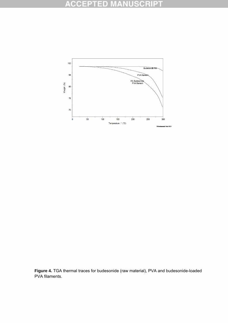
TGA analysis (Figure 4) and DSC data (Figure 5) indicate that budesonide is thermally stable in the temperature range used in both extruding and printing with a mass loss of approximately 0.1% w/w for the raw material and 4% w/w for the drug-loaded filament. These data confirm that budesonide is not degraded during HME, indicating that the lower than expected loading in the filament is attributable to the feedstock preparation and handling. The difference in mass loss seen between the plain PVA and the drug-loaded filaments (2 and 4% w/w, respectively) may be caused by differences in water content of the filaments. DSC of budesonide shows a melting temperature higher than 250°C; the DSC data of the budesonide-loaded filament showed no evidence of melting (Figure 5). PVA melts at a lower temperature than the drug, which may lead to any crystalline drug dissolving in the molten polymer. On the other hand, the presence of two diffraction peaks at around 15° in the XRPD data (Figure 5) indicates some crystalline phases of the drug exist.. DSC results were not conclusive, but the two techniques suggest that the drug may be partially crystalline in the formulation. Nevertheless, the glass transition temperature of the PVA filament is lower for the drug-loaded filament. This may be because the budesonide is acting as a plasticiser interacting with PVA at a molecular level. Budesonide is classified as a small chemically manufactured molecule with a large molecular weight:size ratio. Its plasticisation effects may be the result of its ability to increase free volume between polymer chains (Repka et al., 2007) by interposition and increasing polymer chain motion through intermolecular secondary valence forces. This plasticizing effect was also demonstrated with prednisolone (Skowyra et al., 2015). The budesonide-loaded filament was converted into caplets using the FDM 3D printer (Figure 6). Budesonide loading in the caplets was 4.10 ±0.296%, which is very similar to that of the filament itself, 4.14 ±0.273, indicating that the drug did not degrade during the 3D printing process; this was anticipated and in accordance with the DSC and TGA results. Degradation of the drugs during 3DP was evaluated before, and it is expected if the 3DP process takes place at temperatures higher than the degradation temperature of the drug (Goyanes et al., 2015a). In order to manufacture caplets with the same budesonide content as the Cortiment® product, the dimensions of the caplet were adjusted to print a mass equivalent to 9mg of budesonide per caplet (caplet weights of 220mg). Skowyra et al., (2015) demonstrated a correlation between the theoretical volume of the tablet and its mass on printing using FDM 3DP. In our study, caplets were initially printed with commercial PVA filament (drug free) to estimate the dimensions needed to achieve the target drug weight. This was then repeated with the budesonide-loaded filament, and the scale function was used to produce a smaller or larger dosage form that retained the ratio between the dimensions X, Y and Z. This signifies the potential of FDM 3DP to manufacture tablets of different geometries and infill percentages in tailoring doses accurately and precisely for individual patients where minute increments of dosing may be required. A coat of Eudragit L100 was applied to the 3D printed caplets with the intention of producing a gastro-resistant product (Figure 6). The coated printed product was tested alongside the two commercial budesonide products in the dynamic in vitro model, which simulates intestinal conditions in the GI tract (Goyanes et al., 2015b; Goyanes et al.,
2015c). Figure 7 depicts the release data for all three formulations. The coated 3D printed product is resistant to acidic conditions, and releases after approximately 1 hour in the small intestinal segment of the test, which coincides with a pH of pH 7.2, although the polymer Eudragit® L100 has a theoretical pH threshold of 6. After coat dissolution, drug release from the printed core is sustained and continues in the colonic region. The polymer PVA concentration of the commercial filament determines the swelling ratio of PVA hydrogels (Gupta et al., 2011) with higher concentrations resulting in a reduced swelling ratio. This phenomenon may be responsible for modulating drug release from the drug-loaded 3DP PVA caplets. Drug release rates from water soluble and swellable polymers are governed by the relative contribution of two mechanisms: drug diffusion and polymer dissolution (surface erosion) (Reynolds et al., 2002). Drug solubility and the nature of excipients are factors that affect which mechanism dominates. Previous studies with 3D printed PVA formulations showed that drug release was through an erosion-mediated process (Goyanes et al., 2014; Goyanes et al., 2015a). Entocort® is a commercial medicine that contains budesonide within granules of a matrix of ethylcellulose coated with an enteric coated polymer (Eudragit® L) designed to prevent dissolution at gastric pH. Budesonide release from the Entocort® formulation is rapid (approximately 15 min after gastric emptying), which is in agreement with the results of a previous study investigating the dissolution characteristics of oral steroid formulations (Goyanes et al., 2015b). In a scintgraphic study, Entocort® was observed to dissolve in the duodenum, with the budesonide releasing slowly from the ethylcellulose matrix as it passes through the intestine (Edsbacker et al., 2003). In the in vitro model, the Entocort® formulation releases more than 90% budesonide at the end of the small intestinal phase of the test, with less than 10% of the dose reaching the colon (Goyanes et al., 2015b), and with mean maximal budesonide plasma concentrations seen at 3 h following administration of Entocort® (Edsbacker et al., 2003). Cortiment® (Uceris®) shows a longer lag time and a much slower drug release rate, reaching only 50% drug release after 10h. These dissolution results correspond with those obtained in a study by Nicholls et al (2013) investigating and comparing the in vivo drug release profiles and pharmacokinetics of this product with those of Entocort®. Plasma concentration profiles for both Entocort® and Uceris® demonstrated a similar extent (AUC) of systemic exposure to budesonide, but the time for first appearance of drug in the systemic circulation was significantly delayed for Uceris®. It has been suggested by Nicholls that this initial lag period is attributable to more distal gut dissolution and drug release from the Uceris formulation than Entocort®. The close similarities between the dissolution profiles and the pharmacokinetic results, indicate the predictive value of the dynamic dissolution system. In conclusion, a new modified release 9mg budesonide product was created by combining 3D printing with HME and film coating. The release characteristics of the new product are such that it has potential in the treatment of inflammatory bowel disease.
References Aprecia_Pharmaceuticals, 2015. FDA approves the first 3D printed drug product.
Breitenbach, J., 2002. Melt extrusion: from process to drug delivery technology. Eur. J. Pharm. Biopharm. 54, 107-117.
Crowley, M.M., Zhang, F., Repka, M.A., Thumma, S., Upadhye, S.B., Battu, S.K., McGinity, J.W., Martin, C., 2007. Pharmaceutical applications of hot-melt extrusion: Part I. Drug Dev. Ind. Pharm. 33, 909-926.
Edsbacker, S., Andersson, T., 2004. Pharmacokinetics of budesonide (Entocort (TM) EC) capsules for Crohn's disease. Clin. Pharmacokinet. 43, 803-821.
Edsbacker, S., Bengtsson, B., Larsson, P., Lundin, P., Nilsson, A., Ulmius, J., Wollmer, P., 2003. A pharmacoscintigraphic evaluation of oral budesonide given as controlled-release (Entocort) capsules. Aliment. Pharmacol. Ther. 17, 525-536.
eMC-Cortiment, 2015. Summaries of Product Characteristics: Cortiment 9 mg, prolonged release tablets. https://www.medicines.org.uk/emc/medicine/30253, last accessed 09-2015.
Fadda, H.M., Merchant, H.A., Arafat, B.T., Basit, A.W., 2009. Physiological bicarbonate buffers: stabilisation and use as dissolution media for modified release systems. Int. J. Pharm. 382, 56-60.
Gionchetti, P., Pratico, C., Rizzello, F., Calafiore, A., Capozzi, N., Campieri, M., Calabrese, C., 2014. The role of Budesonide-MMX in active ulcerative colitis. Expert. Rev. Gastroenterol. Hepatol. 8, 215-222.
Goyanes, A., Buanz, A.B., Basit, A.W., Gaisford, S., 2014. Fused-filament 3D printing (3DP) for fabrication of tablets. Int. J. Pharm. 476, 88-92.
Goyanes, A., Buanz, A.B., Hatton, G.B., Gaisford, S., Basit, A.W., 2015a. 3D printing of modified-release aminosalicylate (4-ASA and 5-ASA) tablets. Eur. J. Pharm. Biopharm. 89, 157-162.
Goyanes, A., Hatton, G.B., Basit, A.W., 2015b. A dynamic in vitro model to evaluate the intestinal release behaviour of modified-release corticosteroid products. J. Drug Deliv. Sci. Tec. 25, 36-42.
Goyanes, A., Hatton, G.B., Merchant, H.A., Basit, A.W., 2015c. Gastrointestinal release behaviour of modified-release drug products: Dynamic dissolution testing of mesalazine formulations. Int. J. Pharm. 484, 103-108.
Goyanes, A., Martinez, P.R., Buanz, A., Basit, A., Gaisford, S., 2015d. Effect of geometry on drug release from 3D printed tablets. Int. J. Pharm., DOI 10.1016/j.ijpharm.2015.1004.1069.
Goyanes, A., Wang, J., Buanz, A., Martínez-Pacheco, R., Telford, R., Gaisford, S., Basit, A., 2015e. 3D printing: Engineering novel oral devices with unique design and drug release characteristics. Mol. Pharm., Submited.
Gupta, S., Webster, T.J., Sinha, A., 2011. Evolution of PVA gels prepared without crosslinking agents as a cell adhesive surface. J. Mater. Sci. Mater. Med. 22, 1763-1772.
Katstra, W.E., Palazzolo, R.D., Rowe, C.W., Giritlioglu, B., Teung, P., Cima, M.J., 2000. Oral dosage forms fabricated by three dimensional printing. J. Control. Release 66, 1-9.
Liu, F., Merchant, H.A., Kulkarni, R.P., Alkademi, M., Basit, A.W., 2011. Evolution of a physiological pH 6.8 bicarbonate buffer system: Application to the dissolution testing of enteric coated products. Eur. J. Pharm. Biopharm. 78, 151-157.
McConnell, E.L., Liu, F., Basit, A.W., 2009. Colonic treatments and targets: issues and opportunities. J. Drug Target. 17, 335-363.
Mehuys, E., Remon, J.-P., Vervaet, C., 2005. Production of enteric capsules by means of hot-melt extrusion. Eur. J. Pharm. Sci. 24, 207-212.
Merchant, H.A., Frost, J., Basit, A.W., 2012. Apparatus and method for testing medicaments. PCT/GB2013/051145.
Nicholls, A., Harris-Collazo, R., Huang, M., Hardiman, Y., Jones, R., Moro, L., 2013. Bioavailability profile of Uceris MMX extended-release tablets compared with Entocort EC capsules in healthy volunteers. J. Int. Med. Res. 41, 386-394.
Repka, M.A., Battu, S.K., Upadhye, S.B., Thumma, S., Crowley, M.M., Zhang, F., Martin, C., McGinity, J.W., 2007. Pharmaceutical applications of hot-melt extrusion: Part II. Drug Dev. Ind. Pharm. 33, 1043-1057.
Reynolds, T.D., Mitchell, S.A., Balwinski, K.M., 2002. Investigation of the effect of tablet surface area/volume on drug release from hydroxypropylmethylcellulose controlled-release matrix tablets. Drug Dev. Ind. Pharm. 28, 457-466.
Rowe, C.W., Katstra, W.E., Palazzolo, R.D., Giritlioglu, B., Teung, P., Cima, M.J., 2000. Multimechanism oral dosage forms fabricated by three dimensional printing. J. Control. Release 66, 11-17.
Sanderson, K., 2015. 3D printing: the future of manufacturing medicine? Pharm. J. 294, No 7865.
Schilling, S.U., Shah, N.H., Waseem Malick, A., McGinity, J.W., 2010. Properties of melt extruded enteric matrix pellets. Eur. J. Pharm. Biopharm. 74, 352-361.
Skowyra, J., Pietrzak, K., Alhnan, M.A., 2015. Fabrication of extended-release patient-tailored prednisolone tablets via fused deposition modelling (FDM) 3D printing. Eur. J. Pharm. Sci. 68, 11-17.
Yu, D.G., Shen, X.X., Branford-White, C., Zhu, L.M., White, K., Yang, X.L., 2009. Novel oral fast-disintegrating drug delivery devices with predefined inner structure fabricated by Three-Dimensional Printing. J. Pharm. Pharmacol. 61, 323-329.
Figure Captions
Figure.1. Chemical structure of budesonide.
Figure 2. Caplet design.
Figure 3. SEM images of (A) surface view and (B) cross section of budesonide loaded PVA filament after HME.
Figure 4. TGA thermal traces for budesonide (raw material), PVA and budesonide-loaded PVA filaments.
Figure 5. (A) X-ray powder diffractograms of pure budesonide and 3D printed discs of PVA and budesonide-PVA (B) DSC thermograms for Budesonide, PVA, budesonide-loaded PVA filaments and a physical mixture of PVA and the drug.
Figure 6. Images of 3DP fabricated caplets (A) from left to right, caplet prior to coating, caplet after coating and cross section of coated caplet (scale in cm); (B,C) SEM images of internal structure of cross-section of a coated-3D printed caplet.
Figure 7. Drug release from Cortiment®, Entocort® and coated 3D printed caplets in 0.1M HCl for 2h followed by physiological bicarbonate buffer under dynamic pH conditions (pH 5.6 to 7.4 and then 6.5) controlled by the Auto pH SystemTM. Red line shows the real-time pH values.