Page 1
FABRICATION OF NANO COMPOSITE HYDROGELS BASED
ON POLYVINYL ALCOHOL FOR BIOMEDICAL APPLICATIONS
ALI KARIMI
A THESIS SUBMITTED IN FULFILMENT OF THE
REQUIREMENTS FOR THE DEGREE OF DOCTOR OF
PHILOSOPHY
FACULTY OF ENGINEERING
UNIVERSITY OF MALAYA
KUALA LUMPUR
2016
Page 2
ii
UNIVERSITI MALAYA
ORIGINAL LITERARY WORK DECLARATION
Name of Candidate: Ali Karimi (I.C/Passport No: F26184459)
Registration/Matric No: KHA100092
Name of Degree: Doctor of Philosophy (PhD)
Title of Project Paper/Research Report/Dissertation/Thesis (“this Work”):
FABRICATION OF NANO COMPOSITE HYDROGELS BASED ON
POLYVINYL ALCOHOL FOR BIOMEDICAL APPLICATIONS
Field of Study: Chemical Engineering
I do solemnly and sincerely declare that:
(1) I am the sole author/writer of this Work;
(2) This Work is original;
(3) Any use of any work in which copyright exists was done by way of fair dealing and for
permitted purposes and any excerpt or extract from, or reference to or reproduction of any
copyright work has been disclosed expressly and sufficiently and the title of the Work and its
authorship have been acknowledged in this Work;
(4) I do not have any actual knowledge nor ought I reasonably to know that the making of this work
constitutes an infringement of any copyright work;
(5) I hereby assign all and every rights in the copyright to this Work to the University of Malaya
(“UM”), who henceforth shall be owner of the copyright in this Work and that any reproduction
or use in any form or by any means whatsoever is prohibited without the written consent of UM
having been first had and obtained;
(6) I am fully aware that if in the course of making this Work I have infringed any copyright
whether intentionally or otherwise, I may be subject to legal action or any other action as may be
determined by UM.
Candidate’s Signature Date: 06. 09. 2016
Subscribed and solemnly declared before,
Witness’s Signature Date:
Name: Designation: Chemical Engineering Department, Faculty of Engineering, University of Malaya, Kuala Lumpur, 50603, Malaysia Tel. /Fax: +60 379675206/ +60 379675313
Page 3
iii
ABSTRACT
In order to obtain nontoxic, tissue-compatible and efficient hydrogels for biomedical
applications, Polyvinyl alcohol (PVA) / Na+-montmorillonite (Na
+-MMT)
nanocomposite hydrogels were prepared by a cyclic freeze-thaw process (physical
method). Effect of nanoclay content and sonication mixing on the nanocomposite
structure and morphology as well as its properties (mechanical, thermal), and also its
swelling and deswelling kinetics were investigated.
Glutaraldehyde reacts with PVA to form covalently cross-linked networks. The acetal
linkages were formed between hydroxyl groups of PVA and aldehyde groups of
glutaraldehyde that was used for physicochemical synthesis of nanocomposites. A novel
PVA nanocomposite hydrogel was synthesized by physicochemical method. Effect of
physical and physicochemical cross linking on the structure, morphology, thermal,
mechanical, swelling and deswelling properties of nanocomposite hydrogels were
investigated and were compared together. The results were shown that physicochemical
crosslinking of PVA nanocomposite leads to decreasing of crystallinity and melting
temperature also increasing the Hardness and Water vapor transmission rate (WVTR)
values than physical crosslinked. Swelling and deswelling measurements were done by
gravimetric method and indicated that controlled crosslinking of PVA nanocomposite
hydrogel caused to increase the swelling ratio and also decrease the cumulative amount
of water loss. Sorption and desorption kinetics for both physical and physicochemical
methods were based on diffusion mechanism and obey the Fickian model. As an
important result using the controlled crosslinking can obtain the PVA nanocomposite
hydrogel with higher swelling capacity than conventional PVA nanocomposite
hydrogel. In order to find an optimum amount of nanoclay content for achieving the
optimal Equilibrium water content (EWC) and WVTR properties of nanocomposite
cryogels, my investigations were performed on the barrier and swelling properties of the
Page 4
iv
nanocomposites and it was shown that Na+-MMT may act as a co-crosslinker.
According to the results, the swelling characteristics of nanocomposite cryogels
increases with the nanoclay content up to 1-2% nanoclay, after that they start to
decrease uniformly. In contrast, the water removal from cryogels decreased and its time
of removal prolonged on increasing the nanoclay content. Based on the results of
WVTR measurements, the barrier properties of the nanocomposites can be improved by
increasing the nanoclay content and it is concluded that the optimum range of nanoclay
for having optimum WVTR at 37 °C is up to 1% nanoclay. It was found that the EWC
of PVA nanocomposite cryogel containing 1% nanoclay, having 74% water content
compared to the other nanocomposites at 37 °C. Results of EWC (above 60%) and
WVTR (at about 8.5 g/m2/h) are within the acceptable range for biomedical applications
such as skin treatment and wound dressing.
With the aim of investigation on the antibacterial properties of PVA/Na+-MMT
nanocomposite hydrogels against two types of bacteria, Escherichia coli (E-Coli); as a
gram negative bacteria and Staphylococcus aureus (S-Aureus); as a gram positive
bacteria, polyvinyl pyrrolidone – Iodine (PVP-Iodine) has been used in the hydrogel
network. The effect of nanoclay content on release of antibacterial agent for loaded
hydrogels was also investigated in vitro and found to be dependent on crosslinking
amount due to interaction between PVA and nanoclay.
Page 5
v
ABSTRAK
Dalam usaha untuk mendapatkan hidrogel tanpa toksik, tidak berbahaya, tisu yang
bersesuaian dan berkesan bagi aplikasi bioperubatan, Polivinil alkohol (PVA) / Na+-
montmorillonite (Na+-MMT) Hidrogel nanokomposit telah disediakan mengunakan satu
proses kitaran beku-cair kitaran (kaedah fizikal). Kesan kandungan tanah liat nano dan
campuran sonikast pada struktur nanokomposit dan morfologi serta sifat-sifatnya
(mekanikal, haba), dan juga kinetic swelling dan deswelling yang telah dikaji.
Glutaraldehyde bertindak balas dengan PVA untuk membentuk rangkaian kovalen balas
berkaitan. Hubungan asetal telah dibentuk antara kumpulan hidroksil PVA dan aldehid
kumpulan glutaraldehyde yang digunakan untuk sintesis fizikokimia nanocomposites.
PVA nanokomposit hidrogel telah disintesis melalui kaedah fizikokimia. Kesan
sambung- silang fizikal dan fizikokimia kepada struktur, morfologi, haba, mekanikal,
sifat swelling dan deswelling Hidrogel nanokomposit telah disiasat dan dibandingkan.
Keputusan telah menunjukkan bahawa Sambung-silang fizikokimia PVA nanokomposit
membawa kepada pengurangan penghabluran dan suhu lebur juga meningkatkan
kekerasan dan kadar penghantaran wap air (WVTR) berbanding sambyng-silang fizikal.
ukuran swelling dan deswelling telah dilakukan dengan kaedah gravimetrik dan ia
menunjukkan Sambung-silang terkawal PVA nanokomposit hidrogel disebabkan untuk
meningkatkan nisbah swelling dan juga mengurangkan jumlah kerugian air terkumpul.
Kinetic Erapan dan penyaherapan kinetik bagi kaedah kedua-dua kaedah fizikal dan
fizikokimia adalah berdasarkan mekanisme penyebaran dan mengikut model Fickian.
Sebagai hasil penggunaan Sambung-silang terkawal boleh mendapatkan hidrogel PVA
nanokomposit dengan kapasiti swelling yang lebih tinggi daripada konvensional PVA
nanokomposit hidrogel. Dalam usaha untuk mencari jumlah optimum kandungan tanah
liat nano bagi mencapai kandungan optimum Keseimbangan air (EWC) dan ciri-ciri
WVTR daripada cryogel nanokomposit, kajian telah dilakukan ke atas halangan dan
Page 6
vi
sifat swelling nanokomposit dan ia menunjukkan bahawa Na+ -MMT boleh bertindak
sebagai penyambung-silang. Berdasarkan hasil kajian, ciri-ciri swelling nanokomposit
cryogel meningkat dengan 1-2% kandungan tanah liat nano, dan selepas itu mula
berkurangan secara seragam. Sebaliknya, penyingkiran air dari cryogel menurun dan
masa penyingkiran berpanjangan mengikut peningkatan kandungan tangah liat nano itu.
Berdasarkan hasil pengukuran WVTR, halangan nanokomposit boleh diperbaiki dengan
meningkatkan kandungan tanah liat nano dan julat optimum tanah liat nano bagt
mendapatkan suhu optimum WVTR pada 37° C adalah sehingga 1% tanah liat nano.
Selain itu EWC PVA cryogel nanokomposit yang mengandungi 1% tanah liat nano,
mempunyai 74% kandungan air berbanding dengan nanokomposit lain pada suhu 37° C.
Keputusan EWC (melebihi 60%) dan WVTR (kira-kira 8.5 g / m2 / h) adalah dalam
julat yang boleh diterima bagi aplikasi bioperubatan seperti rawatan kulit dan pembalut
luka.
bagt tujuan kajlan terhadap sifat antibakteria PVA / Na+ -MMT Hidrogel nanokomposit
terhadap dua jenis bakteria, Escherichia coli (E-Coli); sebagai bakteria gram negatif dan
Staphylococcus aureus (S-Aureus); sebagai bakteria gram positif, Polivinil pyrrolidone -
Iodin (PVP-Iodin) telah digunakan dalam rangkaian hidrogel. Kesan kandungan tanah
liat nano terhadap pembebasan agen anti-bakteria untuk Hidrogel juga dikaji secara in-
vitro dan didapati bergantung kepada jumlah sambung-silang berikutan interaksi antara
PVA dan tanah liat nano.
Page 7
vii
ACKNOWLEDGEMENTS
First and foremost, I am very thankful to Allah for his unparalleled grace and guidance
throughout of my life. This research project was not possible without help and kindness
of the Almighty God.
I would like to express my sincere gratitude to my supervisor Professor Dr. Wan Mohd
Ashri Wan Daud, for his guidance, insight and support throughout this research work.
I would like to express my special appreciation and thanks to the Chemical Engineering
Department staff in university of Malaya, for their collaborations during the entire
period of my study. I appreciate to Chemical Engineering Department in the University
of Tehran, especially professor Navid Mostoufi and Dr. Babak Kaffashi, for their
valuable time and collaboration provided in this research project.
I also would like to thank my dear colleague and old friend, Dr. Ahmad Nalbandi for his
help throughout this thesis.
I dedicate this thesis to my father and my brother, for their love and support throughout
my life, although they are not alive anymore. I also grant the dissertation to my dear
mother, my dear wife, Fatemeh and my dear sons Amir Mohammad and Erfan for their
devotion, love and patience.
And finally, I dedicate this thesis to those researchers who generously spend their life to
serve the humanity.
Ali Karimi
Department of Chemical Engineering,
University of Malaya, Kuala Lumpur, Malaysia
Page 8
viii
1 TABLE OF CONTENTS
ABSTRACT iii
ABSTRAK v
ACKNOWLEDGEMENTS vii
TABLE OF CONTENTS viii
LIST OF FIGURES xiv
LIST OF TABLES xviii
LIST OF ABBREVIATIONS viiix
LIST OF APPENDICES xxii
1. CHAPTER 1: GENERAL INTRODUCTION 1
1.1 Background 1
1.2 Problem Statement 5
1.3 Objectives 7
1.4 Outline of the thesis 8
2. CHAPTER 2: REVIEW OF RELATED LITERATURE 10
2.1 Introduction 10
2.2 Structure and properties of nanoclay (Phyllosilicates) 14
2.2.1 Structure and properties of layered silicate
2.2.2. Structure and properties of organically
modified layered silicate (OMLS)
14
16
2.3 Types of polymeric nanocomposites and their
preparative techniques
18
2.3.1 Types of polymeric nanocomposites 18
2.3.2 Polymer/layered silicate (PLS) nanocomposite
technology
20
2.3.3 Preparative techniques 21
Page 9
ix
2.3.3.1 Intercalation of polymer and pre-polymer
from solution
23
2.3.3.2 In situ intercalative polymerization 24
2.3.3.3 Melt intercalation technique 24
2.4 Techniques used for the preparation of PVA
hydrogels and PVA anocomposite hydrogels and their
characterization
27
2.4.1 Covalently crosslinked and cryogels 28
2.4.2 Polyvinyl alcohol nanocomposites and their
characterizations
31
2.5 Summary 48
3. CHAPTER 3: METHODOLOGY 50
3.1 PART 1: Non toxic hydrogels based on polyvinyl
alcohol/Na+-Montmorillonite nanocomposites for
biomedical applications: Fabrication & Characterization
50
3.1.1 Materials 50
3.1.2 Fabrication of nanocomposite hydrogels
(cryogels)
50
3.1.3 Structure and Morphology 52
3.1.4 Thermal and Mechanical Analysis 53
3.2 PART 2: Nanocomposite cryogels based on poly
(vinyl alcohol)/ unmodified Na+-montmorillonite
suitable for wound dressing application: optimizing
nanoclay content
56
3.2.1 Materials 56
Page 10
x
3.2.2 Fabrication of nanocomposite cryogels 56
3.2.3 Morphology and thermomechanical properties 56
3.2.4 Barrier properties 56
3.2.5 Kinetics 56
3.2.5.1 Water sorption kinetics in deionized
water
56
3.2.5.2 Water desorption kinetics for swelled
gels
57
3.3 PART 3: Comparison the Properties of PVA/Na+-
MMT Nanocomposite Hydrogels Prepared by Physical
and Physicochemical Crosslinking
59
3.3.1 Materials 59
3.3.2 Fabrication of Physical Nanocomposite
Hydrogels
59
3.3.3 Fabrication of Physicochemical Nanocomposite
Hydrogels
59
3.3.4 Structure and morphology 60
3.3.5 Thermal and mechanical analysis 60
3.3.6 Kinetics 60
3.3.6.1 Water sorption kinetics in deionized
water
60
3.3.6.2 Water desorption kinetics for swelled
gels
60
3.4. PART 4: Fabrication of (PVA/Na+-MMT/ PVP-
Iodine) nanocomposite hydrogel system and study of in
vitro its antibacterial properties for wound dressing
61
Page 11
xi
application
3.4.1 Materials 61
3.4.2 Preparation of nanocomposite hydrogels 61
3.4.3 Swelling studies 61
3.4.4 Loading of antibacterial agent into hydrogels 61
3.4.5 In vitro release experiment and Evaluation of
antibacterial activity
62
4. CHAPTER 4: RESULTS AND DISCUSSION 63
4.1 Introduction 63
4.2 PART 1: Non toxic hydrogels based on polyvinyl
alcohol/Na+- Montmorillonite nanocomposites for
biomedical applications
69
4.2.1 Structural characterization and morphology 69
4.2.2 Thermal and mechanical analysis 74
4.3. PART 2: Nanocomposite cryogels based on poly
(vinyl alcohol)/ unmodified Na+-montmorillonite
suitable for wound dressing application: optimizing
nanoclay content
81
4.3.1 Effect of nanoclay content on morphology and
thermomechanical properties
81
4.3.2 Effect of nanoclay content on sorption and
Barrier behaviors
86
4.3.2.1 Swelling ratios and Equilibrium water
content
86
4.3.2.2 Barrier properties and Permeation 88
Page 12
xii
analysis
4.3.3 Sorption and desorption kinetics 89
4.3.3.1 Sorption kinetics in deionized water
media
89
4.3.3.2. Kinetics of water desorption for swollen
gels
90
4.4 PART 3: Comparison the Properties of PVA/Na+-
MMT Nanocomposite Hydrogels Prepared by Physical
and Physicochemical Cross linking
94
4.4.1 Structural characterization and morphology 94
4.4.2 Thermal and mechanical analysis 97
4.4.3 Sorption and desorption behavior 103
4.4. 3.1 Water sorption kinetics in deionized
water media
103
4.4.3.2 Water desorption kinetics of swollen gels 105
4.5 PART 4: Fabrication of (PVA/Na+-MMT/ PVP-
Iodine) nanocomposite hydrogel system and study of its
in vitro antibacterial properties for wound dressing
application
108
4.5.1 Equilibrium content and Equilibrium time of
pure PVA hydrogel and its nanocomposite hydrogel
at 37 °C in physiological saline solution
108
4.5.2 Desorption of physiological saline solution
(PSS)
109
Page 13
xiii
4.5.3 Effect of nanoclay content on release of
antibacterial agent
110
CHAPTER 5: CONCLUSION AND RECOMMENDATION 116
5.1 Conclusion 116
5.2 Recommendation for future works 119
REFERENCES 120
APPENDICES 145
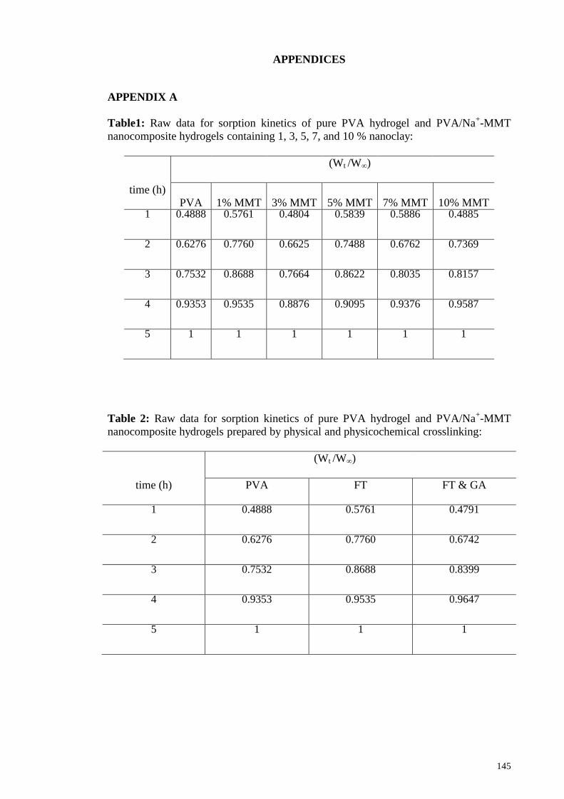
Appendix A 145
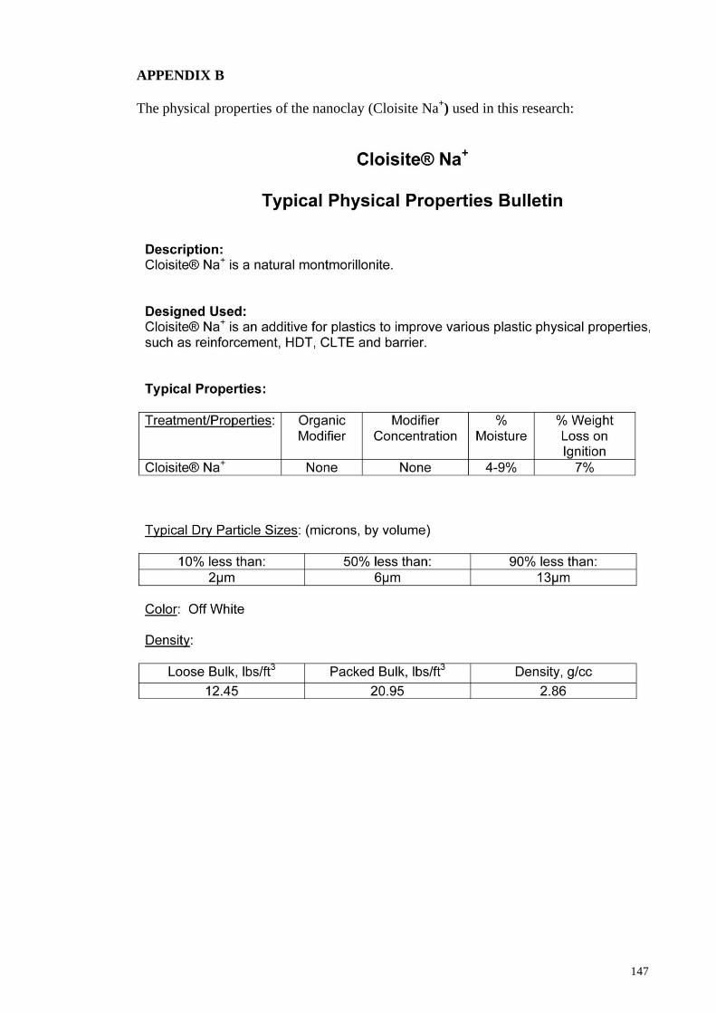
Appendix B 147
Page 14
xiv
LIST OF FIGURES
Figure 1.1: Schematic diagram showing structures of polymeric composites 1
Figure 1.2: Vinyl acetate polymerization and its hydrolysis to polyvinyl alcohol 2
Figure 1.3: Chemical crosslinking of PVA using glutaraldehyde 3
Figure 1.4: Hydrogen bonded poly (vinyl alcohol) organic –inorganic hybrid
structures.
6
Figure 2.1: Interactions among PVA, water, and MMT 10
Figure 2.2: Structure of 2:1 phyllosilicates 15
Figure 2.3: Arrangements of alkyl ammonium ions in mica-type layered silicates
with different layer charges
17
Figure 2.4: Alkyl chain aggregation models 18
Figure 2.5: Schematic illustrations of three different types of thermodynamically
achievable polymer/layered silicate nanocomposites
19
Figure 2.6: Novel crosslinking methods used in hydrogels 28
Figure 2.7: Schematic diagram showing, PVA hydrogels prepared by freeze-
thawing cycles with a PVA-rich region and a PVA-poor region
30
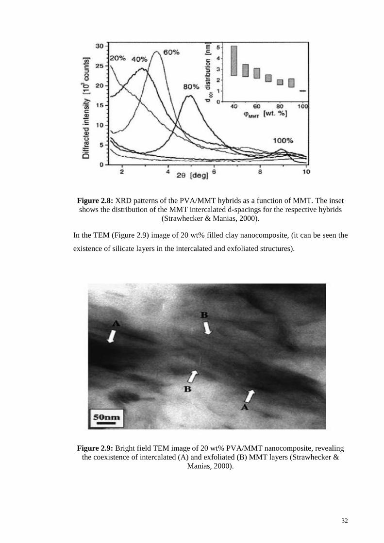
Figure 2.8: XRD patterns of the PVA/MMT hybrids as a function of MMT 32
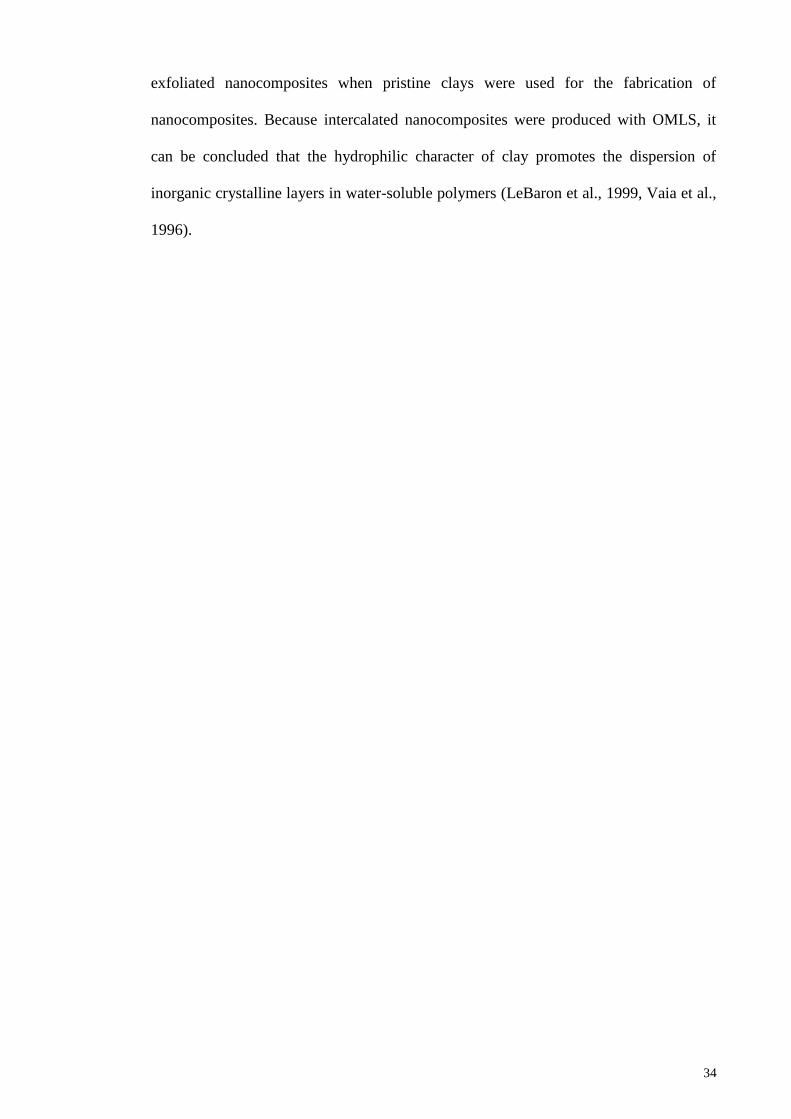
Figure 2.9: Bright field TEM image of 20 wt% PVA/MMT nanocomposite 32
Figure 2.10: XRD patterns of: (a) clays, (b) 4 wt% clays/PVA hybrid films, and
(c) 8 wt% clays/PVA hybrids
35
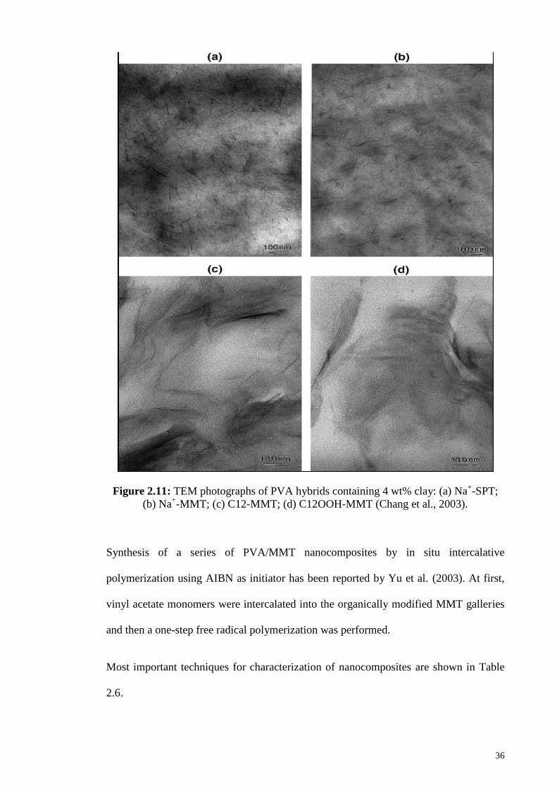
Figure 2.11: TEM photographs of PVA hybrids containing 4 wt% clay: (a) Na+-
SPT; (b) Na+-MMT; (c) C12-MMT; (d) C12OOH-MMT
36
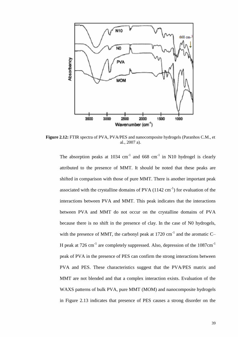
Figure 2.12: FTIR spectra of PVA, PVA/PES and nanocomposite hydrogels 39
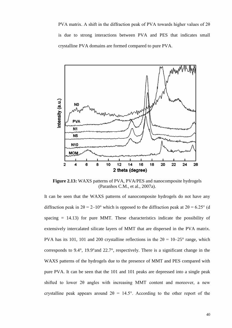
Figure 2.13: WAXS patterns of PVA, PVA/PES and nanocomposite hydrogels 40
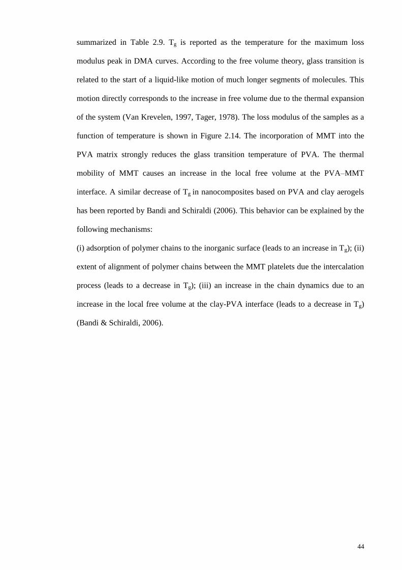
Figure 2.14: Loss modulus of PVA, PVA/PES and nanocomposite hydrogels 45
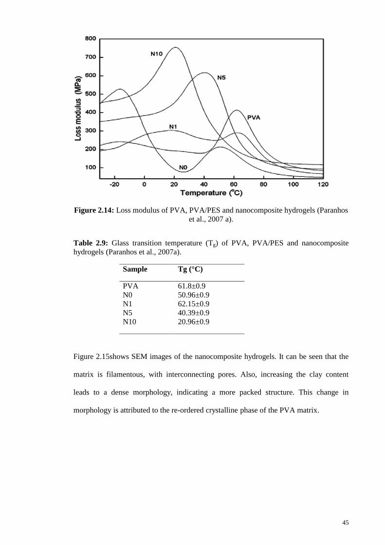
Figure 2.15: SEM micrographs of the nanocomposite hydrogels 46
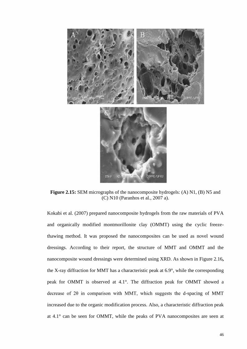
Figure 2.16: XRD of MMT(MONT),OMMT(OMONT) and PVA nanocomposite
hydrogels
47
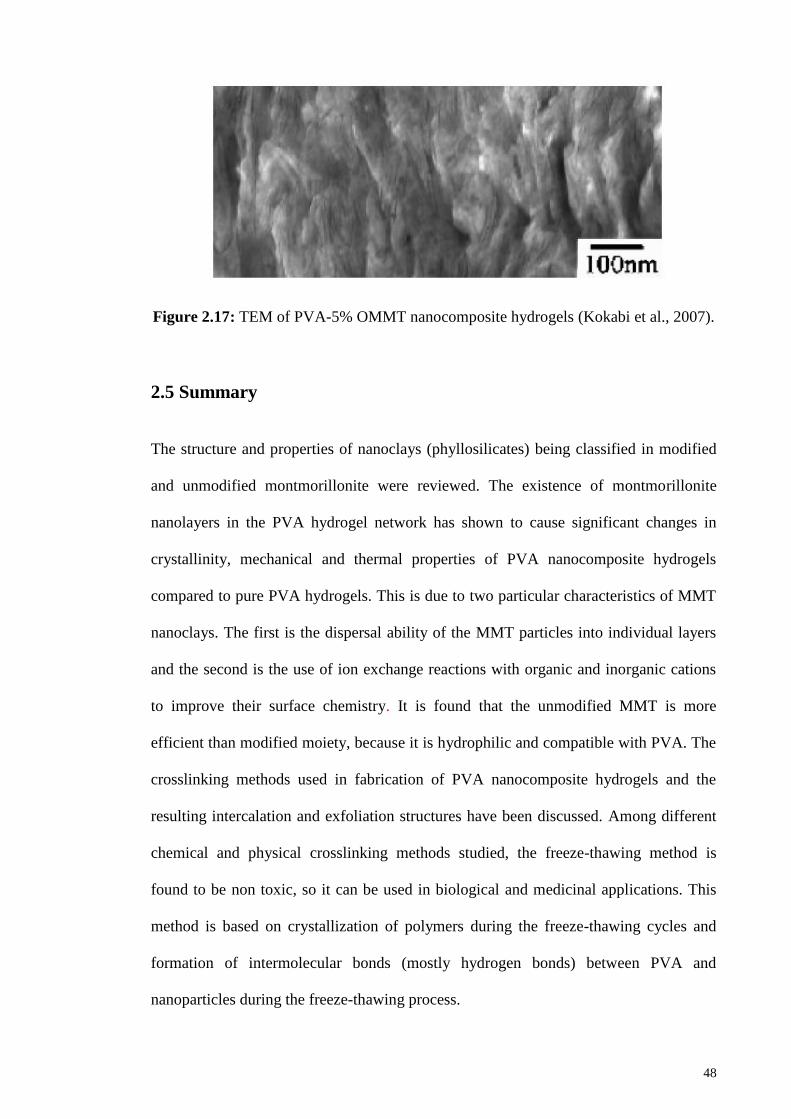
Figure 2.17: TEM of PVA-5% OMMT nanocomposite hydrogels 48
Figure 3.1: Polyvinyl alcohol, PVA 50
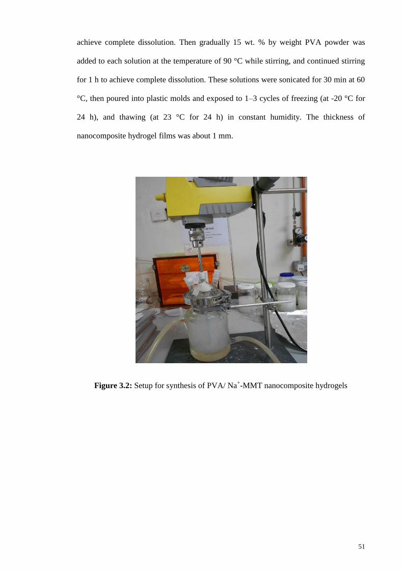
Figure 3.2: Setup for synthesis of PVA/ Na+-MMT nanocomposite hydrogels 51
Page 15
xv
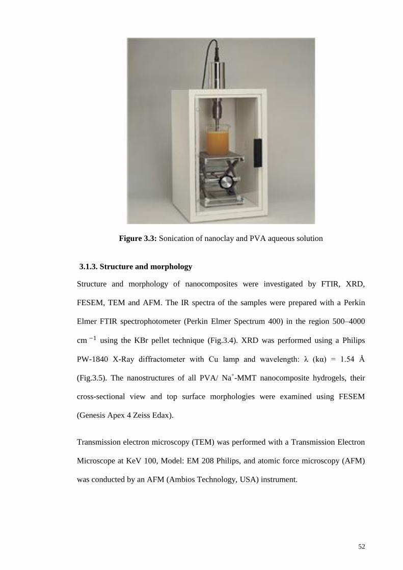
Figure 3.3: Sonication of nanoclay and PVA aqueous solution 52
Figure 3.4: FT-IR spectrophotometer used for structural analysis 53
Figure 3.5: X-Ray diffractometer for morphology analysis 53
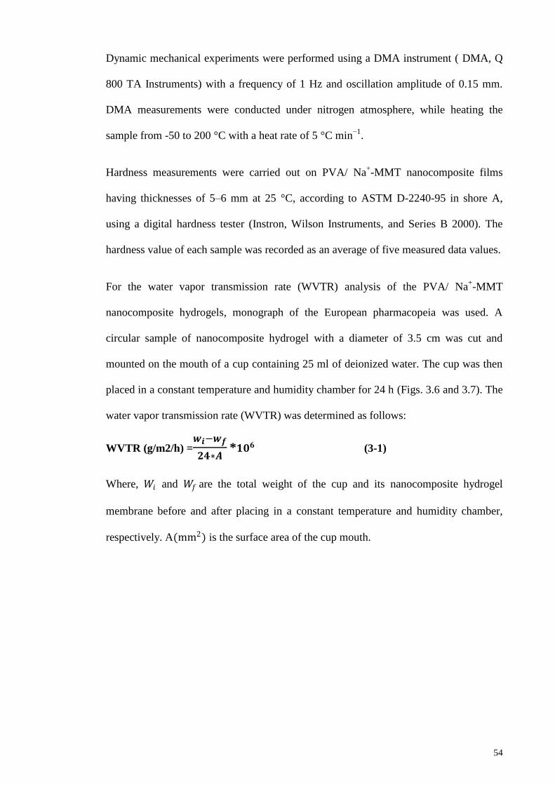
Figure 3.6: Constant temperature and humidity chamber for WVTR analysis 55
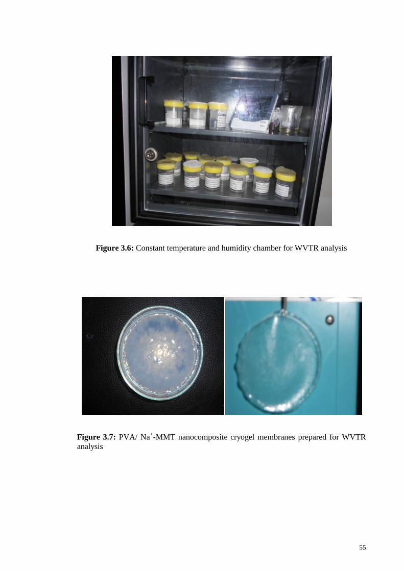
Figure 3.7: PVA/ Na+-MMT nanocomposite cryogel membranes prepared for
WVTR analysis
55
Figure 3.8: Chemical crosslinking of PVA with glutaraldehyde 59
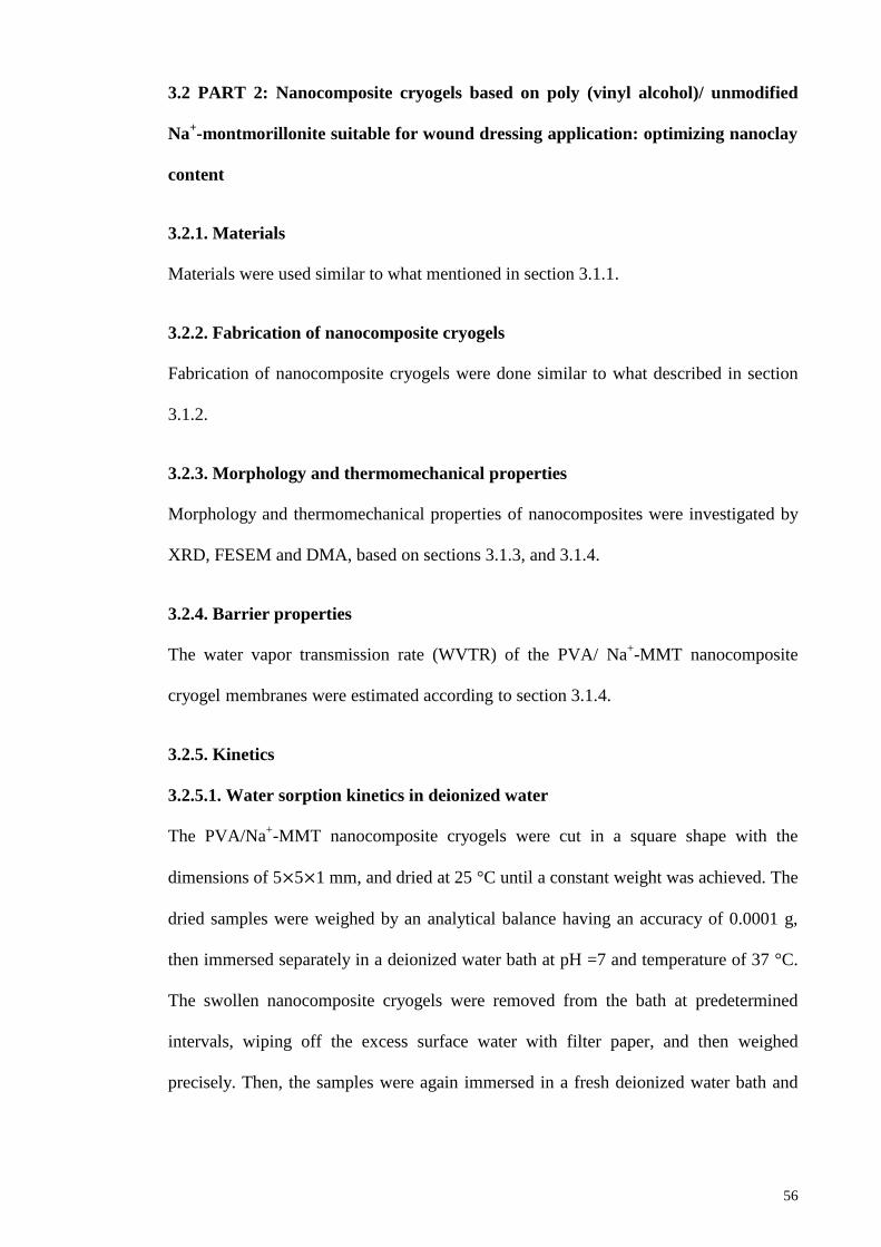
Figure 3.9: Inter chain Hydrogen bonding within a PVA-Na+MMT/PVP–I blend
occurs between carbonyl groups on PVP and hydroxyl groups on PVA and
silanol groups on Na+MMT
62
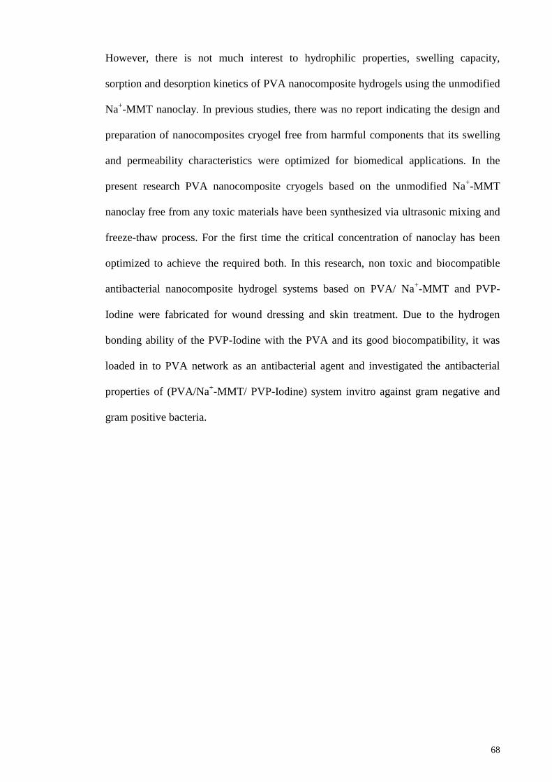
Figure 4.1: FTIR spectra of (A) the pure PVA hydrogel, PVA/ Na+-MMT
hydrogel containing (B) 1, (C) 5and (D)10% nanoclay
70
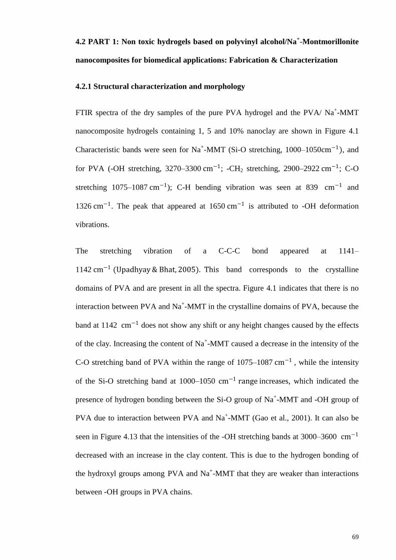
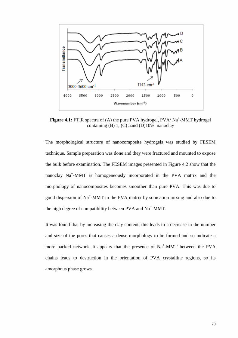
Figure 4.2: FESEM images of pure PVA hydrogel, PVA/ Na+-MMT
nanocomposite hydrogels containing 1, 5 and 10% nanoclay
71
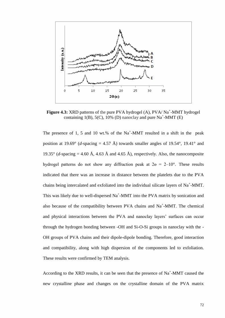
Figure 4.3: XRD patterns of the pure PVA hydrogel (A), PVA/ Na+-MM
hydrogel containing 1(B),5(C), 10% (D)nanoclay and pure Na+-MMT( E)
72
Figure 4.4: TEM of PVA/ Na+-MMT hydrogel containing 5 and 10 wt%
nanoclay
73
Figure 4.5: AFM of PVA/ Na+-MMT hydrogel containing 5 wt% nanoclay 74
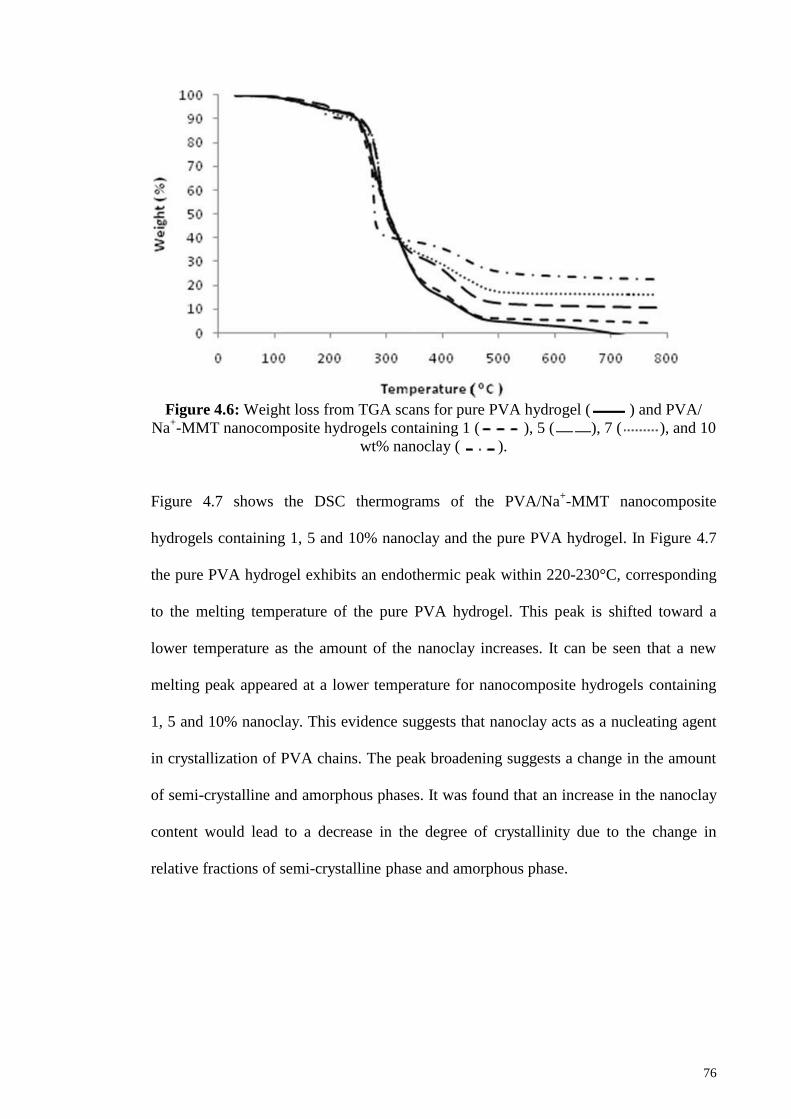
Figure 4.6: Weight loss from TGA scans for pure PVA hydrogel and PVA/ Na+-
MMT nanocomposite hydrogels containing 1, 5, 7 and 10 wt% nanoclay
76
Figure 4.7: DSC curves for (A) the pure PVA hydrogel and PVA/ Na+-
MMTnanocomposite hydrogels containing (B) 1, (C) 5 and (D) 10% nanoclay
77
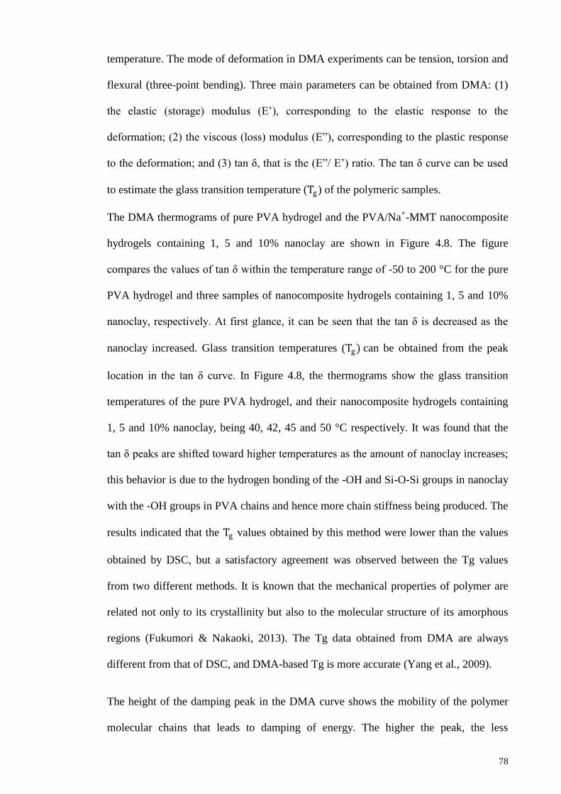
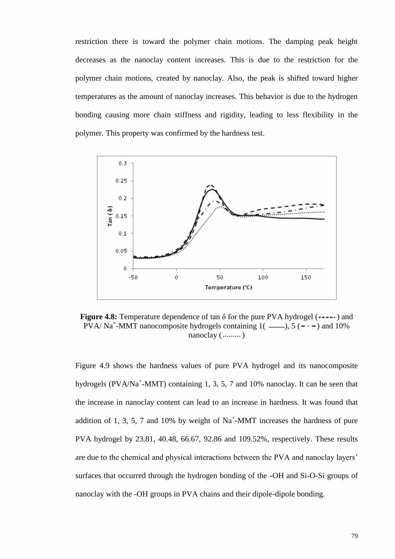
Figure 4.8: Temperature dependence of tan δ for the pure PVA hydrogel and
PVA/ Na+-MMT nanocomposite hydrogels containing 1, 5 and 10% nanoclay
79
Figure 4.9: Hardness of PVA/ Na+-MMT nanocomposite hydrogels containing
0, 1, 3, 5, 7 and 10% nanoclay
80
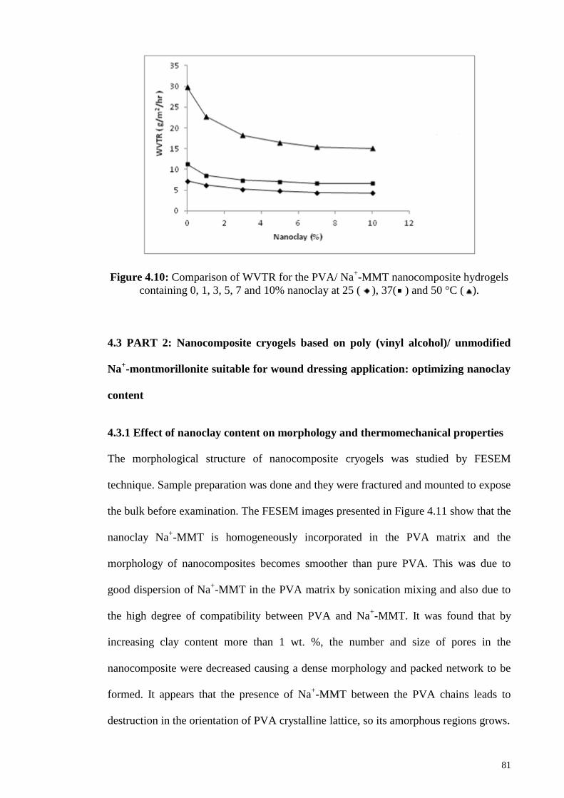
Figure 4.10: Comparison of WVTR for the PVA/ Na+-MMT nanocomposite
hydrogels containing 0, 1, 3, 5, 7 and 10% nanoclay at 25, 37 and 50 °C
81
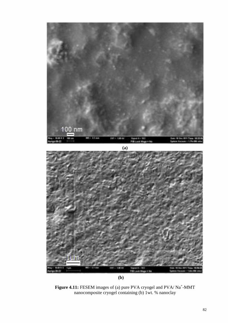
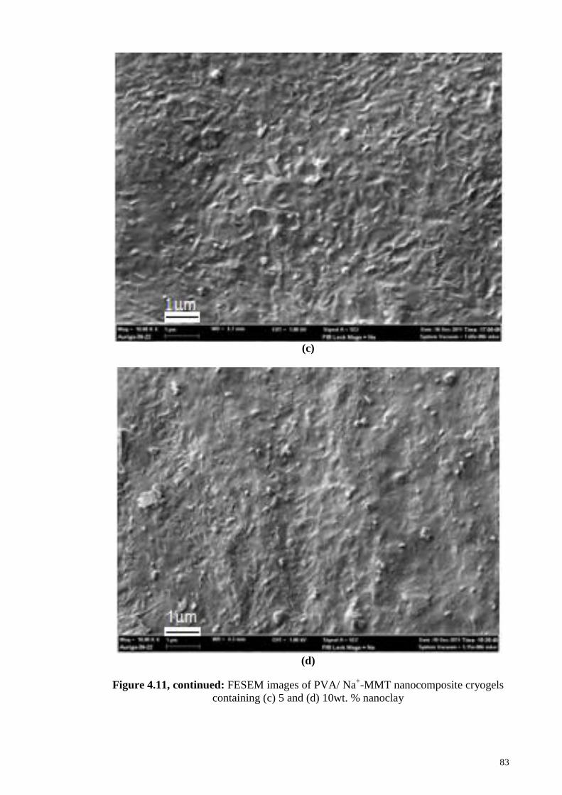
Figure 4.11: FESEM images of (a) pure PVA cryogel, PVA/ Na+-MMT
nanocomposite cryogels containing (b) 1, (c) 5and (d) 10wt. % nanoclay
82
Figure 4.12: XRD patterns of (a) the pure PVA cryogel, (b) PVA/ Na+-MMT
cryogel containing 1, (c) 5, (d) 10 wt. % nanoclay
84
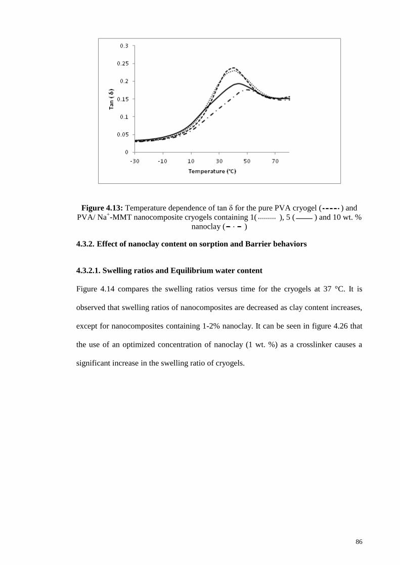
Figure 4.13: Temperature dependence of tan δ for the pure PVA cryogel and
PVA/ Na+-MMT nanocomposite cryogels containing 1, 5 and 10 wt.% nanoclay
86
Page 16
xvi
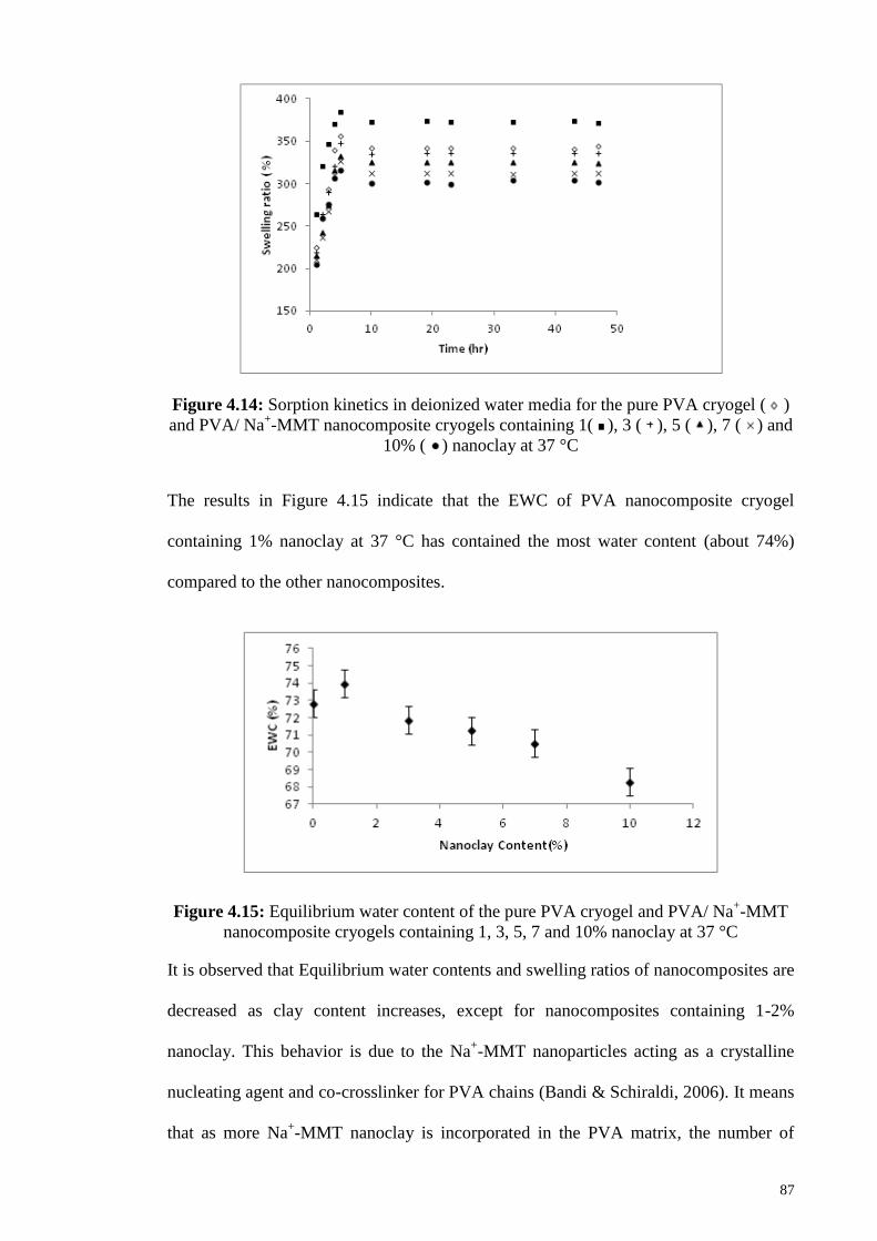
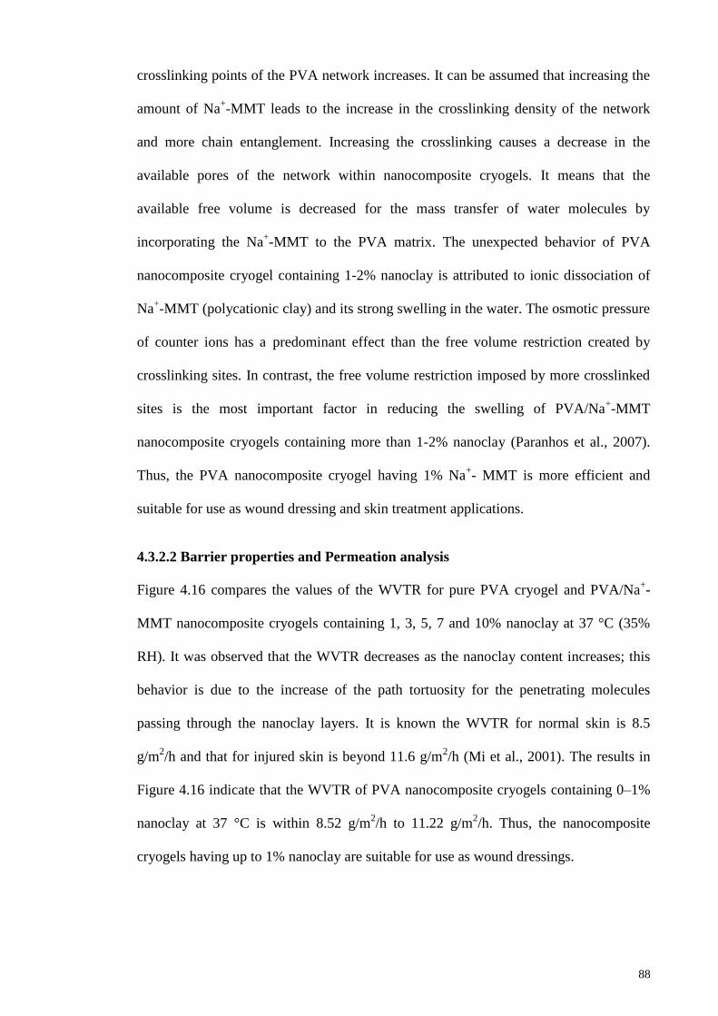
Figure 4.14: Sorption kinetics in deionized water media for the pure PVA
cryogel and PVA/ Na+-MMT nanocomposite cryogels containing 1, 3, 5, 7 and
10% nanoclay at 37 °C
87
Figure 4.15: Equilibrium water content of the pure PVA cryogel and PVA/ Na+-
MMT nanocomposite cryogels containing 1, 3, 5, 7 and 10% nanoclay at 37 °C
87
Figure 4.16: Comparison of WVTR for the PVA/ Na+-MMT nano composite
cryogels containing 0, 1, 3, 5, 7 and 10% nanoclay at 37°C
89
Figure 4.17: Plots of ln (𝑊𝑡 /𝑊∞) versus ln (t) for the pure PVA cryogel and
PVA/ Na+-MMT nanocomposite cryogels containing1, 3, 5, 7 and 10% nanoclay
at 37°C
90
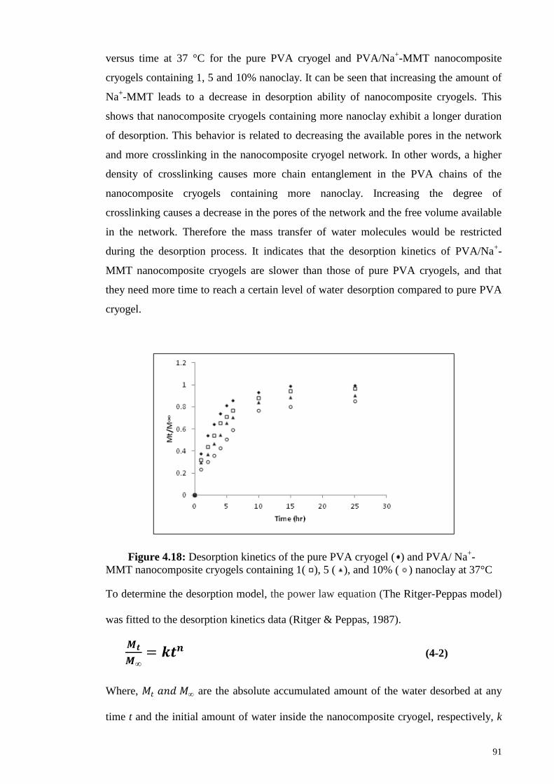
Figure 4.18: Desorption kinetics of the pure PVA cryogel and
PVA/ Na+-MMT nanocomposite cryogels containing 1, 5 and 10% nanoclay at
37°C
91
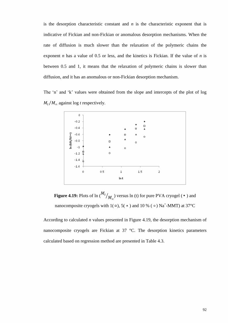
Figure 4.19: Plots of ln (𝑀𝑡
𝑀∞ ) versus ln (t) for pure PVA cryogel and
nanocomposite cryogels with 1, 5 and 10 % Na+-MMT at 37°C
92
Figure 4.20: FT-IR spectra of the pure PVA hydrogel, physical (1N) and
physicochemical (1N & GA) crosslinked PVA/MMT hydrogel containing 1%
nanoclay
95
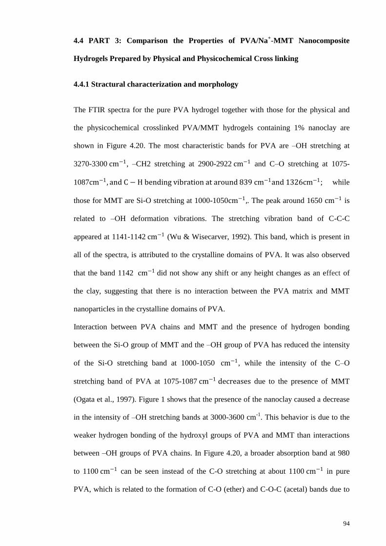
Figure 4.21: XRD patterns of the physical and physicochemical (PVA & GA)
crosslinked pure PVA hydrogel, MMT, and the physical (1N) and
physicochemical (1N & GA) crosslinked PVA/MMT hydrogel containing 1%
nanoclay
96
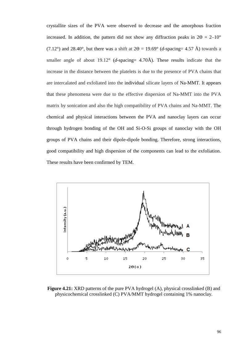
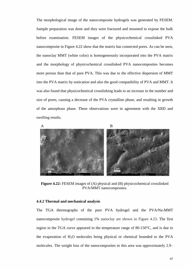
Figure 4.22: FESEM images of (A) physical and (B) physicochemical
crosslinked PVA/MMT nanocomposites.
97
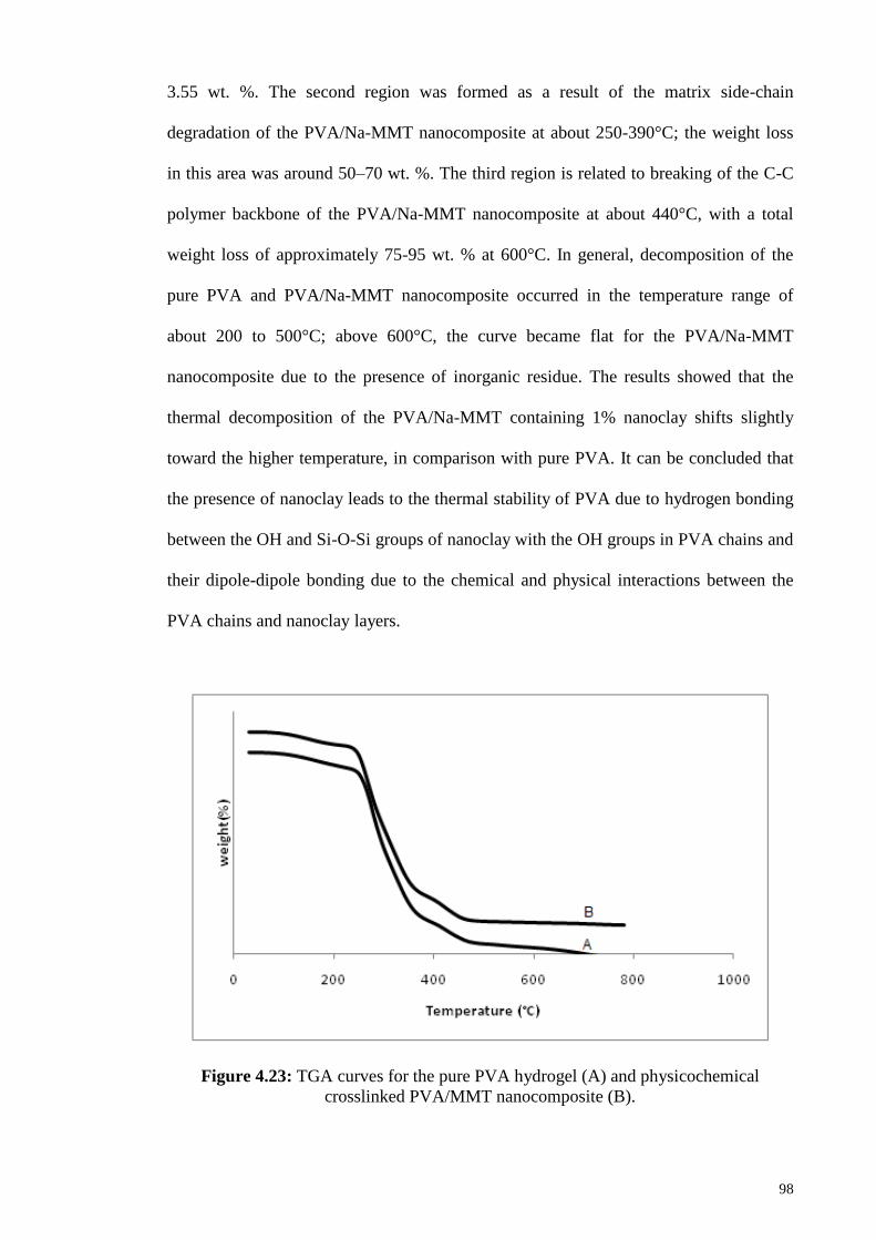
Figure 4.23: TGA curves for the physical (PVA) and physicochemical (1N &
GA) crosslinked PVA/MMT nanocomposites
98
Figure 4.24: DSC curves for the pure PVA hydrogel, and their physical (1N)
and physicochemical (1N & GA) crosslinked nanocomposite hydrogels
99
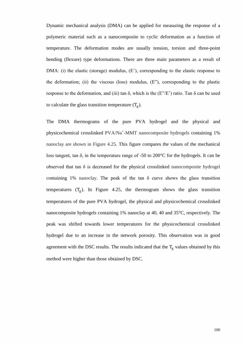
Figure 4.25: Temperature dependence of tan d for the pure PVA hydrogel, their
physical (1N) and physicochemical (PVA & GA) cross linked PVA/MMT
nanocomposite hydrogels
101
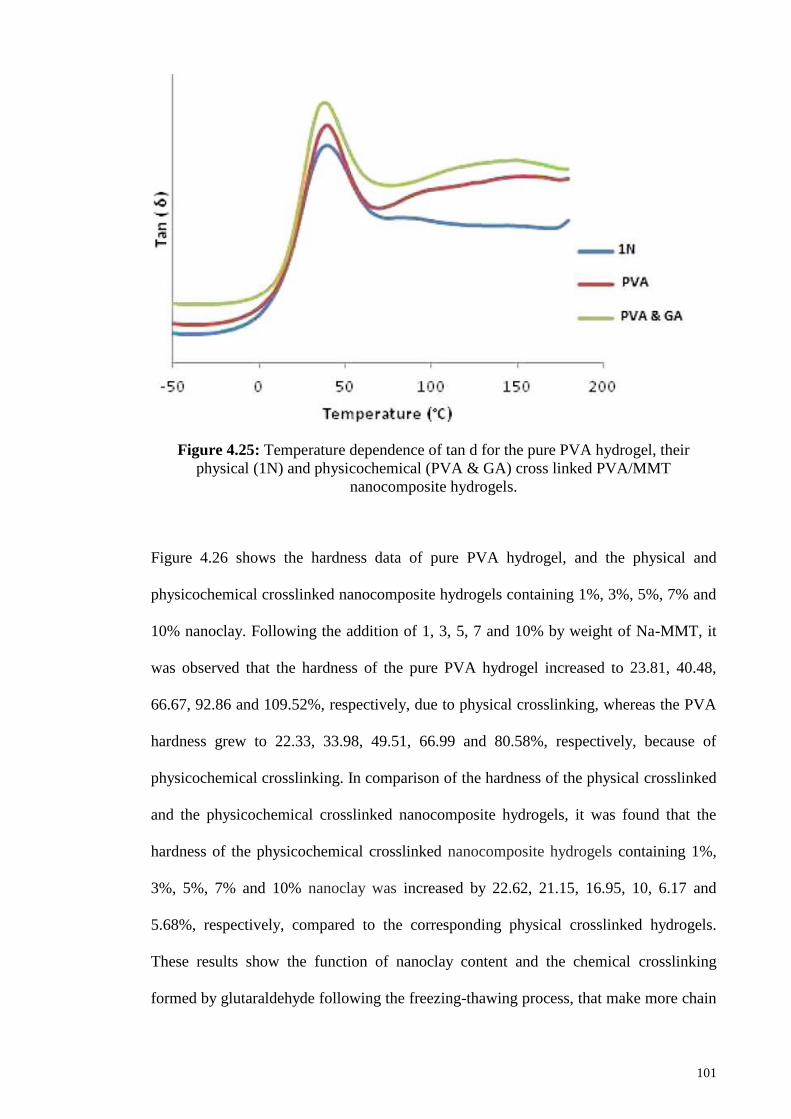
Figure 4.26: Hardness of physical (FT) and physicochemical (FT & GA)
crosslinked PVA/MMT nanocomposite hydrogels versus nanoclay content, those
for pure PVA hydrogels being at 0% nanoclay
102
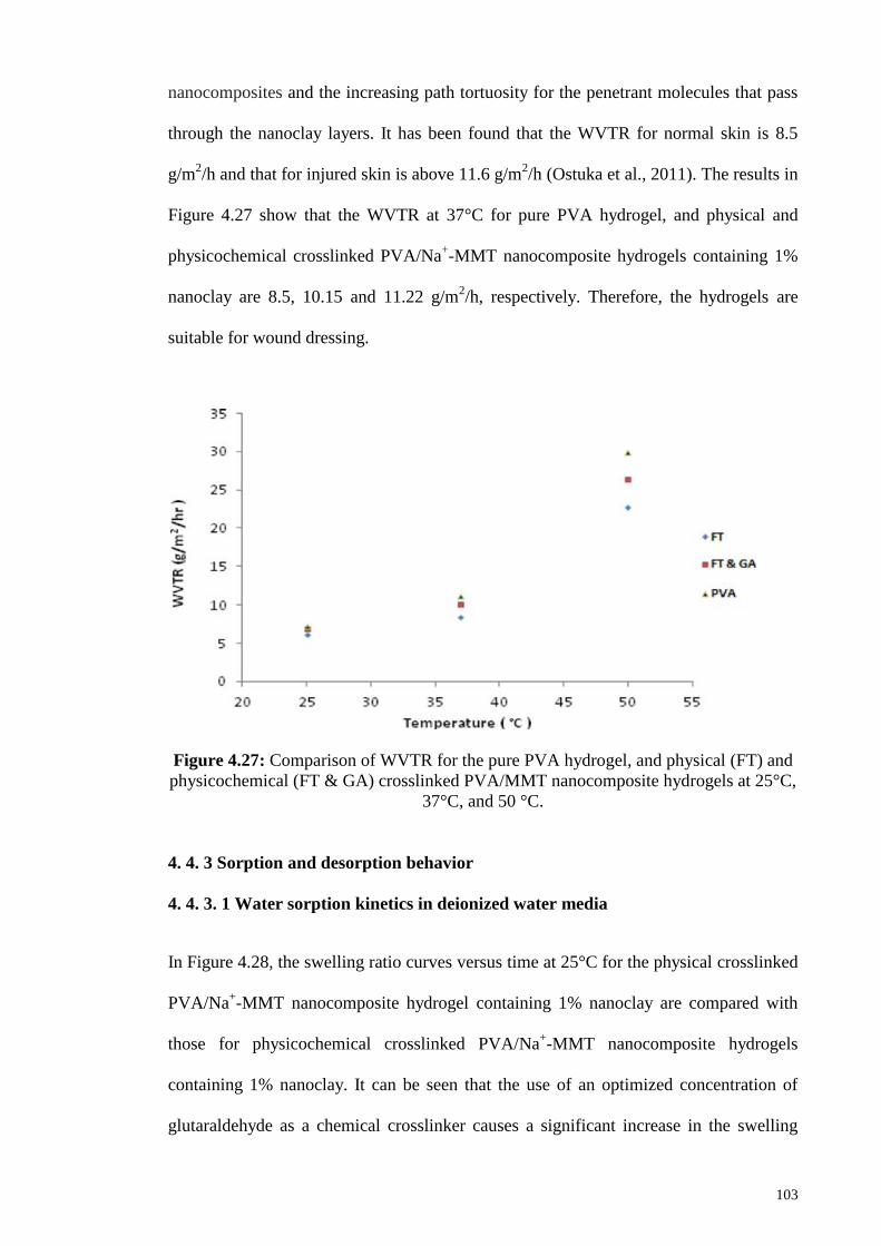
Figure 4.27: Comparison of WVTR for the pure PVA hydrogel, and physical
(FT) and physicochemical (FT & GA) crosslinked PVA/MMT nanocomposite
hydrogels at 25°C, 37°C, and 50 °C
103
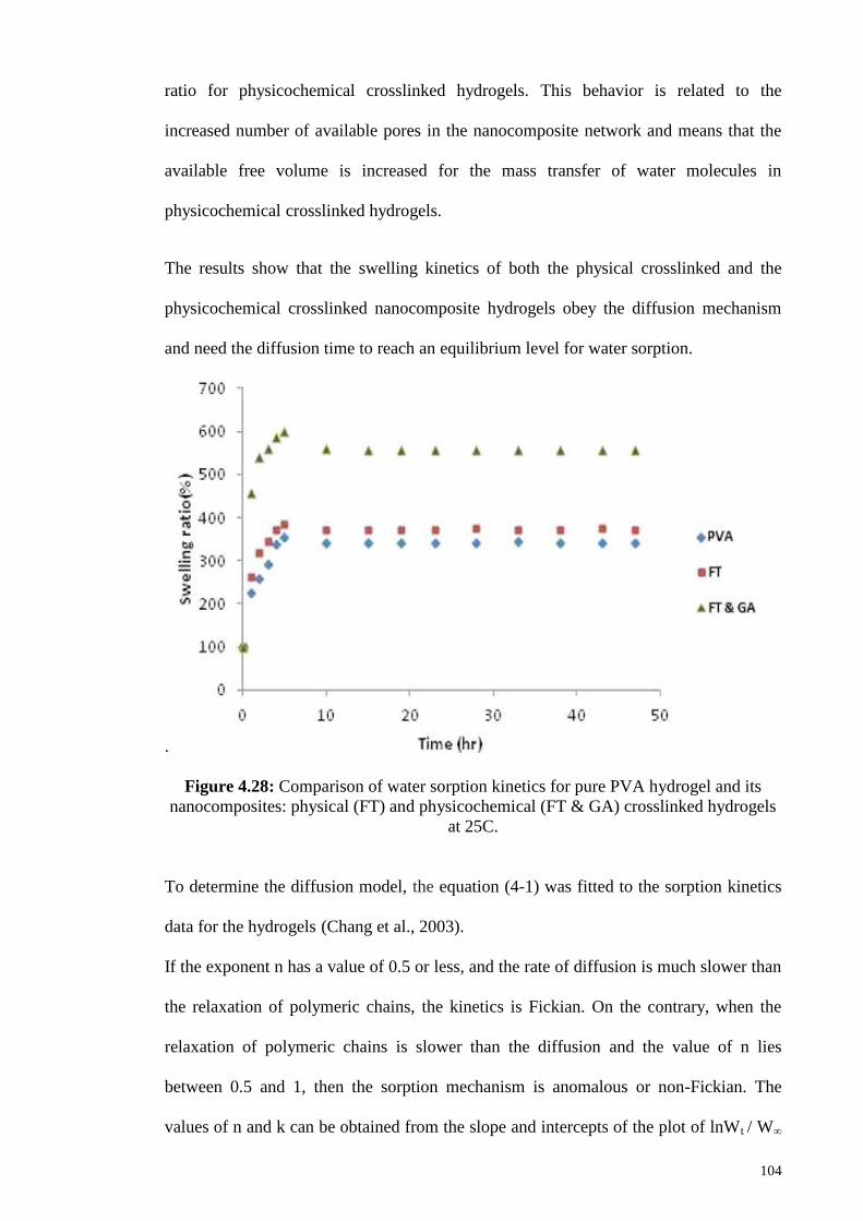
Figure 4.28: Comparison of water sorption kinetics for pure PVA hydrogel an
its nanocomposites: physical (FT) and physicochemical (FT & GA) crosslinked
104
Page 17
xvii
hydrogels at 25°C
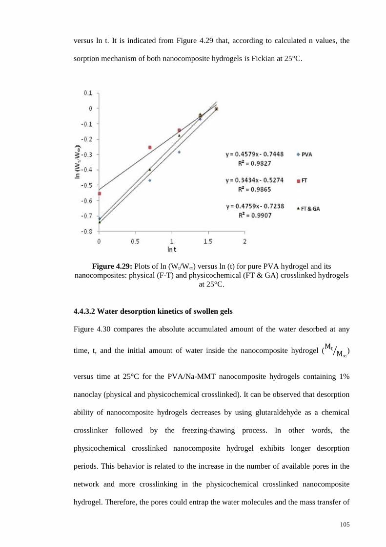
Figure 4.29: Plots of ln (𝑤𝑡 /𝑤∞) versus ln (t) for pure PVA hydrogel and its
nanocomposites: physical (F-T) and physicochemical (FT & GA) crosslinked
hydrogels at 25°C
105
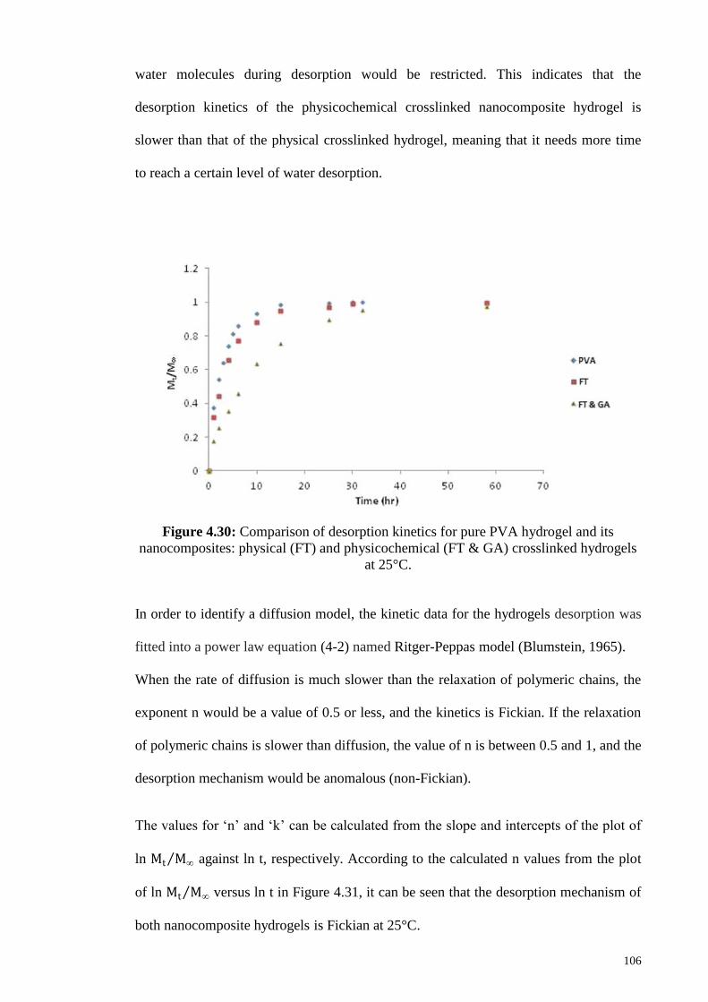
Figure 4.30: Comparison of desorption kinetics for pure PVA hydrogel and its
nanocomposites: physical (FT) and physicochemical (FT & GA) crosslinked
hydrogels at 25°C
106
Figure 4.31: Plots of ln (Mt M∞ ) versus ln (t) for pure PVA hydrogel and its
nanocomposites: physical (F-T) and physicochemical (FT & GA) crosslinked
hydrogels at 25°C
107
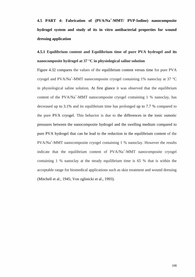
Figure 4.32: Equilibrium content versus time for pure cryogel and PVA/Na+-
MMT nanocomposite cryogel containing 1 % nanoclay at 37 °C in physiological
saline solution
109
Figure 4.33: Residual of physiological saline solution (PSS) amount versus time
at 37 °C for the pure PVA cryogel and PVA/Na+-MMT nanocomposite cryogel
containing 1% nanoclay
110
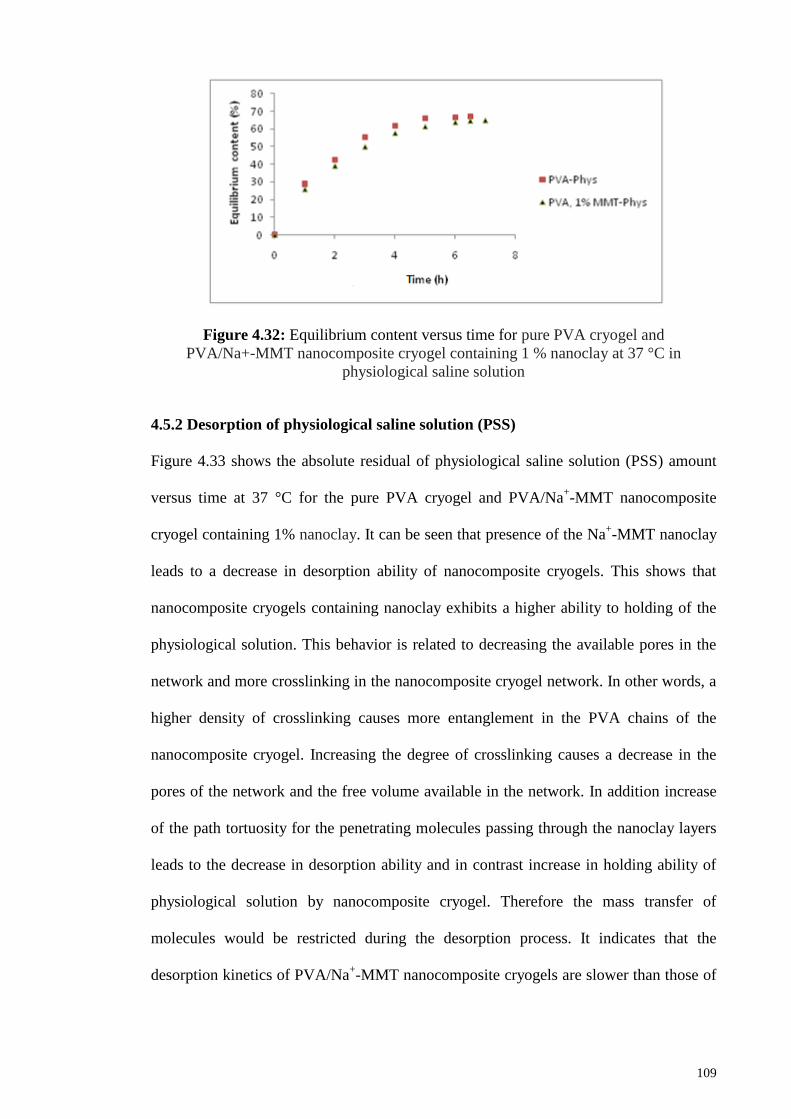
Figure 4.34: Antibacterial activity of pure PVA cryogel and PVA/Na+-MMT
nanocomposite cryogels containing 1, 3, 5, 7, and 10 % nanoclay loaded by
PVP- Iodin at 37 °C after 24 hours
111
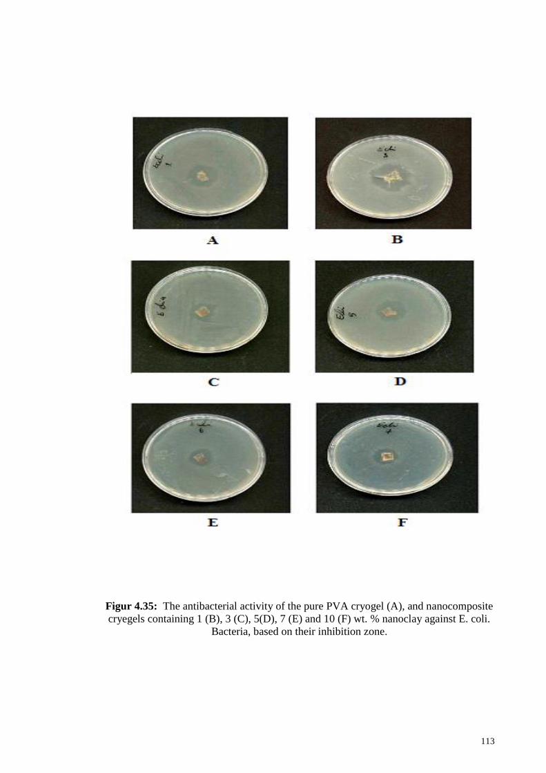
Figure 4.35: The antibacterial activity of the pure PVA cryogel (A), and
nanocomposite cryegels containing 1 (B), 3 (C), 5(D), 7 (E) and 10 (F) wt. %
nanoclay against E. coli. bacteria, based on their inhibition zone
113
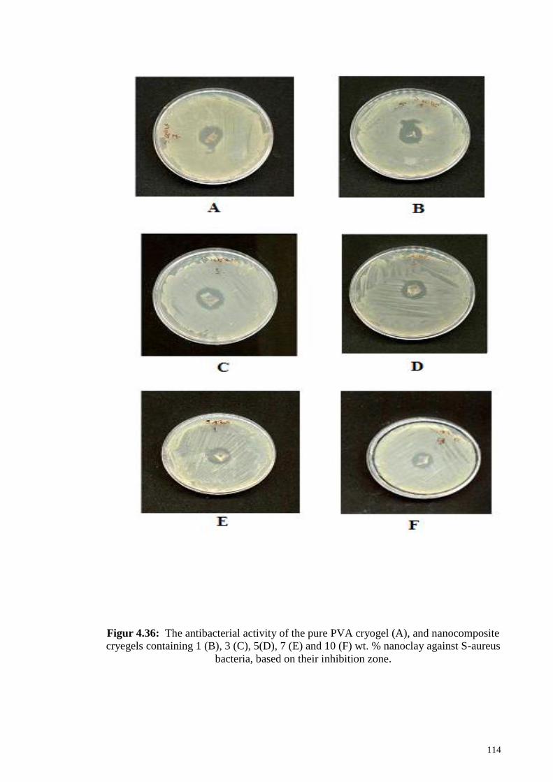
Figure 4.36: The antibacterial activity of the pure PVA cryogel (A), and
nanocomposite cryegels containing 1 (B), 3 (C), 5(D), 7 (E) and 10 (F) wt. %
nanoclay against S-aureus bacteria, based on their inhibition zone
114
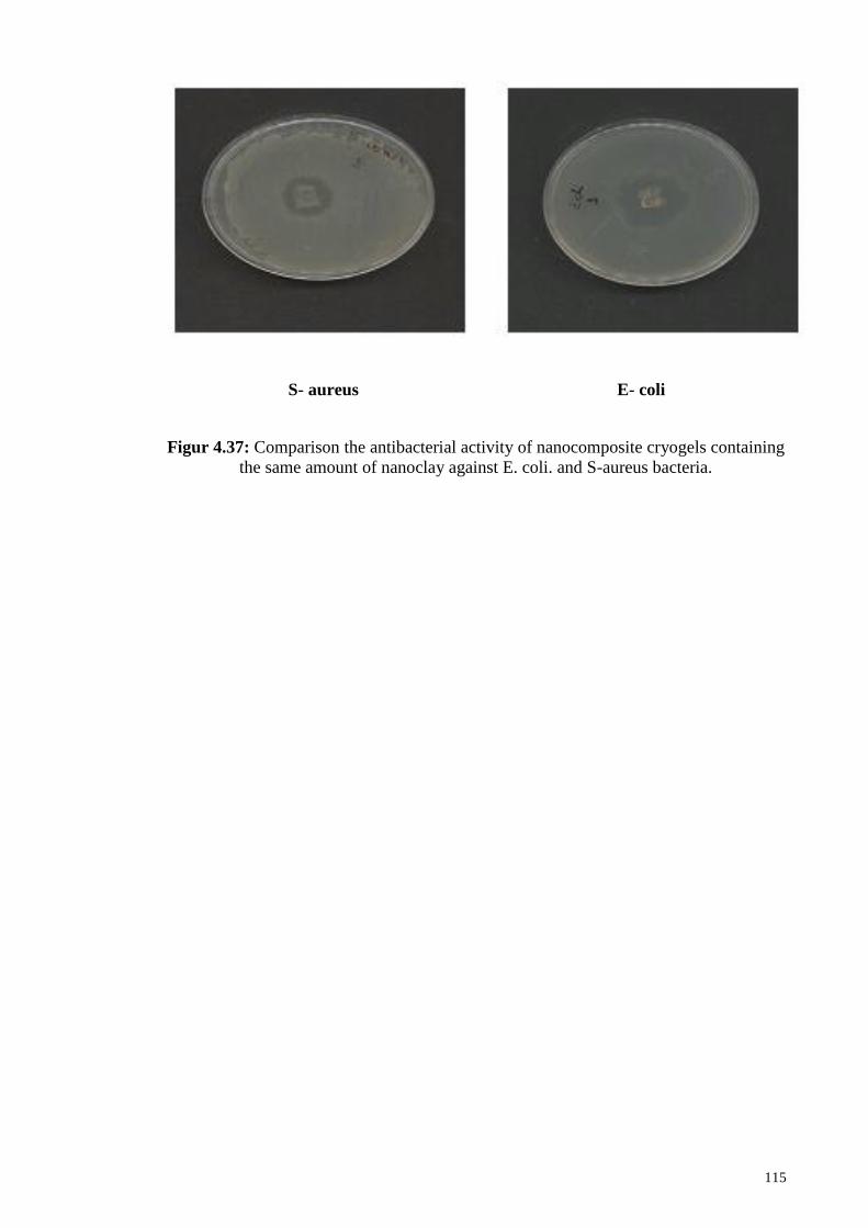
Figure 4.37: Comparison the antibacterial activity of nanocomposite cryogels
containing the same amount of nanoclay against E. coli. and S-aureus bacteria.
115
Page 18
xviii
LIST OF TABLES
Table 2.1: Chemical formula and characterization parameters of commonly used
2:1 phylosilicates 16
Table 2.2: Preparative techniques for polymer-based composite and nano
composite systems. 22
Table 2.3: Advantages and disadvantages for three main groups of Intercalation
methods. 26
Table 2.4: Crosslinking methods for the preparation of nanocomposite hydrogels 29
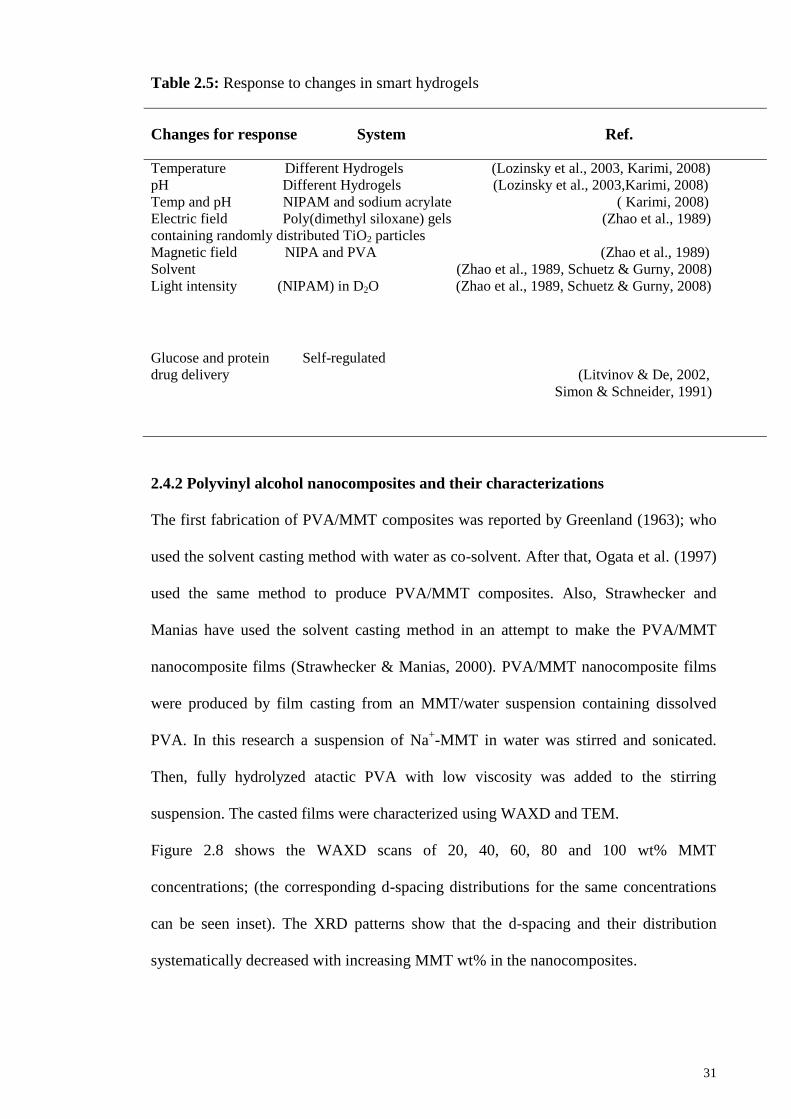
Table 2.5: Response to changes in smart hydrogels 31
Table 2.6: Most important techniques for characterization of nanocomposites 37
Table 2.7: DSC parameters obtained from the nanocomposite hydrogels 43
Table 2.8: Crystalline parameters of PVA matrix as a function of MMT content
in a nanocomposite Hydrogel 43
Table 2.9: Glass transition temperature (Tg) of PVA, PVA/PES and
nanocomposite hydrogels 45
Table 4.1: Weight loss results of TGA for pure PVA hydrogel and PVA/Na+-
MMT nanocomposite hydrogels at various temperatures 75
Table 4.2: Sorption kinetics characteristics for the pure PVA cryogel and
PVA/Na+-MMT nanocomposite cryogels at 37 °C 90
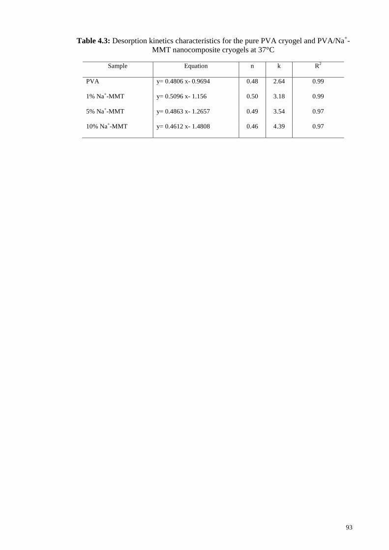
Table 4.3: Desorption kinetics characteristics for the pure PVA cryogel and
PVA/Na+-MMT nanocomposite cryogels at 37°C 93
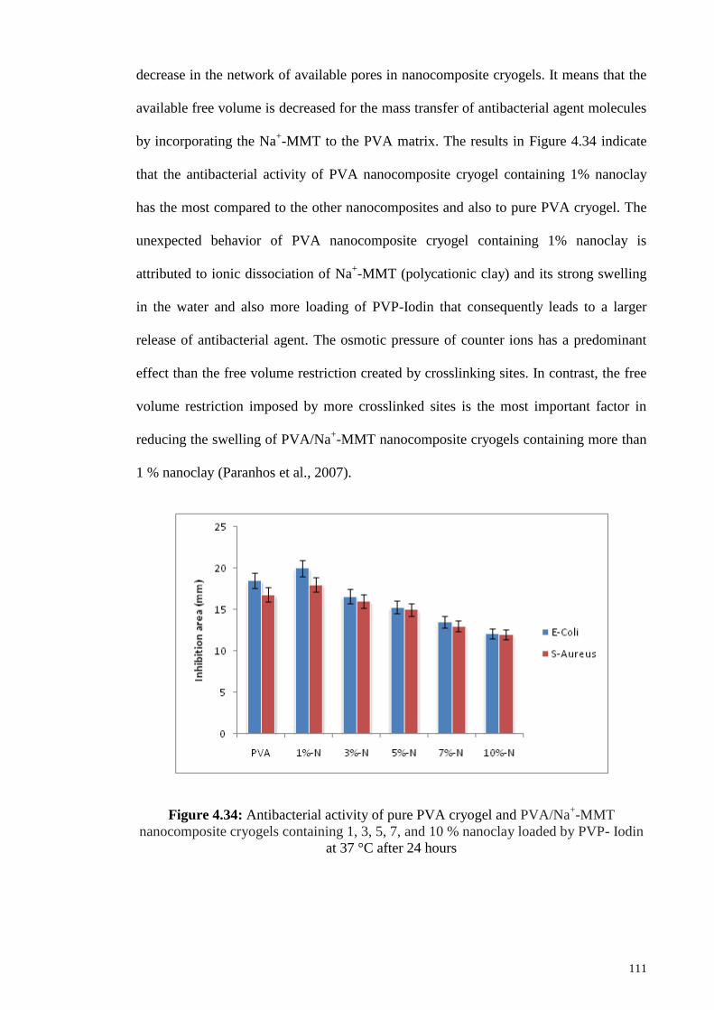
Table 4.4: Antibacterial activity of pure PVA cryogel and PVA/Na+-MMT
nanocomposite cryogels against gram positive and gram negative bacteria 112
Page 19
xix
LIST OF ABBREVIATIONS
AA Acrylic acid
AFM Atomic Force Microscopy
AIBN 2, 2'-Azobis-Isobutyronitrile
C12MMT Dodecylammonium-modified MMT
C12OOHMMT 12-Aminolauric-modified MMT
DMA Dynamic Mechanical Analysis
DMAc N, N-dimethylacetamide
DMTA Dynamic Mechanical Thermal Analysis
DSC Differential Scanning Calorimetry
DW Deionized Water
E‟ Elastic (storage) modulus
E” Viscous (loss) modulus
E-Coli Escherichia Coli
EWC Equilibrium Water Content
FESEM Field Emission Scanning Electron Microscopy
FT Freeze-Thaw
FT & GA Freeze-Thaw & Glutaraldehyde
FTIR Fourier Transform Infrared
GA Glutaraldehyde
HDPE High-Density Poly Ethylene
HPMC Hydroxy Propyl Methyl Cellulose
MDSC Modulated Differential Scanning Calorimetry
MMT Montmorillonite
Na+-MMT Sodium Montmorillonite
NIPA Poly (N-Iso Propyl Acrylamide)
Page 20
xx
NIPAAm N-Iso Propyl Acrylamide
NIPAM N-Iso Propyl Acrylamide
NMR Nuclear magnetic resonance
OMLS Organically Modified Layered Silicate
OMMT Organically Modified Montmorillonite clay
PAA Poly (Acrylic acid)
PAN Poly Acrylonitrile
PANI Poly Aniline
PCL Poly Caprolactone
PDDA Poly (Diallyl Dimethyl Ammonium Chloride)
PEO Poly Eethylene Oxid
PES Poly Ether Sulfone
PET Poly Ethylene Terephthalate
PLA Polyl Actic acid
PLS Polymer Layered Silicate
PP Poly Propylene
PS Poly Styrene
PU Poly Urethane
PVA Poly Vinyl Alcohol
PVA/Na+-MMT Poly (vinyl alcohol) / Sodium Montmorillonite
PVOH Poly Vinyl Alcohol
PVP Poly Vinyl Pyrrolidone
PVP- Iodine poly Vinyl Pyrrolidone – Iodine
SANS Small-Angle Neutron Scattering
S.Aureus Staphylococcus Aureus
SAXS Small Angle X-ray Scattering
Page 21
xxi
SEM Scanning Electron Microscope
SPS Smart polymers
SR Swelling Ratio
TEM Transmission Electron Microscopy
Tg Glass Transition Temperature
TGA Thermogravimetric Analysis
Tm Melting temperature
UV Ultraviolet
WAXD Wide Angle X-ray Diffraction
WVTR Water Vapor Transmission Rate
XRD X-ray Diffraction
ΔHm Enthalpy of melting
χ C Degree of crystallinity
𝑀∞ Initial amount of water inside the nanocomposite
hydrogel
𝑀𝒕 Water desorbed at any time t
Ws Weight of the swollen gel
𝑊∞ Equilibrium water sorption.
𝑊𝑑 Weight of the dry gel
𝑊𝑡 Amount of absorbed water at any time t
𝑚(0) Weight of hydrogel at the initial time zero
𝑚 ∞ Weight of hydrogel at drying time
𝑚 𝑡 Weight of hydrogel at time t
Page 22
xxii
LIST OF APPENDICES
Appendix A 145
Appendix B 147
Page 23
1
CHAPTER 1: GENERAL INTRODUCTION
1.1 Background
In the past years, several attempts have been made to the preparation and
characterization of nano composite hydrogels, the elasticity and permeability of gels
with the reinforcing ability of clays embedded into the hydrogels are combined in these
hydrogels (Bignotti et al., 2004; Ekici et al., 2006; Haraguchi et al., 2003). Also using
nanoclay in nanocomposite hydrogel structures leads to enhanced chemical, physical
and mechanical properties (Haraguchi & Takehisa, 2002; Schexnailder & Schmidt,
2009; Haraguchi, 2007).
Figure 1.1: Schematic diagram showing structures of polymeric composites:
A) Conventional (traditional) B, C) Nanotechnology
Among the natural mineral clays such as bentonite (Lee & Chen, 2004; Huang et al.,
2009), laponite (Abdurrahmanoglu et al., 2008; Liu et al., 2007; Song et al., 2008; Nie
& Oppermann, 2005), hydrotalcite (Lee & Lee, 2006, Zhang et al., 2009) and
montmorillonite (Kasgoz & Durmus, 2008, Lee & Fu, 2003, Sur et al., 2003, Al et al.,
2003), the main natural mineral clay that widely used to prepare nanocomposite
hydrogels, is montmorillonite (MMT). This is due to its good water absorption,
extensive swelling in water and cation exchange capacity (Gao et al., 1999, Gamiz et
Page 24
2
al., 1992, Gao et al., 2001). Due to the presence of silanol groups on the MMT and
ability to hydrogen bonding, montmorillonite can interact with hydrophilic polymers to
participate in ensuring the stability of the nanocomposite systems (Mirzan et al., 2001,
Velazco-Diaz et al., 2005). Thus, MMT can act as a co-crosslinker for hydrophilic
polymers in solution (Mc Gann et al., 2009).
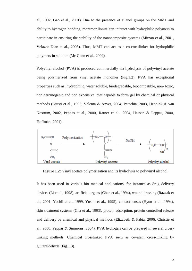
Polyvinyl alcohol (PVA) is produced commercially via hydrolysis of polyvinyl acetate
being polymerized from vinyl acetate monomer (Fig.1.2). PVA has exceptional
properties such as; hydrophilic, water soluble, biodegradable, biocompatible, non- toxic,
non carcinogenic and non expensive, that capable to form gel by chemical or physical
methods (Giusti et al., 1993, Valenta & Anver, 2004, Patachia, 2003, Hennink & van
Nostrum, 2002, Peppas et al., 2000, Ratner et al., 2004, Hassan & Peppas, 2000,
Hoffman, 2001).
Figure 1.2: Vinyl acetate polymerization and its hydrolysis to polyvinyl alcohol
It has been used in various bio medical applications, for instance as drug delivery
devices (Li et al., 1998), artificial organs (Chen et al., 1994), wound dressing (Razzak et
al., 2001, Yoshii et al., 1999, Yoshii et al., 1995), contact lenses (Hyon et al., 1994),
skin treatment systems (Cha et al., 1993), protein adsorption, protein controlled release
and delivery by chemical and physical methods (Elizabeth & Fabia, 2006, Christie et
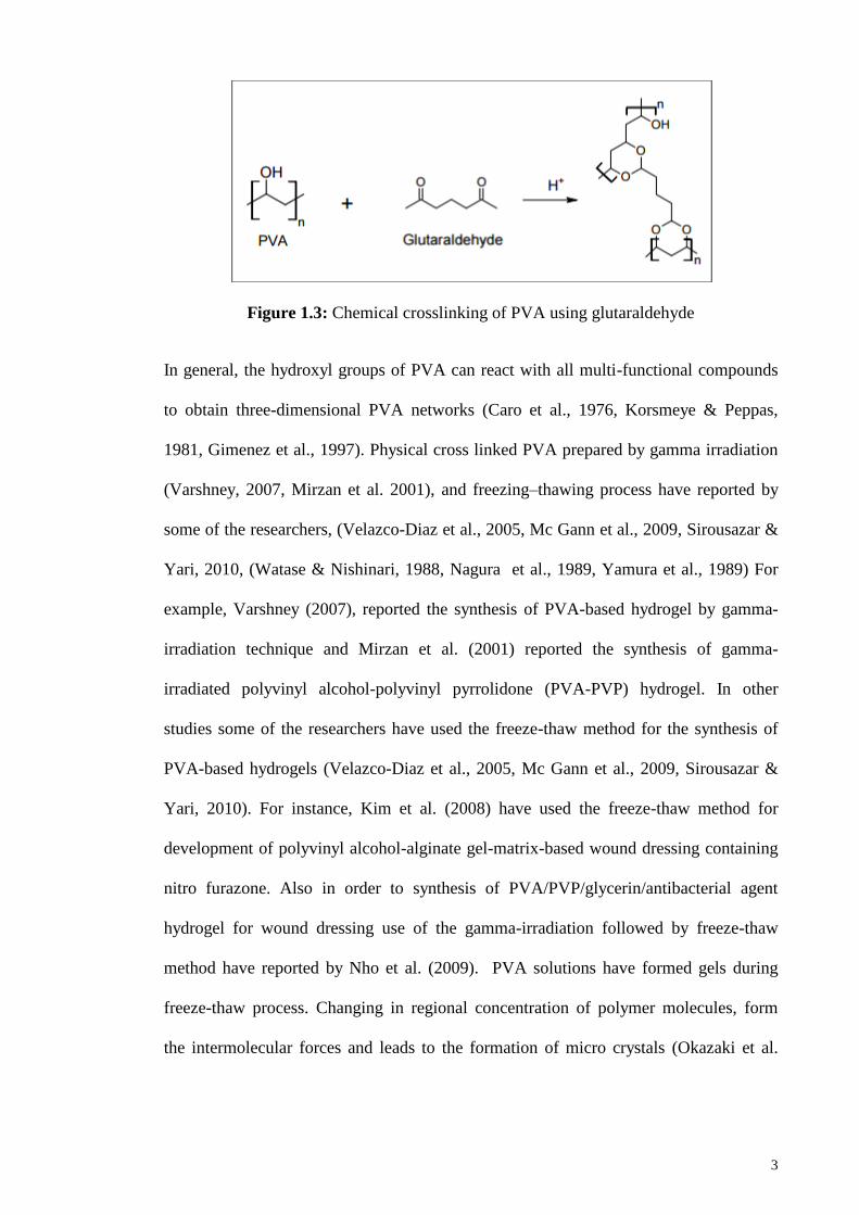
al., 2000, Peppas & Simmons, 2004). PVA hydrogels can be prepared in several cross-
linking methods. Chemical crosslinked PVA such as covalent cross-linking by
glutaraldehyde (Fig.1.3).
Page 25
3
Figure 1.3: Chemical crosslinking of PVA using glutaraldehyde
In general, the hydroxyl groups of PVA can react with all multi-functional compounds
to obtain three-dimensional PVA networks (Caro et al., 1976, Korsmeye & Peppas,
1981, Gimenez et al., 1997). Physical cross linked PVA prepared by gamma irradiation
(Varshney, 2007, Mirzan et al. 2001), and freezing–thawing process have reported by
some of the researchers, (Velazco-Diaz et al., 2005, Mc Gann et al., 2009, Sirousazar &
Yari, 2010, (Watase & Nishinari, 1988, Nagura et al., 1989, Yamura et al., 1989) For
example, Varshney (2007), reported the synthesis of PVA-based hydrogel by gamma-
irradiation technique and Mirzan et al. (2001) reported the synthesis of gamma-
irradiated polyvinyl alcohol-polyvinyl pyrrolidone (PVA-PVP) hydrogel. In other
studies some of the researchers have used the freeze-thaw method for the synthesis of
PVA-based hydrogels (Velazco-Diaz et al., 2005, Mc Gann et al., 2009, Sirousazar &
Yari, 2010). For instance, Kim et al. (2008) have used the freeze-thaw method for
development of polyvinyl alcohol-alginate gel-matrix-based wound dressing containing
nitro furazone. Also in order to synthesis of PVA/PVP/glycerin/antibacterial agent
hydrogel for wound dressing use of the gamma-irradiation followed by freeze-thaw
method have reported by Nho et al. (2009). PVA solutions have formed gels during
freeze-thaw process. Changing in regional concentration of polymer molecules, form
the intermolecular forces and leads to the formation of micro crystals (Okazaki et al.
Page 26
4
1995). The crystalline domains play the role of cross-linking sites in the hydrogel
networks (Takeshita et al. 1999).
PVA gels have physical linkage without any chemical cross-linkers, they are suitable to
design nontoxic and biocompatible, biomedical devices for microorganisms (Okazaki et
al. 1995, Wilcox et al. 1999, Lozinsky et al. 2000, Hassan et al. 2000). Polyvinyl
alcohol nanocomposites have been studied by several authors (Velazco-Diaz et al.,
2005, Mc Gann et al., 2009, Sirousazar & Yari, 2010). The first fabrication of
PVA/MMT composites reported by Greenland (1963) used solvent casting method with
water as a co-solvent. After that Ogata et al., (1997) using the same method have
produced PVA/MMT composites. Also, Strawhecker and Manias have used solvent
casting method in attempts to fabrication of PVA/MMT nanocomposite films. They
produced PVA/MMT nanocomposite films from a MMT/water suspension containing
dissolved PVA by casting and they found a co-existence of silicate layers in the
intercalated and exfoliated states. Also they reported that the properties of PVA
nanocomposite such as the mechanical, thermal and water vapor transmission are
beyond the pure PVA and its conventionally composites (Strawhecker & Manias, 2000).
Preparation of the nanocomposites based on PVA with three different types of clays-
pristine MMT and organically modified MMT, reported by Chang et al. (2003). In order
to prepar nanocomposites they used Na ion exchanged clays (i.e. Na+-saponite and Na
+-
montmorllonite) and alkyl ammonium ion-exchanged clays by the solution intercalation
method. They concluded that the hydrophilic character of clay promotes dispersion of
inorganic crystalline layers in water soluble polymers (Blumstein, 1965, Zhao et al.,
1989). Synthesis of a series of PVA/MMT nanocomposites through in situ intercalative
polymerization method using AIBN as initiator has been reported by Yu et al. (2003).
Kokabi et al. (2007) have reported the nanocomposite hydrogels based on PVA and
organically modified montmorillonite clay were introduced as wound dressings, which
Page 27
5
prepared by the cyclic freezing–thawing method. Investigation of the PVA
hydrogels‟behavior in contact with physiological liquids or at designing electrolyte
sensor shows an intelligent behavior in the presence of electrolyte solutions (Patachia et
al., 2007).
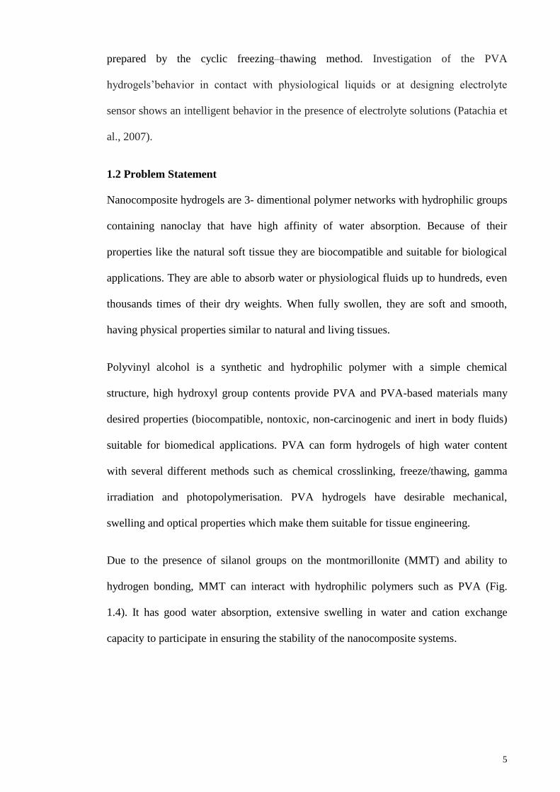
1.2 Problem Statement
Nanocomposite hydrogels are 3- dimentional polymer networks with hydrophilic groups
containing nanoclay that have high affinity of water absorption. Because of their
properties like the natural soft tissue they are biocompatible and suitable for biological
applications. They are able to absorb water or physiological fluids up to hundreds, even
thousands times of their dry weights. When fully swollen, they are soft and smooth,
having physical properties similar to natural and living tissues.
Polyvinyl alcohol is a synthetic and hydrophilic polymer with a simple chemical
structure, high hydroxyl group contents provide PVA and PVA-based materials many
desired properties (biocompatible, nontoxic, non-carcinogenic and inert in body fluids)
suitable for biomedical applications. PVA can form hydrogels of high water content
with several different methods such as chemical crosslinking, freeze/thawing, gamma
irradiation and photopolymerisation. PVA hydrogels have desirable mechanical,
swelling and optical properties which make them suitable for tissue engineering.
Due to the presence of silanol groups on the montmorillonite (MMT) and ability to
hydrogen bonding, MMT can interact with hydrophilic polymers such as PVA (Fig.
1.4). It has good water absorption, extensive swelling in water and cation exchange
capacity to participate in ensuring the stability of the nanocomposite systems.
Page 28
6
Figure 1.4: Hydrogen bonded polyvinyl alcohol organic –inorganic hybrid structures
Despite the widespread use of hydrogels in biological and biomedical applications, due
to the chemical reactions, organical modifications and chemical crossliking most
hydrogels do not provide all of the desired requirements to interact with biological
systems. The hydrogels have some disadvantages such as; presence of chemicals and
residual cross-linking agents in the hydrogel that are toxic and even carcinogenic for the
tissues and organisms. Residuals elimination are expensive that include the increasing
of production costs. Therefore a need was felt to redesign a new non toxic
nanocomposite hydrogel in order to overcome limitations related to fulfilling the above-
mentioned requirements.
Cryogelation is one of the methods of physical hydrogel formation. These gels are
formed through processes which force formation of non-covalent bonds such as
hydrogen bonds, ionic bonds or by basic entanglement of the polymeric chains and
crystallites after freezing and thawing cycles. The gels are very beneficial in the sense
that there is no need for addition of any chemical crosslinker. Cryogels form under
moderate freezing conditions in which frozen solvent causes phase separation and acts
as a porogen, leading to a gel with high water content. Gelation can occur in each of the
three steps of the freeze-thawing process; freezing, storage in frozen state or during
thawing. For PVA the most important step is thawing, since this is where most of the
gel formation occurs. One of the main aspects of cryogelation is that not all of the
solvent freezes under these conditions and there is always a portion of the solvent in the
Page 29
7
liquid phase. The surface tension between the thawed solvent and the gel phase causes
round pores. The freeze/thaw or cryogelation method has an important advantage over
chemical methods. Due to its purely physical nature there is no risk of remnant
chemicals that might compromise the biocompatibility of the final hydrogel. Also, gels
formed with this method are highly elastic and durable, which is quite important for soft
tissue engineering.
The swelling behavior of hydrogels and their swelling kinetics in different media based
on their applications are important. The presence of osmotically active mobile ions
affects the swelling behavior of hydrogels. In vitro studies of biocompatibility
preliminary in simulated physiological fluids have a great importance on the application
of biomaterials. The resemblance of PVA hydrogels to living tissues in their physical
properties because of their relatively high water content as well as soft and rubbery
consistency shows that PVA hydrogels have potential applications in this field and are
excellent candidates for biomedical applications.
Skin is the important body external defense system serving as a mechanical barrier to
prevent bacterial and microorganisms to enter the body. Skin treatment and wound
dressing are examples of hydrogel applications in the biomedical field. Dressings the
wounds with the hydrogels are usually accomplished by directly applying the hydrogels
to the injured skin and wounds. Wound dressings must ideally have characteristics like
maintenance of moisture, permeability of gases, protection against secondary infection,
thermal insulation, elastic, biodegradable and biocompatible, thus the non-toxicity,
biocompatibility and antibacterial properties of the hydrogel must be considered.
1.3 Objectives
The main objective of this thesis is focused on fabricate and characterize the non toxic
nanocomposite cryogels based on polyvinyl alcohol and Na+- montmorillonite via
Page 30
8
physical and physicochemical crosslinking as a novelty and deeply understand their
properties, relating to biomedical applications. For the first time the critical
concentration of nanoclay has been optimized to achieve the required both EWC and
WVTR characteristics in an acceptable range for biomedical applications such as; skin
treatment and wound dressing. The main objectives of this research are:
1- To fabricate of physical PVA/Na+-MMT nanocomposite hydrogels (Cryogels), and
physicochemical PVA/Na+-MMT nanocomposite hydrogels.
2- To compare the properties of PVA/Na+-MMT nanocomposite hydrogels prepared by
physical and physicochemical cross linking.
3- To fabricate and characterize of non toxic hydrogels based on PVA/Na+-MMT
nanocomposites for biomedical applications.
4- To optimize the nanoclay content in acceptable range of Equilibrium water content
(EWC) and Water vapor transmission rate (WVTR) for biomedical applications.
5- To investigate and finding the water sorption and desorption kinetics model of
prepared PVA/Na+-MMT nanocomposite hydrogels.
6- To investigate the effect of nanoclay content on release of antibacterial agent for
loaded PVA/Na+-MMT nanocomposite hydrogels.
1.4 Outline of the thesis
This thesis comprises the following main chapters:
1. Chapter One – GENERAL INTRODUCTION
This chapter includes a brief introduction to the research and objectives of the study.
2. Chapter Two - LITERATURE REVIEW
This chapter gives a comprehensive literature survey for the Materials, preparation, and
characterization of PVA/MMT nanocomposite hydrogels
Page 31
9
3. Chapter Three – METHODOLOGY
This chapter describes the experimental procedure which includes nanocomposite
hydrogels preparation, swelling cxperiments, structural charactrizations and
morphology, thermal and mechanical analysis, Hardness measurements, permeation
analysis and water sorption and desorption kinetics.
4. Chapter Four - RESULTS AND DISCUSSION
The structural characterization and morphology of nanocomposite hydrogels, thermal
and mechanical analysis, Hardness measurements , barrier properties and Permeation
analysis, the swelling behavior of hydrogels which include equilibriurn swelling results,
swelling ratio, swelling rate, water sorption and desorption kinetics, finding the
diffusion kinetics model, are presented in this chapter.
5. Chapter Five – CONCLUSIONS AND RECOMENDATIONS
In the last chapter, the results and findings of this study have summarized and
recommendations for future works have been suggested.
2
3
4
Page 32
10
5 CHAPTER 2: REVIEW OF RELATED LITERATURE
2.1 Introduction
In the past years, several research groups have studied the preparation and
characterization of nanocomposite hydrogel materials. These kinds of hydrogels are
able to combine the elasticity and permeability of gels with the reinforcing ability of
clays embedded in the hydrogels, providing a wide range of application in different
fields for the hydrogels (Haraguchi et al., 2003, Bigotti et al., 2004, Ekici et al., 2006).
Montmorillonite (MMT) is a naturally occurring mineral clay that is widely used to
prepare nanocomposite hydrogels, and this is mainly due to its good water absorption,
extensive swelling in water and cation exchange capacity. Because of its intrinsic
chemical composition, such as; presence of silanol groups (–Si–OH) on the layer
surface of MMT and its ability to form hydrogen bonding (Fig.2.1), the montmorillonite
is able to interact with hydrophilic polymers (Gamiz et al., 1992, Gao et al., 1999,
Chiellini et al., 2000, Gao et al., 2001, Backfolk et al., 2006). Therefore, MMT and
other phyllosilicates can act as co-crosslinkers of hydrophilic polymers in solution (Lin
et al., 2001, Karimi & Wan Daud, 2014).
Figure 2.1: Interactions among PVA, water, and MMT
Page 33
11
These features of MMT were thoroughly studied by Lee and Jou who investigated the
effect of intercalated montmorillonite on the swelling and drug release behaviors of a
nanocomposite constituted by N-isopropylacrylamide (NIPAAm) /acrylic acid (Lee &
Jou, 2004). Liu and co-authors also observed a significant improvement in the tensile
properties of NIPAAm-based nanocomposite hydrogels containing a modified hectorite-
laponite mineral (Liu et al., 2006). An improvement of mechanical and thermal
properties was also reported by Zheng and collaborators (Zheng et al., 2002) and by Lee
and Lee for gelatin hydrogels containing montmorillonite (Lee & Lee, 2006).
Churochkina et al., (1998) have shown that the mechanical properties of the Neutral and
Slightly Charged Poly (acrylamide) Gels are modified with the addition of Na-
montmorillonite (Churochkina et al., 1998). On the other hand, several authors have
studied polyvinyl alcohol (PVA) nanocomposites (Carrado et al., 1996, Yu et al., 2003,
De Bussetti et al., 2004).
PVA has exceptional and interesting properties such as being: hydrophilic, water-
soluble, gas barrier, good chemical resistance, processability, non-toxic, non-
carcinogenic biocompatibile, biodegradabe and inexpensive, and is capable of forming
gel networks by chemical or physical methods (Mahdavi et al., 2013, Valenta & Anver,
2004, Patachia, S., 2003, Hennink & van Nostrum, 2002, Peppas et al., 2000, Ratner et
al., 2004, Hassan & Peppas, 2000, Hoffman, A.S., 2001, Coviello et al., 2007, Silva et
al., 2013). These properties play important roles in the design of pharmaceutical and
biomedical devices. It has been used in various biomedical applications, for instance as
drug delivery devices, artificial organs, wound dressings, contact lenses, antibacterial,
skin treatment systems, protein adsorption, protein controlled release and for delivery
by chemical and physical methods (Mirzan et al., 2001, Paradossi et al., 2003, Chen et
al., 1994, Nacer Khodja et al., 2013, Gonzalez et al., 2012, Kokabi et al., 2007, Abd El-
Mohdy, H.L., 2013, Vicentini et al., 2010, Shalumon et al., 2011, Gonzalez et al., 2011,
Page 34
12
Li et al., 2013, Kenawy et al., 2014, Zhao et al., 2003, Hyon et al., 1994, Cha et al.,
1993, Elizabeth & Fabia, 2006, Christie et al., 2000, Peppas & Simmons, 2004).
In the polymer nanocomposites of PVA and MMT as novel materials, the hydrogen
bonding between the silanol groups and negatively charged on the surface of the MMT
and hydroxyl groups of the PVA has the most important role to the surface interactions
of MMT with the PVA chains (Grunlan et al., 2004, Hernandez et al., 2008). In
addition, because of the existence of metal ions in the MMT lattice, interactions of the
MMT layers with the acetoxy groups in the PVA chains will be increased causing
strong PVA-MMT interactions that lead to enhancement of the MMT layers dispersion
into the PVA matrix (Stathi et al., 2009). These interactions consequently lead to the
intercalated or exfoliated composite structures and their overall performance (Sapalidis
et al., 2011). Due to the presence of MMT the PVA crystallinity is decreased and
nanocomposite biodegradation occurs faster than pure PVA (Lee et al., 2003, Shina.
Ray et al., 2002).
It has been recognized that semi-diluted PVA solutions can form gels under cooling at
low temperatures. Pines & Prins (1973) showed that PVA hydrogels can be formed via
crystallization of PVA chains and or by liquid–liquid phase separation. PVA crystalline
domains can act as crosslinking sites for the network (Takeshita et al., 1999). The
interesting characteristic of PVA solutions has been used to produce PVA hydrogels
without using any chemical crosslinkers that is important to make non-toxic,
biocompatible and biomedical devices (Willcox et al., 1999, Hassan et al., 2000,
Lozinsky & Damshkaln, 2000). PVA gels prepared by the sequential freezing-thawing
process have physical linkages, so they are non toxic for organisms; changes in regional
concentration of polymer molecules form intermolecular forces, leading to the
formation of microcrystals (Okazaki et al., 1995).
Page 35
13
In a number of articles, some PVA cryogels characteristics, their properties and the
cryostructuring conditions have been discussed (Hajizadeh, et al., 2013, Tretinnikov, et
al., 2015). Lozinskii and coworkers investigated changes in the mechanical properties of
these polymeric gels with an increase in temperature from 295°K (normal temp.) to
350-390°K (lyogel melt) range (Lozinsky et al., 2003). Kobayachi et al., (1992) have
prepared the PVA hydrogel with excellent mechanical properties, high clarity and high
water content through crystallization at low temperatures. Also, Won-Ill et al., (1993)
have prepared the PVA hydrogel with elasticity properties below 0°C, as well as good
adhesion and excellent water holding properties, using water and organic solvents. They
prepared the hydrogel from concentrated PVA solutions by a crystallization technique at
low temperatures as a biomaterial for skin treatment systems.
Synthesis and characterization of PVA hydrogels and their hybrids by chemical and
physical methods for protein adsorption, protein controlled release and delivery have
been investigated by some researchers (Christie et al., 2000, Peppas & Simmons, 2004,
Elizabeth & Fabia, 2006). They have also prepared the semi-crystalline PVA films and
their blends with poly (acrylic acid) and poly (ethylene glycol) by freeze-thawing for
drug delivery application. Moreover, the structure and morphology of PVA hydrogels
were investigated (Hassan & Peppas, 2000 a, Hassan & Peppas, (2000) b, Hassan et al.,
2000, Peppas & Tennenhous, 2004)
The conventional hydrogels have some disadvantages such as; presence of chemicals
and residual cross-linking agents in the hydrogel that are toxic and even carcinogenic
for living tissues and organisms. Residuals elimination are expensive that include the
increasing of production costs.
PVA/MMT nanocomposite hydrogels have some advantages compared to the other
conventional and similar systems. They are made of PVA which is a biodegradable,
Page 36
14
biocompatible, non-toxic, non-carcinogenic and inexpensive polymer, as well as
montmorillonite (MMT) which is a low cost, high surface area, hydrophilic and
environmentally friendly and naturally abundant clay, being prepared by physical
methods such as freezing - thawing cycles contains important advantage over chemical
methods. These physical gels have a common benefit that is no need to add any
chemical crosslinker to the hydrogels. Due to its pure physical structure there is no risk
of remnant chemicals that might compromise the biocompatibility of the final hydrogel.
Also, gels formed with this method are non-toxic and highly elastic and durable, which
is quite important for biomedical applications.
2.2 Structure and properties of nanoclay (phyllosilicates)
2.2.1 Structure and properties of layered silicate
The most commonly used layered silicates are Montmorillonite (MMT), hectorite, and
saponite. There are two types of structure for layered silicates: tetrahedral-substituted
and octahedral-substituted moieties. The polymer matrices can interact more readily
with tetrahedrally-substituted layered silicates than with octahedrally-substituted
material due to the negative charges being located on the surface of silicate layers in the
case of tetrahedrally-substituted silicates. The structure and chemical formula of these
layered silicates are shown in Figure 2.2 and Table 2.1.
Page 37
15
Figure 2.2: Structure of 2:1 phyllosilicates
The layered silicates have two particular characteristics for Polymer Layered Silicate
(PLS) nanocomposites. The first characteristic is the dispersal ability of the silicate
particles into individual layers, while the second ability is ion exchange reactions with
organic and inorganic cations to fine-tune their surface chemistry. Of course, these two
characteristics are related to the degree of dispersion of layered silicate in a particular
polymer matrix depending on the interlayer cation.
Page 38
16
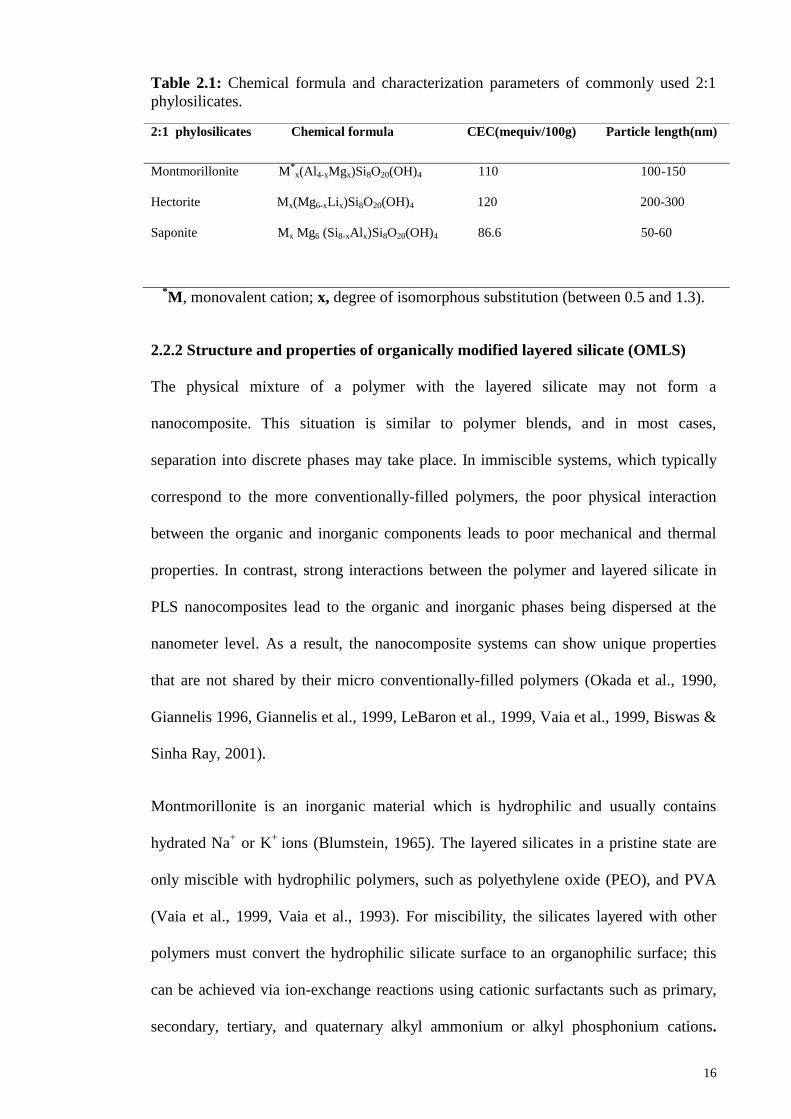
Table 2.1: Chemical formula and characterization parameters of commonly used 2:1
phylosilicates.
*M, monovalent cation; x, degree of isomorphous substitution (between 0.5 and 1.3).
2.2.2 Structure and properties of organically modified layered silicate (OMLS)
The physical mixture of a polymer with the layered silicate may not form a
nanocomposite. This situation is similar to polymer blends, and in most cases,
separation into discrete phases may take place. In immiscible systems, which typically
correspond to the more conventionally-filled polymers, the poor physical interaction
between the organic and inorganic components leads to poor mechanical and thermal
properties. In contrast, strong interactions between the polymer and layered silicate in
PLS nanocomposites lead to the organic and inorganic phases being dispersed at the
nanometer level. As a result, the nanocomposite systems can show unique properties
that are not shared by their micro conventionally-filled polymers (Okada et al., 1990,
Giannelis 1996, Giannelis et al., 1999, LeBaron et al., 1999, Vaia et al., 1999, Biswas &
Sinha Ray, 2001).
Montmorillonite is an inorganic material which is hydrophilic and usually contains
hydrated Na+ or K
+ ions (Blumstein, 1965). The layered silicates in a pristine state are
only miscible with hydrophilic polymers, such as polyethylene oxide (PEO), and PVA
(Vaia et al., 1999, Vaia et al., 1993). For miscibility, the silicates layered with other
polymers must convert the hydrophilic silicate surface to an organophilic surface; this
can be achieved via ion-exchange reactions using cationic surfactants such as primary,
secondary, tertiary, and quaternary alkyl ammonium or alkyl phosphonium cations.
2:1 phylosilicates Chemical formula CEC(mequiv/100g) Particle length(nm)
Montmorillonite M*x(Al4-xMgx)Si8O20(OH)4 110 100-150
Hectorite Mx(Mg6-xLix)Si8O20(OH)4 120 200-300
Saponite Mx Mg6 (Si8-xAlx)Si8O20(OH)4 86.6 50-60
Page 39
17
Alkyl ammonium or alkyl phosphonium cations in organosilicates are responsible for
lowering the surface energy of the inorganic host and improving the wetting properties
of the polymer matrix and the resulting larger interlayer spacing. Also, functional
groups in these cations can react with the polymer matrix to increase the strength of the
interface between the polymer matrix and the inorganic host (Blumstein, 1965, Aranda
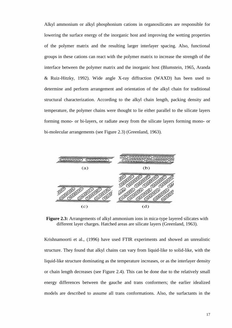
& Ruiz-Hitzky, 1992). Wide angle X-ray diffraction (WAXD) has been used to
determine and perform arrangement and orientation of the alkyl chain for traditional
structural characterization. According to the alkyl chain length, packing density and
temperature, the polymer chains were thought to lie either parallel to the silicate layers
forming mono- or bi-layers, or radiate away from the silicate layers forming mono- or
bi-molecular arrangements (see Figure 2.3) (Greenland, 1963).
Figure 2.3: Arrangements of alkyl ammonium ions in mica-type layered silicates with
different layer charges. Hatched areas are silicate layers (Greenland, 1963).
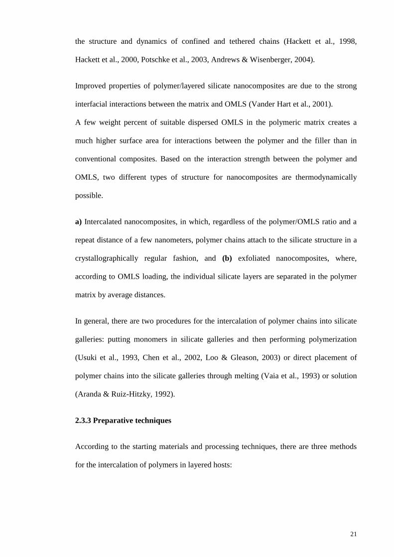
Krishnamoorti et al., (1996) have used FTIR experiments and showed an unrealistic
structure. They found that alkyl chains can vary from liquid-like to solid-like, with the
liquid-like structure dominating as the temperature increases, or as the interlayer density
or chain length decreases (see Figure 2.4). This can be done due to the relatively small
energy differences between the gauche and trans conformers; the earlier idealized
models are described to assume all trans conformations. Also, the surfactants in the
Page 40
18
layered silicate for longer chain length surfactants can show thermal transition caused
by heat, like melting or liquid-crystalline to liquid-like transitions.
Figure 2.4: Alkyl chain aggregation models: (a) in short length chains, the molecules
are effectively isolated from each other; (b) in medium length chains, quasi-discrete
layers form with various degrees of in-plane disorder and inter-digitation between the
layers; and (c) in long length chains, interlayer order increases leading to a liquid-
crystalline polymer environment. Open circles represent the CH2 segments, while
cationic head groups are represented by filled circles (Krishnamoorti et al., 1996).
2.3 Types of polymeric nanocomposites and their preparative techniques
2.3.1 Types of polymeric nanocomposites
Generally, layer thickness in the layered silicates is in the order of 1 nm and a very high
aspect ratio (e.g. 10–1000). Thus, polymer matrix provides much higher surface area for
polymer/filler interaction than conventional composites. There are three different types
of modified or unmodified PLS nanocomposites regarding thermodynamics, depending
on the strength of interfacial interactions between the layered silicate and polymer
matrix (see Figure 2.5):
Page 41
19
Figure 2.5: Schematic illustration of three different types of thermodynamically
achievable polymer/layered silicate nanocomposites (Lagaly, 1986).
a. Intercalated nanocomposites:
Regardless of the clay to polymer ratio, in intercalated nanocomposites, insertion of the
layered silicate structure into a polymer matrix occurs in a crystallographically regular
manner. Normally, intercalated nanocomposites are formed into an interlayer via a few
molecular layers of polymer. Typically, properties of the composites are similar to those
of ceramic materials.
b. Flocculated nanocomposites:
Flocculated nanocomposites are conceptually the same as intercalated nanocomposites.
However, silicate layers are sometimes flocculated due to hydroxylated edge to edge
interactions of the silicate layers.
Page 42
20
c. Exfoliated nanocomposites:
Exfoliated nanocomposites have individual clay layers that depend on clay loading
which are separated in a continuous polymer matrix by average distances. Normally, the
clay content of an intercalated nanocomposite is much higher than that of an exfoliated
nanocomposite.
2.3.2 Polymer/layered silicate (PLS) nanocomposite technology
In the past, in order to improve the polymer performance, the application of inorganic
nanoparticles as additives has been considered. Currently, various nano-reinforcement
materials have been developed such as nanoclay (layered silicates) (Giannelis, 1996,
Giannelis et al., 1999, Vaia et al., 1994, Sinha Ray & Okamoto, 2003, Sinha Ray et al.,
2003), ultrafine layered titanate (LeBaron et al., 1999), cellulose nano-whiskers (Biswas
& Sinha Ray, 2001), and carbon nanotubes (Mitchell et al., 2002, Mohanty et al., 2003,
Hiroi et al., 2004). However, carbon nanotubes-based polymer composites are a
relatively clear case of new nanomaterials that show exceptional thermal, electrical and
mechanical properties (Mitchell et al., 2002). There is a particular interest in
organically-modified polymeric layered silicate (OMLS) nanocomposites, than the
unmodified polymer resin, because OMLS nanocomposites have demonstrated
significant enhancements including a large number of physical properties, such as
thermal and environmental stability, barrier, solvent uptake, flammability resistance and
biodegradability rate of biodegradable polymers (Vaia et al., 1994). Generally, these
improvements are attained at lower silicate contents (65 wt %) than conventional filler-
filled systems. Therefore, the polymer/OMLS nanocomposites are much lighter than
conventional composites, and for specific applications, they are competitive with other
materials. The conventional bulk characterization techniques such as NMR, thermally-
stimulated current, DSC, rheology, and various kinds of spectroscopy are used to study
Page 43
21
the structure and dynamics of confined and tethered chains (Hackett et al., 1998,
Hackett et al., 2000, Potschke et al., 2003, Andrews & Wisenberger, 2004).
Improved properties of polymer/layered silicate nanocomposites are due to the strong
interfacial interactions between the matrix and OMLS (Vander Hart et al., 2001).
A few weight percent of suitable dispersed OMLS in the polymeric matrix creates a
much higher surface area for interactions between the polymer and the filler than in
conventional composites. Based on the interaction strength between the polymer and
OMLS, two different types of structure for nanocomposites are thermodynamically
possible.
a) Intercalated nanocomposites, in which, regardless of the polymer/OMLS ratio and a
repeat distance of a few nanometers, polymer chains attach to the silicate structure in a
crystallographically regular fashion, and (b) exfoliated nanocomposites, where,
according to OMLS loading, the individual silicate layers are separated in the polymer
matrix by average distances.
In general, there are two procedures for the intercalation of polymer chains into silicate
galleries: putting monomers in silicate galleries and then performing polymerization
(Usuki et al., 1993, Chen et al., 2002, Loo & Gleason, 2003) or direct placement of
polymer chains into the silicate galleries through melting (Vaia et al., 1993) or solution
(Aranda & Ruiz-Hitzky, 1992).
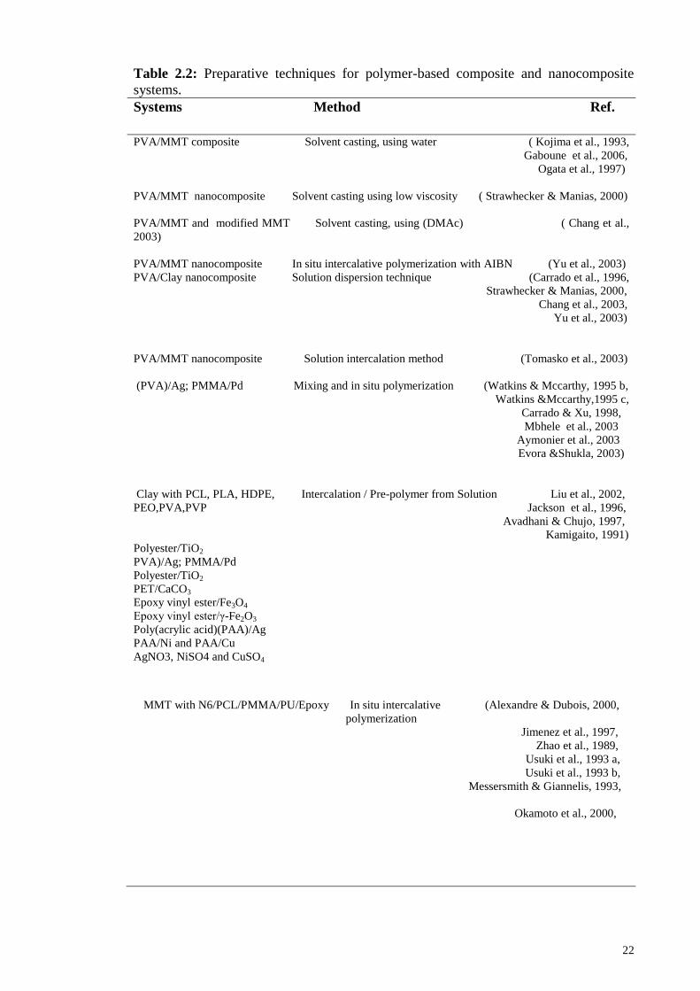
2.3.3 Preparative techniques
According to the starting materials and processing techniques, there are three methods
for the intercalation of polymers in layered hosts:
Page 44
22
Table 2.2: Preparative techniques for polymer-based composite and nanocomposite
systems.
Systems Method Ref.
PVA/MMT composite Solvent casting, using water ( Kojima et al., 1993,
Gaboune et al., 2006,
Ogata et al., 1997)
PVA/MMT nanocomposite Solvent casting using low viscosity ( Strawhecker & Manias, 2000)
PVA/MMT and modified MMT Solvent casting, using (DMAc) ( Chang et al.,
2003)
PVA/MMT nanocomposite In situ intercalative polymerization with AIBN (Yu et al., 2003)
PVA/Clay nanocomposite Solution dispersion technique (Carrado et al., 1996,
Strawhecker & Manias, 2000,
Chang et al., 2003,
Yu et al., 2003)
PVA/MMT nanocomposite Solution intercalation method (Tomasko et al., 2003)
(PVA)/Ag; PMMA/Pd Mixing and in situ polymerization (Watkins & Mccarthy, 1995 b,
Watkins &Mccarthy,1995 c,
Carrado & Xu, 1998,
Mbhele et al., 2003
Aymonier et al., 2003
Evora &Shukla, 2003)
Clay with PCL, PLA, HDPE, Intercalation / Pre-polymer from Solution Liu et al., 2002,
PEO,PVA,PVP Jackson et al., 1996,
Avadhani & Chujo, 1997,
Kamigaito, 1991)
Polyester/TiO2
PVA)/Ag; PMMA/Pd
Polyester/TiO2
PET/CaCO3
Epoxy vinyl ester/Fe3O4
Epoxy vinyl ester/γ-Fe2O3
Poly(acrylic acid)(PAA)/Ag
PAA/Ni and PAA/Cu
AgNO3, NiSO4 and CuSO4
MMT with N6/PCL/PMMA/PU/Epoxy In situ intercalative (Alexandre & Dubois, 2000,
polymerization
Jimenez et al., 1997,
Zhao et al., 1989,
Usuki et al., 1993 a,
Usuki et al., 1993 b,
Messersmith & Giannelis, 1993,
Okamoto et al., 2000,
Page 45
23
Table 2.2, continued
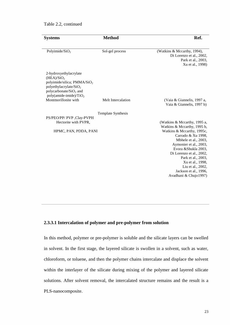
2.3.3.1 Intercalation of polymer and pre-polymer from solution
In this method, polymer or pre-polymer is soluble and the silicate layers can be swelled
in solvent. In the first stage, the layered silicate is swollen in a solvent, such as water,
chloroform, or toluene, and then the polymer chains intercalate and displace the solvent
within the interlayer of the silicate during mixing of the polymer and layered silicate
solutions. After solvent removal, the intercalated structure remains and the result is a
PLS-nanocomposite.
Systems Method Ref.
Polyimide/SiO2 Sol-gel process (Watkins & Mccarthy, 1994),
Di Lorenzo et al., 2002,
Park et al., 2003,
Xu et al., 1998)
2-hydroxyethylacrylate
(HEA)/SiO2
polyimide/silica; PMMA/SiO2
polyethylacrylate/SiO2
polycarbonate/SiO2 and
poly(amide-imide)/TiO2
Montmorillonite with Melt Intercalation (Vaia & Giannelis, 1997 a,
Vaia & Giannelis, 1997 b)
Template Synthesis
PS/PEO/PP/ PVP ,Clay-PVPH
Hectorite with PVPR, (Watkins & Mccarthy, 1995 a,
Watkins & Mccarthy, 1995 b,
HPMC, PAN, PDDA, PANI Watkins & Mccarthy, 1995c,
Carrado & Xu 1998,
Mbhele et al., 2003,
Aymonier et al., 2003,
Evora &Shukla 2003,
Di Lorenzo et al., 2002,
Park et al., 2003,
Xu et al., 1998,
Liu et al., 2002,
Jackson et al., 1996,
Avadhani & Chujo1997)
Page 46
24
In terms of thermodynamics, a negative variation in the Gibbs free energy is required
for the overall process, in which the polymer is exchanged with the previously
intercalated solvent in the gallery. The entropy obtained by desorption of solvent
molecules is the driving force for the polymer intercalation into layered silicate from a
solution, which compensates for the decreased entropy of the confined, intercalated
chains (Vaia & Giannelis, 1997 b). This method is suitable for the intercalation of
polymers with no or low polarity into layered structures, and easily produces thin films
with polymer-oriented clay intercalated layers. However, this method involves the
abundant use of organic solvents, which, from an industrial point of view, are
environmentally unfriendly and economically expensive.
2.3.3.2 In situ intercalative polymerization
In this method, the layered silicate can swell in the liquid monomer or a monomer
solution and the polymer can be formed between the intercalated sheets. Polymerization
reactions can be initiated by radiation or heat, through the diffusion of a suitable
initiator, an organic initiator or catalyst that is fixed inside the interlayer before the
swelling step via cation exchange.
2.3.3.3 Melt intercalation technique
Due to compatibility with recent industrial techniques, the melt intercalation technique
has become a primary choice for the preparation of polymer/OMLS nanocomposites,
and involves annealing the mixture of the polymer and OMLS above the softening point
of the polymer either statically or under shearing. During the annealing, the polymer
chains diffuse into the silicate layer galleries from the mass of melted polymer (Vaia &
Giannelis, 1997a). On the other hand, during polymer intercalation from solution to
replace the imported polymer chains, a relatively large number of solvent molecules
have to be repelled from the host. From a waste view, the absence the solvent makes
Page 47
25
direct melt intercalation an environmentally friendly and economically favorable
method for industries. In addition, direct melt intercalation is highly specific for the
polymer, leading to new hybrids that were previously inaccessible. Therefore, there are
many advantages to direct melt intercalation than solution intercalation. Depending on
the amount of penetration of the polymer chains into the silicate galleries, a range of
nanocomposites can be obtained with intercalated or exfoliated structures. Experimental
results indicate that silicate functionalization and constituent interactions have a critical
influence on the outcome of polymer intercalation.
The researchers have found that (a) an optimal interlayer structure on the OMLS, with
respect to the number per unit area and size of surfactant chains, is the most favorable
for nanocomposite formation, and (b) polymer intercalation depends on the existence of
polar interactions between the OMLS and the polymer matrix.
To understand the thermodynamics of nanocomposite formation, (Vaia & Giannelis,
1997 a & b) used a mean-field statistical lattice model and reported experimental results
and calculations based on the mean field theory that agreed well.
While confinement of the molten polymer with the formation of nanocomposite is
associated with a loss of entropy, this process is allowed because entropy is gained with
the layer separation and results in a net entropy change close to zero. Therefore,
according to the theoretical model, energetic factors, which may be determined from the
surface energies of the polymer and OMLS are the most important for nanocomposite
formation via polymer melt intercalation.
According to the report of (Vaia & Giannelis, 1997 a & b), in order to maximize the
configurational freedom of the functionalizing chains upon layer separation, and to
maximize potential interaction sites at the interlayer surface, the interlayer structure of
the OMLS should first be optimized. In these systems, the desirable structures have a
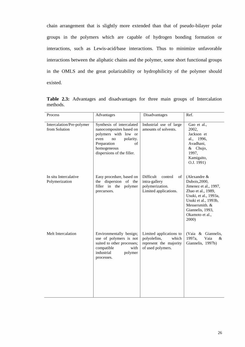
Page 48
26
chain arrangement that is slightly more extended than that of pseudo-bilayer polar
groups in the polymers which are capable of hydrogen bonding formation or
interactions, such as Lewis-acid/base interactions. Thus to minimize unfavorable
interactions between the aliphatic chains and the polymer, some short functional groups
in the OMLS and the great polarizability or hydrophilicity of the polymer should
existed.
Table 2.3: Advantages and disadvantages for three main groups of Intercalation
methods.
Process
Advantages
Disadvantages
Ref.
Intercalation/Pre-polymer
from Solution
In situ Intercalative
Polymerization
Melt Intercalation
Synthesis of intercalated
nanocomposites based on
polymers with low or
even no polarity.
Preparation of
homogeneous
dispersions of the filler.
Easy procedure, based on
the dispersion of the
filler in the polymer
precursors.
Environmentally benign;
use of polymers is not
suited to other processes;
compatible with
industrial polymer
processes.
Industrial use of large
amounts of solvents.
Difficult control of
intra-gallery
polymerization.
Limited applications.
Limited applications to
polyolefins, which
represent the majority
of used polymers.
Gao et al.,
2002, Jackson et
al., 1996,
Avadhani,
& Chujo,
1997, Kamigaito,
O.J. 1991)
(Alexandre &
Dubois,2000, Jimenez et al., 1997, Zhao et al., 1989,
Usuki, et al., 1993a,
Usuki et al., 1993b,
Messersmith. &
Giannelis, 1993,
Okamoto et al.,
2000)
(Vaia & Giannelis,
1997a, Vaia &
Giannelis, 1997b)
Page 49
27
2.4 Techniques used for the preparation of PVA hydrogels and PVA
nanocomposite hydrogels and their characterization.
Hydrogels can be prepared by different methods. The most widely employed methods
are chemical cross-linking using glutaraldehyde as the cross-linking agent (Kawasumi et
al., 1997, Ossipov & Hilborn, 2006), and physical cross-linking by UV radiation
(Benamer et al., 2006), irradiation (Ajji, 2005, Benamer et al., 2006, Martens & Anseth,
2000), and by the use of repeated freeze-thawing cycles (Martens & Anseth, 2000,
Peppas & Mongia, 1997). Hassan & Peppas (2000) and Hassan et al., (2000), have
reported the preparation of hydrogels using physical methods such as freezing and
thawing, chemical methods using a covalent cross-linking agent including boric acid,
glutaraldehyde and formaldehyde and radiation methods using electron beams known as
irradiation. Polyvinyl alcohol hydrogel prepared by freezing and thawing is not toxic,
not carcinogenic and has good biocompatibility. PVA hydrogels were studied by
Stauffer and Peppas for biomedical and pharmaceutical applications (Stauffer & Peppas,
1992). Intermolecular bonds (mostly hydrogen bonds) which form during the freeze-
thawing process of PVA water solutions, act as efficient cross-links. Jolanta Stasko et
al. have reported the influence of PVA molecular weight on the water absorption, gel
formation and the density of gels prepared by the freeze-thawing method (Stasko et al.,
2009). A schematic illustration of crosslinking methods used in hydrogel preparation is
shown below (Hamidi et al., 2008):
Page 50
28
Figure 2.6: Novel crosslinking methods used in hydrogels (Hamidi.et al. 2008)
2.4.1 Covalently crosslinked and cryogels
Most research in smart polymers (SPS) has focused on hydrogels that swell in aqueous
solutions. The smart gels can be synthesized by conventional methods at room
temperature; these prepared hydrogels have small pore sizes. Also, smart macroporous
hydrogels reviewed in several papers (Kopecek, 2003, Rosiak & Ulan ski 1999, Roy &
Gupta 2003), have been synthesized by different methods and with various applications.
Crosslinking methods
Chemical crosslinked Physical crosslinked
Crosslinking by radical polymerization Crosslinking by ion
interactions
Crosslinking by high energy irradiation
Physical crosslinked
hydrogel from
amphiphilic block and
graft copolymers
Crosslinking by
crystallization
Crosslinking using enzymes
Crosslinking by chemical reaction with
complementary groups
Crosslinking with aldehyde
Crosslinking with addition reaction
Crosslinking by condensation
reaction
Crystallization in
homopolymer systems
Crosslinking by
stereocomplex
formation
Page 51
29
Cryogels are large pore size hydrogels which have been synthesized in moderately
frozen conditions with interesting properties (Lozinsky et al., 2003). These gels can be
prepared at temperatures lower than the melting temperature of the solvent. Karimi et al.
have recently reported preparation of a novel physicochemical crosslinked
nanocomposite hydrogel based on PVA and natural Na-montmorillonite (Na+-MMT)
using chemical crosslinking followed by a Freezing-Thawing process ( Karimi & Wan
Daud, 2014).
Table 2.4: Crosslinking methods for the preparation of nanocomposite hydrogels
Preparation methods Ref.
Chemica crosslinking by glutaraldehyde ( Valentın et al., 2009)
Irradiation by gammaradiation (Takeshita et al., 1999,
Morgan & Gilman, 2003)
Irradiation by UV radiation (Sinha Ray et al., 2003)
Freezing/ thawing (Peppas &.Mongia, 1997, Davis et al., 2002,
Lee et al., 1998, Kokabi et al., 2007)
physicochemical crosslinking (Karimi & Wan Daud, 2014)
A short discribtion for the freez-thaw process is presented here. Most of the solvents are
frozen at subzero temperatures, while the dissolved substances are concentrated in small
non-frozen regions known as the “liquid microphase”. The volume of the non-frozen
liquid microphase is much lower than that of the solid phase; the concentration of local
monomer is much higher than the concentration of monomer in the initial reaction
mixture, meaning that gel formation occurs in the liquid microphase and the crystals of
frozen solvents perform in a manner that is similar to porogen. After melting of the ice
crystals, a network of large interconnected pores is formed, meaning that an attractive
system is obtained for surface grafting with large interconnected pores and a high
Page 52
30
surface area that is available for grafting and the efficient mass transport of monomers
(Fig 2.7).
Figure 2.7: Schematic diagram showing, PVA hydrogels prepared by freeze-thawing
cycles with a PVA-rich region and a PVA-poor region. 1, macromolecules in a solution; 2, solvent; 3, low-molecular solutes; 4, polycrystals of frozen
solvent; 5, unfrozen liquid microphase; 6, polymeric framework of a cryogel; 7, macropores; 8,
solvent.
Now, most research is focused on hydrogels that are sensitive to small changes in pH or
temperature (Lozinsky et al., 2003, Karimi, 2008), has reported synthesis of two types
of ionic hydrogels consisting of pH sensitive (copolymer of acrylamide and sodium
acrylate) and temperature and pH sensitive (copolymer of N-isopropylacrylamide and
sodium acrylate) (Karimi, 2008). Moreover the response to changes in solvents, ionic
strength, electric or magnetic fields, and light intensity have been investigated in other
gels. Some gels have also been designed to respond to specific biomolecules such as
glucose or chemical triggers (Traitel et al., 2000, Miyataa et al., 2002). Response to
stimulation makes them useful as smart materials and they have been established in
numerous applications.
Page 53
31
Table 2.5: Response to changes in smart hydrogels
Changes for response System Ref.
Temperature Different Hydrogels (Lozinsky et al., 2003, Karimi, 2008) pH Different Hydrogels (Lozinsky et al., 2003,Karimi, 2008) Temp and pH NIPAM and sodium acrylate ( Karimi, 2008) Electric field Poly(dimethyl siloxane) gels (Zhao et al., 1989) containing randomly distributed TiO2 particles Magnetic field NIPA and PVA (Zhao et al., 1989) Solvent (Zhao et al., 1989, Schuetz & Gurny, 2008) Light intensity (NIPAM) in D2O (Zhao et al., 1989, Schuetz & Gurny, 2008)
Glucose and protein Self-regulated drug delivery (Litvinov & De, 2002, Simon & Schneider, 1991)
2.4.2 Polyvinyl alcohol nanocomposites and their characterizations
The first fabrication of PVA/MMT composites was reported by Greenland (1963); who
used the solvent casting method with water as co-solvent. After that, Ogata et al. (1997)
used the same method to produce PVA/MMT composites. Also, Strawhecker and
Manias have used the solvent casting method in an attempt to make the PVA/MMT
nanocomposite films (Strawhecker & Manias, 2000). PVA/MMT nanocomposite films
were produced by film casting from an MMT/water suspension containing dissolved
PVA. In this research a suspension of Na+-MMT in water was stirred and sonicated.
Then, fully hydrolyzed atactic PVA with low viscosity was added to the stirring
suspension. The casted films were characterized using WAXD and TEM.
Figure 2.8 shows the WAXD scans of 20, 40, 60, 80 and 100 wt% MMT
concentrations; (the corresponding d-spacing distributions for the same concentrations
can be seen inset). The XRD patterns show that the d-spacing and their distribution
systematically decreased with increasing MMT wt% in the nanocomposites.
Page 54
32
Figure 2.8: XRD patterns of the PVA/MMT hybrids as a function of MMT. The inset
shows the distribution of the MMT intercalated d-spacings for the respective hybrids
(Strawhecker & Manias, 2000).
In the TEM (Figure 2.9) image of 20 wt% filled clay nanocomposite, (it can be seen the
existence of silicate layers in the intercalated and exfoliated structures).
Figure 2.9: Bright field TEM image of 20 wt% PVA/MMT nanocomposite, revealing
the coexistence of intercalated (A) and exfoliated (B) MMT layers (Strawhecker &
Manias, 2000).
Page 55
33
At first glance, the d-spacing on the polymer/silicate mass ratio and dependence of the
intercalated structure seems to be in contrast with the theoretical expectations (Carrado
et al., 1996, Doppers et al., 2004, Fornes et al., 2001, Sinha Ray et al., 2002). The
equilibrium nanocomposite structure forecasted from the thermodynamics is related to
the intercalated periodic nanocomposite with d-spacing of around 1.8 nm, which is
expected to be independent of the polymer-to-silicate composition (Hassan & Peppas,
2000). However, thermodynamics is only able to forecast the equilibrium structure.
Actually, in this research, the structure of the nanocomposite is dictated by kinetics; the
layers remain in colloidal suspension in the water solution of PVA and MMT. The
suspension must be dried slowly and the silicate layers remain distributed and
embedded in the polymer gel. All of the water is removed by further drying, although
according to thermodynamics, the MMT layers re-aggregate in an intercalated fashion;
the slow polymer dynamics is a trap for some of the layers and they therefore remain
dispersed in the polymer matrix. Clearly, the kinetic restrictions imposed by the
polymer become less important as the polymer-to-silicate fraction decreases, and
intercalated structures are formed for higher amounts of MMT. For these periodic
structures, the d-spacing changes, with the wt% of MMT representing the different
weight ratios for polymer–silicate, and with an increase in the amount of MMT.
Preparation of PVA-based nanocomposites with three different types of clay-pristine
MMT and organically modified MMT was reported by Chang et al. (2003). In this
work, they used the same solvent casting method for the nanocomposite preparation;
however, in addition to water, N, N-dimethylacetamide (DMAc) was also used as co-
solvent. Dodecylammonium-modified MMT (C12MMT) and 12-aminolauric-modified
MMT (C12OOHMMT) were used as OMLS. XRD patterns of different types of clay
and TEM images of their nanocomposites with PVA are shown in Figure 2.10 and
Figure 2.11 respectively (Chang et al., 2003). These figures show the formation of
Page 56
34
exfoliated nanocomposites when pristine clays were used for the fabrication of
nanocomposites. Because intercalated nanocomposites were produced with OMLS, it
can be concluded that the hydrophilic character of clay promotes the dispersion of
inorganic crystalline layers in water-soluble polymers (LeBaron et al., 1999, Vaia et al.,
1996).
Page 57
35
Figure 2.10: XRD patterns of: (a) clays, (b) 4 wt% clays/PVA hybrid films, and (c) 8
wt% clays/PVA hybrids (Chang et al., 2003).
Page 58
36
Figure 2.11: TEM photographs of PVA hybrids containing 4 wt% clay: (a) Na+-SPT;
(b) Na+-MMT; (c) C12-MMT; (d) C12OOH-MMT (Chang et al., 2003).
Synthesis of a series of PVA/MMT nanocomposites by in situ intercalative
polymerization using AIBN as initiator has been reported by Yu et al. (2003). At first,
vinyl acetate monomers were intercalated into the organically modified MMT galleries
and then a one-step free radical polymerization was performed.
Most important techniques for characterization of nanocomposites are shown in Table
2.6.
Page 59
37
Table 2.6: Most important techniques for characterization of nanocomposites.
Property Techniques Ref.
Thermal analyses DSC (Potschke et al., 2003,
Andrews & Wisenberger, 2004,
VanderHart et al., 2001,
Usuki et al., 1993 b,
Hassan & Peppas, 2000)
Thermal analyses MDSC (Cohen et al., 1992)
(Potschke et al., 2003,
Andrews & Wisenberger, 2004,
VanderHart et al., 2001)
Rheology Rheology (Shaffer & Windle, 1999,
Vaia et al., 1994)
Molecular structure NMR (VanderHart et al., 2001,
Usuki et al., 1993 b,
Chen et al., 2002,
Loo & Gleason2003)
Micro structure Cryo-TEM Lepoittevin et al., 2002)
Micro structure TEM (Zhu et al., 2001, Sur et al., 2001,
Lepoittevin et al., 2002,
Lim et al., 2002,
Mallam et al., 1989,
Kim et al., 1992,
Becker et al., 2002,
Strawhecker & Manias, 2001,
Paranhos et al., 2007a,
Paranhos et al., 2007b, Mathias et al., 1999,
Churochkina et al., 1998)
Phase analyses XRD (Sinha Ray et al., 2003 a,
Morgan & Gilman, 2003,
Sinha Ray et al., 2003b)
Phase analyses SAXS (Bafna et al., 2003, Bafna et al., 2002,
Yu & Xiao, 2008,
Pal et al., 2008,
Mallam et al., 1989,
Cohen et al., 1992,
Schosseler et al., 1994)
Nano Structure SANS (Mallam et al., 1989,
Cohen et al., 1992,
Schosseler et al., 1994
Shibayama et al., 1992)
Thermomechanical DMA (Liu et al., 2002,
Morgan & Gilman, 2003)
Structure FTIR (Loo & Gleason2003)
Micro structure SEM (Yu et al., 2003,
Patachia et al., 2009)
Page 60
38
Table 2.6, continued
Then, for the synthesis of PVA/MMT nanocomposites, the prepared polyvinyl
acetate/OMLS solution was saponified through direct hydrolysis with NaOH solution.
The formation of a mixed intercalated/exfoliated structure of the PVA/MMT
nanocomposites was proven with XRD patterns and TEM images.
Paranhos et al. have studied control of the degree of crystallinity for PVA by changing
clay and sulfonated polyester (PES) contents (Paranhos C.M., et al., (2007 a).
In Figure 2.12, the FTIR spectra of the hydrogels containing 0 (N0), 10% (N10)
nanoclay, bulk PVA and pure MMT (MOM) are shown. It can be seen that the typical
bands of PVA (3273 cm-1
, –OH stretching; 2914 cm-1
, –CH2 stretching; 1087 cm-1
, C–
O stretching) and MMT (1006 cm-1
and 671 cm-1
; Si–O stretching) are confirmed. A
strong interaction is shown between –OH of PVA and silanol groups of MMT.
Property Techniques Ref.
Micro structure OPM (Yu et al., 2003)
Thermal analyses TGA (Paul et al., 2003,
Pantoustier et al., 2002,
Lepoittevin et al.,2002a,
Zhu et al., 2001,
Sur et al., 2001,
Lepoittevin et al., 2002b)
Types of Analyses Various kinds of spectroscopy (Potschke et al., 2003,
Andrews & Wisenberger, 2004,
VanderHart et al., 2001)
Particle size Light scattering (Mallam et al., 1989,
Cohen et al., 1992,
Schosseler et al., 1994
Shibayama et al., 1992)
Swelling Swelling (Yu & Xiao, 2008)
Micro structure AFM ( Patachia et al., 2009)
Phase analyses WAXD (Sinha Ray et al., 2002)
Page 61
39
Figure 2.12: FTIR spectra of PVA, PVA/PES and nanocomposite hydrogels (Paranhos C.M., et
al., 2007 a).
The absorption peaks at 1034 cm-1
and 668 cm-1
in N10 hydrogel is clearly
attributed to the presence of MMT. It should be noted that these peaks are
shifted in comparison with those of pure MMT. There is another important peak
associated with the crystalline domains of PVA (1142 cm-1
) for evaluation of the
interactions between PVA and MMT. This peak indicates that the interactions
between PVA and MMT do not occur on the crystalline domains of PVA
because there is no shift in the presence of clay. In the case of N0 hydrogels,
with the presence of MMT, the carbonyl peak at 1720 cm-1
and the aromatic C–
H peak at 726 cm-1
are completely suppressed. Also, depression of the 1087cm-1
peak of PVA in the presence of PES can confirm the strong interactions between
PVA and PES. These characteristics suggest that the PVA/PES matrix and
MMT are not blended and that a complex interaction exists. Evaluation of the
WAXS patterns of bulk PVA, pure MMT (MOM) and nanocomposite hydrogels
in Figure 2.13 indicates that presence of PES causes a strong disorder on the
Page 62
40
PVA matrix. A shift in the diffraction peak of PVA towards higher values of 2θ
is due to strong interactions between PVA and PES that indicates small
crystalline PVA domains are formed compared to pure PVA.
Figure 2.13: WAXS patterns of PVA, PVA/PES and nanocomposite hydrogels
(Paranhos C.M., et al., 2007a).
It can be seen that the WAXS patterns of nanocomposite hydrogels do not have any
diffraction peak in 2θ = 2–10° which is opposed to the diffraction peak at 2θ = 6.25° (d
spacing = 14.13) for pure MMT. These characteristics indicate the possibility of
extensively intercalated silicate layers of MMT that are dispersed in the PVA matrix.
PVA has its 101, 101 and 200 crystalline reflections in the 2θ = 10–25° range, which
corresponds to 9.4°, 19.9°and 22.7°, respectively. There is a significant change in the
WAXS patterns of the hydrogels due to the presence of MMT and PES compared with
pure PVA. It can be seen that the 101 and 101 peaks are depressed into a single peak
shifted to lower 2θ angles with increasing MMT content and moreover, a new
crystalline peak appears around 2θ = 14.5°. According to the other report of the
Page 63
41
Paranhos et al., increasing MMT content in PVA/MMT hydrogels does not lead to any
significant changes in the (101) crystalline level of PVA (Paranhos et al., 2007 a). These
results suggest the important presence of PES and its role in the crystalline features of
the nanocomposite hydrogels.
The authors have shown that MMT acts as a crystalline nucleation agent for PVA chains
(Bandi & Schiraldi, 2006, Van Krevelen, 1997, Tager, 1978, Kokabi et al., 2007).
However, growing up of the PVA crystallites from PVA/MMT interface are directly
affected by PES. Thus, the crystalline domains of PVA undergo a disarrangement
process and as the result in the WAXS pattern, a constricted crystalline structure can be
seen. A significant change in the crystalline reflections of PVA with sodium MMT
nanocomposite was observed by Strawhecker and Manias (2001); also, Bandi and
Schiraldi reported the same effect in composites of clay/PVA and clay aerogel/PVA
(Bandi & Schiraldi, 2006). They attributed this new crystalline phase to strong specific
interactions between the inorganic surface and the polymer at an interfacial level.
However, the presence and role of PES on the crystallinity of PVA have been
investigated by Paranhos et al. (2007 a).
The addition of MMT into the PVA matrix favors the crystallization of PVA and the
further addition of MMT causes an increase in the tactoid number; consequently, the
mobility and alignment of PVA chains are decreased. This delay mechanism reduces the
crystallite size of PVA.
In the case of PVA/MMT hydrogels, the crystallite size of PVA is independent of the
MMT content (Paranhos et al., 2007 b). These results confirm that the presence of PES
is the main factor causing changes in the crystalline domains of PVA.
According to this research (Paranhos et al., 2007 a), in order to better understand the
changes to the crystalline structure of nanocomposite hydrogels in the presence of PES,
Page 64
42
DSC was employed to quantify the degree of crystallinity of the samples. Table 2.7
summarizes the DSC results obtained from the nanocomposite hydrogels. The presence
of PES in the PVA matrix causes a shift in the melting endotherm peak of PVA to lower
temperatures. This fact is in accordance with the disordered aspect observed in the
WAXS pattern. These features are attributed to interactions between PVA and PES. In
fact, PVA hydroxyls are strongly bonded with PES carbonyls via hydrogen bonding, as
can be seen in FTIR spectra. These interactions cause disruption of the lamellar
arrangement during the formation of PVA crystallites, resulting in a diminution of the
average crystallite size and consequently in a lowering of the PVA melting point,
confirming the WAXS results obtained. From the DSC curves, the enthalpy of melting
(ΔHm) determined from the area of the DSC endothermic peak varied from 37 J g-1
for
pure PVA to 102 J g-1
in presence of PES. Since the degree of crystallization of
PVA/PES is higher than that of pure PVA, and since the average crystallite size is
lower, this indicates that the number of crystallites is higher in PVA/PES than in PVA.
The DSC curves correspond to nanocomposite hydrogels with several MMT contents
also exhibit a lower average melting temperature of crystallites compared to that of pure
PVA. Furthermore, an increase in Tm is apparent in DSC curves, this trend being more
pronounced with increasing MMT contents. This feature reveals an increase in the
average size of the crystalline PVA domains and a decrease in the crystallite size
dispersion following the addition of larger amounts of MMT in PVA/PES. In addition,
it is possible to note that the addition of 1 wt% MMT causes an increase in ΔHm from
102 to 110 J g-1
. For higher MMT amounts, the melting enthalpies decrease. The degree
of crystallinity of the samples were calculated from the ratio between the ΔHm of the
sample and the ΔHm of 100% crystalline PVA (ΔHm = 150.0 J g-1
)
Page 65
43
Table 2.7: DSC parameters obtained from the nanocomposite hydrogels (Paranhos et al.
2007a).
(The experimental errors associated to the thermal analysis experiment are ±1 Jg
-1 , ±1.5% and ±1.8°C for
∆Hm, χC and Tm, respectively).
Paranhos et al. (2007a) have reported that a small amount of MMT favors the
heterogeneous nucleation of the PVA chains. Table 2.8 shows the crystalline parameters
of the PVA matrix as a function of MMT content in nanocomposite hydrogels.
Nevertheless, it seems that increasing the content of MMT induces steric hindrance on
the PVA matrix; thus, the degree of crystallinity of the composites is decreased. The
WAXS analysis is consistent with these results.
Table 2.8: Crystalline parameters of PVA matrix as a function of MMT content in a
nanocomposite Hydrogel (Paranhos et al., 2007 a).
The WAXS patterns show that the MMT platelets are randomly dispersed in the
PVA/PES matrix and are significantly separated. As in the past reported by other groups
(Takeshita et al., 1999, Strawhecker & Manias, 2000), the strong interaction between
MMTand PVA leads to the formation of an interfacial layer. According to this research
(Paranhos et al., 2007 a), the glass transition temperature, Tg of samples was evaluated
to confirm the formation of an interfacial layer between PVA and MMT; the results are
Sample ∆Hm (Jg-1
) χC (%) Tm
(°C)
PVA 37 24.6 227.8
N0 102 68.0 138.7
N1 110 73.3 162.9
N5 94.7 63.1 165.1
N10 84.5 56.6 189.4
Sample Average d-spacing (A°) Average crystallite dimension (A°)
PVA
N1
N5
N10
4.4 ±0.3
5.2 ±0.3
5.2 ±0.3
5.3 ±0.3
45.6 ±1.2
93.4 ±1.2
109.6 ±1.2
72.2 ±1.2
Page 66
44
summarized in Table 2.9. Tg is reported as the temperature for the maximum loss
modulus peak in DMA curves. According to the free volume theory, glass transition is
related to the start of a liquid-like motion of much longer segments of molecules. This
motion directly corresponds to the increase in free volume due to the thermal expansion
of the system (Van Krevelen, 1997, Tager, 1978). The loss modulus of the samples as a
function of temperature is shown in Figure 2.14. The incorporation of MMT into the
PVA matrix strongly reduces the glass transition temperature of PVA. The thermal
mobility of MMT causes an increase in the local free volume at the PVA–MMT
interface. A similar decrease of Tg in nanocomposites based on PVA and clay aerogels
has been reported by Bandi and Schiraldi (2006). This behavior can be explained by the
following mechanisms:
(i) adsorption of polymer chains to the inorganic surface (leads to an increase in Tg); (ii)
extent of alignment of polymer chains between the MMT platelets due the intercalation
process (leads to a decrease in Tg); (iii) an increase in the chain dynamics due to an
increase in the local free volume at the clay-PVA interface (leads to a decrease in Tg)
(Bandi & Schiraldi, 2006).
Page 67
45
Figure 2.14: Loss modulus of PVA, PVA/PES and nanocomposite hydrogels (Paranhos
et al., 2007 a).
Table 2.9: Glass transition temperature (Tg) of PVA, PVA/PES and nanocomposite
hydrogels (Paranhos et al., 2007a).
Sample Tg (°C)
PVA
N0
N1
N5
N10
61.8±0.9
50.96±0.9
62.15±0.9
40.39±0.9
20.96±0.9
Figure 2.15shows SEM images of the nanocomposite hydrogels. It can be seen that the
matrix is filamentous, with interconnecting pores. Also, increasing the clay content
leads to a dense morphology, indicating a more packed structure. This change in
morphology is attributed to the re-ordered crystalline phase of the PVA matrix.
Page 68
46
Figure 2.15: SEM micrographs of the nanocomposite hydrogels: (A) N1, (B) N5 and
(C) N10 (Paranhos et al., 2007 a).
Kokabi et al. (2007) prepared nanocomposite hydrogels from the raw materials of PVA
and organically modified montmorillonite clay (OMMT) using the cyclic freeze-
thawing method. It was proposed the nanocomposites can be used as novel wound
dressings. According to their report, the structure of MMT and OMMT and the
nanocomposite wound dressings were determined using XRD. As shown in Figure 2.16,
the X-ray diffraction for MMT has a characteristic peak at 6.9°, while the corresponding
peak for OMMT is observed at 4.1°. The diffraction peak for OMMT showed a
decrease of 2θ in comparison with MMT, which suggests the d-spacing of MMT
increased due to the organic modification process. Also, a characteristic diffraction peak
at 4.1° can be seen for OMMT, while the peaks of PVA nanocomposites are seen at
Page 69
47
around 2.8°. An increase in the d-spacing of OMMT in the presence of PVA implies
that PVA intercalates between individual silicate layers during the freeze-thawing
process without any serious dependency on OMMT loading.
Figure 2.16: XRD of MMT (MONT), OMMT(OMONT) and PVA
nanocomposite hydrogels (Kokabi et al., 2007).
TEM is a convenient method for observation of the dispersion of OMMT in the PVA
matrix. A TEM micrograph of PVA containing 5% OMMT is shown in Figure 2.17.
The dark lines in the picture are the layers of OMMT. According to XRD patterns, it
can be seen that the intercalation of PVA chains caused the interlayer spacing to
increase and that most OMMT in the PVA-5% OMMT system was still parallel to each
other.
Page 70
48
Figure 2.17: TEM of PVA-5% OMMT nanocomposite hydrogels (Kokabi et al., 2007).
2.5 Summary
The structure and properties of nanoclays (phyllosilicates) being classified in modified
and unmodified montmorillonite were reviewed. The existence of montmorillonite
nanolayers in the PVA hydrogel network has shown to cause significant changes in
crystallinity, mechanical and thermal properties of PVA nanocomposite hydrogels
compared to pure PVA hydrogels. This is due to two particular characteristics of MMT
nanoclays. The first is the dispersal ability of the MMT particles into individual layers
and the second is the use of ion exchange reactions with organic and inorganic cations
to improve their surface chemistry. It is found that the unmodified MMT is more
efficient than modified moiety, because it is hydrophilic and compatible with PVA. The
crosslinking methods used in fabrication of PVA nanocomposite hydrogels and the
resulting intercalation and exfoliation structures have been discussed. Among different
chemical and physical crosslinking methods studied, the freeze-thawing method is
found to be non toxic, so it can be used in biological and medicinal applications. This
method is based on crystallization of polymers during the freeze-thawing cycles and
formation of intermolecular bonds (mostly hydrogen bonds) between PVA and
nanoparticles during the freeze-thawing process.
Page 71
49
A group of analyzing techniques, such as XRD, SEM, TEM, FTIR, DMA and DSC,
which are used for the characterization of PVA nanocomposite hydrogels, have been
reviewed. The XRD patterns and TEM images have proven the intercalated and
exfoliated structures in PVA nanocomposite hydrogels. Investigation of FT-IR spectra
shows the bond formation between OH in PVA chains and silanol groups of MMT.
Also, DSC and DMA showed a decrease in crystallinity and the glass transition
temperature of PVA due to the incorporation of nanoclay, while the loss modulus is
increased compared to pure PVA.
Page 72
50
CHAPTER 3: RESEARCH METHODOLOGY
3.1 PART 1: Non toxic hydrogels based on polyvinyl alcohol/Na+-Montmorillonite
nanocomposites for biomedical applications: Fabrication & Characterization
3.1.1. Materials
Polyvinyl alcohol (PVA) having the chemical structure presented in Fig. 3.1, with a
molecular weight 89000–98000 and degree of hydrolysis more than 99%. was
purchased from Sigma-Aldrich Sdn. Bhd. (Malaysia). Montmorillonite1 (Na
+-MMT)
Cloisite Na+ was supplied by Connell Bros Company (Malaysia). Deionized water (DI
water) was utilized to prepare all aqueous solutions in this research. All materials were
used as received without any purification.
Figure 3.1: Polyvinyl alcohol, PVA
3.1.2. Fabrication of nanocomposite hydrogels (cryogels)
The setup for fabrication of nanocomposite hydrogels was consisted of a one liter two-
walled glass reactor connected to a circolator bath , a mechanical stirrer and a sonication
system ( Fig. 3.2 , 3.3).
Aqueous solutions of Na+-MMT with different concentrations, containing 0, 0.5, 1, 3, 5,
7 and 10 wt.% by weight based on PVA were prepared in deionized water. At first these
solutions were mixed mechanically for 1 h and then were sonicated for 30 min to
1- Montmoeillonite is not available as nanoparticle in the nature but it can be easily delaminated or exfoliated to nano
platelets with a thickness of about 1nm. This Nanoclay has high surface area of 700 – 800 meter square per gram
[Goettler et al. (2007)]. According to Southetn Clay Products data sheet Cloisite Na+, has S.g. 2.86 gm/cc, Moisture
content <= 2%, particle Size: <=2µ about 10%, <=6 µ about 50%, and <=13µ about 90%. (Appendix B)
Page 73
51
achieve complete dissolution. Then gradually 15 wt. % by weight PVA powder was
added to each solution at the temperature of 90 °C while stirring, and continued stirring
for 1 h to achieve complete dissolution. These solutions were sonicated for 30 min at 60
°C, then poured into plastic molds and exposed to 1–3 cycles of freezing (at -20 °C for
24 h), and thawing (at 23 °C for 24 h) in constant humidity. The thickness of
nanocomposite hydrogel films was about 1 mm.
Figure 3.2: Setup for synthesis of PVA/ Na+-MMT nanocomposite hydrogels
Page 74
52
Figure 3.3: Sonication of nanoclay and PVA aqueous solution
3.1.3. Structure and morphology
Structure and morphology of nanocomposites were investigated by FTIR, XRD,
FESEM, TEM and AFM. The IR spectra of the samples were prepared with a Perkin
Elmer FTIR spectrophotometer (Perkin Elmer Spectrum 400) in the region 500–4000
cm −1 using the KBr pellet technique (Fig.3.4). XRD was performed using a Philips
PW-1840 X-Ray diffractometer with Cu lamp and wavelength: λ (kα) = 1.54 Å
(Fig.3.5). The nanostructures of all PVA/ Na+-MMT nanocomposite hydrogels, their
cross-sectional view and top surface morphologies were examined using FESEM
(Genesis Apex 4 Zeiss Edax).
Transmission electron microscopy (TEM) was performed with a Transmission Electron
Microscope at KeV 100, Model: EM 208 Philips, and atomic force microscopy (AFM)
was conducted by an AFM (Ambios Technology, USA) instrument.
Page 75
53
Figure 3.4: FT-IR spectrophotometer used for structural analysis
Figure 3.5: X-Ray diffractometer for morphology analysis
3.1.4. Thermal and mechanical analysis
The thermal properties of the PVA/ Na+-MMT nanocomposites were evaluated using
thermo gravimetric analysis (TGA) and differential scanning calorimetry (DSC). A TA
Instrument Q 500 TGA and a Mettler Toledo STARe DSC1 were used, respectively, for
these analyses. TGA experiments were carried out with the sample weight of 5 mg,
under the flow of nitrogen gas, while heating the sample from 25 to 800 °C with a heat
rate of 10 °C min−1
.
DSC measurements were carried out in a dry nitrogen atmosphere by heating the
samples from -50 to 300 °C with a heat rate of 10 °Cmin−1.
Page 76
54
Dynamic mechanical experiments were performed using a DMA instrument ( DMA, Q
800 TA Instruments) with a frequency of 1 Hz and oscillation amplitude of 0.15 mm.
DMA measurements were conducted under nitrogen atmosphere, while heating the
sample from -50 to 200 °C with a heat rate of 5 °C min−1
.
Hardness measurements were carried out on PVA/ Na+-MMT nanocomposite films
having thicknesses of 5–6 mm at 25 °C, according to ASTM D-2240-95 in shore A,
using a digital hardness tester (Instron, Wilson Instruments, and Series B 2000). The
hardness value of each sample was recorded as an average of five measured data values.
For the water vapor transmission rate (WVTR) analysis of the PVA/ Na+-MMT
nanocomposite hydrogels, monograph of the European pharmacopeia was used. A
circular sample of nanocomposite hydrogel with a diameter of 3.5 cm was cut and
mounted on the mouth of a cup containing 25 ml of deionized water. The cup was then
placed in a constant temperature and humidity chamber for 24 h (Figs. 3.6 and 3.7). The
water vapor transmission rate (WVTR) was determined as follows:
WVTR (g/m2/h) =𝒘𝒊−𝒘𝒇
𝟐𝟒∗𝑨 *𝟏𝟎𝟔 (3-1)
Where, 𝑊𝑖 and 𝑊𝑓 are the total weight of the cup and its nanocomposite hydrogel
membrane before and after placing in a constant temperature and humidity chamber,
respectively. A(mm2) is the surface area of the cup mouth.
Page 77
55
Figure 3.6: Constant temperature and humidity chamber for WVTR analysis
Figure 3.7: PVA/ Na+-MMT nanocomposite cryogel membranes prepared for WVTR
analysis
Page 78
56
3.2 PART 2: Nanocomposite cryogels based on poly (vinyl alcohol)/ unmodified
Na+-montmorillonite suitable for wound dressing application: optimizing nanoclay
content
3.2.1. Materials
Materials were used similar to what mentioned in section 3.1.1.
3.2.2. Fabrication of nanocomposite cryogels
Fabrication of nanocomposite cryogels were done similar to what described in section
3.1.2.
3.2.3. Morphology and thermomechanical properties
Morphology and thermomechanical properties of nanocomposites were investigated by
XRD, FESEM and DMA, based on sections 3.1.3, and 3.1.4.
3.2.4. Barrier properties
The water vapor transmission rate (WVTR) of the PVA/ Na+-MMT nanocomposite
cryogel membranes were estimated according to section 3.1.4.
3.2.5. Kinetics
3.2.5.1. Water sorption kinetics in deionized water
The PVA/Na+-MMT nanocomposite cryogels were cut in a square shape with the
dimensions of 5×5×1 mm, and dried at 25 °C until a constant weight was achieved. The
dried samples were weighed by an analytical balance having an accuracy of 0.0001 g,
then immersed separately in a deionized water bath at pH =7 and temperature of 37 °C.
The swollen nanocomposite cryogels were removed from the bath at predetermined
intervals, wiping off the excess surface water with filter paper, and then weighed
precisely. Then, the samples were again immersed in a fresh deionized water bath and
Page 79
57
this process continued until equilibrium swelling was reached. The weight change was
recorded as a function of time. The measures were made in triplicate.
Equilibrium swelling ratio, the swelling ratio, and the swelling fraction were calculated
by equations 3-2 to 3-4,
Equilibrium swelling ratio = 𝑾𝒔−𝑾𝒅
𝑾𝒅 (3-2)
where 𝑊𝑑 is weight of the dry gel and Ws is weight of the swollen gel in the
equilibrium conditions.
Swelling ratio (SR)=𝐖𝐬
𝐖𝐝 (3-3)
where 𝑊𝑠 is weight of the swollen gel and 𝑊𝑑 is weight of the dry gel, respectively.
Swelling fraction = 𝑾𝒕
𝑾∞ (3-4)
where, 𝑊𝑡 is the amount of absorbed water that was obtained as a function of time, and
𝑊∞is the equilibrium water sorption.
and the Equilibrium water content was determined as follows:
EWC (%) = 𝑾𝒔−𝑾𝒅
𝑾𝑺 (3-5)
where, 𝑊𝑑 is weight of the dry gel, 𝑊𝑠 is weight of the swollen gel at equilibrium
state, 𝑊𝑡 is the amount of absorbed water that was obtained as a function of time
and 𝑊∞ is the equilibrium sorption respectively.
3.2.5.2. Water desorption kinetics for swelled gels
Each sample that had reached the swelling equilibrium was removed from the deionized
water bath. Its excess surface water was removed by a filter paper and the gel was
weighed using an analytical balance with an accuracy of 0.0001 g as the swollen weight
(Ws). Then, the gels were placed separately in an atmosphere at 25 °C with constant
humidity for predetermined interval times. This process continued until the gels were
Page 80
58
completely dried and reached a constant weight. The amount of water desorption was
recorded as a function of time. The measurements were made in triplicate.
The water desorption was calculated as follows:
𝑴𝒕
𝑴∞=
𝒎 𝒕 −𝒎(𝟎)
𝒎 ∞ −𝒎(𝟎) (3-6)
where, 𝑀𝒕 is the water desorbed at any time t, 𝑀∞ is the initial amount of water inside
the nanocomposite hydrogel, 𝑚 𝑡 , 𝑚(0) and 𝑚 ∞ are the weights of hydrogel at the
time of t, the initial time zero and at the time of complete drying of samples,
respectively.
Page 81
59
3.3 PART 3: Comparison the Properties of PVA/Na+-MMT Nanocomposite
Hydrogels Prepared by Physical and Physicochemical Crosslinking
3.3.1. Materials
Materials were used similar to what mentioned in section 3.1.1 except for
glutaraldehyde.
3.3.2 Fabrication of Physical Nanocomposite Hydrogels (Cryogels)
Fabrication of nanocomposite hydrogels were done similar to that in section 3.1.2.
3.3.3 Fabrication of Physicochemical Nanocomposite Hydrogels
The physical nanocomposite hydrogels were immersed in 0.04% aqueous solution of
glutaraldehyde (hydrogel weight to glutaraldehyde solution volume, 1 g/100 cc) for 12 h
at 5 °C, and then kept in an acidic solution (Fig. 3.8) for 1 h at 40°C. The gels were
rinsed in deionized water and neutralized to pH 7.
Figure 3.8: Chemical crosslinking of PVA with glutaraldehyde
Page 82
60
3.3.4. Structure and morphology
Structure and morphology of nanocomposites were investigated according to section
3.1.3.
3.3.5. Thermal and mechanical analysis
Thermal and mechanical properties of nanocomposites were investigated according to
section 3.1.4. The water vapor transmission rate (WVTR) was determined base on
equation 3-1.
3.3.6 Kinetics
3.3.6.1 Water sorption kinetics in deionized water
Water sorption kinetics in deionized water for pure PVA cryogel and The PVA/Na+-
MMT nanocomposite cryogels at pH =7 and temperature of 25 °C were determined
according to section 3.2.5.1.
3.3.6.2. Water desorption kinetics for swelled gels
Water desorption kinetics for swelled gels at 25 °C were determined based on section
3.2.5.2. The water desorption was calculated according to the equation 3-6.
.
Page 83
61
3.4 PART 4: Fabrication of (PVA/Na+-MMT/ PVP-Iodine) nanocomposite
hydrogel system and study of in vitro its antibacterial properties for wound
dressing application
3.4.1. Materials
Materials were used similar to what mentioned in section 3.1.1 except for Polyvinyl
pyrrolidone- Iodin (10% aqueous solution PVP- I) that was taken as antiseptic solution
and physiological saline solution.
3.4.2. Preparation of nanocomposite hydrogels
Fabrication of nanocomposite hydrogels were done similar to that in section 3.1.2.
3.4.3. Swelling studies
The dried samples with the size 5*5*1 mm, were weighed by an analytical balance with
an accuracy of 0.0001g, and were immersed in pH 7 physiological solution bath at
37°C, the swollen nanocomposite hydrogels were removed of the bath at determined
interval times, wiping off the excess surface water with filter paper and weighed
carefully. The samples were then immersed again in the fresh physiological solution
bath and this process continued until equilibrium swelling. The weight change was
recorded as a function of time. The measurements were made in triplicate.
Equilibrium swelling ratio was obtained by the equation 3-2.
3.4.4. Loading of antibacterial agent into hydrogels
The PVA/MMT nanocomposite hydrogels were cut in square of size 5*5*1mm, and
dried at 25°C until constant weight. A 10% aqueous solution of polyvinyl pyrrolidone-
Iodin (Povidone-Iodine) as an antibacterial agent was loaded into the nanocomposite
hydrogels (Fig. 3.9) by immersion the dry hydrogels in the solution until equilibrium
Page 84
62
swelling. The swollen hydrogels were taken and dried at room temperature until they
were reached to constant weights.
Figure 3.9: Inter chain Hydrogen bonding within a PVA-Na+MMT/PVP–I blend
occurs between carbonyl groups on PVP and hydroxyl groups on PVA and silanol
groups on Na+MMT
3.4.5. In vitro release experiment and Evaluation of antibacterial activity
According to Kirby-Bauer test, using the agar diffusion method, halos of poor bacterial
growth surround some wafers indicating susceptibility to the antimicrobial agent. The
zone of inhibition on agar media was used for determination of the antibacterial effects
of Povidone-iodine nanocomposite hydrogels against Escherichia coli (E-coli); as a
gram negative bacteria and Staphylococcus aureus (S-aureus); as geram positive
bacteria respectively.
Povidone-iodine nanocomposite hydrogels were placed on Mueller-Hinton agar plates
of bacteria with 4 mm deep, 60 mm diameter and the pH between 7.2 to 7.4. Inoculation
is made with a broth culture diluted to match a 0.5 Mc Farland turbidity standard, which
is roughly equivalent to 150 million Cells per ml. Then the plats were incubated at 37°C
for 24hours. After that the zone of inhibition on the agar plates were examined and the
diameters of inhibition zone surrounding the nanocomposite hydrogels were measured.
Each assay was made in triplicate on two separate experimental runs.
Page 85
63
CHAPTER 4: RESULTS AND DISCUSSIONS
4.1. Introduction
Nanocomposite hydrogels are polymer networks containing nanoclay that have high
affinity of absorption the water or physiological fluids up to hundreds, even thousands
times of their dry weights. Polyvinyl alcohol (PVA) nanocomposites have been studied
by several researchers (Velazco-Diaz et al., 2005, Mc Gann et al., 2009, Sirousazar &
Yari, 2010).
The first fabrication of PVA/MMT composites was reported by Greenland (1963). He
used a solvent casting method and utilized water as a co-solvent. After that Ogata et al.,
(1997), used the same method to produce PVA/MMT composites. In the past,
Strawhecker and Manias have used the solvent casting method, in an attempt to
fabricate PVA/MMT nanocomposite films. They reported the properties of PVA
nanocomposite films such as the mechanical, thermal and water vapor transmission rate,
and found that the properties are far beyond those of pure PVA and its conventional
composites (Strawhecker & Manias, 2000). Preparation of nanocomposites of PVA with
two different types of clays, consisting of pristine-MMT and organically-modified
MMT, were reported by Chang et al. (2003). They concluded that the hydrophilic
character of the clays promotes dispersion of inorganic crystalline layers in water-
soluble polymers. A solvent casting method for synthesis of PVA-based mixed
uncrosslinked membranes containing Na-montmorillonite has reported (Nho Aet al.,
2009). Moreover, Doppers et al. (2004) have investigated the solution intercalation
method and in situ diffusion of acetone and water into nanocomposites for synthesis of
PVA/MMT nanocomposite. They found that the acetone diffusion rate reduced
significantly with clay loading while it had little influence on the water uptake.
Page 86
64
PVA polymer has exceptional properties, including hydrophilic, water soluble,
biodegradable, biocompatible, nontoxic, noncarcinogenic, nonexpensive, and able to
form the gel via chemical and physical methods (Giusti et al., 1993, Valenta & Anver,
2004, Patachia, 2003, Hennink & van Nostrum, 2002, Peppas et al., 2000, Ratner et al.,
2004, Hassan & Peppas, 2000, Hoffman, 2001). PVA has been used in various
biomedical applications such as drug delivery devices (Li et al., 1998), artificial organs
(Chen et al., 1994), wound dressing (Razzak et al., 2001, Yoshii et al., 1999, Yoshii et
al., 1995), contact lenses (Hyon et al., 1994), skin treatment systems (Cha et al., 1993),
protein adsorption, protein controlled release, and delivery devices, using one of the
chemical or physical methods of crosslinking (Elizabeth & Fabia, 2006, Christie et al.,
2000, Peppas & Simmons, 2004). PVA hydrogels can be prepared by several
crosslinking methods. Chemical crosslinking of PVA includes covalent crosslinking
using crosslinking agents such as formaldehyde, glutaraldehyde, terephthalaldehyde,
and hexamethylenediamine to increase the gel strength (Varshney, 2007, Mirzan et al.,
2001, Kenawy et al., 2013). In general, the hydroxyl groups in PVA can react with a
number of multifunctional compounds creating three dimensional PVA networks (Caro
et al., 1976, Korsmeye & Peppas, 1981, Gimenez et al., 1997). Physical cross linked
PVA prepared by gamma irradiation and freezing thawing processes has been reported
by many researchers (Velazco-Diaz et al., 2005, Mc Gann et al., 2009, Sirousazar &
Yari, 2010, Watase & Nishinari, 1988, Nagura et al., 1989, Yamura et al., 1989). For
example, Varshney (2007) reported the synthesis of PVA-based hydrogel and Mirzan et
al., (2001), reported the preparation of the gamma-irradiated polyvinyl alcohol-
polyvinyl pyrrolidone (PVA-PVP) hydrogel by gamma-irradiation technique. They
determined the amount of gel fraction, mechanical properties, water content, and water
absorption capacity of the hydrogel for wound dressing applications. In a number of
studies, several researchers have used the freeze-thaw method for the synthesis of PVA-
Page 87
65
based hydrogels (Velazco-Diaz et al., 2005, Mc Gann et al., 2009, Sirousazar & Yari,
2010). For instance, Kim et al. (2008) used the freeze-thaw method for the development
of a polyvinyl alcohol-alginate gel-matrix based wound dressing containing nitro
furazone. Also, Nho et al. (2009), used gamma-irradiation followed by the freeze-thaw
method to synthesize a PVA/PVP/glycerin/ antibacterial agent hydrogel for wound
dressing.
The freezing-thawing technique basically consists of freezing a homogeneous aqueous
solution containing polymer at low temperatures and then thawing back to room
temperature (Stauffer & Peppas, 1992). This process can be repeated for a few times.
The freeze-thawing method for PVA results in a stable physical cross-linked PVA
cryogel. During the freezing steps the formation of ice crystals within the aqueous PVA
system leads to the macroporous structure that is mostly imprinted by formation of ice
crystals. Due to drive out the amorphous polymer segments by the ice crystals, the PVA
aqueous solution separate into polymer-rich parts and polymer-poor parts of a porous
polymer network (Peppas, 1975, Lozinsky, 1998, Lozinsky et al., 2003, Lozinsky et al.,
1984, Wu & Wisecarver, 1992). Changes in the regional concentration of polymer
molecules generate intermolecular forces and leads to the formation of microcrystals
(Ogata et al., 1997). Polymer chain folded microcrystallites that formed in polymer-rich
phases, play the role of crosslinking sites in the hydrogel networks (Peppas, 1975,
Lozinsky, 1998, Lozinsky et al., 2003, Lozinsky et al., 1984, Wu & Wisecarver, 1992,
Strawhecker, K.E. & Manias, E. (2000).
Chemical crosslinked hydrogels have some disadvantages, including the existence of
residual crosslinking agents in the hydrogel which can be toxic to the tissues and
organs. Residual elimination of the crosslinkers is sometimes expensive, which has
caused production costs to be increased.
Page 88
66
PVA solutions can form gels during the freeze-thaw process. Changes in regional
concentration of polymer molecules upon sublimation of solvent through freeze-thaw
process create intermolecular forces, leading to the formation of micro crystals. The
crystalline domains play the role of crosslinking sites in the hydrogel networks
(Takeshita et al. 1999).
Incorporation of nanoclay in PVA hydrogel leads to enhancement of its chemical,
physical and mechanical properties (Haraguchi & Takehisa, 2002; Schexnailder &
Schmidt, 2009; Haraguchi, 2007). Montmorillonite (MMT) is the most widely used
mineral in preparation of nanocomposite hydrogels. This is due to its good water
absorption, extensive swelling and cation exchange ability in water (Gao et al., 1999,
Gamiz et al., 1992, Gao et al., 2001). Due to the presence of silanol groups on the
surface of MMT and their ability to form hydrogen bonds, MMT can interact with
hydrophilic polymers, participating in stability improvement of the nanocomposite
systems (Mirzan et al., 2001, Velazco-Diaz et al., 2005). Thus, MMT may act as a co-
crosslinker for hydrophilic polymers (Mc Gann et al., 2009).
The swelling behavior of hydrogels and theirs swelling kinetics in different media based
on their applications are important as well as in vitro studies of biocompatibility
preliminary in simulated physiological fluids have a great importance on the application
of biomaterials. The resemblance of PVA hydrogels to living tissues in their physical
properties because of their relatively high water content as well as soft and rubbery
consistency shows that PVA hydrogels have potential applications in this field and are
excellent candidates for biomedical applications. Skin treatment and wound dressing are
examples of hydrogel applications in the biomedical field. Dressings the wounds with
the hydrogels are usually accomplished by directly applying the hydrogels to the injured
skin and wounds, thus the non-toxicity, biocompatibility and antibacterial properties of
the hydrogel must be considered.
Page 89
67
Physical prepared PVA gels have physical linkages without any hazardous ingredients
such as crosslinking agents, catalysts, organic solvents, emulsifiers, and others (Hickey
& Peppas, 1995, Ostuka et al., 2011), which are usually used in chemical crosslinking.
This makes them suitable to design nontoxic, biocompatible and biomedical devices for
organisms (Okazaki et al. 1995, Wilcox et al. 1999, Lozinsky et al. 2000, Hassan et al.
2000). Several researchers have reported physical crosslinked PVA, prepared by gamma
irradiation and freeze-thaw processes (Sur et al., 2003, Al et al., 2008, Gao et al., 1999,
Lozinsky, 1998, Gonzalez et al., 2012, Lozinsky et al., 1984, Wu & Wisecarver, 1992,
Elizabeth & Fabia, 2006). Moreover, Mirzan et al. (2001) reported the synthesis of
gamma-irradiated polyvinyl alcohol-polyvinyl pyrrolidone copolymer (PVA-PVP)
hydrogel.
The physicochemical crosslinking method used in this research consisted using a very
small amount of glutaraldehyde followed by repeated freezing and thawing cycles. This
method in addition to having the advantages of freezing-thawing method increases
(improves) the structural strength and swelling properties of the gel. However, synthesis
of PVA nanocomposite hydrogels by physicochemical methods has not been reported
yet. In this research, for the first time we have synthesized PVA nanocomposite
hydrogels using the unmodified Na+-MMT nanoclay via a physicochemical method.
Also, a new mixing technique (controlled sonication followed by mechanical mixing)
was used for the preparation of nanocomposite hydrogels. In previous studies, there is
not enough attention paid to hydrophilic and non toxic properties of unmodified MMT
compared to modified MMT. Additionally, the role of ultrasonic mixing to disperse
MMT nanoparticles has not been investigated. In this work we have investigated for the
first time the use of unmodified Na+-MMT nanoclay for fabrication of non toxic PVA
nanocomposite hydrogels by freeze-thaw technique used for skin treatment and wound
dressing applications.
Page 90
68
However, there is not much interest to hydrophilic properties, swelling capacity,
sorption and desorption kinetics of PVA nanocomposite hydrogels using the unmodified
Na+-MMT nanoclay. In previous studies, there was no report indicating the design and
preparation of nanocomposites cryogel free from harmful components that its swelling
and permeability characteristics were optimized for biomedical applications. In the
present research PVA nanocomposite cryogels based on the unmodified Na+-MMT
nanoclay free from any toxic materials have been synthesized via ultrasonic mixing and
freeze-thaw process. For the first time the critical concentration of nanoclay has been
optimized to achieve the required both. In this research, non toxic and biocompatible
antibacterial nanocomposite hydrogel systems based on PVA/ Na+-MMT and PVP-
Iodine were fabricated for wound dressing and skin treatment. Due to the hydrogen
bonding ability of the PVP-Iodine with the PVA and its good biocompatibility, it was
loaded in to PVA network as an antibacterial agent and investigated the antibacterial
properties of (PVA/Na+-MMT/ PVP-Iodine) system invitro against gram negative and
gram positive bacteria.
Page 91
69
4.2 PART 1: Non toxic hydrogels based on polyvinyl alcohol/Na+-Montmorillonite
nanocomposites for biomedical applications: Fabrication & Characterization
4.2.1 Structural characterization and morphology
FTIR spectra of the dry samples of the pure PVA hydrogel and the PVA/ Na+-MMT
nanocomposite hydrogels containing 1, 5 and 10% nanoclay are shown in Figure 4.1
Characteristic bands were seen for Na+-MMT (Si-O stretching, 1000–1050cm−1), and
for PVA (-OH stretching, 3270–3300 cm−1; -CH2 stretching, 2900–2922 cm−1; C-O
stretching 1075–1087 cm−1); C-H bending vibration was seen at 839 cm−1 and
1326 cm−1. The peak that appeared at 1650 cm−1 is attributed to -OH deformation
vibrations.
The stretching vibration of a C-C-C bond appeared at 1141–
1142 cm−1 (Upadhyay & Bhat, 2005). This band corresponds to the crystalline
domains of PVA and are present in all the spectra. Figure 4.1 indicates that there is no
interaction between PVA and Na+-MMT in the crystalline domains of PVA, because the
band at 1142 cm−1 does not show any shift or any height changes caused by the effects
of the clay. Increasing the content of Na+-MMT caused a decrease in the intensity of the
C-O stretching band of PVA within the range of 1075–1087 cm−1 , while the intensity
of the Si-O stretching band at 1000–1050 cm−1 range increases, which indicated the
presence of hydrogen bonding between the Si-O group of Na+-MMT and -OH group of
PVA due to interaction between PVA and Na+-MMT (Gao et al., 2001). It can also be
seen in Figure 4.13 that the intensities of the -OH stretching bands at 3000–3600 cm−1
decreased with an increase in the clay content. This is due to the hydrogen bonding of
the hydroxyl groups among PVA and Na+-MMT that they are weaker than interactions
between -OH groups in PVA chains.
Page 92
70
Figure 4.1: FTIR spectra of (A) the pure PVA hydrogel, PVA/ Na+-MMT hydrogel
containing (B) 1, (C) 5and (D)10% nanoclay
The morphological structure of nanocomposite hydrogels was studied by FESEM
technique. Sample preparation was done and they were fractured and mounted to expose
the bulk before examination. The FESEM images presented in Figure 4.2 show that the
nanoclay Na+-MMT is homogeneously incorporated in the PVA matrix and the
morphology of nanocomposites becomes smoother than pure PVA. This was due to
good dispersion of Na+-MMT in the PVA matrix by sonication mixing and also due to
the high degree of compatibility between PVA and Na+-MMT.
It was found that by increasing the clay content, this leads to a decrease in the number
and size of the pores that causes a dense morphology to be formed and so indicate a
more packed network. It appears that the presence of Na+-MMT between the PVA
chains leads to destruction in the orientation of PVA crystalline regions, so its
amorphous phase grows.
Page 93
71
Figure 4.2: FESEM images of pure PVA hydrogel (A), PVA/Na1-MMT
nanocomposite hydrogels containing 1 (B), 5 (C), and 10 wt% (D) nanoclay
Figure 4.3 shows the XRD patterns of Na+-MMT, pure PVA hydrogel, and PVA/ Na
+-
MMT nanocomposite hydrogels containing 1, 5 and 10% nanoclay. At first glance, the
existence of nanoclay causes significant changes to the crystalline forms of the PVA.
The XRD pattern of PVA hydrogel in the 2ө = 2–25° showed a broad peak at about 20°
that is attributed to the semi-crystalline nature of PVA (Nanda et al. 2010, Bhargav et al.
2009). The pure Na+-MMT showed two peaks at 19.69° and 7.17° respectively, but the
PVA did not indicate any peaks in the region of 2–10°.
Page 94
72
Figure 4.3: XRD patterns of the pure PVA hydrogel (A), PVA/ Na+-MMT hydrogel
containing 1(B), 5(C), 10% (D) nanoclay and pure Na+-MMT (E)
The presence of 1, 5 and 10 wt.% of the Na+-MMT resulted in a shift in the peak
position at 19.69° (d-spacing = 4.57 Å) towards smaller angles of 19.54°, 19.41° and
19.35° (d-spacing = 4.60 Å, 4.63 Å and 4.65 Å), respectively. Also, the nanocomposite
hydrogel patterns do not show any diffraction peak at 2ө = 2–10°. These results
indicated that there was an increase in distance between the platelets due to the PVA
chains being intercalated and exfoliated into the individual silicate layers of Na+-MMT.
This was likely due to well-dispersed Na+-MMT into the PVA matrix by sonication and
also because of the compatibility between PVA chains and Na+-MMT. The chemical
and physical interactions between the PVA and nanoclay layers‟ surfaces can occur
through the hydrogen bonding between -OH and Si-O-Si groups in nanoclay with the -
OH groups of PVA chains and their dipole-dipole bonding. Therefore, good interaction
and compatibility, along with high dispersion of the components led to exfoliation.
These results were confirmed by TEM analysis.
According to the XRD results, it can be seen that the presence of Na+-MMT caused the
new crystalline phase and changes on the crystalline domain of the PVA matrix
Page 95
73
(Strawhecker & Manias, 2000, Jeong et al. 2005). From the literature, MMT has been
found having a role as crystalline nucleating agent for PVA chains (Yu et al., 2003,
Carrado et al., 1996, Bandi & Schiraldi, 2006, Paranhos et al., 2007). Also, it has been
found that the crystallinity of PVA is decreased with increasing the clay content, while
its amorphous phase is increased.
In order to analysis the morphology of nanocomposites, the TEM results were combined
with those of the XRD's. A TEM image of the PVA/ Na+-MMT nanocomposite
hydrogel containing 5% nanoclay is shown in Figure 4.4 It can be observed that the clay
nanoparticles are highly dispersed into the polymer matrix and the silicate layers are
mostly in the exfoliated state.
Figure 4.4: TEM of PVA/ Na1-MMT hydrogels containing (A) 5 wt% and (B) 10 wt%
nanoclay
These results are obtained because of having high dispersion of nanoclay upon
sonication, and achiveing good interactions and compatibility of nanoclay with PVA
chains. The observations are in good agreement with the XRD results.
The three-dimensional and topographic images of the PVA/ Na+-MMT nanocomposite
hydrogels were studied by AFM technique. Figure 4.5 shows an AFM image of
PVA/Na+-MMT nanocomposite hydrogel containing 5% nanoclay. As shown in this
Page 96
74
image the Na+-MMT nanoparticles were uniformly dispersed in the PVA matrix. This
was due to good distribution of nanoclay in the PVA matrix by high dispersion of
nanoclay via sonication, and good interactions and compatibility of nanoclay with PVA
chains. The results were in agreement with the TEM observations.
Figure 4.5: AFM images of PVA/ Na+-MMT hydrogel containing 5% nanoclay
4.2.2 Thermal and mechanical analysis
TGA thermographs of the dry samples of the pure PVA hydrogel and the PVA/Na+-
MMT nanocomposite hydrogels containing 1, 5 and 10% nanoclay are shown in Figure
4.6. At first glance, three main weight loss regions are seen in the TGA curves. The first
region, at the temperature range of 80–150 °C, appeared due to the evaporation of H2O
molecules, showing that they are physical and chemical bounded; the weight loss of the
nanocomposites in this area was about 2.9–3.55 wt.%.
The second region, at about 250–390 °C, was due to the side chain degradation of the
polymer matrix in the PVA/Na+-MMT nanocomposite; the weight loss calculated in this
area was approximately 50–70 wt. %. The third region at about 440 °C, with a total
weight loss of about 75–95 wt. % up to 500 °C and 77-100 wt. % up to 700 °C
corresponds to the cleavage of the C-C bonds of PVA backbone existed in the
PVA/Na+-MMT nanocomposite as listed in Table 4.1:
Page 97
75
Table 4.1: Weight loss results of TGA for pure PVA hydrogel and PVA/Na+-MMT
nanocomposite hydrogels at various temperatures Weight loss (%)
Types 100°C 300°C 500°C 700°C
PVA hydrogel 0.82 48.03 95.30 99.84
PVA/1 wt% Na+-MMT 0.86 48.12 94.18 95.39
PVA/5 wt% Na+-MMT 0.43 50.74 87.27 88.81
PVA/7 wt% Na+-MMT 0.83 49.94 82.91 83.95
PVA/10 wt% Na+-MMT 0.81 59.21 74.25 77.0
In general, the structural decomposition of the PVA occurs in the range of about 200 to
500 °C, and after 600 °C the curve flattens due to the inorganic residue. These results
indicate that the thermal decomposition region of the PVA/Na+-MMT nanocomposites
shifts toward the higher temperatures compared to the PVA. Therefore it could be
concluded that an increase in nanoclay content may lead to an improvement in thermal
stability of the PVA due to the existence of chemical and physical interactions between
the PVA and nanoclay layers‟ surfaces that can occur through the hydrogen bonding of
the -OH and Si-O-Si groups of nanoclay with the -OH groups of PVA chains and their
dipole-dipole bonding. There is an opposite trend in the thermal stability of 10%
nanoclay content. This behavior can be attributed to the insulating role and mass
transfer barrier of the intercalated and agglomerated nanoclay in relation with the
volatile products generated during decomposition. This heat barrier acts inversely after a
certain degree of decomposition. It means that the accumulated heat of the nanoclay will
be released and acts as a new heat source with the outside heat source simultaneously
(Chang et al., 2003). Thus, after a certain temperature, the nanocomposite containing
higher nanoclay shows lower thermal stability.
Page 98
76
Figure 4.6: Weight loss from TGA scans for pure PVA hydrogel ( ) and PVA/
Na+-MMT nanocomposite hydrogels containing 1 ( ), 5 ( ), 7 ( ), and 10
wt% nanoclay ( ).
Figure 4.7 shows the DSC thermograms of the PVA/Na+-MMT nanocomposite
hydrogels containing 1, 5 and 10% nanoclay and the pure PVA hydrogel. In Figure 4.7
the pure PVA hydrogel exhibits an endothermic peak within 220-230°C, corresponding
to the melting temperature of the pure PVA hydrogel. This peak is shifted toward a
lower temperature as the amount of the nanoclay increases. It can be seen that a new
melting peak appeared at a lower temperature for nanocomposite hydrogels containing
1, 5 and 10% nanoclay. This evidence suggests that nanoclay acts as a nucleating agent
in crystallization of PVA chains. The peak broadening suggests a change in the amount
of semi-crystalline and amorphous phases. It was found that an increase in the nanoclay
content would lead to a decrease in the degree of crystallinity due to the change in
relative fractions of semi-crystalline phase and amorphous phase.
Page 99
77
Figure 4.7: DSC curves for (A) the pure PVA hydrogel and PVA/ Na+-MMT
nanocomposite hydrogels containing (B) 1, (C) 5 and (D) 10% nanoclay
The presence of nanoclay in the PVA hydrogel structure causes two melting peaks to
appear in the DSC thermogram. The peak corresponding to the higher melting point is
related to pure PVA, and the peak corresponding to the lower melting point is attributed
to the intercalated and exfoliated structures formed by PVA/nanoclay interactions. It can
be said that the first melting region is related to the pure PVA crystalline phase and the
second melting region is attributed to the newly formed crystalline phase of "PVA plus
nanoclay". The newly formed crystalline phase is due to the interaction between PVA
with the intercalated and exfoliated nanoclay structures that are formed during the
freeze-thaw process.
The hydrogen bonding between pure PVA molecules is stronger than those between
PVA chains and nanoclay. The presence of nanoclay causes the hydrogen bonding
between PVA chains as well as van der Waals‟ forces to be weakened. Thus, the
melting point would be shifted toward lower temperatures. In addition, the melting point
decreases with increasing nanoclay content.
Dynamic mechanical analysis (DMA) is usually run on polymeric materials such as
nanocomposites to measure their responses toward a cyclic deformation as a function of
Page 100
78
temperature. The mode of deformation in DMA experiments can be tension, torsion and
flexural (three-point bending). Three main parameters can be obtained from DMA: (1)
the elastic (storage) modulus (E‟), corresponding to the elastic response to the
deformation; (2) the viscous (loss) modulus (E”), corresponding to the plastic response
to the deformation; and (3) tan δ, that is the (E”/ E‟) ratio. The tan δ curve can be used
to estimate the glass transition temperature (Tg) of the polymeric samples.
The DMA thermograms of pure PVA hydrogel and the PVA/Na+-MMT nanocomposite
hydrogels containing 1, 5 and 10% nanoclay are shown in Figure 4.8. The figure
compares the values of tan δ within the temperature range of -50 to 200 °C for the pure
PVA hydrogel and three samples of nanocomposite hydrogels containing 1, 5 and 10%
nanoclay, respectively. At first glance, it can be seen that the tan δ is decreased as the
nanoclay increased. Glass transition temperatures (Tg) can be obtained from the peak
location in the tan δ curve. In Figure 4.8, the thermograms show the glass transition
temperatures of the pure PVA hydrogel, and their nanocomposite hydrogels containing
1, 5 and 10% nanoclay, being 40, 42, 45 and 50 °C respectively. It was found that the
tan δ peaks are shifted toward higher temperatures as the amount of nanoclay increases;
this behavior is due to the hydrogen bonding of the -OH and Si-O-Si groups in nanoclay
with the -OH groups in PVA chains and hence more chain stiffness being produced. The
results indicated that the Tg values obtained by this method were lower than the values
obtained by DSC, but a satisfactory agreement was observed between the Tg values
from two different methods. It is known that the mechanical properties of polymer are
related not only to its crystallinity but also to the molecular structure of its amorphous
regions (Fukumori & Nakaoki, 2013). The Tg data obtained from DMA are always
different from that of DSC, and DMA-based Tg is more accurate (Yang et al., 2009).
The height of the damping peak in the DMA curve shows the mobility of the polymer
molecular chains that leads to damping of energy. The higher the peak, the less
Page 101
79
restriction there is toward the polymer chain motions. The damping peak height
decreases as the nanoclay content increases. This is due to the restriction for the
polymer chain motions, created by nanoclay. Also, the peak is shifted toward higher
temperatures as the amount of nanoclay increases. This behavior is due to the hydrogen
bonding causing more chain stiffness and rigidity, leading to less flexibility in the
polymer. This property was confirmed by the hardness test.
Figure 4.8: Temperature dependence of tan δ for the pure PVA hydrogel ( ) and
PVA/ Na+-MMT nanocomposite hydrogels containing 1( ), 5 ( ) and 10%
nanoclay ( )
Figure 4.9 shows the hardness values of pure PVA hydrogel and its nanocomposite
hydrogels (PVA/Na+-MMT) containing 1, 3, 5, 7 and 10% nanoclay. It can be seen that
the increase in nanoclay content can lead to an increase in hardness. It was found that
addition of 1, 3, 5, 7 and 10% by weight of Na+-MMT increases the hardness of pure
PVA hydrogel by 23.81, 40.48, 66.67, 92.86 and 109.52%, respectively. These results
are due to the chemical and physical interactions between the PVA and nanoclay layers‟
surfaces that occurred through the hydrogen bonding of the -OH and Si-O-Si groups of
nanoclay with the -OH groups in PVA chains and their dipole-dipole bonding.
Page 102
80
Figure 4.9: Hardness of PVA/ Na+-MMT nanocomposite hydrogels containing 0, 1, 3,
5, 7 and 10% nanoclay
Figure 4.10 compares the values of the WVTR for pure PVA hydrogel and PVA/Na+-
MMT nanocomposite hydrogels containing 1, 3, 5, 7 and 10% nanoclay at 25 °C (43%
RH), 37 °C (35% RH) and 50 °C (18% RH). It was observed that the WVTR decreased
as the nanoclay content increases; this behavior is due to the increase of the path
tortuosity for the penetrating molecules passing through the nanoclay layers. On the
contrary, it was found that the WVTR is increased by increasing the temperature,
because the kinetic energy of the molecules increases with increasing temperature. It is
known that the WVTR for normal skin is 8.5g/m2/h and that for injured skin is beyond
11.6 g/m2/h (Mi et al., 2001). The results in Figure 10 indicate that the WVTR of PVA
nanocomposite hydrogels containing 0–1% nanoclay at 37 °C are 8.52g/m2/h to
11.22g/m2/h. Thus, the nanocomposite hydrogels may be a suitable candidate for use as
wound dressings.
Page 103
81
Figure 4.10: Comparison of WVTR for the PVA/ Na+-MMT nanocomposite hydrogels
containing 0, 1, 3, 5, 7 and 10% nanoclay at 25 ( ), 37( ) and 50 °C ( ).
4.3 PART 2: Nanocomposite cryogels based on poly (vinyl alcohol)/ unmodified
Na+-montmorillonite suitable for wound dressing application: optimizing nanoclay
content
4.3.1 Effect of nanoclay content on morphology and thermomechanical properties
The morphological structure of nanocomposite cryogels was studied by FESEM
technique. Sample preparation was done and they were fractured and mounted to expose
the bulk before examination. The FESEM images presented in Figure 4.11 show that the
nanoclay Na+-MMT is homogeneously incorporated in the PVA matrix and the
morphology of nanocomposites becomes smoother than pure PVA. This was due to
good dispersion of Na+-MMT in the PVA matrix by sonication mixing and also due to
the high degree of compatibility between PVA and Na+-MMT. It was found that by
increasing clay content more than 1 wt. %, the number and size of pores in the
nanocomposite were decreased causing a dense morphology and packed network to be
formed. It appears that the presence of Na+-MMT between the PVA chains leads to
destruction in the orientation of PVA crystalline lattice, so its amorphous regions grows.
Page 104
82
(a)
(b)
Figure 4.11: FESEM images of (a) pure PVA cryogel and PVA/ Na+-MMT
nanocomposite cryogel containing (b) 1wt. % nanoclay
Page 105
83
(c)
(d)
Figure 4.11, continued: FESEM images of PVA/ Na+-MMT nanocomposite cryogels
containing (c) 5 and (d) 10wt. % nanoclay
Page 106
84
Figure 4.12 shows the XRD patterns of Na+-MMT, pure PVA cryogel, and PVA/ Na
+-
MMT nanocomposite cryogels containing 1, 5 and 10 wt% nanoclay. At first glance, the
existence of nanoclay causes significant changes to be made in crystalline arrangement
of the PVA. The XRD pattern of PVA cryogel at 2ө = 2–25° showed a broad peak at
about 20° that is attributed to the semi-crystalline nature of PVA (Nanda et al. 2010,
Bhargav et al. 2009).
Figure 4.12: XRD patterns of (a) the pure PVA cryogel, (b) PVA/ Na+-MMT cryogel
containing 1, (c) 5, (d) 10 wt. % nanoclay
Physical interactions between PVA and nanoclay layers‟ surfaces can occur through the
hydrogen bonding between Si-O-Si and -OH groups in nanoclay with the -OH groups of
PVA chains and their dipole-dipole bonding. According to the XRD results, it can be
seen that the presence of Na+-MMT caused the new crystalline phase and changes on
the crystalline domain of the PVA matrix (Strawhecker & Manias, 2000, Jeong et al.
2005). From the literature, MMT has been found having a role as crystalline nucleating
agent for PVA chains (Bandi & Schiraldi, 2006, Paranhos et al., 2007). It was also
found that the crosslinking with nanoclay leads to a decrease in crystalline phase of
PVA, in other words, causes an expansion in amorphous regions. These findings were in
good agreement with the swelling results.
Page 107
85
DMA thermograms of pure PVA cryogel and the PVA/Na+-MMT nanocomposite
cryogels containing 1, 5 and 10 wt. % nanoclay are shown in Figure 4.13. The figure
compares the values of tan δ within the temperature range of -50 to 200 °C for the pure
PVA cryogel and three samples of nanocomposite cryogels containing 1, 5 and 10 wt.%
nanoclay, respectively. At first glance, it can be seen that the tan δ is decreased as the
nanoclay increased. Glass transition temperatures (Tg) can be obtained from the peak
position in the tan δ curve. In Figure 4.13, the thermograms show the glass transition
temperatures of the pure PVA cryogel, and their nanocomposite cryogels containing 1, 5
and 10 wt. % nanoclay to be 40, 42, 45 and 50 °C respectively. It was found that the tan
δ peak positions are shifted toward higher temperatures as the amount of nanoclay
increases; this behavior is due to the hydrogen bonding of the -OH and Si-O-Si groups
in nanoclay with the -OH groups in PVA chains and hence more chain stiffness being
produced. The height of the damping peak in the DMA curve shows the amount of
mobility for the polymer molecular chains that leads to damping of energy. The higher
the peak, the less restriction there is toward the polymer chain motions. The damping
peak height decreases as the nanoclay content increases. The thermograms show that the
tan δ values of the pure PVA cryogel, and nanocomposite cryogels containing 1, 5 and
10 wt. % nanoclay to be 0.24, 0.23, 0.19 and 0.17 respectively.This is due to the
restriction to the polymer chain motions, created by nanoclay. Also, the peak is shifted
toward higher temperatures as the amount of nanoclay increases. This behavior is due to
the hydrogen bonding causing to free volume restriction created by crosslinking sites in
the polymer network. More crosslinking leads to more free volume restriction and
decreasing the mass transfer of water molecules which consequently caused to
restriction for sorption and desorption of water.
Page 108
86
Figure 4.13: Temperature dependence of tan δ for the pure PVA cryogel ( ) and
PVA/ Na+-MMT nanocomposite cryogels containing 1( ), 5 ( ) and 10 wt. %
nanoclay ( )
4.3.2. Effect of nanoclay content on sorption and Barrier behaviors
4.3.2.1. Swelling ratios and Equilibrium water content
Figure 4.14 compares the swelling ratios versus time for the cryogels at 37 °C. It is
observed that swelling ratios of nanocomposites are decreased as clay content increases,
except for nanocomposites containing 1-2% nanoclay. It can be seen in figure 4.26 that
the use of an optimized concentration of nanoclay (1 wt. %) as a crosslinker causes a
significant increase in the swelling ratio of cryogels.
Page 109
87
Figure 4.14: Sorption kinetics in deionized water media for the pure PVA cryogel ( )
and PVA/ Na+-MMT nanocomposite cryogels containing 1( ), 3 ( ), 5 ( ), 7 ( ) and
10% ( ) nanoclay at 37 °C
The results in Figure 4.15 indicate that the EWC of PVA nanocomposite cryogel
containing 1% nanoclay at 37 °C has contained the most water content (about 74%)
compared to the other nanocomposites.
Figure 4.15: Equilibrium water content of the pure PVA cryogel and PVA/ Na+-MMT
nanocomposite cryogels containing 1, 3, 5, 7 and 10% nanoclay at 37 °C
It is observed that Equilibrium water contents and swelling ratios of nanocomposites are
decreased as clay content increases, except for nanocomposites containing 1-2%
nanoclay. This behavior is due to the Na+-MMT nanoparticles acting as a crystalline
nucleating agent and co-crosslinker for PVA chains (Bandi & Schiraldi, 2006). It means
that as more Na+-MMT nanoclay is incorporated in the PVA matrix, the number of
Page 110
88
crosslinking points of the PVA network increases. It can be assumed that increasing the
amount of Na+-MMT leads to the increase in the crosslinking density of the network
and more chain entanglement. Increasing the crosslinking causes a decrease in the
available pores of the network within nanocomposite cryogels. It means that the
available free volume is decreased for the mass transfer of water molecules by
incorporating the Na+-MMT to the PVA matrix. The unexpected behavior of PVA
nanocomposite cryogel containing 1-2% nanoclay is attributed to ionic dissociation of
Na+-MMT (polycationic clay) and its strong swelling in the water. The osmotic pressure
of counter ions has a predominant effect than the free volume restriction created by
crosslinking sites. In contrast, the free volume restriction imposed by more crosslinked
sites is the most important factor in reducing the swelling of PVA/Na+-MMT
nanocomposite cryogels containing more than 1-2% nanoclay (Paranhos et al., 2007).
Thus, the PVA nanocomposite cryogel having 1% Na+- MMT is more efficient and
suitable for use as wound dressing and skin treatment applications.
4.3.2.2 Barrier properties and Permeation analysis
Figure 4.16 compares the values of the WVTR for pure PVA cryogel and PVA/Na+-
MMT nanocomposite cryogels containing 1, 3, 5, 7 and 10% nanoclay at 37 °C (35%
RH). It was observed that the WVTR decreases as the nanoclay content increases; this
behavior is due to the increase of the path tortuosity for the penetrating molecules
passing through the nanoclay layers. It is known the WVTR for normal skin is 8.5
g/m2/h and that for injured skin is beyond 11.6 g/m
2/h (Mi et al., 2001). The results in
Figure 4.16 indicate that the WVTR of PVA nanocomposite cryogels containing 0–1%
nanoclay at 37 °C is within 8.52 g/m2/h to 11.22 g/m
2/h. Thus, the nanocomposite
cryogels having up to 1% nanoclay are suitable for use as wound dressings.
Page 111
89
Figure 4.16: Comparison of WVTR for the PVA/ Na+-MMT nanocomposite cryogels
containing 0, 1, 3, 5, 7 and 10% nanoclay at 37 °C
4.3.3 Sorption and desorption kinetics
4.3.3.1. Sorption kinetics in deionized water media
The results show that the sorption kinetics of the pure PVA cryogel and nanocomposite
PVA cryogels follows the diffusion mechanism. In order to find a diffusion model for
the cryogels, the following equation was fitted to the swelling data (Berens &
Hopfenberg, 1978):
𝑾𝒕
𝑾∞
= 𝒌𝒕𝒏 (4-1)
where, 𝑤𝑡 and 𝑤∞ are the water intakes at time t and at the equilibrium time; k is a
constant and n is the characteristic exponent describing the transport mode of water in
the nanocomposites. This can indicate whether it is a Fickian, non-Fickian or anomalous
sorption mechanism.When the rate of diffusion is much slower than the relaxation of
polymeric chains, the exponent n has a value of 0.5 or below, and the kinetics is
Fickian. When the relaxation of polymeric chains is slower than diffusion, the value of n
can be a number within the range of 0.5 to 1. This is then described as an anomalous or
non-Fickian sorption mechanism. The „n‟ and „k‟ can be calculated from the slope and
intercepts of the plot of log 𝑊𝑡 /𝑊∞ against log t.
Page 112
90
Figure 4.17: Plots of ln (𝑊𝑡 /𝑊∞) versus ln (t) for the pure PVA cryogel ( ) and PVA/
Na+-MMT nanocomposite cryogels containing 1 ( ), 5 ( ) and 10% ( ) nanoclay at
37°C
According to calculated n values from plots of ln (𝑊𝑡 /𝑊∞) versus ln (t) in Figure 4.17,
the sorption mechanism of nanocomposite cryogels at 37 °C for pure PVA cryogel and
nanocomposite PVA cryogels is Fickian. The sorption kinetics parameters using
regression method are summarized in Table 4.2.
Table 4.2: Sorption kinetics characteristics for the pure PVA cryogel and PVA/Na+-
MMT nanocomposite cryogels at 37 °C Sample Equation n k R
2
PVA y= 0.4579 x- 0.7448 0.45 2.11 0.98
1% Na+-MMT y= 0.3434 x- 0.5247 0.34 1.69 0.99
3% Na+-MMT y= 0.4469 x- 0.7339 0.44 2.08 0.99
5% Na+-MMT y= 0.3281 x- 0.5282 0.32 1.69 0.99
7% Na+-MMT y= 0.3420 x- 0.5683 0.34 1.76 0.96
10% Na+-MMT y= 0.4456 x- 0.6802 0.44 1.97 0.98
4.3.3.2. Kinetics of water desorption for swollen gels
Figure 4.18 shows the ratio of the absolute accumulated amount of the water desorbed
at any time t to the initial amount of water inside the nanocomposite cryogel ( Mt
M∞ )
-0.8
-0.7
-0.6
-0.5
-0.4
-0.3
-0.2
-0.1
0
0 0.5 1 1.5 2
ln (
wt/
w∞
)
ln t
Page 113
91
versus time at 37 °C for the pure PVA cryogel and PVA/Na+-MMT nanocomposite
cryogels containing 1, 5 and 10% nanoclay. It can be seen that increasing the amount of
Na+-MMT leads to a decrease in desorption ability of nanocomposite cryogels. This
shows that nanocomposite cryogels containing more nanoclay exhibit a longer duration
of desorption. This behavior is related to decreasing the available pores in the network
and more crosslinking in the nanocomposite cryogel network. In other words, a higher
density of crosslinking causes more chain entanglement in the PVA chains of the
nanocomposite cryogels containing more nanoclay. Increasing the degree of
crosslinking causes a decrease in the pores of the network and the free volume available
in the network. Therefore the mass transfer of water molecules would be restricted
during the desorption process. It indicates that the desorption kinetics of PVA/Na+-
MMT nanocomposite cryogels are slower than those of pure PVA cryogels, and that
they need more time to reach a certain level of water desorption compared to pure PVA
cryogel.
Figure 4.18: Desorption kinetics of the pure PVA cryogel ( ) and PVA/ Na+-
MMT nanocomposite cryogels containing 1( ), 5 ( ), and 10% ( ) nanoclay at 37°C
To determine the desorption model, the power law equation (The Ritger-Peppas model)
was fitted to the desorption kinetics data (Ritger & Peppas, 1987).
𝑴𝒕
𝑴∞= 𝒌𝒕𝒏 (4-2)
Where, 𝑀𝑡 𝑎𝑛𝑑 𝑀∞ are the absolute accumulated amount of the water desorbed at any
time t and the initial amount of water inside the nanocomposite cryogel, respectively, k
Page 114
92
is the desorption characteristic constant and n is the characteristic exponent that is
indicative of Fickian and non-Fickian or anomalous desorption mechanisms. When the
rate of diffusion is much slower than the relaxation of the polymeric chains the
exponent n has a value of 0.5 or less, and the kinetics is Fickian. If the value of n is
between 0.5 and 1, it means that the relaxation of polymeric chains is slower than
diffusion, and it has an anomalous or non-Fickian desorption mechanism.
The „n‟ and „k‟ values were obtained from the slope and intercepts of the plot of log
𝑀𝑡 𝑀∞ against log t respectively.
Figure 4.19: Plots of ln (𝑀𝑡
𝑀∞ ) versus ln (t) for pure PVA cryogel ( ) and
nanocomposite cryogels with 1( ), 5( ) and 10 % ( ) Na+-MMT) at 37°C
According to calculated n values presented in Figure 4.19, the desorption mechanism of
nanocomposite cryogels are Fickian at 37 °C. The desorption kinetics parameters
calculated based on regression method are presented in Table 4.3.
Page 115
93
Table 4.3: Desorption kinetics characteristics for the pure PVA cryogel and PVA/Na+-
MMT nanocomposite cryogels at 37°C
Sample Equation n k R2
PVA y= 0.4806 x- 0.9694 0.48 2.64 0.99
1% Na+-MMT y= 0.5096 x- 1.156 0.50 3.18 0.99
5% Na+-MMT y= 0.4863 x- 1.2657 0.49 3.54 0.97
10% Na+-MMT y= 0.4612 x- 1.4808 0.46 4.39 0.97
Page 116
94
4.4 PART 3: Comparison the Properties of PVA/Na+-MMT Nanocomposite
Hydrogels Prepared by Physical and Physicochemical Cross linking
4.4.1 Stractural characterization and morphology
The FTIR spectra for the pure PVA hydrogel together with those for the physical and
the physicochemical crosslinked PVA/MMT hydrogels containing 1% nanoclay are
shown in Figure 4.20. The most characteristic bands for PVA are –OH stretching at
3270-3300 cm−1, –CH2 stretching at 2900-2922 cm−1 and C–O stretching at 1075-
1087cm−1, and C − H bending vibration at around 839 cm−1and 1326cm−1; while
those for MMT are Si-O stretching at 1000-1050cm−1,. The peak around 1650 cm−1 is
related to –OH deformation vibrations. The stretching vibration band of C-C-C
appeared at 1141-1142 cm−1 (Wu & Wisecarver, 1992). This band, which is present in
all of the spectra, is attributed to the crystalline domains of PVA. It was also observed
that the band 1142 cm−1 did not show any shift or any height changes as an effect of
the clay, suggesting that there is no interaction between the PVA matrix and MMT
nanoparticles in the crystalline domains of PVA.
Interaction between PVA chains and MMT and the presence of hydrogen bonding
between the Si-O group of MMT and the –OH group of PVA has reduced the intensity
of the Si-O stretching band at 1000-1050 cm−1, while the intensity of the C–O
stretching band of PVA at 1075-1087 cm−1 decreases due to the presence of MMT
(Ogata et al., 1997). Figure 1 shows that the presence of the nanoclay caused a decrease
in the intensity of –OH stretching bands at 3000-3600 cm-1
. This behavior is due to the
weaker hydrogen bonding of the hydroxyl groups of PVA and MMT than interactions
between –OH groups of PVA chains. In Figure 4.20, a broader absorption band at 980
to 1100 cm−1 can be seen instead of the C-O stretching at about 1100 cm−1 in pure
PVA, which is related to the formation of C-O (ether) and C-O-C (acetal) bands due to
Page 117
95
the crosslinking reaction of PVA with glutaraldehyde (Strawhecker & Manias, 2000).
Moreover, duplet absorption with two peaks at about 2800 cm−1 to 2900 cm−1 is
related to the alkyl chain (Reisa, et al., 2006, Hickey & Peppas, 1995). The C–H
stretching bands are attributed to aldehydes. These evidences prove the chemical
crosslinking of glutaraldehyde with PVA.
Figure 4.20: FT-IR spectra of the pure PVA hydrogel (A), physical (B) and
physicochemical (C) crosslinked PVA/MMT hydrogel containing 1% nanoclay.
Figure 4.21 shows the X-ray diffraction patterns (XRD) of MMT, physical crosslinked
pure PVA hydrogel, physicochemical crosslinked pure PVA hydrogel, physical
crosslinked PVA/MMT hydrogel containing 1% nanoclay and physicochemical
crosslinked PVA/MMT hydrogel containing 1% nanoclay. At first glance, the presence
of glutaraldehyde causes a significant decrease in the crystalline domain of the PVA so
there is an increase in its amorphous phase. In the case of physicochemical crosslinked
PVA/MMT nanocomposite hydrogel containing 1% nanoclay, the crystallinity and
Page 118
96
crystallite sizes of the PVA were observed to decrease and the amorphous fraction
increased. In addition, the pattern did not show any diffraction peaks in 2Ѳ = 2–10°
(7.12°) and 28.40°, but there was a shift at 2Ѳ = 19.69° (d-spacing= 4.57 Å) towards a
smaller angle of about 19.12° (d-spacing= 4.70Å). These results indicate that the
increase in the distance between the platelets is due to the presence of PVA chains that
are intercalated and exfoliated into the individual silicate layers of Na-MMT. It appears
that these phenomena were due to the effective dispersion of Na-MMT into the PVA
matrix by sonication and also the high compatibility of PVA chains and Na-MMT. The
chemical and physical interactions between the PVA and nanoclay layers can occur
through hydrogen bonding of the OH and Si-O-Si groups of nanoclay with the OH
groups of PVA chains and their dipole-dipole bonding. Therefore, strong interactions,
good compatibility and high dispersion of the components can lead to the exfoliation.
These results have been confirmed by TEM.
Figure 4.21: XRD patterns of the pure PVA hydrogel (A), physical crosslinked (B) and
physicochemical crosslinked (C) PVA/MMT hydrogel containing 1% nanoclay.
Page 119
97
The morphological image of the nanocomposite hydrogels was generated by FESEM.
Sample preparation was done and they were fractured and mounted to expose the bulk
before examination. FESEM images of the physicochemical crosslinked PVA
nanocomposite in Figure 4.22 show that the matrix has connected pores. As can be seen,
the nanoclay MMT (white color) is homogeneously incorporated into the PVA matrix
and the morphology of physicochemical crosslinked PVA nanocomposites becomes
more porous than that of pure PVA. This was due to the effective dispersion of MMT
into the PVA matrix by sonication and also the good compatibility of PVA and MMT. It
was also found that physicochemical crosslinking leads to an increase in the number and
size of pores, causing a decrease of the PVA crystalline phase, and resulting in growth
of the amorphous phase. These observations were in agreement with the XRD and
swelling results.
Figure 4.22: FESEM images of (A) physical and (B) physicochemical crosslinked
PVA/MMT nanocomposites.
4.4.2 Thermal and mechanical analysis
The TGA thermographs of the pure PVA hydrogel and the PVA/Na-MMT
nanocomposite hydrogel containing 1% nanoclay are shown in Figure 4.23. The first
region in the TGA curve appeared in the temperature range of 80-150°C, and is due to
the evaporation of H2O molecules being physical or chemical bounded to the PVA
molecules. The weight loss of the nanocomposites in this area was approximately 2.9–
Page 120
98
3.55 wt. %. The second region was formed as a result of the matrix side-chain
degradation of the PVA/Na-MMT nanocomposite at about 250-390°C; the weight loss
in this area was around 50–70 wt. %. The third region is related to breaking of the C-C
polymer backbone of the PVA/Na-MMT nanocomposite at about 440°C, with a total
weight loss of approximately 75-95 wt. % at 600°C. In general, decomposition of the
pure PVA and PVA/Na-MMT nanocomposite occurred in the temperature range of
about 200 to 500°C; above 600°C, the curve became flat for the PVA/Na-MMT
nanocomposite due to the presence of inorganic residue. The results showed that the
thermal decomposition of the PVA/Na-MMT containing 1% nanoclay shifts slightly
toward the higher temperature, in comparison with pure PVA. It can be concluded that
the presence of nanoclay leads to the thermal stability of PVA due to hydrogen bonding
between the OH and Si-O-Si groups of nanoclay with the OH groups in PVA chains and
their dipole-dipole bonding due to the chemical and physical interactions between the
PVA chains and nanoclay layers.
Figure 4.23: TGA curves for the pure PVA hydrogel (A) and physicochemical
crosslinked PVA/MMT nanocomposite (B).
Page 121
99
Figure 4.24 shows the DSC thermograms of the PVA/MMT nanocomposite hydrogel
containing 1% nanoclay (physical crosslinking and physicochemical crosslinking using
the freezing-thawing process and glutaraldehyde) and the pure PVA hydrogel. In Figure
4.24, the pure PVA hydrogel exhibits an endothermic peak at 223°C, corresponding to
the melting temperature of the pure PVA hydrogel. This peak is shifted to 217°C and
205°C for the physical and physicochemical crosslinked nanocomposite hydrogel
containing 1% nanoclay, respectively. This provides evidence for the role of nanoclay
as a nucleating agent in the crystallization of PVA chains. Two new melting peaks
appeared at lower temperatures for physicochemical crosslinked nanocomposite
hydrogels containing 1% nanoclay. These peaks at 159°C and 180°C were broader,
showing a change between the semi-crystalline phase and the amorphous phase due to
crosslinking by glutaraldehyde. It was also found that physicochemical crosslinking led
to a decrease in crystallinity due to a change between a semi-crystalline phase and an
amorphous phase; these results were in agreement with the observations from XRD.
Figure 4.24: DSC curves for the pure PVA hydrogel, and their physical (1N) and
physicochemical (1N & GA) crosslinked nanocomposite hydrogels.
Page 122
100
Dynamic mechanical analysis (DMA) can be applied for measuring the response of a
polymeric material such as a nanocomposite to cyclic deformation as a function of
temperature. The deformation modes are usually tension, torsion and three-point
bending (flexure) type deformations. There are three main parameters as a result of
DMA: (i) the elastic (storage) modulus, (E‟), corresponding to the elastic response to
the deformation; (ii) the viscous (loss) modulus, (E”), corresponding to the plastic
response to the deformation, and (iii) tan δ, which is the (E”/E‟) ratio. Tan δ can be used
to calculate the glass transition temperature (Tg).
The DMA thermograms of the pure PVA hydrogel and the physical and
physicochemical crosslinked PVA/Na+-MMT nanocomposite hydrogels containing 1%
nanoclay are shown in Figure 4.25. This figure compares the values of the mechanical
loss tangent, tan δ, in the temperature range of -50 to 200°C for the hydrogels. It can be
observed that tan δ is decreased for the physical crosslinked nanocomposite hydrogel
containing 1% nanoclay. The peak of the tan δ curve shows the glass transition
temperatures (Tg). In Figure 4.25, the thermogram shows the glass transition
temperatures of the pure PVA hydrogel, the physical and physicochemical crosslinked
nanocomposite hydrogels containing 1% nanoclay at 40, 40 and 35°C, respectively. The
peak was shifted towards lower temperatures for the physicochemical crosslinked
hydrogel due to an increase in the network porosity. This observation was in good
agreement with the DSC results. The results indicated that the Tg values obtained by this
method were higher than those obtained by DSC.
Page 123
101
Figure 4.25: Temperature dependence of tan d for the pure PVA hydrogel, their
physical (1N) and physicochemical (PVA & GA) cross linked PVA/MMT
nanocomposite hydrogels.
Figure 4.26 shows the hardness data of pure PVA hydrogel, and the physical and
physicochemical crosslinked nanocomposite hydrogels containing 1%, 3%, 5%, 7% and
10% nanoclay. Following the addition of 1, 3, 5, 7 and 10% by weight of Na-MMT, it
was observed that the hardness of the pure PVA hydrogel increased to 23.81, 40.48,
66.67, 92.86 and 109.52%, respectively, due to physical crosslinking, whereas the PVA
hardness grew to 22.33, 33.98, 49.51, 66.99 and 80.58%, respectively, because of
physicochemical crosslinking. In comparison of the hardness of the physical crosslinked
and the physicochemical crosslinked nanocomposite hydrogels, it was found that the
hardness of the physicochemical crosslinked nanocomposite hydrogels containing 1%,
3%, 5%, 7% and 10% nanoclay was increased by 22.62, 21.15, 16.95, 10, 6.17 and
5.68%, respectively, compared to the corresponding physical crosslinked hydrogels.
These results show the function of nanoclay content and the chemical crosslinking
formed by glutaraldehyde following the freezing-thawing process, that make more chain
Page 124
102
entanglements and crosslinking, causing the hardness of the polymer network to be
increased. Also, the results are due to the chemical and physical interactions between
the PVA and nanoclay layer surfaces occurring through hydrogen bonding between the
OH and Si-O-Si groups in nanoclay with the OH groups in PVA chains and their dipole-
dipole bonding.
Figure 4.26: Hardness of physical (FT) and physicochemical (FT & GA) crosslinked
PVA/MMT nanocomposite hydrogels versus nanoclay content, those for pure PVA
hydrogels being at 0% nanoclay.
Barrier properties
Figure 4.27 compares the values of WVTR for pure PVA hydrogel with those for the
physical and physicochemical crosslinked PVA/Na+-MMT nanocomposite hydrogels
containing 1% nanoclay at 25°C (43%, RH), 37°C (35%, RH) and 50°C (18%, RH). It
was found that WVTR was increased by elevating the temperature, because the kinetic
energy of the molecules increases at higher temperatures. On the other hand, it was
found that WVTR was increased by using the glutaraldehyde as a chemical crosslinker
but conversely, it was shown to decrease when using the 1% nanoclay; these behaviors
are due to the increase in the network porosity of physicochemical crosslinked
Page 125
103
nanocomposites and the increasing path tortuosity for the penetrant molecules that pass
through the nanoclay layers. It has been found that the WVTR for normal skin is 8.5
g/m2/h and that for injured skin is above 11.6 g/m
2/h (Ostuka et al., 2011). The results in
Figure 4.27 show that the WVTR at 37°C for pure PVA hydrogel, and physical and
physicochemical crosslinked PVA/Na+-MMT nanocomposite hydrogels containing 1%
nanoclay are 8.5, 10.15 and 11.22 g/m2/h, respectively. Therefore, the hydrogels are
suitable for wound dressing.
Figure 4.27: Comparison of WVTR for the pure PVA hydrogel, and physical (FT) and
physicochemical (FT & GA) crosslinked PVA/MMT nanocomposite hydrogels at 25°C,
37°C, and 50 °C.
4. 4. 3 Sorption and desorption behavior
4. 4. 3. 1 Water sorption kinetics in deionized water media
In Figure 4.28, the swelling ratio curves versus time at 25°C for the physical crosslinked
PVA/Na+-MMT nanocomposite hydrogel containing 1% nanoclay are compared with
those for physicochemical crosslinked PVA/Na+-MMT nanocomposite hydrogels
containing 1% nanoclay. It can be seen that the use of an optimized concentration of
glutaraldehyde as a chemical crosslinker causes a significant increase in the swelling
Page 126
104
ratio for physicochemical crosslinked hydrogels. This behavior is related to the
increased number of available pores in the nanocomposite network and means that the
available free volume is increased for the mass transfer of water molecules in
physicochemical crosslinked hydrogels.
The results show that the swelling kinetics of both the physical crosslinked and the
physicochemical crosslinked nanocomposite hydrogels obey the diffusion mechanism
and need the diffusion time to reach an equilibrium level for water sorption.
.
Figure 4.28: Comparison of water sorption kinetics for pure PVA hydrogel and its
nanocomposites: physical (FT) and physicochemical (FT & GA) crosslinked hydrogels
at 25C.
To determine the diffusion model, the equation (4-1) was fitted to the sorption kinetics
data for the hydrogels (Chang et al., 2003).
If the exponent n has a value of 0.5 or less, and the rate of diffusion is much slower than
the relaxation of polymeric chains, the kinetics is Fickian. On the contrary, when the
relaxation of polymeric chains is slower than the diffusion and the value of n lies
between 0.5 and 1, then the sorption mechanism is anomalous or non-Fickian. The
values of n and k can be obtained from the slope and intercepts of the plot of lnWt / W∞
Page 127
105
versus ln t. It is indicated from Figure 4.29 that, according to calculated n values, the
sorption mechanism of both nanocomposite hydrogels is Fickian at 25°C.
Figure 4.29: Plots of ln (Wt/W∞) versus ln (t) for pure PVA hydrogel and its
nanocomposites: physical (F-T) and physicochemical (FT & GA) crosslinked hydrogels
at 25°C.
4.4.3.2 Water desorption kinetics of swollen gels
Figure 4.30 compares the absolute accumulated amount of the water desorbed at any
time, t, and the initial amount of water inside the nanocomposite hydrogel (Mt
M∞ )
versus time at 25°C for the PVA/Na-MMT nanocomposite hydrogels containing 1%
nanoclay (physical and physicochemical crosslinked). It can be observed that desorption
ability of nanocomposite hydrogels decreases by using glutaraldehyde as a chemical
crosslinker followed by the freezing-thawing process. In other words, the
physicochemical crosslinked nanocomposite hydrogel exhibits longer desorption
periods. This behavior is related to the increase in the number of available pores in the
network and more crosslinking in the physicochemical crosslinked nanocomposite
hydrogel. Therefore, the pores could entrap the water molecules and the mass transfer of
Page 128
106
water molecules during desorption would be restricted. This indicates that the
desorption kinetics of the physicochemical crosslinked nanocomposite hydrogel is
slower than that of the physical crosslinked hydrogel, meaning that it needs more time
to reach a certain level of water desorption.
Figure 4.30: Comparison of desorption kinetics for pure PVA hydrogel and its
nanocomposites: physical (FT) and physicochemical (FT & GA) crosslinked hydrogels
at 25°C.
In order to identify a diffusion model, the kinetic data for the hydrogels desorption was
fitted into a power law equation (4-2) named Ritger-Peppas model (Blumstein, 1965).
When the rate of diffusion is much slower than the relaxation of polymeric chains, the
exponent n would be a value of 0.5 or less, and the kinetics is Fickian. If the relaxation
of polymeric chains is slower than diffusion, the value of n is between 0.5 and 1, and the
desorption mechanism would be anomalous (non-Fickian).
The values for „n‟ and „k‟ can be calculated from the slope and intercepts of the plot of
ln Mt M∞ against ln t, respectively. According to the calculated n values from the plot
of ln Mt M∞ versus ln t in Figure 4.31, it can be seen that the desorption mechanism of
both nanocomposite hydrogels is Fickian at 25°C.
Page 129
107
Figure 4.31: Plots of ln (Mt/M∞) versus ln (t) for pure PVA hydrogel and its
nanocomposites: physical (F-T) and physicochemical (FT & GA) crosslinked hydrogels
at 25°C.
Page 130
108
4.5 PART 4: Fabrication of (PVA/Na+-MMT/ PVP-Iodine) nanocomposite
hydrogel system and study of its in vitro antibacterial properties for wound
dressing application
4.5.1 Equilibrium content and Equilibrium time of pure PVA hydrogel and its
nanocomposite hydrogel at 37 °C in physiological saline solution
Figure 4.32 compares the values of the equilibrium content versus time for pure PVA
cryogel and PVA/Na+-MMT nanocomposite cryogel containing 1% nanoclay at 37 °C
in physiological saline solution. At first glance it was observed that the equilibrium
content of the PVA/Na+-MMT nanocomposite cryogel containing 1 % nanoclay, has
decreased up to 3.1% and its equilibrium time has prolonged up to 7.7 % compared to
the pure PVA cryogel. This behavior is due to the differences in the ionic osmotic
pressures between the nanocomposite hydrogel and the swelling medium compared to
pure PVA hydrogel that can be lead to the reduction in the equilibrium content of the
PVA/Na+-MMT nanocomposite cryogel containing 1 % nanoclay. However the results
indicate that the equilibrium content of PVA/Na+-MMT nanocomposite cryogel
containing 1 % nanoclay at the steady equilibrium time is 65 % that is within the
acceptable range for biomedical applications such as skin treatment and wound dressing
(Mitchell et al., 1945; Von zglinicki et al., 1993).
Page 131
109
Figure 4.32: Equilibrium content versus time for pure PVA cryogel and
PVA/Na+-MMT nanocomposite cryogel containing 1 % nanoclay at 37 °C in
physiological saline solution
4.5.2 Desorption of physiological saline solution (PSS)
Figure 4.33 shows the absolute residual of physiological saline solution (PSS) amount
versus time at 37 °C for the pure PVA cryogel and PVA/Na+-MMT nanocomposite
cryogel containing 1% nanoclay. It can be seen that presence of the Na+-MMT nanoclay
leads to a decrease in desorption ability of nanocomposite cryogels. This shows that
nanocomposite cryogels containing nanoclay exhibits a higher ability to holding of the
physiological solution. This behavior is related to decreasing the available pores in the
network and more crosslinking in the nanocomposite cryogel network. In other words, a
higher density of crosslinking causes more entanglement in the PVA chains of the
nanocomposite cryogel. Increasing the degree of crosslinking causes a decrease in the
pores of the network and the free volume available in the network. In addition increase
of the path tortuosity for the penetrating molecules passing through the nanoclay layers
leads to the decrease in desorption ability and in contrast increase in holding ability of
physiological solution by nanocomposite cryogel. Therefore the mass transfer of
molecules would be restricted during the desorption process. It indicates that the
desorption kinetics of PVA/Na+-MMT nanocomposite cryogels are slower than those of
Page 132
110
pure PVA cryogels, and that they need more time to reach a certain level of water
desorption compared to pure PVA cryogel.
Figure 4.33: Residual of physiological saline solution (PSS) amount versus time at 37
°C for the pure PVA cryogel and PVA/Na+-MMT nanocomposite cryogel containing
1% nanoclay
4. 5. 3 Effect of nanoclay content on release of antibacterial agent
Figure 4.34 compares the antibacterial activity zone of pure PVA cryogel and
PVA/Na+-MMT nanocomposite cryogel containing 1, 3, 5, 7, and 10 % nanoclay loaded
by PVP- Iodin as an antibacterial agent at 37 °C after 24 hours. At first glance it is
observed that the antibacterial activity of nanocomposites against Escherichia coli (E-
coli); as a gram negative bacteria and Staphylococcus aureus (S- aureus); as a gram
positive bacteria are decreased as clay content increases, except for nanocomposite
containing 1% nanoclay. This behavior is due to the Na+-MMT nanoparticles acting as a
crystalline nucleating agent and co-crosslinker for PVA chains (Bandi & Schiraldi,
2006). It means that as more Na+-MMT nanoclay is incorporated in the PVA matrix, the
number of crosslinking points of the PVA network increases. It can be assumed that
increasing the content of Na+-MMT leads to the increase in the crosslinking density of
the network and more chain entanglement. Increasing the crosslinking density causes a
Page 133
111
decrease in the network of available pores in nanocomposite cryogels. It means that the
available free volume is decreased for the mass transfer of antibacterial agent molecules
by incorporating the Na+-MMT to the PVA matrix. The results in Figure 4.34 indicate
that the antibacterial activity of PVA nanocomposite cryogel containing 1% nanoclay
has the most compared to the other nanocomposites and also to pure PVA cryogel. The
unexpected behavior of PVA nanocomposite cryogel containing 1% nanoclay is
attributed to ionic dissociation of Na+-MMT (polycationic clay) and its strong swelling
in the water and also more loading of PVP-Iodin that consequently leads to a larger
release of antibacterial agent. The osmotic pressure of counter ions has a predominant
effect than the free volume restriction created by crosslinking sites. In contrast, the free
volume restriction imposed by more crosslinked sites is the most important factor in
reducing the swelling of PVA/Na+-MMT nanocomposite cryogels containing more than
1 % nanoclay (Paranhos et al., 2007).
Figure 4.34: Antibacterial activity of pure PVA cryogel and PVA/Na+-MMT
nanocomposite cryogels containing 1, 3, 5, 7, and 10 % nanoclay loaded by PVP- Iodin
at 37 °C after 24 hours
Page 134
112
Figures 4.34, 4.35 and 4.36 also show that the pure PVA cryogel and PVA/Na+-MMT
nanocomposite cryogels have good sensitivity against the two types of bacteria but they
are more sensitive to E-coli as a gram negative bacteria compared to the S- aureus as a
gram positive bacteria. The figure 4.37 compares the antibacterial activity of the
nanocomposite cryegels containing same nanoclay content against E. coli. and S-aureus
bacteria.The results are summarized in table 4.4.
Table 4.4 Antibacterial activity of pure PVA cryogel and PVA/Na+-MMT
nanocomposite cryogels against gram positive and gram negative bacteria
Sample Inhibition zone (mm)
gram positive gram negative
(S-aurouse) (E-coli)
PVA
1 % Na+-MMT
3 % Na+-MMT
5 % Na+-MMT
7 % Na+-MMT
10 % Na+-MMT
16.8
18
16
15
13
12
18.5
20
16.6
15.3
13.5
12.1
Page 135
113
Figur 4.35: The antibacterial activity of the pure PVA cryogel (A), and nanocomposite
cryegels containing 1 (B), 3 (C), 5(D), 7 (E) and 10 (F) wt. % nanoclay against E. coli.
Bacteria, based on their inhibition zone.
Page 136
114
Figur 4.36: The antibacterial activity of the pure PVA cryogel (A), and nanocomposite
cryegels containing 1 (B), 3 (C), 5(D), 7 (E) and 10 (F) wt. % nanoclay against S-aureus
bacteria, based on their inhibition zone.
Page 137
115
S- aureus E- coli
Figur 4.37: Comparison the antibacterial activity of nanocomposite cryogels containing
the same amount of nanoclay against E. coli. and S-aureus bacteria.
Page 138
116
CHAPTER 5: CONCLUSION AND RECOMMENDATION FOR FUTUR
WORKS
5.1 CONCLUSION
In the present thesis in order to obtain nontoxic, tissue-compatible and efficient
hydrogels for biomedical applications, a new series of nanocomposite hydrogels were
prepared by a cyclic freeze-thaw process (physical method), using polyvinyl alcohol
(PVA) as polymer matrix and 0–10 wt.% of hydrophilic natural Na-montmorillonite
(Na+-MMT), free from any modification, as nanocomposite aggregates. Investigations
were performed on the nanostructure, morphology, barrier and swelling properties of
the nanocomposites and it was shown that Na+-MMT can act as a co-crosslinker.
According to the results, the swelling characteristics of nanocomposite cryogels
increases with the nanoclay content up to 1-2 wt.% nanoclay, after that they start to
decrease uniformly. In contrast, the water removal from cryogels decreased and its time
of removal prolonged on increasing the nanoclay content. These results are in good
agreement with FESEM findings. Based on the results of WVTR measurements, it is
concluded that the barrier properties of the nanocomposites can be improved by
increasing the nanoclay content. According to the kinetics results, the sorption and
desorption mechanisms for all of the nanocomposites are Fickian. The results of EWC
(above 60%) and WVTR (at about 8.5 g/m2/h) are within the acceptable range for
biomedical applications. Thus achieving the optimal swelling and WVTR properties of
nanocomposite cryogels that required for desired applications, the nanoclay content and
ultrasonic mixing conditions can be optimized. As the results show an optimum amount
of nanoclay content for maximized EWC was observed at 1%, also it was found that the
optimum range of nanoclay for having optimum WVTR is up to 1% nanoclay. Thus it is
concluded that the optimum amount of nanoclay that is most suitable for wound healing
Page 139
117
can be the same as nanoclay concentration (1 wt. %) that makes both properties
optimum.
In order to study the effect of physical crosslinking and physicochemical crosslinking
on the structure, morphology, thermal, mechanical, swelling and deswelling properties
of nanocomposite hydrogels, a novel PVA nanocomposite hydrogel was synthesized by
a physicochemical method and its behaviors were compared with those of a PVA
nanocomposite hydrogel obtained using the physical method. The XRD and TEM
results showed that Na+- MMT nanoparticles formed intercalated and exfoliated
structures with PVA matrix which act as crystallization nuclei and co-crosslinkers.
Interactions between the silanol groups in MMT and the hydroxyl groups of PVA
chains was evaluated by FT-IR, DSC, and XRD. Using 0.04% of glutaraldehyde as a
chemical crosslinker in PVA caused the polymer crystallinity to decrease and its
amorphous phase to increase; these results were confirmed by XRD and DSC. The
FESEM and DMA results showed that chemical crosslinking with glutaraldehyde
followed by physical crosslinking in a physicochemical crosslinked nanocomposite
hydrogel led to an increase in the number of pores in the nanocomposite network. The
swelling, water vapor transmission rate, and hardness measurements showed that the
physicochemical crosslinked nanocomposite hydrogel has a higher swelling ratio, lower
cumulative water loss, higher hardness, and WVTR than the physical crosslinked
nanocomposite hydrogel. These behaviors are due to the high dispersion of nanoclay by
sonication and controlled chemical crosslinking of PVA hydrogel containing 1wt. %
nanoclay. The sorption and desorption kinetics in both methods were based on diffusion
mechanisms and obey the Fickian model. According to the results obtained it is possible
to optimize the unmodified nanoclay content, sonication process and controlled
chemical crosslinking followed by physical crosslinking to obtain suitable products for
biomedical applications, especially those with higher swelling capacity.
Page 140
118
In order to investigate the possibility of biomedical applications of polyvinyl
alcohol/Na+-Montmorillonite (PVA/Na
+-MMT) nanocomposite hydrogels, the swelling
behavior and swelling kinetics of PVA/Na+-MMT nanocomposite hydrogels in
deionized water and in the physiological saline solution at 37 ºC were examined. The
isothermal kinetic curves of the swelling of a PVA/Na+-MMT nanocomposite hydrogel
in deionized water and in physiological saline solution at 37 ºC were determined. The
differences in the ionic osmotic pressures between the nanocomposite hydrogel and the
swelling medium can be lead to the reduction in the equilibrium water content and
residual water of the nanocomposite hydrogel in the physiological saline solution
compared to swelling in deionized water. Moreover, the increase of the initial swelling
rate in the physiological saline solution compared to swelling in deionized water might
be explained by an increased density of charges at the network and so increasing the
affinity of the hydrogel network towards the water molecules. The obtained results were
within the acceptable range for biomedical applications such as skin treatment and
wound dressing.
The effect of nanoclay content on release of antibacterial agent for loaded hydrogels by
poly (vinyl pyrrolidone) – Iodine (PVP- Iodine) was also investigated in vitro and found
to be dependent on crosslinking amount due to interaction between PVA and nanoclay.
It was concluded that nanocomposite hydrogel based on PVA/ Na+-MMT loaded by
PVP- Iodine has antibacterial effects against Escherichia coli (E-Coli); as a gram
negative bacteria and Staphylococcus aureus (S-Aureus); as a gram positive bacteria
respectively. Moreover, it is non toxic and suitable for wound dressing and skin
treatment.
Therefore, it was summed up that according to the results, upon using the polyvinyl
alcohol and unmodified Na+-montmorillonite, controlled physical or physicochemical
crosslinking, optimized the unmodified Na+-MMT nanoclay content and suitable
Page 141
119
ultrasonic mixing, it is possible to prepare a non toxic, biocompatible and efficient
hydrogel. Also it was concluded that regarding to the in vitro sorption and desorption of
this nanocomposite in physiological saline solution as well as in vitro their antibacterial
properties, it can be considered being a good candidate for biomedical applications such
as; wound dressing, skin treatment and tissue engineering.
5.2 RECOMMENDATION FOR FUTUR WORKS
The following recommendations are suggested for future works:
The research experiments in ionic media
The swelling behavior of hydrogels and their swelling kinetics are important in different
media based on their applications. Due to effects of ions on the swelling behavior of
hydrogels, the influence of electrolyte media on the swelling behavior of hydrogels can
be investigated.
Considering other kinetics models
In this research sorption and desorption kinetics for both physical and physicochemical
methods of preparation were investigated based on diffusion mechanism and it is found
that they obey the Fickian model in the deionized water and physiological solution.
Other kinetic mechanism and models can be considered for the behavior study of the
hydrogels, based on their applications.
Optimization of these hydrogel properties for other biomedical applications
PVA/ Na+-MMT nanocomposite hydrogels that prepared in this research have many
desired properties such as; biocompatible, nontoxic, non-carcinogenic and inert in body
fluids. In addition these hydrogels have desirable mechanical, swelling and optical
properties which make them suitable for example as drug delivery applications.
Page 142
120
REFERENCES
Abd El-Mohdy, H.L. (2013). Radiation synthesis of nanosilver/polyvinyl
alcohol/cellulose acetate/gelatin hydrogels for wound dressing. J. Polym. Res.,
20: 177–188.
Abdurrahmanoglu, S., Can, V. & Okay, O. (2008). Equilibrium swelling behavior and
elastic properties of polymer-clay nanocomposite hydrogels. J. Appl. Polym.
Sci., 109: 3714-3724
Adoor, S.G., Sairam, M., Manjeshwar, L.S., Raju, K.V.S.N. & Aminabhavi, T.M.
(2006). Sodium montmorillonite clay loaded novel mixed matrix membranes of
poly(vinyl alcohol) for pervaporation dehydration of aqueous mixtures of
isopropanol and 1,4-dioxane. J Membr Sci, 285:182–95.
Ajji, Z. (2005). Preparation of poly (vinyl alcohol) hydrogels containing citric or
succinic acid using gamma radiation. Radiat Phys Chem, 74: 36-41
Al, E., Guclu, G., Iyim,T.B., Emik, S. & Ozgumus, S. (2008). Synthesis and properties
of starch-graft-acrylic acid/Namontmorillonite superabsorbent nanocomposite
hydrogels. J. Appl. Polym. Sci., 109: 16-22.
Alexandre,. M. & Dubois, P. (2000). Polymer layered silicate nanocomposites:
preparation, properties and uses of a new class of materials.Mater Sci Eng, 28(1-
2); 1-63.
Andrews, R. & Wisenberger, M.C. (2004). Carbon nanotube polymer composites Curr
Opin Solid State Mater Sci, 8: 31-7.
Aranda, P. & Ruiz-Hitzky, E. (1992). Poly(ethylene oxide)- silicate intercalation
materials. Chem Mater, 4: 1395-1403.
Avadhani, C.V. & Chujo, Y. (1997). Polyimide–Silica Gel Hybrids Containing Metal
Salts: Preparation via the Sol–Gel Reaction. Appl Organomet Chem., 11(2): 153-
161.
Aymonier, C., Bortzmeyer, D. & Thomann, R.M. Lhaupt. (2003). Poly (methyl
methacrylate)/palladium nanocomposites: synthesis and characterization of the
morphological, thermomechanical, and thermal properties. Chem Mater, 15 (25):
4874-4878.
Page 143
121
Backfolk,K., Rosenholm, J.B., Husband, J.& Eklund, D. (2006). the influence of surface
chemical properties of kaolin surfaces on the adsorption of poly ( vinyl alcohol).
Colloid Surf A, 275: 133.
Bafna, A., Beaucage, G., Mirabella, F., & Mehata, S. (2002). Shear induced orientation
and associated property enhancement in polymer/clay nanocomposites. Proc
Nanocomposites, September 23–25. San Diego, California, USA, ECM
Publication.
Bafna, A., Beaucage, G., Mirabella, F., & Mehata, S. (2003). 3D hierarchical orientation
in polymer-clay nanocomposite films. Polymer, 44: 1103-15.
Bandi, S., & Schiraldi, D.A. (2006). Glass Transition Behavior of Clay/Poly(vinyl
alcohol) Composites. Macromolecules, 39: 6537.
Becker, O., Varley, R., & Simon, G., (2002). Morphology, thermal relaxation and
mechanical properties of layered silicate nanocomposites based upon
highfunctionality epoxy resins. Polymer, 43: 4365-73.
Benamer, S., Mahlous, M., Boukrif, A., Masouri, B.& Larbi, Y. S. (2006). Synthesis
and Characterisation of Hydrogels Based on Poly(Vinyl Pyrrolidone). Nucl
Instrum Methods Phys Res B,248: 284-290.
Berens A. R., & Hopfenberg, H. B. (1978). Diffusion and relaxation in glassy polymer
powders: 2. Separation of diffusion and relaxation parameters. Polymer, 19: 489.
Bhajantri,S. F., Ravindachary, V., Harisha, A., Crasta, V., Nayak, S.P. & Poojary B.
(2006). Microstructural studies on BaCl2 doped poly (vinyl alcohol). Polymer,
47: 3598.
Bhargav, P. B., Mohan, V. M., Sharma, A. K. & Rao, V. (2007). Structural and
electrical studies of sodium iodide doped poly(vinyl alcohol) polymer electrolyte
films for their application in electrochemical cells. Ionics, 13, 173–178.
Bhargav, PB., Mohan, VM., Sharma, AK., & Rao, VVRN. (2009). Investigations on
electrical properties of (PVA:NaF) Polymer electrolytes for electrochemical cell
applications. Current Applied Physics, 9 (1): 165–171.
Page 144
122
Bignotti, F., Sartore, L., Penco, M., Ramorino, G. & Peroni, I. (2004). Effect of
montmorillonite on the properties of thermosensitive poly(N-
isopropylacrylamide) composite hydrogels. J Appl Polym Sci, 93:1964.
Biswas,M.& Sinha Ray, S. (2001). Recent Progress in Synthesis and Evaluation of
Polymer-Montmorillonite Nanocomposites. Adv Polym Sci, 155: 167-221.
Blumstein, A. (1965). Polymerization of adsorbed monolayers: II. Thermal degradation
of the inserted polymers. J Polym Sci A, 3:2665–73.
Caro, V., Paik Sung, C.S. & Merrill, E.W. (1976). Reaction of hexamethylene
diisocyanate with poly(vinyl alcohol) films for biomedical applications. J Appl
Polym Sci, 20:3241.
Carrado, K.A. & Xu, L.Q. (1998). In situ synthesis of polymer-clay nanocomposites
from silicate gels. Chemistry of Materials, 10(5): 1440-1445.
Carrado, K.A., Thiyagarajan, P. & Elder, D.L. (1996). Polyvinyl alcohol-clay
complexes formed by direct synthesis. Clay Clay Miner, 44: 506.
Cha, Won –Ill., Hyon,Suong-Hyu., Graiver, Daniel., & Ikada, Yoshito. (1993). Sticky
poly (vinyl Alcohol) Hydrogels. J.of.Appl. Poly. Sci, 47:339-343.
Chang, J., Jang, T., Ihn, KJ., Lee, W., & Sur, G.S. (2003). Poly (vinyl alcohol)
nanocomposites with different clays: Pristine clays and organoclays. J. Appl.
Polym. Sci, 90: 3208-3214.
Chang, J.H., Seo, B.S., & Hwang, D.H. (2002). An exfoliation of organoclay in
thermotropic liquid crystalline polyester nanocomposites.Polymer, 43: 2969-74.
Chen, DH,, Leu, J.C. & Huang TC. (1994). Transport and hydrolysis of urea in a
reactor–separator combining an anion exchange membrane and immobilized
urease. J Chem Technol Biotechnol., 61:351–357.
Chen, J.S., Poliks, M.D., Ober, C.K., Zhang, Y., Wiesner, U. & Giannelis, E.P. (2002).
Study of the interlayer expansion mechanism and thermal-mechanical properties
of surface-initiated epoxy nanocomposites. Polymer, 43: 4895-904.
Page 145
123
Chiellini,E., Corti, A., Politi, B. & Solaro, R. (2000). Adsorption/Desorption of
Polyvinyl Alcohol on Solid Substrates and Relevant Biodegradation. J Polym
Environ, 8: 67-79
Christie, M.H., Jennifer, E.S & Nikolaos, A.P. (2000). Diffusional characteristics of
freeze/thawed poly(vinyl alcohol) hydrogels: Applications to protein controlled
release from multilaminate devices. European Journal of Pharmaceutics and
Biopharmaceutics. 49: 161-165.
Churochkina, N.A., Starodoubtsev. S.G. & Khokhlov, A.R. (1998). Swelling and
Collapse of the Gel Composite Based on Neutral and Slightly Charged Poly
(acrylamide) Gels Containing Na-Montmorilonite. plym. GELS. Networks, 6:
205-215.
Cohen, Y., Ramon, O., Kopelman, I.J., & Mizrahi, S.J. (1992). Characterization of
inhomogeneous polyacrylamide hydrogels. Polym Sci Polym Phys Ed, 30: 1055-
1067. 33.
Coviello, T., Matricardi, P., Marianecci, C., & Alhaique, F. (2007). Polysaccharide
hydrogels for modified release formulations. J. Controlled Release, 119: 5-24.
Davis, C.H., Mathias, L.J., Gilman, J.W., Schiraldi, D.A., Shields, J.R., Trulove, P.,
Sutto, T.E., & Delong, H.C. (2002). Effects of melt-processing conditions on the
quality of poly (ethylene terephthalate) montmorillonite clay nanocomposites. J
Polym Sci Part B Polym Phys, 40: 2661-2666.
De Bussetti, S.G. & Ferreiro, E.A. (2004). Adsorption of Poly (Vinyl Alcohol)
onMontmorillonite. Clay and Clay Miner, 52(3): 334 – 340
Di Lorenzo, M.L., Errico, M.E. & Avella, M. (2002). Thermal and morphological
characterization of poly (ethylene terephthalate)/calcium carbonate
nanocomposites. J. Mater Sci, 37(11): 2351 2358.
Doppers, L-M., Breen, Ch. & Sammon, Ch. (2004). Diffusion of water and acetone into
poly(vinyl alcohol)–clay nanocomposites using ATR-FTIR. Vibrat Spectrosc,
35:27–32.
Ekici, S., Isikver, Y. & Saraydın, D. (2006). Poly(Acrylamide-Sepiolite) Composite
Hydrogels: Preparation, Swelling and Dye Adsorption Properties. Polym Bull,
57:231.
Page 146
124
Elizabeth, F.D.R. & Fabia, S.C. (2006). Synthesis and characterization of poly (vinyl
alcohol) hydrogels and hybrids for rMPB70 protein adsorption. Mat.Res. 9 (2):
185-191.
Evora, V.M.F. &Shukla, A. (2003). Fabrication, characterization, and dynamic behavior
of polyester/TiO2 nanocomposites. Mater Sci Eng, 361(1-2): 358-366.
Fornes, T.D., Yoon, P.J., Hunter, D.L., Keskkula, H., & Paul, D.R. (2002). Effect of
organoclay structure on nylon-6 nanocomposite morphology and properties.
Polymer, 43: 5915-33.
Fornes, T.D., Yoon, P.J., Keskkula, H., & Paul, D.R. (2001). Nylon 6nanocomposites:
the effect of matrix molecular weight. Polymer, 42: 9929-40.
Fukumori, T., & Nakaoki, T. (2013). Significant Improvement of Mechanical Properties
for Polyvinyl Alcohol Film Prepared from Freeze/Thaw Cycled Gel
Sci. Res., 3, 110-116.
Gaboune, A., Sinha Ray, S., Ait-Kadi, A., Riedl, B. & Bousmina, M. (2006).
Polyethylene/Clay Nanocomposites Prepared by Polymerization Compounding
Method. Journal of nanoscience and nanotechnology, 6 (2): 530-535.
Gamiz, E., Linares, J., Delgado, R. (1992). Assessment of two Spanish bentonites for
pharmaceutical uses. Appl Clay Sci. 6:359.
Gao, D., Heimann, R.B., Lerchner, J., Seidel, J.& Wolf, G. (2001). Development of a
novel moisture sensor based on superabsorbent poly(acrylamide)-
montmorillonite composite hydrogels. J Mater Sci. 36:4567-4571.
Gao, D., Heimann, R.B., Williams, M.C., Wardhaugh, L.T., Muhammad, M. (1999).
Rheological properties of poly(acrylamide)-bentonite composite hydrogels, J
Mater Sci 34:1543-1552.
Giannelis, E.P. Krishnamoorti, R. & Manias, E. (1999). Polymer-silicate
nanocomposites: model systems for confined polymers and polymer brushes.
Adv Polym Sci, 138: 107-47.
Giannelis, E.P. (1996). Polymer Layered Silicate Nanocomposites. Adv Mater, 8: 29-35.
Page 147
125
Gilmann, . J.W. (1999). Flammability and thermal stability studies of polymer- layered
–silicate (clay) nanocomposites. Appl Clay Sci, 15(1-2): 31-49.
Gimenez, V., Mantecon, A., Ronda, J.C. & Cadiz, V. (1997). Poly(vinyl alcohol)
modified with carboxylic acid anhydrides: crosslinking through carboxylic
groups. J Appl Polym Sci,65:1643-1651
Ginzburg, V.V. & Balazs, A.C. (1999). Calculating phase diagram of polymer–platelet
mixtures using density functional theory: implication for polymer/clay
composites. Macromolecules, 32: 5681-5688.
Giusti, P., Lazzeri, L., Barbani, N., Narducci, P., Bonaretti, A., Palla, M. & L. Lelli,
(1993). Hydrogels of poly (vinyl alcohol) and collagen as new bioartificial
materials. J. Mater. Sci.: Materials in Medicine, 4: 538.
Gonzalez, J. S., Maiolo, A. S., Hoppe, C. E., & Alvarez, V. A. (2012). Composite gels
based on poly (vinyl alcohol) for biomedical uses. Proc. Mater. Sci, 1: 483.
Gonzalez, J.S., Maiolo, A.S., Ponce, A.G., & Alvarez, V.A. (2011). Composites based
on poly(vinyl alcohol) hydrogels for wound dressing. The Argentine Congress of
Bioengineering and Clinical Engineering Conference VII, SABI, 1
Greenland,D.J. (1963). Adsorption of poly(vinyl alcohols) by montmorillonite.J.Colloid
Sci, 18: 647– 64.
Grunlan, J.C., Grigorian, A., Hamilton, C.B., & Mehrabi, A.R. (2004). Effect of clay
concentration on the oxygen permeability and optical properties of a modified
poly(vinyl alcohol. J. Appl. Polym. Sci., 93: 1102.
Hackett, E., Manias, E. & Giannelis, E.P. (2000). Computer simulation studies of
PEO/layered silicate nanocomposites. Chem. Mater, 12: 2161-2167.
Hackett, E., Manias, E. & Giannelis, E.P. (1998). Molecular dynamics simulations of
organically modified layered silicates. J Chem. Phys, 108: 7410-7415.
Hajizadeh, S., Xu, C., Kirsebom, H., Ye, L. & Mattiasson, B. (2013). Cryogelation of
molecularly imprinted nanoparticles: A macroporous structure as affinity
chromatography column for removal of β-blockers from complex samples.
Journal of Chromatography A, 1274: 6-12.
Page 148
126
Hamidi, M.et al. (2008). Hydrogel nanoparticles in drug delivery. Adv Drug Del Rev,
60: 1638-1649.
Haraguchi, K. & Takehisa, T. (2002). Nanocomposite Hydrogels: A Unique Organic–
Inorganic Network Structure with Extraordinary Mechanical, Optical, and
Swelling/De-swelling Properties. Adv. Mater, 14: 1120-1124.
Haraguchi, K. (2007). Nanocomposite hydrogels. Curr. Opin. Solid State Mater. Sci,
11: 47-54.
Haraguchi, K., Farnworth, R., Ohbayashi, A. & Takehisa, T.( 2003). Compositional
Effects on Mechanical Properties of Nanocomposite Hydrogels Composed of
Poly (N,N-dimethylacrylamide) and Clay. Macromolecules, 36: 5732-5741.
Hassan, C. M. & Peppas, N. A. (2000). Structure and applications of poly (vinyl
alcohol) hydrogels produced by conventional crosslinking or by freezing-
thawing methods. Advances in Polymer Science, 153: 37–65.
Hassan, C. M., Stewart, J. E., & Peppas, N. A. (2000). Diffusional characteristics of
freeze/thawed poly (vinyl alcohol) hydrogels: applications to protein controlled
release from multilaminate devicesEur. J. Pharm. Biopharm, 49: 161.
Hassan, C.M. & Peppas, N.A. (2000). Structure and morphology of freeze/thawed PVA
hydrogels. Macromolecules., 33: 2472-2479.
Hassan, C.M., Ward, J.H. & Peppas, N.A. (2000). Modeling of crystal dissolution of
poly (vinyl alcohol) gels produced by freezing/thawing processes. Polymer,
41:6729.
Hatakeyema, T., Uno, J., Yamada, C., Kishi, A. & Hatakeyama, H. Thermochim.
(2005). Gel-sol transition of poly(vinyl alcohol) hydrogels formed by freezing
and thawing. Thermochim. Acta, 431:144-148.
Hennink, W. E. & van Nostrum, C. F. (2002). Novel crosslinking methods to design
hydrogels. Advanced Drug Delivery Reviews, 54: 13–36.
Hernandez, M.C., Suarez, N., Martınez, L.A., Feijoo, J.L., Monaco, S.L., & Salazar, N.
(2008). Effects of nanoscale dispersion in the dielectric properties of poly(vinyl
alcohol)-bentonite nanocomposites, Phys. Rev. E, 77: 051801.
Page 149
127
Hickey A. S., & Peppas, N. A. (1995). Screening of potentially mucoadhesive polymer
microparticles in contact with rat intestinal mucosa. J. Membr. Sci, 107: 229.
Hiroi,R., Sinha Ray, Okamoto, M. & Shiroi, T. (2004). Organically modified layered
titanate: a new nanofiller to improve the performance biodegradable polylactide.
Macromol Rapid Commun, 25: 1359-63.
Hoffman, A. S. (2001). Hydrogels for biomedical applications. Annals of the New York
Academy of Science, 944:62–73.
Huang, X., Xu, S., Zhong, M., Wang, J.,Feng, S. & Shi, R. (2009). Modification of Na-
bentonite by polycations for fabrication of amphoteric semi- IPN nanocomposite
hydrogels, J. Appl. Polym. Sci, 42: 455-459.
Hyon, S.H., Cha, W.I., Ikada, Y., Kita, M., Ogura, Y. & Honda, Y. (1994). Poly(vinyl
alcohol) hydrogels as soft contact lens material. J Biomater Sci Polym Ed,
5:397–406.
Iwaseya, M., Katsuyama, N., Yamaura, K. & Dai L-X. (2006). Effect of degree
saponification on the properties of films obtained from PVA/NaCl/H2O systems.
Journal of Materials Science, 41, 1979–1982.
Iwaseya, M., Watanabe, M., Yamaura, K., Dai, L-X. & Noguchi, H. (2005). High
performance films obtained from PVA/Na2SO4/H2O and
PVA/CH3COOONa/H2O systems. Journal of Materials Science, 40:5695–5698.
Jackson, C.L., Bauer, B.J., Nakatani, J. A.I. & Barnes, D. (1996). Synthesis of hybrid
organic-inorganic materials from interpenetrating polymer network
chemistry.Chem Mater, 8(3): 727-733.
Jeong H. M., Kim B. C., Kim E.H.(2005). Structure and properties of
EVOH/organoclay nanocomposites. J. Mater. Sci, 40 (14) 3783-3787.
Jimenez, G., Ogata, N., Kawai, H. & Ogihara, T. (1997). Structure and thermal/
mechanical properties of poly (ε-caprolactone) - clay blend. J Appl Polymer Sci,
64, 2211-2220.
Kamigaito, O.J. (1991). What can be improved by nanometer composites?. Japan Soc
Powder Metallurgy, 38: 315-321.
Page 150
128
Kamoun, E. A., Kenawy, E.-R., Tamer, T. M., El- Meligy, M. A., & Mohy Eldin, M. S.
(2015). Poly (vinyl alcohol)-alginate physically crosslinked hydrogel
membranes for wound dressing applications: characterization and bio-
evaluation. Arab. J. Chem, 8 (1): 38–47.
Karimi, A. (2008). Investigation the Effect of Cross Linking Agent on Equilibrium
Swelling and Kinetics of Water Absorption and Desorption for pH and pH-
Temperature Sensitive Hydrogels. Asian Journal of Chemistry, 20 (5): 3482-
3488.
Karimi, A. & Wan Daud, WMA. (2014). Comparison the Properties of PVA/Na+-MMT
Nanocomposite Hydrogels Prepared by Physical and Physicochemical Cross-
Linking. Polymer Composites, DOI: 10.1002/pc.
Kasgoz, H. & Durmus, A. (2008). Dye removal by a novel hydrogel-clay
nanocomposite with enhanced swelling properties. Polym. Adv. Technol, 19:
838-845.
Kawasumi, M., Hasegawa, N., Kato, M., Usuki, A. & Okada, A. (1997). Preparation
and mechanical properties of polypropylene-clay hybrids. Macromolecules,
30(20): 6333-6338.
Kenawy, E.-R., El-Newehy, M. H., & Al-Deyab, S. S. (2010). Controlled release of
atenolol from freeze/thawed poly(vinyl alcohol) hydrogel. J. Saudi Chem. Soc,
14: 237-240.
Kenawy, E.-R., Kamoun, E. A., Mohy Eldin, M. S., & El-Meligy, M. A. (2014).
Physically crosslinked poly(vinyl alcohol)-hydroxyethyl starch blend hydrogel
membranes: Synthesis and characterization for biomedical applications. Arab. J.
Chem, 7: 372–380.
Kickelbick, G. (2003). Concepts for the incorporation of inorganic building blocks into
organic polymers on a nanoscale. Progr Polym Sci, 28(1):83-114.
Kim, J. O., Park, J. K., Kim J. H., Giujin, S., Yong, C. S., Li, D. X., Choi, J. Y., Woo, J.
S., Yoo, B. K., Lyoo, W. S., Jung-Ae, K.& Han-Gon, C. (2008). Development of
polyvinyl alcohol–sodium alginate gel-matrix-based wound dressing system
containing nitrofurazone. Int, J, Pharm, 359: 79-86
Kim, S.W., Bae, Y.H., & Okano, T., (1992). Hydrogels: Swelling, drug loading and
release. Pharm Res, 9(3): 283-290.
Page 151
129
Kobayashi, H.et al. (1992). Tissue reactions induced by modified poly(vinyl alcohol)
hydrogels in rabbit cornea. J. Biomed Mater Res, 26: 1583-1598.
Kojima, Y., Usuki, A., Kawasumi, M., Okada, A., Fukushima, Y., Kurauchi, T.et al.
(1993). Mechanical properties of nylon 6-clay hybrid. J Mater Res, 8: 1185-9.
Kokabi, M., Sirousazar M., & Hassan, Z.M. (2007). PVA-clay nanocomposite
hydrogels for wound dressing. European Polymer Journal, 43: 773-781.
Komiya, S., Otsuka, E., Hirashima, Y. & Suzuki A. (2011). Salt effects on formation of
microcrystallites in poly(vinyl alcohol) gels prepared by cast-drying. Progress in
Natural Science: Materials International. 21: 375-379.
Kopecek, J. (2003). Smart and genetically engineered biomaterials and drug delivery
systems. Eur J Pharm Sci, 20: 1-16.
Korsmeye, R.W. & Peppas, N.A. (1981). Effect of the morphology of hydrophilic
polymeric matrices on the diffusion and release of water soluble drugs. J Membr
Sci, 9 (3):211-227.
Krishnamoorti,R., Vaia, R.A. & Giannelis, E.P. (1996). Structure and dynamics of
polymer-layered silicate nanocomposites.Chem. Mater, 8: 1728-1734.
Lagaly, G. (1986). Interaction of alkylamines with different types of layered
compounds. Solid State Ionics, 22:43-51.
Lavin E., & Snelgrove, L. (1983). “Vinyl Polymers,” in Encyclopedia of Chemical
Technology, K. Othmer, Ed., John Wiley, New York, 808.
LeBaron, P.C., Wang, Z.& Pinnavaia, T.J. (1999). Polymer-layered silicate
nanocomposites: an overview. Appl Clay Sci, 15: 11-29.
Lee, J.H., Park, T.G., Park, H.S., Lee, D.S., Lee, Y.K., Yoon, S.C., & Nam, J.D. (2003).
Thermal and mechanical characteristics of poly (L-lactic acid) nanocomposite
scaffold, Biomaterials, 24: 2773.
Lee, J.Y., Baljon, A.R.C., Loring, R.F.& Panagiopoulos, A.Z. (1998). Simulation of
polymer melt intercalation in layered nanocomposites. J Chem Phy, 109: 10321-
30.
Page 152
130
Lee, W.& Jou, L. (2004). Effect of the intercalation agent content of montmorillonite on
the swelling behavior and drug release behavior of nanocomposite hydrogels. J
Appl Polym Sci, 94: 74.
Lee, W.F. & Chen, Y.C. (2004). Effect of bentonite on the physical properties and drug-
release behavior of poly(AA-co-PEGMEA)/bentonite nanocomposite hydrogels
for mucoadhesive. J. Appl. Polym. Sci, 91: 2934-1941.
Lee, W.F. & Lee, S.C. (2006). Effect of hydrotalcite on the swelling and mechanical
behaviors for the hybrid nanocomposite hydrogels based on gelatin and
hydrotalcite. J. Appl. Polym. Sci, 100: 500-507.
Lee, W.F.& Fu, Y.T. (2003). Effect of montmorillonite on the swelling behavior and
drug release behavior of nanocomposite hydrogels. J. Appl. Polym. Sci., 89:
3652-3660.
Lepoittevin, B., Devalckenaere, M., Pantoustier, N., Alexandre, M., Kubies, D., Calberg
C., Jerome, R. & Dubois, P. (2002). Poly (1-caprolactone)/clay nanocomposites
prepared by melt intercalation: mechanical, thermal and rheological properties.
Polymer, 43: 4017-4023.
Lepoittevin, B., Pantoustier, N., Devalckenaere, M., Alexandre, M., Kubies, D., Calberg
C.,et al., (2002). Pol(e-caprolactone)/clay nanocomposites prepared by melt
intercalation: mechanical thermal and rheological properties. Macromolecules,
35: 8385-8390. 44.
Li, C., Fu, R., Yu, C., Li, Z., Guan, H., Hu, D., Zhao, D., & Lu, L. (2013). Silver
nanoparticle/chitosan oligosaccharide/poly(vinyl alcohol) nanofibers as wound
dressing: a preclinical study. Int. J. Nanomed., 8: 4131.
Li, J.K., Wang, N. & Wu, X.S. (1998). Poly(vinyl alcohol) nanoparticles prepared by
freezing–thawing process for protein/peptide drug delivery. J Control Rel,
56:117–126.
Lim, S.T., Hyun, Y.H., Choi, H.J., & Jhon, M.S. (2002). Synthetic biodegradable
aliphatic polyester/montmorillonite nanocomposites. Chem. Mater, 14: 1839-
1844.
Lin, J.M., Wu, J.H., Yang, Z.F. & Pu, M.L. (2001). Synthesis and Properties of
Poly(acrylic acid)/Mica Superabsorbent Nanocomposite. Macromol Rapid
Commun, 22: 422.
Page 153
131
Litvinov, V. M. & De, P. P. (2002). Spectroscopy of Rubbers and RubberyMaterials.
Rapra, Shawbury, U.K.
Liu, J., Gao, Y., Wang, F., Li, D . & Xu, J. (2002). Preparation and characteristic of a
new class of silica/polyimide nanocomposites. J Mater Sci, 37(14): 3085-3088.
Liu, Y., Zhu, M., Liu, X., Zhang, W., Sun, B.,Chen, Y. et al. (2006). High clay content
nanocomposite hydrogels with surprising mechanical strength and interesting
deswelling kinetics. Polymer, 47: 1.
Liu,Y., Zhu, M., Liu, X., Jiang, Y.M., Ma, Y., Qin, Z.Y., Kuckling, D. & Adler, H.J.P.
(2007). Mechanical Properties and Phase Transition of High Clay Content
Clay/Poly(N-isopropylacrylamide) Nanocomposite Hydrogel. Macromol. Symp,
254: 353-360.
Loo, L.S. & Gleason, K.K. (2003). Fourier transforms infrared investigation of the
deformation behavior of montmorillonite in nylon 6/nanoclay nanocomposites.
Macromolecules, 36: 2587-2590.
Lozinsky, V. I. (1998). Cryotropic gelation of poly (vinyl alcohol) solutions.Russ.
Chem. Rev, 67: 573.
Lozinsky, V. I., Domotenko, L. V., Zumbov, A.L. & Simenel, I.A. (1996). Study of
cryostructuration of polymer systems. XII. Poly(vinyl alcohol) cryogels:
Influence of low-molecular electrolytes. Journal of Applied Polymer Science,
61: 1991–1998.
Lozinsky, V. I., Korotaeva, G. F, . Vainerman, E. S., & Rogozhin, S. V. (1984). Study
of cryostructurization of polymer systems. Colloid Polym. Sci, 262: 617.
Lozinsky, V.I. & Damshkaln, L.G. (2000). Study of cryostructuration of polymer
systems. XVII. Poly(vinyl alcohol) cryogels: Dynamics of the cryotropic gel
formation. J Appl Polym Sci, 77:2017.
Lozinsky, V.L., Galaev, I.Yu ., Plieva, F.M., Savina, I.N., Jungvid, H. & Mattiasson, B.
(2003). Polymeric cryogels as promising materials of biotechnological interest.
Trends Biotechno, 21: 445–451.
Page 154
132
Mahdavi, H., Mirzadeh, H., Zohuriaan-Mehr, M. J., & Talebnezhad, F. (2013).
Poly(vinyl alcohol)/chitosan/clay nanocomposite films: Preparation, properties
and anti-bacterial activities. J. Am. Sci, 9(8): 203.
Mallam, S., Horkay, F., Hecht, A.M., & Geissler, E. (1989). Scattering and swelling
properties of inhomogeneous polyacrylamide gels. Macromolecules, 22: 3356-
3361.
Mansur, H. S., & Mansur, A. P. (2005). Small Angle X-Ray Scattering, FTIR and SEM
Characterization of Nanostructured PVA/TEOS Hybrids by Chemical
Crosslinking Mater. Res. Soc. Symp. Proc, 873E (K1.9.1), 20.
Mansur, H. S., Sadahira, C. M., Souza, A. N., & Mansur, A. A. P. (2008). FTIR
spectroscopy characterization of poly (vinyl alcohol) hydrogel with different
hydrolysis degree and chemically crosslinked with glutaraldehyde. Mater. Sci.
Eng. C, 28 (4) 539–548.
Martens, P. & Anseth, K. S. (2000). Characterization of hydrogels formed from acrylate
modified poly(vinyl alcohol) macromers. Polymer, 41: 7715-7722.
Mathias, L.J., Davis R.D., & Jarrett, W.L. (1999). Observation of a- and g-crystal
forms and amorphous regions of nylon 6-clay nanocomposites using solid-state
15N nuclear magnetic resonance.Macromolecules, 32: 7958-7960.
Mbhele, Z.H., Salemane, M.G., Van Sittert, C.G.C.E., Nedeljkovic, J.M., Djokovic, V.
& Luyt, A.S. (2003). Fabrication and characterization of silver-polyvinyl
alcohol nanocomposites. Chem. Mater, 15 (26): 5019-5024.
Mc Gann, M. J., Higginbotham, C. L., Geever, L. M. & Nugent, M. J. D. (2009). The
synthesis of novel pH-sensitive poly(vinyl alcohol) composite hydrogels using a
freeze/thaw process for biomedical applications. Int. J. Pharm, 372:154.
Messersmith,P.B. & Giannelis, E.P. (1993). Polymer Layered Silicate Nanocomposites:
in situ intercalative polymerization of ε–caprolactone in layered silicates. Chem.
Mater, 5(8): 1064-1066.
Messersmith, P.B. & Giannelis, E.P. (1994). Synthesis and Characterization of Layered
Silicate-Epoxy Nanocomposites. Chem. Mater, 6 (10): 1719-1725.
Page 155
133
Mi, F.-L., Shyu, S. S., Wu, Y. B., Lee, S. T., Shyong, J. Y., & Huang, R. N. (2001).
Fabrication and characterization of a sponge-like asymmetric chitosan
membrane as a wound dressing. Biomaterials, 22: 165-173.
Michael Nugent . J.D., et al., (2007). Preparation of a novel freeze thawed poly(vinyl
alcohol) composite hydrogel for drug delivery applications. European Journal of
Pharmaceutics and biopharmaceutics, 67: 377-386.
Mirzan, T. R., Darmawan, D.& Zainuddin, S. (2001). Irradiation of polyvinyl alcohol
and polyvinyl pyrrolidone blended hydrogel for wound. Radiat. Phys. Chem, 62:
107-113.
Mitchell, C.A., Bahr, J.L., Arepalli, S., Tour, J.M. & Krishnamoorti, R. (2002).
Dispersion of functionalized carbon nanotubes in polystyrene. Macromolecules,
35: 8825-8830.
Mitchell, H.H., Hamilton, T. S., Steggerda, F. R., & Bean, H. W. (1945). The Chemical
Composition of the Adult Human Body and Its Bearing on the Biochemistry of
Growth. J. Biol. Chem, 158:625-637.
Miyataa, T., Uragamia, T. & Nakamaeb, K (2002). Biomoleculesensitive hydrogels.
Adv Drug Deliv Rev, 54: 79-98.
Mohanty, A.K., Drzal, L.T. & Misra, M. (2003). Nano-reinforcement of bio-based
polymers-the hope and reality. Polym Mater Sci Eng, 88: 60-61.
Morgan, A.B. & Gilman, J.W. (2003). Characterization of poly-layered silicate (clay)
nanocomposites by transmission electron microscopy and X-ray diffraction: a
comparative study. J Appl Polym Sci, 87: 1329-38.
Mouhoub, L., Sedgelmaci, M., Ammi, N., Benslimane Mansouri, M., & Mameri,S.
(2013). Evaluation of healing activity of PVA/chitosan hydrogels on deep
second degree burn: Pharmacological and toxicological tests. BURNS, 39: 98-
104.
Nacer Khodja, A., Mahlous, M.,Tahtat, D., Benamer, S., Larbi Youcef, S., Chader, H.,
Nagura, M., Hamano, T. & Ishikawa, H. (1989). Structure of poly(vinyl alcohol)
hydrogel prepared by repeated freezing and melting. Polymer, 30:762-765
Page 156
134
Nacer Khodja, A., Mahlous, M., Tahtat, D., Benamer, S., Larbi Youcef, S., Chader, H.,
Mouhoub, L., Sedgelmaci, M., Ammi, N., Benslimane Mansouri M., & Mameri,
S. (2013). Evaluation of healing activity of PVA/chitosan hydrogels on deep
second degree burn: Pharmacological and toxicological tests, BURNS,39: 98.
Nam, . P.H., Maiti, P., Okamoto, M., Kotaka, T., Nakayama, T., Takada, M., Ohshima,
M., Usuki, A., Hasegawa, N., & Okamoto, H. (2002). Foam processing and
cellular structure of polypropylene/clay nanocomposites. Polym Eng Sci, 42:
1907-18.
Nanda, P., De, SK., Manna, S., De, U., & Tarafdar, S. (2010). Effect of gamma
irradiation on a polymer electrolyte: Variation in crystallinity, viscosity and ion-
conductivity with dose. Nuclear Instruments and Methods in Physics Research
Section B 268 (1): 73–78.
Nho, Y. C., Lim, Y. M., Gwon, H. & Choi, E. K. (2009). Preparation and
characterization of PVA/PVP/glycerin/antibacterial agent hydrogels using γ-
irradiation followed by freeze-thawing. Korean J. Chem. Eng. , 26: 1675-1678.
Nie, J., Du, B. & Oppermann, W. (2005). Swelling, Elasticity, and Spatial
Inhomogeneity of Poly(N-isopropylacrylamide)/Clay Nanocomposite
Hydrogels. Macromolecules, 38: 5729-5736.
Nuget, M. J. D., Hanley, A., Tomkins, P. T. & Higginbotham, C. L. (2005).
Investigation of a novel freeze-thaw process for the production of drug delivery
hydrogels. Journal of Materials Science: Materials in Medicine, 16: 1149–1158.
DOI: 10.1007/s10856-005-4722-7
Ogata, N., Kawakage, S. & Ogihara, T. (1997). Poly(vinyl alcohol)-clay and
poly(ethylene oxide)–clay blend prepared using water as solvent. J Appl Polym
Sci, 66:573–81.
Okada,A., Kawasumi, M., Usuki, A., Kojima, Y., Kurauchi, T. & Kamigaito, O. (1990).
Synthesis and properties of nylon-6/clay hybrids. MRS Symposium Proceedings,
Pittsburgh,171: 45-50.
Okamoto, M., Morita, S., Kotaka, T. (2001). Dispersed structure and ionic conductivity
of smectic clay/polymer nanocomposites. Polymer, 42(6); 2685-2688.
Page 157
135
Okamoto, M., Morita, S., Taguchi, H., Kim, Y.H., Kotaka, T. & Tateyama, H. (2000).
Synthesis and structure of smectic clay/poly(methyl methacrylate) and
clay/polystyrene nanocomposites via in situ intercalative polymerization.
Polymer, 41(10): 3887-3890.
Okazaki, M., Hamada, T., Fujii, H., Mizobe, A. & Matsuzawa,S. (1995) . Development
of PVA Hydrogel for Wast Water Cleaning.I.Study of PVA Gel as a carrier for
Immobilizing Microorganism. J.of.Appl.Poly. Sci, 58: 2235-2241.
Ossipov, D. A. & Hilborn, J. (2006). Poly(vinyl alcohol)-based hydrogels formed by
“click chemistry‟‟. Macromolecules, 39: 1709-1718.
Ostuka, E., Sugiyama, M., & Suzuki, A. (2011). Formation and destruction of physical
crosslinks by mild treatments in chemically crosslinked poly(vinyl alcohol) gels.
Polym. Bull. 67: 1215-1226.
Pal, K., Banthia, A.K., & Majumdar, D.K. (2008). Effect of heat treatment of starch on
the properties of the starch hydrogels. Mater Lett, 62; 215-218.
Pantoustier, N., Lepoittevin, B., Alexandre, M., Kubies, D., Calberg, C., Jerome, R. et
al., (2002). Biodegradable polyester layered silicate nanocomposites based on
poly(e-caprolactone). Biodegradable Polym Eng Sci, 42: 1928-37.
Paradossi, G., Cavalieri, F., & Chiessi, E. (2003). Poly(vinyl alcohol) as versatile
biomaterial for potential biomedical applications. J. Mater. Sci. Mater. Med, 14:
687-691.
Paranhos, C.M., et al., (2007a). Microstructure and free volum evaluation of poly (vinyl
alcohol) nanocomposite hydrogels. Eur Polymer J, 43: 4882-4890.
Paranhos C.M., Soares, B.G., Oliveira, R.N., & Pessan, L.A. (2007b). Poly (vinyl
alcohol)/clay‐based nanocomposite hydrogels: swelling behavior and
characterization, Macromol Mater Eng, 292: 620-626.
Park, J.-S., Park, J.-W., & Ruckenstein, E. (2001). Thermal and dynamic mechanical
analysis of PVA/MC blend hydrogels. Polymer, 42: 4271-4280.
Park, S.S., Bernet, N., De La Roche, S. & Hanh, H.T. (2003). Processing of iron oxide-
epoxy vinyl ester nanocomposites J Comp Mater, 37(5): 465-465.
Page 158
136
Patachia S.,Florea C., Friedrich Chr. & Thomann Y. (2005). e XPRESS Polymer Letters
3(5) : 320-331.
Patachia, S. (2003). Blends based on poly(vinyl alcohol) and the products based on this
polymer. in: „Handbook of polymer blends and composites‟ (eds.: Vasile C.,
Kulshreshtha A. K.). Rapra Technology, Shawbury, 288–365.
Patachia, S., et al., (2009). Tailoring of poly(vinyl alcohol) cryogels properties by salts
addition, eXPRESS Polymer Letters, 3(5): 320-331.
Patachia, S., Valente, A.J.M. & Baciu, C. (2007). European Polymer Journal, 43
(2):460-467.
Paul, M-A., Alexandre, M., Degee, P., Henrist, C., Rulmont A., & Dubois, P. (2003).
New nanocomposite materials based on plasticized poly(L-lactide) and organo-
modified montmorillonites: thermal and morphological study. Polymer, 44: 443-
50.
Peppas, N. A. & Mongia, N. K. (1997). Ultrapure poly (vinyl alcohol) hydrogels with
mucoadhesive drug delivery characteristics. Eur J Pharm Biopharm, 43; 51-58.
Peppas, N. A., Bures, P., Leobandung, W. & Ichikawa, H. (2000). Hydrogels in
pharmaceutical formulations. European Journal of Pharmaceutics and
Biopharmaceutics, 50: 27–46.
Peppas, N.A. & Tennenhous, D. (2004). Semicrystalline poly (vinyl alcohol) films and
their blends with poly (acrylic acid) and poly (ethylene glycol) for drug delivery
applications. J. Drug. Del. Sci. Tech, 14 (4): 291-297.
Peppas, N.A.& Simmons, R.E.P. (2004). Mechanistic analysis of protein delivery from
porous poly (vinyl alcohol) systems. J. Drug Del. Sci. Tech., 14(4): 285-289.
Pines, E.& Prins, W. (1973). Structure- property Relations of Thermoreversible
Macromolecular Hydrogels. Macromolecules,6:888-895.
Potschke, P., Bhattacharyya, A., Janke, A. & Goering, H. (2003). Melt-mixing of
polycarbonate/multi-wall carbon nanotube composites. Compos Interfaces, 10:
389-404.
Page 159
137
Purss, K. H., Qiao G. G. & Solomon, D. H. (2005). Effect of “glutaraldehyde”
functionality on network formation in poly(vinyl alcohol) membranes. J. Appl
Polym Sci, 96: 780-792.
Ratner, B., Hoffman, A. S., Schoen, F. J. & Lemons, J. E. (2004). Biomaterials science:
An introduction to materials in medicine. Elsevier Academic Press, San Diego.
Razzak, M.T., Darwis, D. & Sukirno, Z. (2001). Irradiation of poly(vinyl alcohol) and
poly(vinyl pyrrolidone) belended hydrogel for wound dressing. Radiat Phys
Chem. 62:107–13.
Reisa, E. F., Camposa, F. S., Lagea, A. P., Leitea, R. C., Heneineb, L. G.,
Vasconcelosc, W. L., Lobatoa, Z. P., & Mansurc, H. S. (2006).Synthesis and
Characterization of Poly(Vinyl Alcohol) Hydrogels and Hybrids for Rmpb70
Protein Adsorption. Mater. Res, 9(2): 185-191.
Ricciardi, R., Gailet, C., Ducouret, G., Lafuma, F. & Lauporete, F. (2003). Investigation
of the relationships between the chain organization and rheological properties of
atactic poly(vinyl alcohol) hydrogels. Polymer, 44: 3375-3380
Ritger P. L., & Peppas, N. A. (1987). A Simple Equation for Description of Solute
Release I Fickian and Non-Fickian Release from Non-Swellable Devices in the
form of Slabs, Spheres, Cylinders or Discs. J. Control. Release, 5: 23.
Rosiak, J.M.& Ulan ski, P. (1999). Synthesis of hydrogels by irradiation of polymers in
aqueous solution.Radiat Phys Chem, 55: 139-51.
Roy, I. & Gupta, M.N. (2003). Smart polymeric materials:emerging biochemical
applications. Chem Biol, 10: 1161-71.
Sapalidis, A.A., Katsaros, F.K., & Kanellopoulos, N.K. (2011). PVA /Montmorillonite
Nanocomposites: Development and Properties, in Nanocomposites and
Polymers with Analytical Methods, ISBN: 978-953-307-352-1, InTech.
Schexnailder, P. & Schmidt, G. (2009). Nanocomposite Polymer Hydrogels. Colloid
Polym. Sci, 287: 1-11.
Schosseler, F., Skouri, R., Munch, J.P., & Candau , S.J.. (1994). Swelling and cross-
linking density effects on the structure of partially ionized gels. J Phys II, 4:
1221-1239.
Page 160
138
Schuetz, Y.B., Gurny, R.& Jordan, O. (2008). A novel thermoresponsive hydrogel of
chitosan. Eur. J. Pharm. Biopharm, 68:19-25.
Shaffer, M.S.P., & Windle, A.H. (1999). Fabrication and characterization of carbon
nanotube/poly (vinyl alcohol) composites. Adv Mater, 11: 937-941.
Shaheen, S. M., Ukai, K., Dai, L.-X., & Yamaura, K. Properties of hydrogels of atactic
poly (vinyl alcohol)/NaCl/H2O system and their application to drug release,
Polymer International. (2002) 51 (12): 1390-1397.
Shaheen, S.M. & Yamaura, K. (2002). Preparation of theophylline hydrogels of atactic
poly(vinyl alcohol)/NaCl/H2O system for drug delivery system. J. Controlled
Release, 81: 367-377.
Shaheen, S.M. & Yamaura, K. (2006). In vitro Parameters Evaluation of Theophyline
Release from the Hydrogels of a-PVA/NaCl/H2O. International Journal of
Pharmacology, 2 (3): 286-292, 42. K.T.
Shalumon, K.T., Anulekha, K.H., Nair, S.V., Chennazhi, K.P., & Jayakumar, R. (2011).
Sodium alginate/poly(vinyl alcohol) nano ZnO composite nanofibers for
antibacterial wound dressing, Int. J. Biol. Macromol., 49: 247.
Shibayama, M., Tanaka, T., & Han, C.C. (1992). Small‐angle neutron scattering study
on weakly charged temperature sensitive polymer gels. J Chem Phys, 97: 6842-
6854. 34.
Silva FEF, Di-Medeiros MCB, Batista KA, & Fernandes KF. (2013).
PVA/polysaccharides blended films: mechanical properties. J. Mater. 2013: 1-6.
Simon, G .& Schneider, H. (1991). MC-determination in elastomers by 1H-NMR
relaxation and 2H-NMR spectroscopy. Makromol Chem Macromol Symp,
52:233-246.
Sinha Ray, Okamoto, K., Okamoto, M. (2003). Structure–propertyrelationship in
biodegradable poly(butylene succinate)/layered silicate nanocomposites.
Macromolecules, 36: 2355-2367.
Sinha Ray, Okamoto, S. M. (2003). Polymer/layered silicate nanocomposites: a review
from preparation toprocessing. Prog Polym Sci, 28: 1539-641.
Page 161
139
Sinha Ray, S., Maiti, P., Okamoto, M., Yamada, K., & Ueda, K. (2002). New
polylactide/layered silicate nanocomposites. 1. Preparation, characterization and
properties.Macromolecule, 35: 3104-3110.
Sinha Ray, S., Yamada, K., Okamoto, M., & Ueda, K. (2002). Polylactide-layered
silicate nanocomposite: A novel biodegradable material. Nano Lett., 2: 1093-
1096.
Sinha Ray, S., Yamada, K., Okamoto, M., Ogami, A., & Ueda, K. (2003). New
polylactide/layered silicate nanocomposites. 3. High performance biodegradable
materials. Chem Mater, 15: 1456-1465.
Sirousazar, M..& Yari, M. (2010). Dehydration Kinetics of Polyvinyl Alcohol Hydrogel
Wound Dressing During The Wound Healing Process. Chinese J. Polym. Sci,
28: 573-580.
Song, L., Zhu, M., Chen, Y. & Haraguchi K. (2008). Temperature- and pH-Sensitive
Nanocomposite Gels with Semi-Interpenetrating Organic/Inorganic Networks.
Macromol. Chem. Phys, 209: 1564-1575.
Stasko, J., Kalniņs M., Dzene, A. & Tupureina, V. (2009). Poly(vinyl alcohol)
hydrogels. Proc Estonian Acad Sci, 58(1): 63-66.
Stathi, P., Papadas, I.T., Enotiadis, A., Gengler, R.Y.N., Gournis, D., Rudolf, P., &
Deligiannakis, Y. (2009). Effects of Acetate on Cation Exchange Capacity of a
Zn- Containing Montmorillonite: Physicochemical Significance and Metal
Uptake. Langmuir., 25: 6825-6833.
Stauffer, S.R. & Peppas, N.A. (1992). Poly (vinyl alcohol) hydrogels prepared by
freezing-thawing cyclic processing. Polymer, 33(18): 3932-3936.
Strawhecker K.E., & Manias, E. (2001). AFM of Poly(vinyl alcohol) Crystals Next to
an Inorganic Surface. Macromolecules, 34: 8475-8482.
Strawhecker, K.E.,&Manias,E. (2000). Structure and properties of poly (vinyl
alcohol)/Na+-montmorillonite. chem. Mater, 12:2943-2949.
Sur, G.S., Lyu, S.C. & Chang, J.H. (2003). Synthesis and LCST behavior of
thermosensitive poly (N-isopropylacrylamide)-clay nanocomposites. J. Ind. Eng.
Chem, 9: 58-62
Page 162
140
Sur, G.S., Sun, H.L., Lyu, S.G., & Mark, J.E. (2001). Synthesis, structure, mechanical
properties,and thermal stability of some polysulfone/organoclay
nanocomposites. Polymer, 42: 9783-9789.
Tager. A. (1978). Physical chemistry of polymers, Moscow (Russia), Mir Publishers.
Takeshita, H., Kanaya, T., Nishida, N. & Kaji, K. (1999). Gelation Process and Phase
Separation of PVA Solutions As Studied by a Light Scattering Technique.
Macromolecules, 32:7815-7819.
Tomasko, D.L., Han, X., Liu, D.H. & Gao, W. (2003). Supercritical fluid applications in
polymer nanocomposites. Curr Opin Solid State Mater Sci, 7(4-5): 407-412.
Traitel, T., Cohen, Y. & Kost, J. (2000). Characterization of glucosesensitive insulin
release systems in simulated in vivo conditions. Biomaterials, 21: 1679-1687.
Tretinnikov, O.N., Sushko, N.I. & Zagorskaya, S.A. (2015). Effect of salt concentration
on the structure of Poly (vinyl alcohol) cryogels obtained from aqueous salt
solutions. Appl.Spectrosc, 82 (1) : 40-45.
Upadhyay, D.J., & Bhat, N.V. (2005). Separation of azeotropic mixture using modified
PVA membrane, J. Membr. Sci, 255: 181-186.
Usuki, A., Kawasumi, M., Kojima, Y., Okada, A., Kurauchi, T. & Kamigaito, O.
(1993). Swelling behavior of montmorillonite cation exchanged for x-amine acid
by x-caprolactam. J Mater Res, 8: 1174-1184.
Usuki, A., Kojima, Y., Kawasumi, M., Okada, A., Fukushima, Y., Kurauchi, T. &
Kamigaito, O. (1993). Synthesis of Nylon-6-clay hybrid. Synthesis of Nylon-6-
clay hybrid. J Mater Res, 8(5): 1179-1183.
Vaia, R.A. & Giannelis, E.P. (1997). Polymer Melt Intercalation in Organically-
Modified Layered Silicates: Model Predictions and Experiment.
Macromolecules, 30: 7990-7999.
Vaia, R.A. & Giannelis, E.P. (1997). Polymer melt intercalation in organically
modified layered silicates: model predictions and experiment. Macromolecules,
30: 8000-8009.
Page 163
141
Vaia, R.A. Vasudevan, S., Krawiec, W., Scanlon, L.G. & Giannelis, E.P. (1995). New
polymer electrolyte nanocomposites: melt intercalation of poly (ethylene oxide)
in mica-type silicates. Adv Mater, 7(2): 154-156.
Vaia, R.A., Jant, K.D., Kramer, E.J., & Giannelis, E.P. (1996). Microstructural
evaluation of melt-intercalated polymer-organically modified layered silicate
nanocomposites. Chem. Mater, 8: 2628-2635.
Vaia, R.A., Ishii, H.& Giannelis, E.P. (1993). Synthesis and properties of two-
dimensional nanostructures by direct intercalation of polymer melts in layered
silicates. Chem. Mater, 5: 1694-1696.
Vaia, R.A., Price, G., Ruth, P.N., Nguyen, H.T.& Lichtenhan, (1999). Polymer/layered
silicate nanocomposites as high performance ablative materials. J. Appl Clay Sci,
15: 67-92.
Vaia, R.A., Teukolsky, R.K.& Giannelis, E.P. (1994). Interlayer structure and molecular
environment of alkylammonium layered silicates. Chem. Mater, 6: 1017-22.
Valenta, C. & Anver, B. G., (2004). The use of polymers for dermal and transdermal
delivery.Eur. J. Pharm. Biopharm., 58: 279-289.
Valentine et al., (2009). Structure of Poly(vinyl alcohol) Cryo-Hydrogels as Studied by
Proton Low-Field NMR Spectroscop. Macromolecules, 42: 263-272.
Van Krevelen. D.W. (1997). Properties of polymers, Amsterdam (The Netherlands),
Elsevier.
Vander Hart, D.L., Asano, A. & Gilman, J.W. (2001). NMR measurements related to
clay dispersion quality and organic-modifier stability in nylon 6/clay
nanocomposites.Macromolecule, 38: 3819-22.
Varshney,L. (2007). Role of natural polysaccharides in radiation formation of PVA–
hydrogel wound dressing. Nucl. Instrum. Methods Phys. Res. Sec B: Beam
Interactions with Materials and Atoms, 255: 343-349.
Velazco-Diaz, M., Ruiz, F. A., Doria-Serrano, M. C., Gonzàlez-Montiel, A. &
Zolotukin, M. (2005). Synthesis and Characterization of Hydrogels Based on
Poly(vinyl alcohol)-g-Poly(styrene) Copolymers.Ind. Eng. Chem. Res., 44:
7092-7097.
Page 164
142
Vicentini, D.S., Smania Jr, A.. & Laranjeira, M.C.M. (2010). Chitosan/poly(vinyl
alcohol) films containing ZnO nanoparticles and plasticizers. Mater. Sci.
Eng., C 30: 503-508.
Von zglinicki, T., Lindberg, M., Roomans, G. M., & Forslind, B. (1993). Water and Ion
Distribution Profiles in Human Skin. Acta Derm Venereol (Stockh), 73: 340-343.
Watase, M. & Nishinari, K. (1988). Thermal and rheological properties of poly(vinyl
alcohol) hydrogels prepared by repeated cycles of freezing and thawing.
Makromol Chem, 189:871-880.
Watkins, J.J. & Mccarthy, T.J. (1994). Polymerization in supercritical fluid-swollen
polymers: a new route to polymer blends. Macromolecules, 27(17): 4845-4847.
Watkins, J.J. & Mccarthy, T.J. (1995 a). Abstract of Papers of the American Chemical
Society, 210: 84 84.
Watkins, J.J. & Mccarthy, T.J. (1995 b). Polymer/metal nanocomposite synthesis in
supercritical CO2.Chem Mater, 7(11): 1991-1994.
Watkins, J.J. & Mccarthy, T.J. (1995 c). Polymerization of styrene in supercritical CO2-
swollen poly (chlorotrifluoroethylene). Macromolecules, 28(12): 4067-4074.
Willcox, P.J., Howie, Jr. D.W., Rohr, K.S., Hoagland, D.A., Gido,S.P., Pudjianto, S., et
al. (1999). Microstructure of poly(vinyl alcohol) hydrogels produced by
freeze/thaw cycling . J Polym Sci, Part B: Polym Phys, 37:3438-3454.
Wu, K. A., & Wisecarver, K. D. (1992). Cell immobilization using PVA crosslinked
with boric acid, Biotechnol. Bioeng, 39: 447-449.
Xu, X., Yin, Y., Ge, X ., Wu, H. & Zhang, Z. (1999). -Radiation Synthesis of Poly
(acrylic acid)-Metal Nanocomposites. Mater Letters, 37(6): 354-358.
Yamaura, K. & Naitoh, M. (2002). Preparation of high performance films from poly
(vinyl alcohol)/NaCl/H2O systems. Journal of Materials Science, 37: 705–708.
Yamura, K., Itoh, M., Tanigami, T.& Matsuzawa, S. (1989). Properties of gels obtained
by freezing/thawing of poly (vinyl alcohol) / water/ dimethyl sulfoxide, J Appl
Polym Sci, 37:2709-2718.
Page 165
143
Yang, C.-C., Lee, Y.-J., & Yang, J. M. (2009). Direct methanol fuel cell (DMFC) based
on PVA/MMT composite polymer membranes, J. Polym. Sour, 188: 30-37.
Yao, K.J., Song, M., Hourston, D.J. & Luo, D.Z. (2002). Polymer/layered clay
nanocomposites: 2- polyurethane nanocomposites. Polymer, 43(3): 1017-1020.
Yoshii, F., Makunchi, K. Darwis, D., Iriawan, T., Razzak, M.T. & Rosiak, J.M. (1995).
Heat resistance poly (vinyl alcohol) hydrogel. Radiat Phys Chem, 46(2):169–
174
Yoshii, F., Zhanshan, Y., Isobe, K., Shiozaki, K. & Makunchi, K. (1999). Electron
beam crosslinked PEO and PEO/PVA hydrogels for wound dressing. Radiat
Phys Chem.55:133–138.
Yu, H., & Xiao, C., (2008). Synthesis and properties of novel hydrogels from oxidized
Konjac glucomannan cross linked gelation for in-vitro drug delivery. Carbohyd
Polym, 72:479-489.
Yu, Y-H., Lin, Ch-Y., Yeh, J-M, Lin, W-H. (2003). Preparation and properties of
poly(vinyl alcohol)–clay nanocomposite materials.Polymer,44:3553–3560.
Zhang, Li-Zhi., Wang, Yuan-Yuan., Wang, Cai-Ling. & Xiang, Hui. (2008). Synthesis
and characterization of a PVA/LiCl blend membrane for air dehumidification. J.
Membr. Sci, 308: 198-206.
Zhang, Y.T., Zhi, t.t., Zhi, T.T., Zhang, L., Huang, H. & Chen, H.L. (2009).
Immobilization of carbonic anhydrase by embedding and covalent coupling into
nanocomposite hydrogel containing hydrotalcite. Polymer, 50: 5693-5700.
Zhao, L., Mitomo, H., Zhai, M., Yoshii, F., Nagasawa, N., & Kume, T. (2003).
Synthesis of antibacterial PVA/CM-chitosan blend hydrogels with electron beam
irradiation. Carbohydr. Polym., 53: 439-446.
Zhao, X., Urano, K. & Ogasawara, S. (1989) Adsorption of poly (ethylene vinyl
alcohol) from aqueous solution on montmorillonite clays. Colloid Polym Sci,
267:899–906.
Zheng, J.P., Li, P. & Yao, K.D. (2002). Preparation and characterization of
gelatin/montmorillonite nanocomposite. J Mater Sci Lett, 21: 779.
Page 166
144
Zhu, J., Morgan, A.B., Lamelas F.J., & Wilkie, C.A. (2001). Fire properties of
polystyrene–clay nanocomposites. Chem. Mater, 13: 3774-80.
Page 167
145
APPENDICES
APPENDIX A
Table1: Raw data for sorption kinetics of pure PVA hydrogel and PVA/Na+-MMT
nanocomposite hydrogels containing 1, 3, 5, 7, and 10 % nanoclay:
(Wt /W∞)
time (h)
PVA
1% MMT
3% MMT
5% MMT
7% MMT
10% MMT 1 0.4888 0.5761 0.4804 0.5839 0.5886 0.4885
2 0.6276 0.7760 0.6625 0.7488 0.6762 0.7369
3 0.7532 0.8688 0.7664 0.8622 0.8035 0.8157
4 0.9353 0.9535 0.8876 0.9095 0.9376 0.9587
5 1 1 1 1 1 1
Table 2: Raw data for sorption kinetics of pure PVA hydrogel and PVA/Na+-MMT
nanocomposite hydrogels prepared by physical and physicochemical crosslinking:
(Wt /W∞)
time (h)
PVA
FT
FT & GA
1 0.4888 0.5761 0.4791
2 0.6276 0.7760 0.6742
3 0.7532 0.8688 0.8399
4 0.9353 0.9535 0.9647
5 1 1 1
Page 168
146
Table3: Raw data for desorption kinetics of pure PVA hydrogel and PVA/Na+-MMT
nanocomposite hydrogels containing 1, 5 and 10 % nanoclay:
(Mt/M∞)
time (h)
PVA
1% MMT
3% MMT
5% MMT
1 0.3753 0.3188 0.2961 0.2354
2 0.5396 0.4395 0.3684 0.3050
3 0.6412 0.5415 0.4695 0.3567
4 0.7403 0.6544 0.5478 0.4258
5 0.8149 0.7134 0.6521 0.5075
Table4: Raw data for desorption kinetics of pure PVA hydrogel and PVA/Na+-MMT
nanocomposite hydrogels prepared by physical and physicochemical crosslinking.
(Mt/M∞)
time (h)
PVA
FT
FT & GA
1 0.3753 0.3188 0.1982
2 0.5396 0.4395 0.2520
3 0.6412 - -
4 0.7403 0.6544 0.3544
5 0.8149 - -
6
0.7687 0.4561
8
- -
10
0.8795 0.6061
Page 169
147
APPENDIX B
The physical properties of the nanoclay (Cloisite Na+) used in this research:
Page 170
148
LIST OF PUBLICATIONS AND PRESENTED PAPERS
Academic Journals
1) Ali Karimi, Wan Mohd Ashri Wan Daud, Comparison the Properties of PVA/Na+-
MMT Nanocomposite Hydrogels Prepared by Physical and Physicochemical Cross
linking, Polymer composites, Article first published online : 3 OCT 2014, DOI:
10.1002/pc.23248
2) Ali Karimi, Wan Mohd Ashri Wan Daud, Harmless hydrogels based on polyvinyl
alcohol/Na+-Montmorillonite nanocomposites for biomedical applications: Fabrication
& Characterization, Polymer Composites, Article first published online: 16 JUL 2015,
DOI: 10.1002/pc.23676
3) Ali Karimi, Wan Mohd Ashri Wan Daud, Materials, preparation, and
characterization of PVA/MMT nanocomposite hydrogels: A review, Polymer
Composites, Article first published online: 21 AUG 2015, DOI: 10.1002/pc.23671
4) Ali Karimi, Wan Mohd Ashri Wan Daud, Nanocomposite cryogels based on poly
(vinyl alcohol)/ unmodified Na+-montmorillonite suitable for wound dressing
application: optimizing nanoclay content, under review.
5) Ali Karimi, Wan Mohd Ashri Wan Daud, Fabrication of (PVA/Na+-MMT/ PVP-
Iodine) nanocomposite hydrogel system and study of its in vitro antibacterial properties
for wound dressing application, submitted.
Page 171
149
Conference Proceedings
1) Ali Karimi, Wan Mohd Ashri Wan Daud, Preparation techniqus and characterization
of Poly (vinyl alcohol) – clay nanocomposite hydrogels, 4th
Regional Conference on
Chemical Engineering, Kuala Lumpur, Malaysia. February, 2012.
2) Ali Karimi, Wan Mohd Ashri Wan Daud, Babak Kaffashi, Ahmad Nalbandi,
Comparison of the sorption and desorption kinetics of PVA/Na+-MMT nanocomposite
hydrogels in deionized water and physiological saline solution, Fourth International
Conference on Multifunctional, Hybrid and Nanomaterials, Spain (Barcelona), March,
2015.
3) Ali Karimi, Wan Mohd Ashri Wan Daud, Fatemeh Mirsalimi, Behnam
Rasekh,Antibacterial wound dressing based on PVA/ Na+-MMT nanocomposite
hydrogels, Fourth International Conference on Multifunctional, Hybrid and
Nanomaterials, Spain (Barcelona), March, 2015.