Page 1
FABRÍCIO PALERMO BRENELLI
SEGURANÇA ONCOLÓGICA E MANIFESTAÇÕES RADIOLÓGICAS DO ENXERTO AUTÓLOGO DE GORDURA EM PACIENTES COM ANTECEDENTE DE CIRURGIA CONSERVADORA DA MAMA: UMA AVALIAÇÃO PROSPECTIVA
ONCOLOGICAL SAFETY AND RADIOLOGICAL FEATURES OF THE AUTOLOGOUS FAT GRAFTING IN PATIENTS WITH PREVIOUS BREAST
CONSERVATIVE TREATMENT: A PROSPECTIVE EVALUATION
CAMPINAS 2012
Page 2
i
UNIVERSIDADE ESTADUAL DE CAMPINAS Faculdade de Ciências Médicas
FABRÍCIO PALERMO BRENELLI
SEGURANÇA ONCOLÓGICA E MANIFESTAÇÕES RADIOLÓGICAS DO ENXERTO AUTÓLOGO DE GORDURA EM PACIENTES COM ANTECEDENTE DE CIRURGIA CONSERVADORA DA MAMA: UMA AVALIAÇÃO PROSPECTIVA
Orientador: Prof. Dr. AARÃO MENDES PINTO NETO Coorientadora: Profª. Drª. FRANCESCA DE LORENZI
ONCOLOGICAL SAFETY AND RADIOLOGICAL FEATURES OF THE AUTOLOGOUS FAT GRAFTING IN PATIENTS WITH PREVIOUS BREAST
CONSERVATIVE TREATMENT: A PROSPECTIVE EVALUATION
Tese de Doutorado apresentada ao Programa de Pós-Graduação em Tocoginecologia da Faculdade de Ciências Médicas da Universidade
Estadual de Campinas para obtenção do título de Doutor em Ciências da Saúde, na área de concentração em Oncologia Ginecológica e Mamária.
Doctorate thesis submitted to the Programme of Obstetrics and Gynecology of the
Unicamp’s Faculdade de Ciências Médicas for obtaining the title of Doctor in Health Sciences in the concentration area of Breast and Gynecologic Oncology.
ESTE EXEMPLAR CORRESPONDE À VERSÃO FINAL DA TESE DEFENDIDA PELO ALUNO FABRÍCIO PALERMO BRENELLI E ORIENTADA PELO PROF. DR. AARÃO MENDES PINTO NETO
Assinatura do Orientador
Campinas, 2012
Page 3
ii
FICHA CATALOGRÁFICA ELABORADA POR MARISTELLA SOARES DOS SANTOS – CRB8/8402
BIBLIOTECA DA FACULDADE DE CIÊNCIAS MÉDICAS UNICAMP
Informações para Biblioteca Digital Título em inglês: Oncological safety and radiological features of the autologous fat grafting in patients with
previous breast conservative treatment: a prospective evaluation. Palavras-chave em inglês:
Stem cells Mastectomy, Segmental Neoplasm recurrence, Local Oncoplastic surgery Lipofilling
Área de concentração: Oncologia Ginecológica e Mamária Titulação: Doutor em Tocoginecologia Banca examinadora:
Aarão Mendes Pinto Neto [Orientador] César Cabello dos Santos Luiz Otávio Sarian Vicente Renato Bagnoli Cicero de Andrade Urban
Data da defesa: 14 – 12 – 2012 Programa de Pós-Graduação: Tocoginecologia
Diagramação e arte-final: Assessoria Técnica do CAISM (ASTEC)
Brenelli, Fabrício Palermo, 1977- B75s Segurança oncológica e manifestações radiológicas do enxerto
autólogo de gordura em pacientes com antecedente de cirurgia conservadora da mama : uma avaliação prospectiva / Fabrício Palermo Brenelli. – Campinas, SP : [s.n.], 2012.
Orientador: Aarão Mendes Pinto Neto. Coorientador: Francesca de Lorenzi. Tese (Doutorado) – Universidade Estadual de Campinas,
Faculdade de Ciências Médicas. 1. Células-tronco. 2. Mastectomia segmentar. 3. Recidiva local
de neoplasia. 4. Cirurgia oncoplástica. 5. Lipoenxertia. I. Pinto-Neto, Aarão Mendes. II. Lorenzi, Francesca de. III. Universidade Estadual de Campinas. Faculdade de Ciências Médicas. IV. Título.
Page 5
iv
Dedico este trabalho...
...à minha família:
Rosely Palermo Brenelli, minha mãe, que sempre foi exemplo de dedicação à família e à
atividade acadêmica, conciliando magistralmente algo que apenas a condição
sobrenatural de “mãe” é capaz de realizar. Obrigado por tudo!
Henrique Benedito Brenelli, meu pai, meu mestre. Agradeço o exemplo de médico e ser
humano ímpar que desde cedo ensinou com seus exemplos o verdadeiro sentido da
palavra médico, desprovido de preconceitos morais, sociais ou religiosos. Obrigado
pela confiança e incentivo constante.
Henrique Brenelli e Ferdinando Palermo, Vô Henrique e Vô Nando. Peças fundamentais
na minha história e formação. Tenho muitas saudades de vocês. Obrigado por tudo!
Margarida Palermo e Regina Brenelli, Vó Lila e Vó Regina (in memoriam).
Obrigado por sempre terem dado tanto amor e incentivo. Vocês são o meu exemplo.
A Patricia Brenelli de Almeida e Fabiola Brenelli Von Zuben, minhas irmãs. Obrigado pelo
apoio e pelos sobrinhos João Pedro, Helena, Luiza, Laura e Sofia, a alegria de nossas famílias.
Professor Umberto Veronesi, representando todo o Instituto Europeu de Oncologia, que
me acolheu como um de seus membros e me permitiu aprimorar a arte da mastologia e
da cirurgia plástica reconstrutora. Não existem palavras para expressar gratidão, mas
sim a certeza de um trabalho sério e honesto em prol da ciência e do bem-estar das
pacientes portadoras de câncer de mama.
A todos meus professores - da Faculdade de Medicina da PUC-Campinas e do
Departamento de Tocoginecologia da UNICAMP. Sem exceção, peças fundamentais na
minha formação de médico e principalmente de ser humano. Muito obrigado!
A todas as pacientes que humildemente entregaram, de alguma forma, parte de suas
vidas em minhas mãos durante a minha formação, durante este trabalho e no dia a dia
da nossa atividade. Dedico tudo: a coragem, a confiança e a esperança inabalável
contidas em cada olhar e gesto de vocês. Muito obrigado!
Page 6
v
Agradecimentos
Prof. Dr. Aarão Mendes Pinto Neto
Meu orientador, meu amigo. Obrigado por ter aceitado o pedido de orientação ainda
quando estava “longe”. Obrigado pela confiança no trabalho e pelos ensinamentos.
Dr. Mario Rietjens
Meu mestre, meu amigo, muitas vezes “um pai”, aconselhando-me nos rumos profissionais
de minha vida. Obrigado por permitir que este trabalho fosse realizado. Obrigado
pelos ensinamentos da arte da medicina e da reconstrução mamária de forma tão
aberta e desprovida de mistérios. Sua generosidade com seus “aprendizes” o torna
peça fundamental no desenvolvimento da cirurgia reparadora da mama em todo o
mundo. Muito obrigado, mestre!
Prof. Dra. Francesca DeLorenzi
Coorientadora deste trabalho, mas - mais que isso - peça essencial no incentivo da
realização do mesmo e parte fundamental na minha formação. Uma grande amiga e
um exemplo de dedicação na área da cirurgia plástica reconstrutora.
Prof. Jean Yves Petit
Um visionário. Obrigado pelos ensinamentos, opiniões e pelo exemplo de que nunca
devemos nos conformar com as informações existentes. Devemos buscar sempre mais.
Fábio Rossetto
Data-manager da divisão de cirurgia plástica e reconstrutora do IEO (Milão).
Obrigado por toda a ajuda despendida.
Dr. Stefano Martella
Pela amizade e trabalho duro durante os cinco anos no IEO (Milão). Muito obrigado!
Dra. Giovanna Gatti
Pelo auxílio e inestimáveis ensinamentos em relação à produção científica.
Dr. Alberto Luini
Diretor da Divisão de Mastologia do IEO (Milão). Pela confiança, ensinamento e
espaço a mim proporcionados dentro desta grande instituição.
Page 7
vi
Dr. Daniel Barbalho
Pela ajuda na coleta de dados, muito obrigado.
A todo staff de médicos, enfermeiros, auxiliares, recepcionistas... do IEO
Muito obrigado por tudo.
A todas as pacientes que participaram deste trabalho
A coragem e confiança de vocês são os motores do avanço da ciência. Muito obrigado.
Prof. Dr. Renato Torresan
Pela parceria, amizade e ajuda contínua. Obrigado pelas orientações apresentadas
na qualificação
Dr. Fernando Brandão
Pelos conselhos e apoio
Prof. Dr. Sérgio Mendes
Pelo acolhimento, amizade e possibilidade de continuar a passos largos nesta caminhada.
Prof. Dr. Antônio Luiz Frasson
Pela amizade, exemplo, ensinamentos e conselhos durante todo este processo.
Dr.Fábio Bagnoli
Pela amizade, parceria e ajuda mútua.
Prof. Dr. César Cabello dos Santos
Pela amizade, ensinamentos e pela ajuda na melhoria do trabalho durante a
qualificação
Prof. Dr. Luiz Otávio Sarian
Pela amizade e os conselhos na qualificação deste trabalho
Sra. Sirlei Siani Morais
Realizou as análises estatísticas. Muito obrigado
CAISM
Todos os funcionários, enfermeiros, técnicos, limpeza, recepção... a todos, sem exceção,
muito obrigado!
ASTEC
Pela elaboração e finalização deste projeto. Muito obrigado!
Dra. Mariana Porto
Pela paciência, compreensão e apoio incondicional, mesmo que isso significasse a minha
ausência em momentos importantes. Muito obrigado!
Page 8
vii
“Meu filho, aceita a instrução desde teus jovens anos;
ganharás uma sabedoria que durará até a velhice. Vai ao
encontro dela, como aquele que lavra e semeia, espera
pacientemente seus excelentes frutos, terás alguma pena
em cultivá-la, mas, em breve, comerás os seus frutos.”.....
...”Meu filho, se me ouvires com atenção, serás instruído,
se submeteres o teu espiríto, tornar-te-ás sábio.”
Eclesiástico, cap. 6 e 6.7, versículo 18-20 e 33, in Biblia Sagrada, 55 edição, editora Ave Maria, 1987, pg. 870-971.
Page 9
viii
Sumário
Símbolos, Siglas e Abreviaturas .................................................................................................... ix
Resumo .......................................................................................................................................... x
Summary ....................................................................................................................................... xii
1. Introdução ............................................................................................................................... 14
1.1. Cirurgia Conservadora da mama .................................................................................... 14
1.2. Enxerto autólogo de gordura ou lipoenxertia .................................................................. 18
1.3. Enxerto Autólogo de Gordura e Cirurgia Conservadora da Mama ................................. 20
2. Objetivos ................................................................................................................................. 25
2.1. Artigo 1 ............................................................................................................................ 25
2.2. Artigo 2 ............................................................................................................................ 25
3. Publicações ............................................................................................................................. 26
3.1. Artigo 1 ............................................................................................................................ 27
3.2. Artigo 2 ............................................................................................................................ 48
4. Discussão ................................................................................................................................ 74
5. Conclusões.............................................................................................................................. 82
6. Referências Bibliográficas ....................................................................................................... 83
7. Anexos .................................................................................................................................... 95
7.1. Anexo 1 – Ficha de Coleta de Dados.............................................................................. 95
7.2. Anexo 2 – Consenso Informato ....................................................................................... 97
7.3. Anexo 3 – Carta do Comitê de Ética ............................................................................... 98
Page 10
Símbolos, Siglas e Abreviaturas ix
Símbolos, Siglas e Abreviaturas
ACR – American College of Radiology
ADMSC – Adipose Derived Mesenchymal Stem Cell
ADSC – Adipose Derived Stem Cell
ASPRS – American Society of Plastic and Reconstructive Surgeons
ASPS – American Society of Plastic Surgeons
BI-RADS® – Breast Imaging Report and Data System
CAP – Complexo Aréolo-Papilar
CCM – Cirurgia Conservadora da Mama
CDIS – Carcinoma Ductal in situ
E.U.A. – Estados Unidos da América
EUSOMA – European Society of Breast Cancer Specialists
INCA – Instituto Nacional do Câncer
IEO – Instituto Europeu de Oncologia
RL – Recidiva Local
RNM – Ressonância Nuclear Magnética
UNICAMP – Universidade Estadual de Campinas
VEGF – VascularEndothelialGrowthFactor
Page 11
Resumo x
Resumo
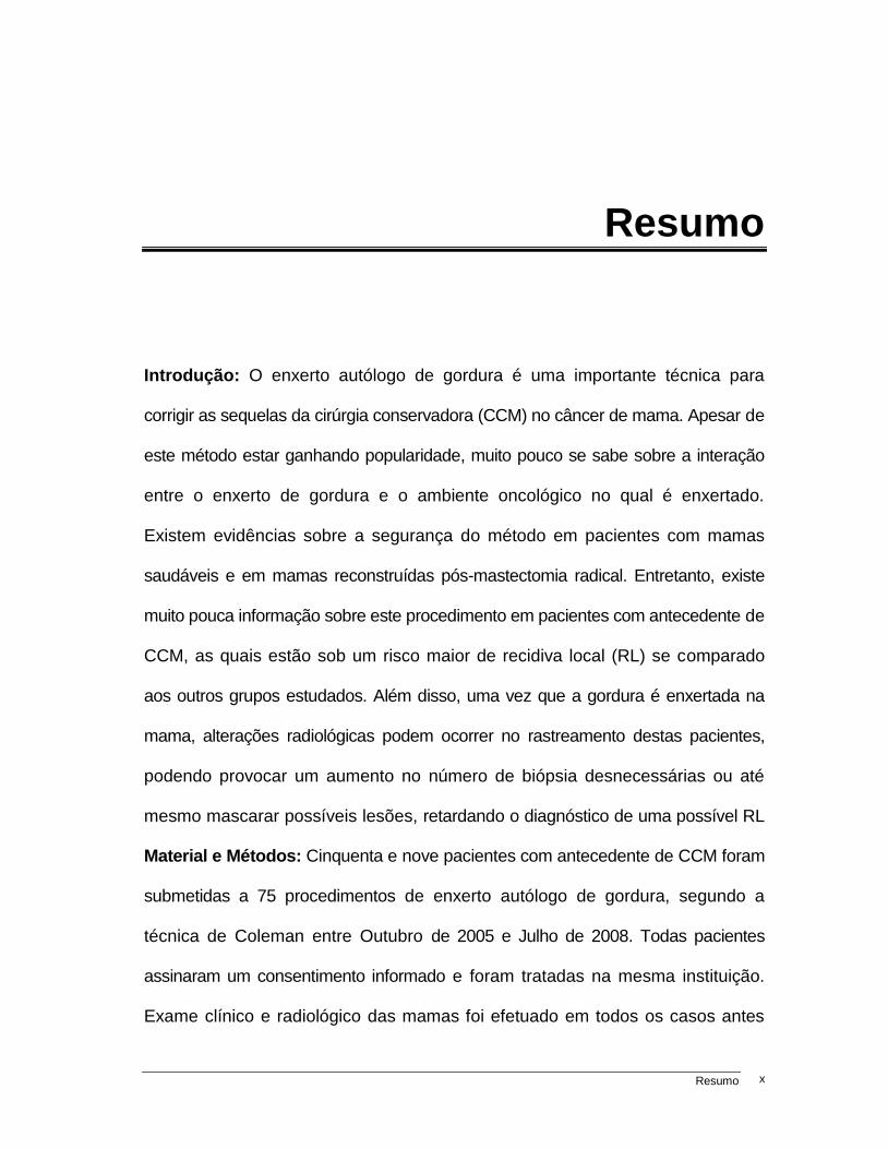
Introdução: O enxerto autólogo de gordura é uma importante técnica para
corrigir as sequelas da cirúrgia conservadora (CCM) no câncer de mama. Apesar de
este método estar ganhando popularidade, muito pouco se sabe sobre a interação
entre o enxerto de gordura e o ambiente oncológico no qual é enxertado.
Existem evidências sobre a segurança do método em pacientes com mamas
saudáveis e em mamas reconstruídas pós-mastectomia radical. Entretanto, existe
muito pouca informação sobre este procedimento em pacientes com antecedente de
CCM, as quais estão sob um risco maior de recidiva local (RL) se comparado
aos outros grupos estudados. Além disso, uma vez que a gordura é enxertada na
mama, alterações radiológicas podem ocorrer no rastreamento destas pacientes,
podendo provocar um aumento no número de biópsia desnecessárias ou até
mesmo mascarar possíveis lesões, retardando o diagnóstico de uma possível RL
Material e Métodos: Cinquenta e nove pacientes com antecedente de CCM foram
submetidas a 75 procedimentos de enxerto autólogo de gordura, segundo a
técnica de Coleman entre Outubro de 2005 e Julho de 2008. Todas pacientes
assinaram um consentimento informado e foram tratadas na mesma instituição.
Exame clínico e radiológico das mamas foi efetuado em todos os casos antes
Page 12
Resumo xi
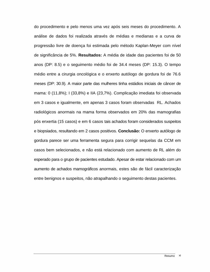
do procedimento e pelo menos uma vez após seis meses do procedimento. A
análise de dados foi realizada através de médias e medianas e a curva de
progressão livre de doença foi estimada pelo método Kaplan-Meyer com nível
de significância de 5%. Resultados: A média de idade das pacientes foi de 50
anos (DP: 8.5) e o seguimento médio foi de 34.4 meses (DP: 15.3). O tempo
médio entre a cirurgia oncológica e o enxerto autólogo de gordura foi de 76.6
meses (DP: 30.9). A maior parte das mulheres tinha estádios iniciais de câncer de
mama: 0 (11,8%); I (33,8%) e IIA (23,7%). Complicação imediata foi observada
em 3 casos e igualmente, em apenas 3 casos foram observadas RL. Achados
radiológicos anormais na mama forma observados em 20% das mamografias
pós enxertia (15 casos) e em 6 casos tais achados foram considerados suspeitos
e biopsiados, resultando em 2 casos positivos. Conclusão: O enxerto autólogo de
gordura parece ser uma ferramenta segura para corrigir sequelas da CCM em
casos bem selecionados, e não está relacionado com aumento de RL além do
esperado para o grupo de pacientes estudado. Apesar de estar relacionado com um
aumento de achados mamográficos anormais, estes são de fácil caracterização
entre benignos e suspeitos, não atrapalhando o seguimento destas pacientes.
Page 13
Summary xii
Summary
Background: Autologous fat graft to the breast is a useful tool to correct defects
after breast conservative treatment (BCT). Although this procedure gains
popularity, little is known about the interaction between the fat graft and the prior
oncological environment. Evidences of safety of this procedure in healthy breast
and after postmastectomy reconstruction exist. However, there is paucity of data
among patients who underwent BCT which are hypothetically under a higher risk of
local recurrence (LR). Moreover, since fat is injected in the breast, this technique
can potentially produce radiological features that could increase numbers of
unnecessary biopsies or even mask suspicious hidden lesions. Material and
Methods: Fifty nine patients, with prior BCT, underwent 75 autologous fat graft
procedures using the Coleman´s technique, between October 2005 and July
2008. All patients signed an informed consent and were treated at the same
institution. Radiological and clinical examination was performed in all cases prior
of the procedure. Follow up was made by clinical and radiological examination at
least once, after 6 months of the procedure. Statistical analysis was performed
by means and medians and progression free survival was estimated by the
Kaplan-Meyer method with significance level of 5%. Results: Mean age was
Page 14
Summary xiii
50±8.5 years and mean follow up was 34.4 ±15.3 months. Mean time from
oncological surgery to the first fat grafting procedure was 76.6± 30.9 months. Most of
patients were at initial stage 0 (11,8%), I (33,8%) or IIA (23,7%). Immediate
complication was observed in 3 cases and LR was observed in only 3 cases of true
LR. Abnormal breast images were present in 20% of the post-operative
mammograms (15 cases) and in six cases biopsy was warranted resulting positive
for LR in two cases. Conclusion: Autologous fat graft seems to be a safe tool to
correct defects after BCT without increasing the expected rates of LR, in low risk
and selected cases. Although it increases the rate of abnormal mammographic
findings, those are easily distinguished between benign and suspicious lesion by
a trained radiologist and do not interfere with the patient´s follow up.
Page 15
Introdução 14
1. Introdução
1.1. Cirurgia Conservadora da mama
O câncer de mama é o segundo tipo de câncer mais frequente no mundo, e o
principal tipo de câncer entre as mulheres, respondendo por aproximadamente 22%
de casos novos ao ano (1). Nos Estados Unidos da América (EUA) estima-se
que 226.870 mulheres serão diagnosticadas com carcinoma invasivo de mama
em 2012 e um adicional de aproximadamente 39.520 casos novos de carcinoma
in situ (CDIS) (2). No Brasil, a estimativa do Instituto Nacional do Câncer (INCA)
para o ano de 2012 é de 52.680 casos novos de carcinoma invasivo, acarretando
aproximadamente mais de 12.870 mortes ao ano (3).
O tratamento local do câncer de mama é eminentemente cirúrgico (associado
muitas vezes à radioterapia locorregional), pois a extirpação total do tumor da
mama, quando o mesmo não é metastático, confere á paciente ganho na sobrevida
global. Tal conceito não existia até o final do século XIX, quando Sir. William Halsted
publicou, de forma científica, em 1894, (4) seus resultados com a mastectomia
radical, que consistia na retirada de toda a mama, com incisão cutânea ampla,
Page 16
Introdução 15
junto com os músculos peitorais maior e menor e toda cadeia ganglionar axilar,
sendo que o fechamento da parede deveria ser feito com enxerto de pele (5).
A partir de então, modificações no tipo de mastectomia para técnicas
mais radicais, como as mastectomias alargadas de Urban e Veronesi, ou mais
conservadoras, como as mastectomias radicais modificadas por Patey, em
1948 (6), e Madden, em 1965 (7), passaram a ser utilizadas como única forma
de tratamento cirúrgico do câncer de mama.
Foi apenas no final da década de 1970 e início da década de 1980 que o
paradigma da extensão da cirurgia mamária mudou. Já em 1977, a primeira
publicação de Veronesi et al. apontava para os bons resultados da cirurgia
conservadora da mama (CCM) (8) e, após a publicação final do mesmo, em
1981 (9), e a publicação de Fisher et al. dos resultados do estudo NSABP-06
(10), assim como a publicação de Aitkin & Minton (11) na Inglaterra, a CCM
consagrou-se como tratamento-padrão dos tumores iniciais da mama.
Assim, a CCM popularizou-se e em todo o mundo um número crescente de
mulheres com câncer de mama passou a ser submetido a tal procedimento, que
consistia na retirada de todo o quadrante da mama afetado pelo tumor associado á
remoção de pele e da fáscia do músculo peitoral maior, conforme proposto por
Veronesi, ou apenas na retirada do nódulo tumoral com margem de segurança
livre, conforme proposto por Fisher, seguido sempre de radioterapia adjuvante.
Apesar de este modelo cirúrgico ter sido estudado com critérios de seleção
precisos e os resultados de atualização de mais de 20 anos de seguimento
Page 17
Introdução 16
demonstrarem taxas de recidiva local (RL) que variam entre 15% a 20%
(12,13), adaptações na técnica cirúrgica foram aparecendo com a evolução dos
conhecimentos, e a CCM passou a ser considerada sempre nos casos em que
fosse possível retirar o tumor com margem de segurança e o resultado
cosmético final fosse satisfatório (14).
Atualmente, a CCM representa a escolha principal de tratamento cirúrgico nos
EUA, com índices que variam de 10% a 67%, dependendo da instituição (15-17).
Entretanto, estes índices vêm caindo em consequência do aumento paradoxal no
número de mastectomias, relacionado ao emprego da Ressonância Nuclear Magnética
(RNM) no estadiamento pré-operatório (aumentando os achados de doença
multicêntrica e de lesões falso positivo), às modernas técnicas de reconstrução
mamária com melhor resultado cosmético e à falsa impressão para as pacientes de
que a mastectomia confere menor risco de recorrência sistêmica da doença (18).
Por outro lado, dados recentes da European Society of Breast Cancer Specialists
(EUSOMA), apresentados por Garcia-Ettienne et al., mostram justamente o contrário:
um declínio nas indicações de mastectomias e aumento nas indicações de CCM, com
tentativa de CCM em 84% dos casos e sucesso em 78% das vezes (19).
O objetivo da CCM, além de tratar o câncer, é deixar uma mama residual que
mantenha sua forma e função cosmética. Apesar da popularidade do procedimento
e da alta taxa de satisfação das pacientes com o resultado cosmético final, que varia
de 75% a 96% (15-18), estima-se que uma assimetria ou deformidade severa ocorra
em aproximadamente 30% dos casos (20-22). Estas sequelas são muitas
Page 18
Introdução 17
vezes difíceis de serem tratadas, dependendo do tipo de defeito residual e da
presença de radioterapia, o que ocorre na quase totalidade dos casos.
Clough et al (23) dividiram e classificaram estas sequelas em três tipos: tipo 1
- quando existe apenas assimetria da mama contralateral em relação à operada, o
que seria facilmente resolvido com uma mamoplastia de simetrização; tipo 2 -
quando além da assimetria existe um defeito no contorno e no volume glandular,
provocando muitas vezes também o mau posicionamento do complexo aréolo-
papilar (CAP); e o defeito de tipo 3 é aquele em que o contorno e o volume da
mama estão muito alterados, o aspecto glandular parece marmorizado e não
existe tecido glandular adequado para uma mamoplastia e nem pele com
vascularização adequada para tal.
Assim, sendo o defeito tipo 1 de fácil resolução e o tipo 3 de resolução
quase que impossível, a não ser com uma mastectomia de “resgate cosmético” com
reconstrução, o defeito tipo 2 torna-se o mais difícil de se manejar. Este requer
muitas vezes mais de um procedimento de mamoplastia, com muitos resultados
desagradáveis e complicações pós-operatórias, uma vez que a mamoplastia em
mama irradiada tem alto risco de complicações e maus resultados (22,23).
Desta maneira, se o defeito não é prevenido na cirurgia oncológica com
técnicas de reconstrução glandular e de mamoplastias aplicadas à cirurgia
oncológica, o que se denomina cirurgia oncoplástica (24-27), restam poucas opções
para a sua correção. É neste cenário que o enxerto autólogo de gordura, ou
lipoenxertia, teria papel na correção destes defeitos, corrigindo o contorno,
Page 19
Introdução 18
aumentando o volume e melhorando a forma da mama, além de melhorar a
qualidade da pele prejudicada pela radioterapia.
1.2. Enxerto autólogo de gordura ou lipoenxertia
O tecido adiposo vem sendo utilizado como material de transplante ou
enxerto autólogo para a correção de defeitos de preenchimento da superfície
corpórea há mais de um século. A gordura autóloga pode ser considerada o
enxerto ideal, uma vez que é abundante, não tem custo, é compatível com o
receptor e pode ser obtido de maneira fácil e repetidamente, sem maiores danos ao
paciente (autodoador) (28).
O primeiro relato da utilização de um enxerto autólogo de gordura para
corrigir um defeito mamário foi feito por Vincenz Czerny, que em 1895 descreveu a
remoção de um lipoma da região dorsal de uma paciente e o implantou na mama
(29). Apesar de diversos relatos da utilização do enxerto de gordura autóloga em
pacientes com lipodistrofia facial datando de 1912 (30) e da descrição, em 1926, da
utilização de cânulas para realizar a aspiração e a lipoenxertia (31), foi apenas a
partir de meados da década de 1980 que esta técnica passou a ser mais bem
explorada e difundida, estimulada pela inovação cirúrgica da lipoaspiração na
cirurgia estética. Nesta, o cirurgião encontrava-se ao final do procedimento com
tecido adiposo abundante que deveria ser desprezado e alguns, ao invés de fazê-lo,
reinjetavam a gordura em regiões do corpo que entendessem adequadas para
melhorar o contorno (lipoescultura) ou para corrigir defeitos de diferentes ordens
(traumáticos, congênitos e etc.) (32).
Page 20
Introdução 19
Inúmeras publicações demonstraram a eficácia do método na correção
de defeitos da face, mãos, lábios e na lipoescultura (33-36), enquanto apenas
poucos artigos aludiram a possibilidade da utilização da lipoenxertia para correção
de defeitos mamários e realização de mamoplastia de aumento. A explicação
para tal fato deve-se à heterogeneidade dos dados apresentados com respeito
à quantidade de tecido adiposo que é reabsorvido após o procedimento, que
pode variar de 20% a 90% (37).
A falta de padronização do método de obtenção e enxertia da gordura na
mama, associado a poucos dados na literatura sobre a eficácia do método, que
na maior parte das publicações foi avaliado como pobre e decepcionante, fez
com que o interesse sobre o procedimento diminuísse e sua utilização na mama
fosse praticamente abandonada (38-39).
Entretanto, alguns cirurgiões continuaram a acreditar no método e passaram
a avaliá-lo de maneira criteriosa e científica, propondo diferentes modelos e
padronizações na técnica de obtenção do material, no seu processamento e na fase
de enxertia. Desta maneira, Colleman (40,41) foi o primeiro a padronizar todo este
processo e estudar a curto e médio prazos os resultados da lipoenxertia na mama.
Como seus resultados foram positivos, a lipoenxertia na mama passou novamente a
fazer parte do arsenal de técnicas na cirurgia cosmética e reparadora da mama.
A padronização da técnica de Colleman (40,41) consiste na aspiração da
gordura com cânulas finas de 2mm a 3mm a baixa pressão, centrifugação da
gordura por 3 minutos, separação das partes oleosa e de sangue dos adipócitos
e enxerto da gordura em pequenos planos com uma cânula de 17G. Desta
Page 21
Introdução 20
maneira, resultados a médio e longo prazo, foram obtidos com baixas taxas de
reabsorção do enxerto (cerca de 30%). Outros autores descreveram inúmeras
outras técnicas que parecem ser tão eficazes quanto a descrita acima, porém
foi esta padronização que permitiu que a técnica fosse considerada efetiva e
pequenas modificações são realizadas por diferentes autores.
Assim, a lipoenxertia passou a ser utilizada em casos de mamoplastia de
aumento sem a utilização de prótese, correção de defeitos congênitos como
síndrome de Polland e mama tuberosa e em casos de complicações destas
cirurgias (40-45). Além disso, o enxerto de gordura não apenas recompõe o defeito
de volume mamário, mas também melhora substancialmente a qualidade da
pele adjacente ao tecido enxertado, especialmente em casos de pele irradiada,
graças à ação das células-tronco derivadas do tecido aiposo (ADSC), que
regeneram o tecido estimulando a neoangiogênese local (46,47).
Portanto, o enxerto de gordura autólogo seria a ferramenta ideal para
corrigir os defeitos tipo 2 e, em alguns casos, tipo 3 decorrentes da CCM, uma
vez que não requer mobilização de pele nem deslocamento glandular, evitando
complicações como deiscências, necrose gordurosa da mama e necroses de
pele que não são infrequentes quando utilizadas as técnicas tradicionais para a
correção destes defeitos (48,49).
1.3. Enxerto Autólogo de Gordura e Cirurgia Conservadora da Mama
Apesar de existirem poucas dúvidas quanto à eficácia e à aplicabilidade do
enxerto autólogo de gordura na mama, o fator de maior preocupação é referente á
Page 22
Introdução 21
segurança da utilização desta técnica em mamas que foram submetidas a
tratamento oncológico (50-53), especialmente naquelas submetidas à CCM.
Sociedades médicas europeias e americanas expressaram sua preocupação
em relação à utilização da lipoenxertia neste grupo de pacientes, uma vez que
os dados científicos sobre o procedimento em pacientes oncológicos ainda não
são claros (54,55). Devido a isso, em 2009 a Sociedade Americana de Cirurgia
Plástica (ASPS) convocou uma força-tarefa de especialistas para elaborar um
relatório sobre a aplicabilidade, técnica, indicações e segurança do enxerto
autólogo de gordura na mama (54). A conclusão dessa força tarefa foi que quase
tudo o que se sabe sobre lipoenxertia na mama é baseada em trabalhos como série
de casos ou opiniões de experts, e que dados sobre a segurança oncológica da
lipoenxertia deveriam ser pesquisados em estudos prospectivos.
Entretanto, nenhuma evidência entre lipoenxertia e recidiva local ou a
distância foi encontrada na literatura, podendo então o procedimento ser realizado,
desde que as pacientes entendessem que este é um procedimento experimental,
aconselhando-as a participarem de estudos clínicos prospectivos.
A recidiva local é mais frequente em pacientes submetidas à CCM do
que as submetidas à mastectomia radical modificada clássica, sem impacto na
mortalidade (9-12,56). Este fato pode ser explicado pela existência de células
tumorais dormentes presentes no parênquima mamário, mesmo após radioterapia e
terapia sistêmica (47,57-59). Hipoteticamente, o enxerto de ADSC no parênquima
mamário poderia estimular células tumorais dormentes a se multiplicarem,
acelerando ou mesmo induzindo a um processo de recidiva local (57-59, 63, 69).
Page 23
Introdução 22
Alguns estudos in vitro mostram relação entre a presença de ADSC e o
estímulo da proliferação de células tumorais (60-64), e outros mostram a ação
protetora dos derivados das ADSC (65). Além disso, estudos usando modelos
animais demonstraram estímulo de células tumorais após a injeção de ADSC e
seus derivados, assim como aceleração no processo de crescimento do tumor
mamário quando comparados com animais não enxertados (66-68). Isto acontece
por mecanismos distintos, pois a ADSC sofre transformações até se tornar uma
célula capaz de acumular gordura (adipócito). Para isso é necessária a
liberação de susbtâncias angiogênicas e fatores de crescimento celular, que
poderiam agir também em células tumorais dormentes ou células pré-cancerosas e
se diferenciarem em célula tumoral (62,63). Além disso, hormônios liberados
por estes adipócitos como Leptina e Adiponectina podem promover a proliferação
celular, assim como promover aromatização periférica dos androgênios em
Estrona e Estradiol, podendo influenciar no controle local ou a distância do
câncer de mama (60-68).
Apesar destas possibilidades aventadas na pesquisa básica, nenhum
estudo de série de casos demonstrou sinais claros de associação entre lipoenxertia
mamária em pacientes previamente tratadas por câncer de mama e aumento nas
taxas de recidiva local, incluindo uma série de casos muito grande de Dealy E.
et al com 880 casos (69) e séries exclusivamente de pacientes oncológicas como de
Rietjens et al (70) e um estudo multicêntrico de Petit J.Y. et al (71). Entretanto,
nestas séries foram incluídos, na sua maioria, pacientes com antecedentes de
mastectomia e reconstrução, e apenas poucos pacientes com CCM prévia.
Page 24
Introdução 23
Portanto, ainda falta muita informação sobre a relação entre lipoenxertia,
CCM e recidiva local, pois poucos estudos focaram neste grupo de pacientes,
que, como discutido acima, apresentaria um maior risco teórico de recidiva local
relacionado ao procedimento de lipoenxertia.
Outro fator pertinente á lipoenxertia na mama é a questão sobre possíveis
alterações radiológicas que este procedimento poderia ocasionar. A injeção de
gordura no parênquima mamário pode causar alterações radiológicas decorrentes
dos processos de reabsorção da gordura, inflamação local e principalmente da
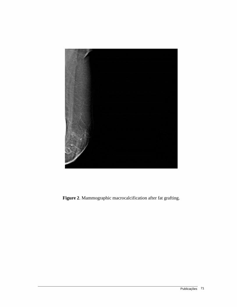
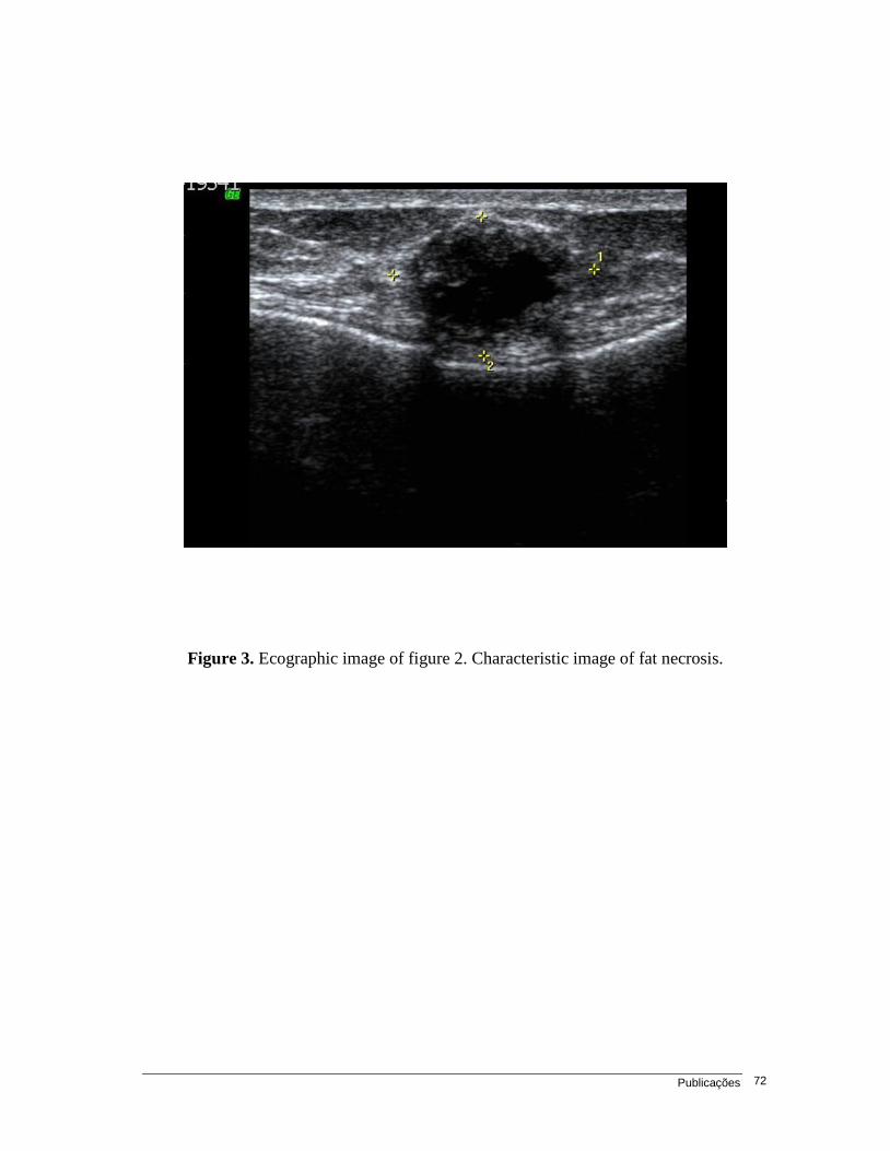
necrose gordurosa ou esteatonecrose (72,73). Tais processos podem levar à
formação de imagens que poderiam, eventualmente, mimetizar uma lesão
suspeita ou então mascarar uma lesão já presente na mama, retardando um
diagnóstico de recidiva local.
Devido a este risco teórico, em 1987 a Sociedade Americana de Cirurgia
Plástica e Reconstrutiva (ASPRS) estabeleceu um comitê específico para avaliar a
utilização da lipoenxertia na mama. Neste comitê ficou definido por unanimidade
que esta técnica era “deplorável” e sua utilização para mamoplastia de aumento
poderia gerar alterações radiológicas na mama, que de alguma maneira poderiam
mascarar lesões existentes, retardando o diagnóstico precoce de um câncer,
sendo, portanto, danosa à saúde pública (74).
Fato interessante foi que inesse mesmo ano um estudo retrospectivo sobre
aspectos mamográficos em pacientes submetidas à mamoplastia redutora
apontava que diferentes tipos de lesões (macrocalcificações, microcalcificações,
cistos) estavam presentes em 50% das pacientes submetidas à cirurgia, e, apesar
Page 25
Introdução 24
disso, o procedimento foi considerado seguro (75). Desde então se discutem as
alterações radiológicas provocadas por procedimentos mamários invasivos, e
atualmente padrões de suspeita ou benignidade são facilmente identificados e
classificados pelos radiologistas (76-78).
Assim, 20 anos após considerar o enxerto autólogo de gordura na mama
como técnica deplorável, a mesma ASPS considerou, em 2009, que as alterações
radiológicas relacionadas ao procedimento não estariam relacionadas a alterações
mamográficas que pudessem trazer prejuízo às pacientes (54). Entretanto, mais uma
vez, a conclusão é baseada em dados que em sua maioria remonta à lipoenxertia em
mamas sadias. Apenas algumas séries avaliaram os aspectos radiológicos pós-
lipoenxertia em pacientes com CCM prévia. Tais estudos não demonstraram
associação da técnica com número elevado de achados suspeitos, sendo a maioria
das alterações relacionadas a lesões típicas de esteatonecrose (52,53,79).
Finalmente, é inquestionável que a lipoenxertia é um excelente método para
corrigir defeitos pós-CCM. Seu uso vem se popularizando, apesar de faltarem
ainda evidências claras da segurança do método neste grupo de pacientes. Desta
maneira, o presente estudo apresenta uma série prospectiva de 59 pacientes com
antecedente de CCM submetidas a 75 procedimentos de lipoenxertia mamária,
realizados em uma única instituição e pela mesma equipe cirúrgica, com enfoque
nos seguimentos oncológico e radiológico destas pacientes.
Page 26
Objetivos 25
2. Objetivos
2.1. Artigo 1
Avaliar as taxas de recidiva local em pacientes submetidas a enxerto
autólogo de gordura para correção de defeitos decorrentes da cirurgia
conservadora da mama.
Comparar as taxas de recidiva local encontradas com dados da literatura e
avaliar se o enxerto autólogo de gordura pode estar ou não associado a
este evento
Identificar e avaliar as complicações imediatas e tardias decorrentes do
enxerto autólogo de gordura nestas pacientes
2.2. Artigo 2
Descrever as alterações radiológicas mamárias decorrentes do enxerto
autólogo de gordura em pacientes com antecedente de cirurgia conservadora
da mama.
Avaliar se este procedimento provoca aumento no número de biópsias
mamárias desnecessárias.
Identificar se existe um padrão mamográfico para as recidivas locais
encontradas após o procedimento neste grupo de pacientes.
Page 27
Publicações 26
3. Publicações
Artigo 1 – Oncological safety of autologous fat grafting after breast
conservative treatment: A prospective evaluation
Fabricio Brenelli M.D., Mario Rietjens M.D., Francesca De Lorenzi PhD., Aarão
Pinto-Neto PhD., Fabio Rossetto, Stefano Martella, José R.P. Rodrigues M.D.,
Visnu Lohsiriwat M.D., Daniel Barbalho M.D., Jean Yves Petit PhD.
Artigo 2 – Radiological Features of Autologous Breast Fat Grafting After
Conservative Treatment: Results of a Prospective Study
Fabricio Brenelli M.D., Mario Rietjens M.D., Aarão Pinto-Neto PhD., Francesca
De Lorenzi PhD., Fabio Rossetto, Daniel Barbalho
Page 28
Publicações 27
3.1. Artigo 1
Oncological safety of autologous fat grafting after breast conservative
treatment: A prospective evaluation
a ricio renelli ario iet ens rancesca e orenzi
PhD.**, Aarão Pinto-Neto PhD.*, Fabio Rossetto**, Stefano Martella**, José
R.P. Rodrigues M.D.***, Daniel Barbalho M.D.**
* State University of Campinas (Unicamp), Department of Gyanecology and
Obstetrics – Breast Oncology Division, Campinas – São Paulo, Brazil
eneficencia Portuguesa de São Paulo São José’s Hospital reast Surgery
|Division, São Paulo, SP, Brazil.
** European Institute of Oncology, Division of Plastic and Reconstructive
Surgery, Milan, Italy
*** State University of São Paulo (Unesp), Botucatu- São Paulo, Brazil
Corresponding author
Fabricio Brenelli: R Elvino Silva 30, Campinas – SP, Brazil
Zip: 13092-559
Email: [email protected]
Original Article
Page 29
Publicações 28
Abstract
Background: Autologous fat graft to the breast is a useful tool to correct defects
after breast conservative treatment (BCT). Although this procedure gains
popularity, little is known about the interaction between the fat graft and the prior
oncological environment. Evidences of safety of this procedure in healthy breast
and after postmastectomy reconstruction exist. However, there is paucity of data
among patients who underwent BCT which are hypothetically under a higher risk
of local recurrence (LR). Methods: Fifty nine patients, with prior BCT,
underwent 75 autologous fat graft procedures using the Coleman´s technique,
between October 2005 and July 2008. Follow up was made by clinical and
radiological examination at least once, after 6 months of the procedure.
Results: Mean age was 50±8.5 years and mean follow up was 34.4 ±15.3
months. Mean time from oncological surgery to the first fat grafting procedure
was 76.6± 30.9 months. Most of patients were at initial stage 0 (11,8%), I
(33,8%) or IIA (23,7%). Immediate complication was observed in 3 cases (4%).
Only 3 cases of true LR (4%) associated with the procedure were observed
during the follow up. Abnormal breast images were present in 20% of the post-
operative mammograms and in 8% of the cases biopsy was warranted.
Conclusion: Autologous fat graft is a safe procedure to correct breast defects
after BCT, with low post-operative complications. Although it was not associated
with increased risk of LR in the group of patients studied, prospective trials are
needed to certify that it does not interfere in patient’s oncological prognosis
Page 30
Publicações 29
Introduction
Breast conservative treatment (BCT) is a standard of care for early breast
cancer. As definition, BCT intends to give local treatment as effective as
mastectomy, but with better cosmetic results once the breast tissue is spared (1, 2).
Since the publications of the Milan I Trial and the NSABP-06 Trial, increasing
numbers of women sought BCS (3-5). Rates of BCS vary from 10 to 67% in
different U.S centers (6-9 espite the high quote of patient’s satisfaction on these
studies (75% to 96%), severe asymmetry is noted in almost 30% of then (10).
Correction of breast asymmetry after BCT may be very challenging, especially
in type 2 and 3 cosmetic sequelae as described by Clough KB et al. (11). It is in this
scenario that autologous fat graft seems to be a good alternative to fill the defects
and improve cosmetic outcome of BCS (12-14). Despite the discussion about
the technique used and predictability of results, the main concern among
surgeons is on its safety regarding the oncological aspects, especially in those
who underwent BCT (12, 15-17).
Medical societies throughout the world expressed the concern about
surgeons performing this procedure without clear evidence of its safety (18,19).
In 2009 de American Society of Plastic Surgery (ASPS) set up a task force to
assess the indications, safety and efficacy of autologous fat transfer (18). The
conclusion of this task force was that most of what is known comes from case
series and expert´s opinion, which means a low grade of scientific evidence.
However, the task force did not find any association between breast fat graft and
local recurrence (LR), so they were not able to give further recommendations
Page 31
Publicações 30
addressing this issue than considering this experimental in breast cancer
patients and that prospective controlled studies should be performed.
Hypothetically, the transfer of adipose derived stem cells (ADSC) or adipose
derived mesenchymal stem cell (ADMSC) could induce silent tumor cells to
reproduce and predispose to “In vitro” and animal models asic researches
are conflictive, and show positive and also negative association with breast
cancer cell proliferation (20-24).
In the other hand, case series did not demonstrate an association
between autologous fat graft and breast cancer recurrence, including large
individuals’ series and multi institutional studies 25-27). However, most of these
series focused on fat grafting after mastectomy and reconstruction, and few
cases are dedicated to study its impact on BCT patients.
Local recurrence is more frequent in patients with BCT compared to
mastectomy, without impact on mortality (1-4). This fact maybe due to the
existence of dormant tumor cells in the breast parenchyma (24, 28). Therefore, if
autologous fat graft can stimulate those dormant tumor cells, it should happen
indeed more frequently in patients treated with breast conservation rather than
mastectomy. However, this information lacks in many studies.
It is in this scenario of lack of strong scientific evidence that we present a
prospective evaluation of 59 patients with prior BCT undergoing 75 autologous
breast fat graft procedure and analyze the oncological results along time,
focusing in procedure safety, local control and disease free survival.
Page 32
Publicações 31
Material and methods
Patient’s selection
From October 2005 to July 2008 we prospectively evaluated 59 patients
that underwent 75 breast fat grafting procedures at the European Institute of
Oncology (Milan, Italy). All patients had been submitted to a previous BCT for
oncological reasons, which lead to an aesthetical breast defect. All patients were
visited by a single surgeon who indicated the procedure. Only patients free of
breast locoregional disease were considered eligible to the procedure after an
accurate breast clinical and instrumental evaluation. The presence of stable
bone metastasis was not an exclusion criteria.
Pre-operative evaluation
All patients scheduled for the fat grafting procedure were evaluated pre
operatively with clinical and breast image exams. Bilateral mammogram and
breast ultrasound (U.S.) were requested for all patients. After explanation of the
procedure and the signature of an informed consent, all patients agreed to
undergo the surgery. Pre operative pictures were taken in all cases and the
breast defect was measured by a centimeter on its 2 major axis, and finally on
its depth by an approximate measurement. If there were one or more defects,
they were measured and documented as defect 1, 2, 3 and so on.
Surgical technique
The procedure was performed under local or general anesthesia, depending
on the patient’s clinical conditions and risks ocal anesthesia was prefera le and
Page 33
Publicações 32
general anesthesia was recommended in cases which there was a need of
harvesting a great amount of fat tissue, when an associated procedure was indicated
(prosthesis exchange and capsulotomy, for instance), or when the patient
showed extreme anxiety.
The whole procedure of fat harvesting and grafting was performed according
to the Coleman’s technique (12) with minimal modifications.
Follow up
ollow up was made at the outpatient’s clinics at least once after 6
months of the procedure. It consisted of clinical evaluation. Mammogram was
requested every year associated with breast ultrasound when needed. When
patient could not be reached, telephone contact was made.
Statistical analysis
Progression free survival curve was estimated by the Kaplan-Meyer method
with significance level of 5%. Time between the fat grafting and last follow up was
studied by mean time and median. The software used was SAS, version 9.2.
Results
Mean age of the women at the time of fat grafting was 50±8.5 years and
mean follow up was 34.4 ±15.3 months, and more than 75% of them had follow
up greater than 45.9 months. Procedure was conducted under local anesthesia
in 58 cases (77.3%) and under general anesthesia in 17 cases (22.7%). Ninety
eight percent of the defects were localized exclusively in the breast parenchyma,
and just in one patient defect was also present in the axilla. Right and left breast
Page 34
Publicações 33
were equally affected (50.7 vs. 48%, respectively). Population´s characteristics
by patient and by procedure are described in table 1.
Only 5 patients had more than one defect in the breast, and two of them had 3
defects. Mean volume of fat preparation injected was 52.3 ±28.7 cc. Second and
third defects had a mean volume injected of 32.8c and 52.8cc, respectively. In
78.7% of the cases, fat was harvested from the abdomen and in 10.7% from the hips,
in the other cases association between hips, abdomen and knees were made.
Immediate complication was observed in 3 cases (4%) and consisted of
fat necrosis in 2 patients and cellulites in 1 patient. All cases were managed
clinically with no further complications.
Follow-up occurred at least once 6 months after the procedure. In 15
cases (20%), the mammogram was normal before the procedure and exhibited
abnormalities afterwards. Lesions were considered suspicious in 6 cases and breast
biopsy was performed. In 2 cases biopsies resulted positive and in 4 cases
negative (table 2). In 8 cases (10, 6%) post-operative breast images were missing.
Most of patients were at initial stage 0 (11,8%), I (33,8%) or IIA (23,7%)
(figure 1). Thirty five patients (59.3%) had undergone quadrantectomy with axillary
dissection (QUART), and 14 patients (23.7%) underwent quadrantectomy with
sentinel node biopsy (SNB). Only 10 patients (16.9%) underwent quadrantectomy
alone. Oncoplastic techniques were used in just 9 patients (15.3%). External
radiotherapy was present in 56 patients (94.9%).
During the follow up, there were 4 cases of ipsilateral breast recurrence.
However, in one case LR was suspected in the day of the procedure and was
confirmed by histology one week after it. Therefore, we do not associate this to the
Page 35
Publicações 34
fat grafting procedure. All 3 LR were invasive ductal carcinoma and patient. Mean
time from oncological surgery to the first fat grafting procedure was 76.6± 30.9
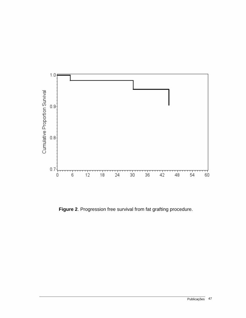
months. Considering that we found just 3 cases of true local recurrence (4%) in 34
months of follow up, this would produce a rate of 1,4% LR per year. The progression
free survival from time of fat grafting to local recurrence is shown in figure 2.
Discussion
Autologous fat graft is a valuable and promising tool to help breast and
plastic surgeons to correct defects after BCT. Procedure is simple and can be
done mostly under local anesthesia, which means no hospital stay and low cost.
Complication’s rate was a out 4% and was of simple management any papers
confirm this low rate of post-operative complications which makes autologous fat
graft to be a popular choice for surgeons and patients (29-31).
Unfortunately, efficacy of the procedure is not predictable. Despite of our
experience and others from the literature (25-27, 29-33), it is still very difficult to
predict whether the fat graft will attach or not. In this study, only in 16.8% of the
cases a further fat graft procedure was performed. This could means that a good
result was achieved in most of the cases. However, those numbers are
underestimated, once in the follow up there are patients already scheduled to a
new procedure or others who still show the breast defect but do not want to
undergo another procedure. The lack of an objective evaluation of the efficacy is
a weak point of his paper, as it is in review articles, which shows a difficulty in
assessing its efficacy (18, 31, 33-34).
Page 36
Publicações 35
A great concern of our study was about the abnormal radiological features
that fat graft could produce in the breast. Although some evidences in literature
shows that breast fat graft is not correlated to features that could mask hidden
lesions or increase the number of unnecessary biopsies (13-14, 16- 17, 34-36), this
was a critical argument to be evaluated. Actually, we found no significant increase in
abnormal breast images after the procedure (20%) if we compare to other types
of breast intervention such as reduction mammoplasty, which can produce up to
85% of post-operative radiological features (37-39). Most abnormal breast
images associated with fat graft are macrocalcification and oil’s cyst which are
easily recognized as benign lesions by trained radiologists (39, 40).
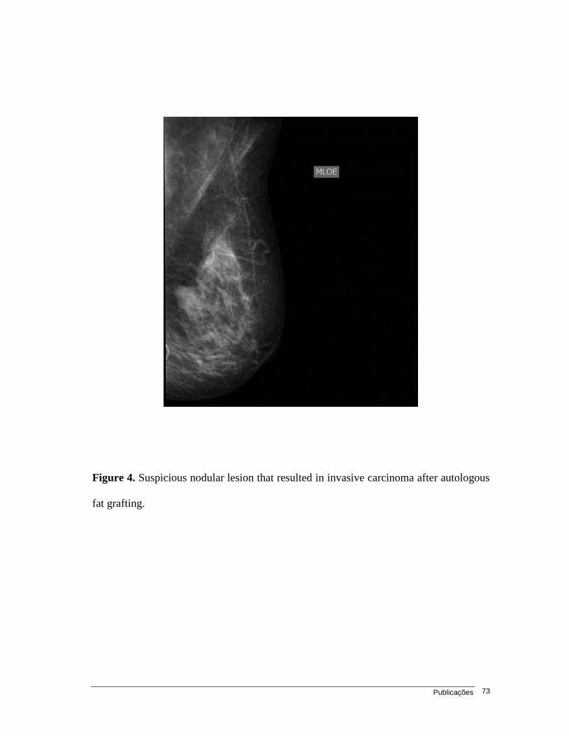
From 6 cases warranting biopsies in our study, only 2 irregular nodules
resulted in LR. Little information exists on LR following autologous fat graft after
BCT, therefore, there is no specific radiological pattern of it. Based on our
observation, we suggest that suspicious nodular lesions after autologous fat
graft should be biopsied to rule out LR. However, it is evident that all suspicious
lesions should be biopsied.
Local recurrence was the main goal of this study. Although the use of
autologous fat graft is rising in clinical practice, the real oncological impact on
patients who have had breast cancer is still unknown (28-31, 33-34, 41-44).
Delay et al (25) published data on 10 years follow up of more than 880 fat
grafting procedures, including 42 cases after BCT and found no increased rate
of LR. Illouz et al (32) in his personal experience with 820 patients undergoing
breast fat grafting found no LR at all in the group of 30 patients with previous
BCT. Two recent systematic reviews addressed the oncological issue and found
Page 37
Publicações 36
no clinical evidence of increased LR in any study, although they consider that
safety should be objective of prospective and controlled studies (31, 34).
In our study LR was observed only in 3 cases out of 75 procedures (4%). It is
acceptable in the literature that LR occurs in the rate of 1 to 1,5%% per year (1-4). In
this series, the mean time between oncologic surgery to fat graft was 76 months, and
mean follow up time was 34 months. So, 4% of LR in 34 months produces a rate of
1,4% LR per year, which seems acceptable for BCT. Moreover, timing of recurrence
was very different in the 3 cases (figure 2), showing that there is no pattern of LR,
linking these facts much more to a chance rather than to the fat grafting procedure.
Patient’s selection and the a sence of a control group to match the results
can be considered a bias to this study. The majority of patients in the study were
patients of good oncological prognosis presenting with initial breast cancer
pathological stages (0, I and IIA), evidencing a selection for LR low risk patients.
Recently, Petit JY et al (45) published data about autologous fat transfer in
previous breast cancer patients treated with mastectomy or BCT and matched the
follow up with a control group. There was no difference in LR in patients with previous
invasive carcinoma. However, LR in patients with previous ductal carcinoma in situ
(DCIS) was significantly higher. Difference between mastectomy and BCT group
was not of statistical significance. Despite of the findings, the authors conclude
that breast lipofilling seems to be safe, but this higher incidence of LR in the
DCIS group must be better studied once it can be biased by many factors,
including the design of the study.
The fact is that the role of ADSC in a previous cancerous environment is still
unknown. Bench basic research shows conflicting results about the interaction
Page 38
Publicações 37
between ADSC with breast tissues and cancer cells. There is data that suggest
A SC promoting or causing reast cancer in “in vitro” and animal models 46-48).
Mechanisms of this interaction may be multiple, including liberation of adiponectin
and leptin from ADSC promoting cellular proliferation, increase in the peripheral
aromatization and liberation of angiogenic factors such as vascular endothelial
growth factor (VEGF) (20-21, 23-24, 47). This could theoretically induce the
awakening of a dormant tumor cell or even interfere with adjuvant hormonal therapies.
Moreover, Pearl et al (49) published a recent review on stem cell biology
and its behavior in the breast environment. It seems that ADMSC interacts with
adjacent stroma and potentially promotes proliferation of fibroblasts and other
cell lines promoting as occurs in prostate cancer with the “fi ro last
associated carcinoma” 49 50 ecently has een published a case report of a
osteosarcoma treated 13 years before with surgery and chemotherapy, and after
18 months of a autologous fat graft to correct the surgical defect a LR happened
(51). The authors discuss the interaction between ADSC and the LR as this
pattern of late recurrence is extremely rare.
In contrast, there are some studies that show ADSC inhibiting tumor
growth and metastasis of breast cancer in animal models (22, 24, and 52).
Therefore, the exact role and intrinsic interaction between ADSC, normal breast
and breast cancer cell and its environment must be further investigated.
Conclusion
Autologous fat graft is a promising tool to correct defects after BCT.
Procedure is simple and is associated with low rates of postoperative complications.
Page 39
Publicações 38
LR rate was not higher than the expected for this low risk group of patients. The
role of ADSC in previous cancer environment is still unclear. Further investigation is
needed, although prospective randomized trials will be difficult to be done since
there is not another technique or “filler” that can su stitute the role of fat grafting in
correcting breast defects. Data should be collected and gathered from multicenter
institution to enlarge the evidences of the safety of fat grafting after BCT.
Conflict of interests
The authors declare no conflicts of interests or sponsorships, grants and
patents. Study was approved by the ethics committee and all patients were
given an informed consent.
References
1. Veronesi U, Cascinelli N, Mariani L, et al. Twenty-year follow-up of a
randomized study comparing breast-conserving surgery with radical mastectomy
for early breast cancer. N Engl J Med. 2002 Oct 17; 347(16):1227-32.
2. Fisher B, Anderson S, Bryant J, et al. Twenty-year follow-up of a randomized trial
comparing total mastectomy, lumpectomy, and lumpectomy plus irradiation for
the treatment of invasive breast cancer. N Engl J Med. 2002 Oct 17;
347(16):1233-41.
3. Veronesi U, Saccozzi R, Del Vecchio M, et al. Comparing radical mastectomy
with quadrantectomy, axillary dissection, and radiotherapy in patients with
small cancers of the breast. N Engl J Med. 1981 Jul 2;305(1):6-11.
4. Fisher B, Bauer M, Margolese R, et al. Five-year results of a randomized
clinical trial comparing total mastectomy and segmental mastectomy with or
Page 40
Publicações 39
without radiation in the treatment of breast cancer. N Engl J Med. 1985 Mar
14; 312(11):665-73.
5. Coleman SR, Saboeiro AP. Fat grafting to the breast revisited: safety and
efficacy. Plast Reconstr Surg. 2007 Mar;119(3):775-85
6. Wang HT, Barone CM, Steigelman MB, et al. Aesthetic outcomes in breast
conservation therapy. Aesthet Surg J. 2008 Mar-Apr;28(2):165-70.
7. Kotwall CA, Covington DL, Rutledge R, et al. Patient, hospital, and surgeon
factors associated with breast conservation surgery: a statewide analysis in
North Carolina. Ann Surg 1996;224:419–429
8. Parviz MA, Cassel JB, Kaplan BJ, et al. Breast conservation therapy rates
are no different in medically indigent versus insured patients with early stage
breast cancer. J Surg Oncol 2004;84:57–62.
9. Kelemen JJ III, Poulton T. Surgical treatment of early-stage breast cancer in the
Department of Defense healthcare system. J Am Coll Surg 2001; 192:293–297.
10. McGuire KP, Santillan AA, Kaur P, et al. Are mastectomies on the rise? A
13-year trend analysis of the selection of mastectomy versus breast conservation
therapy in 5865 patients. Ann Surg Oncol. 2009 Oct; 16(10):2682-90.
11. Bajaj AK, Kon PS, Oberg KC, Miles DAG. Aesthetic outcomes in patients
undergoing breast conservation therapy for the treatment of localized breast
cancer. Plast Reconstr Surg 2004; 14:1442–1449.
12. Clough KB, Thomas SS, Fitoussi AD, et al. Reconstruction after conservative
treatment for breast cancer: cosmetic sequelae classification revisited. Plast
Reconstr Surg. 2004 Dec; 114(7):1743-53.
13. Gosset J, Flageul G, Toussoun G, et al. Lipomodelling for correction of breast
conservative treatment sequelae. Medicolegal aspects. Expert opinion on
five problematic clinical cases. Ann Chir Plast Esthet. 2008 Apr; 53(2):190-8.
14. Delay E, Gosset J, Toussoun G, et al. Efficacy of lipomodelling for the
management of sequelae of breast cancer conservative treatment. Ann Chir
Plast Esthet. 2008 Apr; 53(2):153-68.
Page 41
Publicações 40
15. Missana MC, Laurent I, Barreau L et al. Autologous fat transfer in reconstructive
breast surgery: indications, technique and results. EJSO 2007; 33:685-90.
16. Gosset J, Guerin N, Toussoun G, et al. Radiological evaluation after
lipomodelling for correction of breast conservative treatment sequelae. Ann
Chir Plast Esthet. 2008 Apr; 53(2):178-89.
17. Amar O, Bruant-Rodier C, Lehmann S, et al. Fat tissue transplant: restoration of
the mammary volume after conservative treatment of breast cancers, clinical and
radiological considerations. Ann Chir Plast Esthet. 2008 Apr; 53(2):169-77.
18. Gutowski KA; ASPS Fat Graft Task Force. Current applications and safety
of autologous fat grafts: a report of the ASPS fat graft task force. Plast
Reconstr Surg. 2009 Jul; 124(1):272-80.
19. Delay E, Sinna R, Delaporte T. Patient information before aesthetic lipomodeling
(lipoaugmentation): a French plastic surgeon's perspective. Aesthet Surg J.
2009 Sep-Oct; 29(5):386-95.
20. Schaffler A, Scholmerich J, Buechler C. Mechanisms of disease: adipokines and
breast cancer - endocrine and paracrine mechanisms that connect adiposity
and breast cancer. Nature Clin Pract Endocrinol Metab 2007; 3:345-54.
21. Dieudonne MN, Machinal-Quelin F, Serazin-Leroy V, et al. Leptin mediates
a proliferative response in human MCF7 breast cancer cells. Biochem
Biophys Res Commun. 2002 Apr 26; 293(1):622-8.
22. Dieudonne MN, Bussiere M, Dos Santos E, et al. Adiponectin mediates
antiproliferative and apoptotic responses in human MCF7 breast cancer
cells. Biochem Biophys Res Commun. 2006 Jun 23; 345(1):271-9.
23. Iyengar P, Espina V, Williams TW, et al. Adipocyte-derived collagen VI affects
early mammary tumor progression in vivo, demonstrating a critical interaction in
the tumor/stroma microenvironment. J Clin Invest. 2005 May; 115(5):1163-76.
24. Lohsiriwat V, Curigliano G, Rietjens M, Goldhirsch A, Petit JY. Autologous fat
transplantation in patients with breast cancer: "silencing" or "fueling" cancer
recurrence? Breast. 2011 Aug;20(4):351-7. Epub 2011 Feb 5.
Page 42
Publicações 41
25. Delay E, Garson S, Tousson G, Sinna R. Fat injection to the breast: technique,
results, and indications based on 880 procedures over 10 years. Aesthet
Surg J. 2009 Sep-Oct; 29(5):360-76.
26. Rietjens M, De Lorenzi F, Rossetto F, Brenelli F, et al. Safety of fat grafting
in secondary breast reconstruction after cancer. J Plast Reconstr Aesthet
Surg. 2011 Apr; 64(4):477-83.
27. Petit JY, Lohsiriwat V, Clough KB, Sarfati I, Ihrai T, Rietjens M, Veronesi P,
Rossetto F, Scevola A, Delay E. The oncologic outcome and immediate surgical
complications of lipofilling in breast cancer patients: a multicenter study--
Milan-Paris-Lyon experience of 646 lipofilling procedures. Plast Reconstr
Surg. 2011 Aug;128(2):341-6.
28. Rigotti G, Marchi A, Sbarbati A. Adipose-derived mesenchymal stem cells:
past, present, and future. Aesthetic Plast Surg. 2009 May; 33(3):271-3.
29. ELFadl D, Garimella V, Mahapatra TK,et al. Lipomodelling of the breast: a
review. Breast. 2010 Jun; 19(3):202-9.
30. Mizuno H, Hyakusoku H. - Fat grafting to the breast and adipose-derived
stem cells: recent scientific consensus and controversy. Aesthet Surg J.
2010 May-Jun; 30(3):381-7. Review.
31. Claro F Jr, Figueiredo JC, Zampar AG, Pinto-Neto AM. Applicability and
safety of autologous fat for reconstruction of the breast. Br J Surg. 2012
Jun;99(6):768-80. doi: 10.1002/bjs.8722. Epub 2012 Apr 4.
32. Illouz YG, Sterodimas A. Autologous fat transplantation to the breast: a
personal technique with 25 years of experience. Aesthetic Plast Surg 2009;
33(5):706–715.
33. Chan CW, McCulley SJ, Macmillan RD. Autologous fat transfer--a review of the
literature with a focus on breast cancer surgery. J Plast Reconstr Aesthet
Surg. 2008 Dec; 61(12):1438-48.
34. Rosing JH, Wong G, Wong MS, Sahar D, Stevenson TR, Pu LL. Autologous
fat grafting for primary breast augmentation: a systematic review. Aesthetic
Plast Surg. 2011 Oct;35(5):882-90. Epub 2011 Apr 1. Review.
Page 43
Publicações 42
35. Pierrefeu-Lagrange AC, Delay E, Guerin N, et al. Radiological evaluation of
breasts reconstructed with lipomodeling. Ann Chir Plast Esthet. 2006 Feb;
51(1):18-28.
36. Pulagam SR, Poulton T, Mamounas EP. Long-term clinical and radiologic
results with autologous fat transplantation for breast augmentation: case reports
and review of the literature. Breast J. 2006 Jan-Feb; 12(1):63-5.
37. Veber M, Tourasse C, Toussoun G, Moutran M, Mojallal A, Delay E.
Radiographic findings after breast augmentation by autologous fat transfer.
Plast Reconstr Surg. 2011 Mar;127(3):1289-99.
38. Carvajal J, Patiño JH. Mammographic findings after breast augmentation
with autologous fat injection. Aesthet Surg J. 2008 Mar-Apr;28(2):153-62.
39. Rubin JP, Coon D, Zuley M, Toy J, Asano Y, Kurita M, Aoi N, Harii K, Yoshimura
K. Mammographic changes after fat transfer to the breast compared with
changes after breast reduction: a blinded study. Plast Reconstr Surg. 2012
May;129(5):1029-38.
40. Chala LF, de Barros N, de Camargo Moraes P, Endo E, Kim SJ, Pincerato
KM, Carvalho FM, Cerri GG. Fat necrosis of the breast: mammographic,
sonographic, computed tomography, and magnetic resonance imaging findings.
Curr Probl Diagn Radiol. 2004 May-Jun;33(3):106-26. Review.
41. Fraser JK, Hedrick MH, Cohen SR. Oncologic risks of autologous fat
grafting to the breast. Aesthet Surg J. 2011 Jan 1; 31(1):68-75.
42. Yoshimura K, Sato K, Aoi N, et al. Cell-assisted lipotransfer for cosmetic
breast augmentation: supportive use of adipose-derived stem/stromal cells.
Aesthetic Plast Surg. 2008 Jan;32(1):48-55
43. Petit JY, Clough K, Sarfati I et al. Lipofilling in breast cancer patients: from
surgical technique to oncologic point of view. Plast Reconstr Surg
2010;126(5): 262–263
44. Rigotti G, Marchi A, Stringhini P et al. Determining the oncological risk of
autologous lipoaspirate grafting for post-mastectomy breast reconstruction.
Aesthetic Plast Surg 2010; 34(4): 475–480.
Page 44
Publicações 43
45. Petit JY, Botteri E, Lohsiriwat V, Rietjens M, De Lorenzi F, Garusi C, Rossetto F,
Martella S, Manconi A, Bertolini F, Curigliano G, Veronesi P, Santillo B,
Rotmensz N. Locoregional recurrence risk after lipofilling in breast cancer
patients. Ann Oncol. 2012 Mar;23(3):582-8. Epub 2011 May 24.
46. Manabe Y, Toda S, Miyazaki K, Sugihara H (2003) Mature adipocytes, but not
preadipocytes, promote the growth of breast carcinoma cells in collagen gel
matrix culture through cancer–stromal cell interactions. J Patholol 201:221–228
47. Yu JM, Jun ES, Bae YC, Jung JS (2008) Mesenchymal stem cells derived
from human adipose tissues favor tumor cell growth in vivo. Stem Cells Dev
17:463–473
48. Muehlberg FL, Song YH, Krohn A, Pinilla SP, Droll LH, Leng X, Seidensticker M,
Ricke J, Altman AM, Devarajan E, Liu W, Arlinghaus RB, Alt EU. Tissue-
resident stem cells promote breast cancer growth and metastasis.
Carcinogenesis. 2009 Apr;30(4):589-97. Epub 2009 Jan 30.
49. Pearl RA, Leedham SJ, Pacifico MD. The safety of autologous fat transfer in
breast cancer: lessons from stem cell biology. J Plast Reconstr Aesthet
Surg. 2012 Mar;65(3):283-8.
50. Olumi AF, Grossfeld GD, Hayward SW, Carroll PR, Tlsty TD, Cunha GR.
Carcinoma-associated fibroblasts direct tumor progression of initiated
human prostatic epithelium. Cancer Res. 1999 Oct 1;59(19):5002-11.
51. Perrot P, Rousseau J, Bouffaut AL, Rédini F, Cassagnau E, Deschaseaux F,
Heymann MF, Heymann D, Duteille F, Trichet V, Gouin F. Safety Concern
between Autologous Fat Graft, Mesenchymal Stem Cell and Osteosarcoma
Recurrence. PLoS One. 2010 Jun 8;5(6):e10999.
52. Sun B, Roh KH, Park JR, Lee SR, Park SB, Jung JW, Kang SK, Lee YS, Kang
KS. Therapeutic potential of mesenchymal stromal cells in a mouse breast
cancer metastasis model. Cytotherapy 11:289–298 281 p following 298.
Page 45
Publicações 44
Table 1. Population´s charactheristics by procedures and patients
PROCEDURES PATIENTS
PATIENTS n % n %
AGE
N 75 59
Mean 49,6 50,0
FOLLOW UP
N 75 59
Mean 34,2 34,4
STAGE
0 8 10,6 7 11,8
I 25 33,3 20 33,8
IIA 19 25,3 14 23,7
IIB 3 4 2 3,4
IIIA 2 2,6 2 3,4
Unknown 18 24 14 23,7
SURGERY
QUAD+SNB 16 21,3 14 23,7
QUAD 13 17,3 10 16,9
QUAD+AD 46 61,3 35 59,3
ONCOPLASTIC
Dorsal flap 1 1,3 1 1,7
Mastopexy 1 1,3 1 1,7
Prosthesis 10 13,3 7 11,9
No 63 84,0 50 84,7
RADIOTHERAPY
No 4 5,3 3 5,1
Yes 71 94,7 56 94,9
ANESTESIA
General 17 22,7 14 23,7
Local 58 77,3 45 76,3
COMPLICATION
No 72 96,0 56 94,9
Yes 3 4,0 3 5,1
LOCAL RECURRENCE
No 72 96,0 56 94.9
Yes 3 4,0 3 5.1
METASTASIS
No 73 97,3 58 98,3
Yes 2 2,7 1 1,7
Page 46
Publicações 45
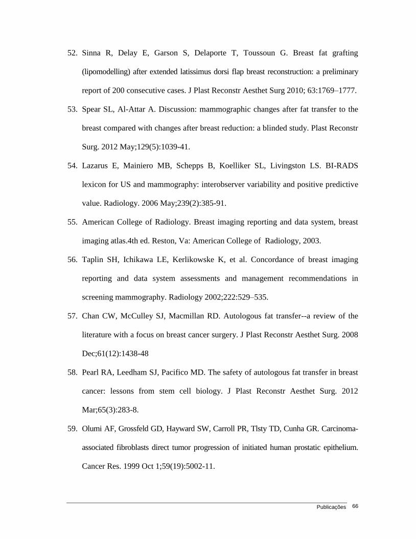
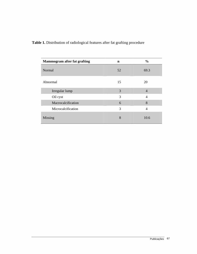
Table 2. Radiological features of the 15 abnormal mammograms after fat
grafting procedure
Mammographic features n % Biopsy LR*
Oil cyst 3 20 No 0
Irregular lump 3 20 Yes 2
Macrocalcification 6 40 No 0
Microcalcification 3 20 Yes 0
*LR: Local Recurrence
Page 47
Publicações 46
P atholog ic al S tag e
0
5
10
15
20
25
30
35
40
S tage 0 S tage I S tage IIA S tage IIB S tage IIIA Unknown
Figure 1. Distribution of pathological breast cancer stage of the 59 patients.
Page 48
Publicações 47
Figure 2. Progression free survival from fat grafting procedure.
Page 49
Publicações 48
3.2. Artigo 2
Radiological Features of Autologous Breast Fat Grafting After Conservative
Treatment: Results of a Prospective Study
Fabricio P. Brenelli MD.*, Mario Rietjens MD.**, Aarão M. Pinto-Neto PhD.*
Francesca De Lorenzi PhD.**, Fabio Rossetto**, Daniel Barbalho MD
* State University of Campinas (Unicamp), Department of Gynecology and Obstetrics –
Breast Oncology Division, Campinas – São Paulo, Brazil
** European Institute of Oncology, Division of Plastic and Reconstructive Surgery,
Milan, Italy
Running head: Radiological Features of Breast Fat Grafting
Corresponding authors:
Fabricio Brenelli
R Elvino Silva 30, Campinas – SP, Brazil
Zip: 13092-559. Email: [email protected]
Commercial and Financial Disclosure
Authors declares having neither conflict of interest nor funding support for publishing
this paper.
Page 50
Publicações 49
Abstract
Background: Autologous fat graft to the breast is a useful tool to correct defects after breast
conservative treatment (BCT). Although this procedure gains popularity, little is known about
the interaction between the fat graft and the prior oncological environment. Since fat is
injected into the breast, it can potentially produce radiological features that could increase
numbers of unnecessary biopsies or even mask suspicious hidden lesions in patients with
prior BCT, who are at a higher risk of local recurrence (LR). Materials and Methods: Fifty
nine patients, with prior BCT, underwent 75 autologous fat graft procedures using the
Coleman´s technique, between October 2005 and July 2008. Radiological and clinical
examination was performed in all cases before the procedure. Follow up was made by
clinical and radiological examination. Results: Mean follow up was 34.4 ±15.3 months. Mean
time from oncological surgery to the first fat grafting procedure was 76.6± 30.9 months.
Abnormal breast images were present in 20% of the post-operative mammograms and
consisted mostly of non-suspicious lesions (60%). Lesions warranting biopsies were
microcalcification (3 cases) and irregular lumps (3 cases). Two cases of irregular lump
resulted in local recurrence (LR). Conclusion: Autologous fat grafting is a useful tool
to correct breast defects. Although it increases the rate of abnormal mammographic
findings, as it happens with other types of breast surgery, it was not associated to
unnecessary biopsies. Despite the two LR were irregular lumps, it was not possible to
identify a specific radiological pattern of LR associated with this procedure.
Level of evidence
Study: Therapeutics study
Level of evidence: Level IV
Page 51
Publicações 50
Introduction
Breast conservative treatment (BCT) is a standard of care for early-stage breast
cancer. Its purpose is to offer women local treatment as effective as mastectomy, but with
better cosmetic outcomes (1, 2). Since the publications of the classics randomized trials
(Milan trial and NSABP-B06 trial) an increasing number of women have sought BCT
(3-5) and its rate varies from 10 to 67% in different U.S centers (6-9). Despite the high
level of patient satisfaction reported (75% to 96%), severe asymmetry is observed in
almost 30% of these studies (10).
Correction of breast asymmetry after BCT can be very challenging, especially in
type 2 and 3 cosmetic sequelae, as described by Clough KB et al. (11). Autologous fat
graft seems to be a good alternative for BCT patients, filling defects and improving
cosmetic outcome (12-14).
However, fat transfer to the breast can produce abnormal breast imaging related to fat
reabsorption, inflammation and necrosis (15-16). In 1987, the American Society of Plastic and
Reconstructive Surgeons (ASPRS) set up an ad hoc committee to evaluate new
procedures contemplating fat transfer to the breast. The committee was “unanimous in
deploring the use of fat injection for breast augmentation because it can inhibit early detection
of breast cancer and is therefore hazardous to public health” (17-18). Fortunately, several
subsequent studies mainly based on case reports have disagreed with that statement. There is
indeed an association between autologous fat grafting and abnormal breast images.
However, those features do not appear to mask hidden breast lesions. Breast images resulting
from this procedure are mostly oil cysts and macrocalcifications (13-14, 18-19).
Therefore, in 2009 the American Society of Plastic Surgery (ASPS) set up a task
force to assess the indications, safety and efficacy of autologous fat transfer. The task
Page 52
Publicações 51
force concluded that knowledge about the procedure was mainly acquired from case series
and expert opinion, considered a low grade of scientific evidence. Based on current scientific
evidence, the task force concluded that although fat grafting can potentially interfere
with breast cancer detection, there is no evidence suggesting that it really happens (20).
Most studies are focused on fat grafting in the healthy breast or after mastectomy and
reconstruction. Few studies have been dedicated to assess the impact of fat grafting on BCT
patients. Hypothetically, the transfer of adipose derived stem cells (ADSC) or Adipose
derived mesenchymal stem cell (ADMSC) could induce reproduction of silent tumor cells,
predisposing to local recurrence (LR). ). “In vitro” and animal models basic researches with
ADSC and breast cancer environment are conflictive, as many shows positive association and
some negative association with tumor cell proliferation (21-25).
Therefore, it is crucial to understand the role of autologous fat graft in this group of
patients. It is important to identify whether this procedure increases the risk of masking
hidden lesions that are actually LRs, or results in features mimicking suspicious lesions and
leading to higher rates of unnecessary biopsies. In this scenario, we present a prospective
evaluation of 59 patients with prior BCT undergoing 75 autologous breast fat graft procedure
followed with clinical and imaging examination to help clarify this important issue.
Material and methods
Patient selection
From October 2005 to July 2008, we prospectively evaluated 59 patients undergoing
75 breast fat grafting procedures at the European Institute of Oncology (Milan, Italy).
All patients had undergone previous BCT for oncological reasons, leading to a cosmetic
breast defect. A single surgeon visited all patients and indicated the procedure. Only
Page 53
Publicações 52
patients free from locorregional breast disease were considered eligible for the procedure
after accurate clinical and instrumental breast evaluation. The presence of stable bone
metastasis was not a criterion for exclusion.
Preoperative evaluation
All patients scheduled for the fat grafting procedure were evaluated preoperatively
with clinical and breast imaging tests. Mammogram was mandatory for all patients and
breast ultrasound (U.S.) ordered eventually and all exams were evaluated by a senior
radiologist. After receiving an explanation about the procedure and signing an informed
consent term, all patients agreed to undergo surgery.
Surgical technique
The procedure was performed under local or general anesthesia, depending on the
patient’s clinical conditions and risks. Local anesthesia was preferable. General anesthesia
was recommended when a large amount of fat tissue needed to be harvested, an
associated procedure was indicated (prosthesis exchange and capsulotomy, for instance),
or the patient was extremely anxious.
The whole procedure of fat harvesting and grafting was performed, according to
the Coleman technique (12) with minimal modifications.
Follow-up
Follow-up visits with clinical evaluation occurred in the outpatient clinic, at least
once after 6 months following the procedure. Mammograms were ordered annually, in
association with breast ultrasound (US) when required. Mammograms and US were
Page 54
Publicações 53
considered normal if there was no suspicious image, and abnormal if there was groups of
calcifications, calcified cysts, or any other image that was not present in a previous
examination. Magnetic Resonance Imaging (MRI) was not used in any of the cases
because it was judged inapplicable by the clinician an radiologist.
Results
The mean age of the women at the time of fat grafting was 50±8.5 years and the mean
follow-up period was 34.4 ±15.3 months. More than 75% of these women had follow-up
periods greater than 45.9 months. The procedure was conducted under local anesthesia in 58
cases (77.3%) and under general anesthesia in 17 cases (22.7%). Ninety eight percent of the
defects were located exclusively in the breast parenchyma. One patient also had a defect
in the axilla. The right and left breast were equally affected (50.7 vs. 48%, respectively).
Most patients (83.2%) underwent only one surgical procedure. Only 16.8% of the
patients required more than one session to achieve results. Immediate complications
were observed in 3 cases (4%): liponecrosis in 2 patients and cellulitis in 1 patient. All
cases were managed clinically with no further complications.
Most patients (60%) had Stage 0 or I tumors. Thirty-five patients (59.3%) had
undergone quadrantectomy with axillary dissection (QUART), and 14 patients (23.7%) had
undergone quadrantectomy with sentinel node biopsy (SNB). Only 10 patients (16.9%)
underwent quadrantectomy alone. Oncoplastic techniques were used in only 9 patients
(15.3%). External radiation therapy was administered to 56 patients (94.9%). The mean time
period from oncological surgery to the first fat grafting procedure was 76.6± 30.9 months.
Follow-up visits took place at least once after 6 months since the procedure.
Mammograms and or breast US, if necessary, were ordered. In 15 cases (20%), the
Page 55
Publicações 54
mammogram was normal before the procedure and abnormal afterwards. In 8 cases
(10.6%), information about the postoperative mammogram was missing (table 1). Patterns of
abnormal breast images are described in table 1 and 2.
Of the 75 procedures performed, images were considered suspicious in 6 cases
(8%) and all were biopsied, corresponding to 40% of the abnormal images. Of these 6
cases, 2 resulted positive (Table 3).
During the follow-up period, there were 4 cases of ipsilateral breast recurrence.
However, LR was suspected in one case on the day of the procedure and was confirmed
a week later by histology. Therefore, we did not associate this with the fat grafting
procedure, finding only 3 cases of true local recurrence (4%).
Local recurrence was diagnosed by breast imaging in 2 cases and clinically in
one case. All three patients were treated with total mastectomy. It was also noteworthy
that more than 75% of the patients had a follow-up period longer than 45.9 months.
Therefore, the LR rate may be lower than 1% per year.
Three patients were metastatic (bone metastasis) before the procedure. During
the follow-up period, only one patient had disease progression with lung and liver
metastasis, but was still alive at the time of the last consultation.
Discussion
Autologous fat graft is a useful and promising tool to help breast and plastic
surgeons in the correction of defects after BCT. The whole procedure is simple and
reproducible by trained surgeons. Most cases can be performed under local anesthesia in a
day-hospital regimen, reducing surgical costs, as reported in other series (29).
Complication rates are about 4% and are simple to manage. Many studies confirm this
Page 56
Publicações 55
low rate of postoperative complications, making autologous fat graft a popular choice
for surgeons and patients (26-27).
Efficacy of the procedure is unpredictable. Despite our experience, as well as
other reports from the literature (28-32), it is still very difficult to predict whether a fat
graft will attach or not. In this study, a subsequent fat graft procedure was performed in
only16.8% of the cases, meaning that good results were achieved in most cases. This
number, however, is probably underestimate. There are patients under follow-up who
have already been scheduled for a new procedure and others who still exhibit a breast
defect but refuse to undergo another procedure.
We found no correlation between autologous fat graft and the risk of ipsilateral
breast recurrence (IBR) or distant recurrence. Although there was not a comparison
group, our result is in agreement with many other authors who published data on
oncological aspects of fat grafting (15, 18, 25, 28-32). However, most papers addressed
the procedure after mastectomy and immediate reconstruction and few studies have
evaluated the safety after BCT. Therefore, our series contributes to enlarge the evidence
that the procedure may be safe in selected group of patients with prior BCT.
Concerning radiological findings after breast fat graft, it is worth mentioning that
this procedure can cause inflammation, fat reabsorption and fat necrosis, leading to breast
imaging of oil cysts, scarring, macro and microcalcification and nodules or architectural
distortion (13, 18, 33). In our series, lesion morphology was mainly composed of oil cyst
and macrocalcification (60%). Microcalcification and nodular lesion corresponded to
40% of the findings (6 cases).
Fat necrosis along with its radiological features occur in every type of breast
surgery, such as: breast biopsy (33), implant augmentation (34-37), reduction mammoplasty
Page 57
Publicações 56
(33,38-40) and breast reconstruction (41-43). Therefore it is not different with breast fat
grafting. In fact, this technique was thought to promote more radiological findings than
any other breast intervention, since fat is injected directly into the breast with no
vascular pedicle, taking between 7 to 21 days to the graft process be completed (44-46).
However, most of the evidence with fat grafting to breast shows that the rate of post-
operative radiological findings are not higher than those found in other types of breast
intervention (12-16, 18-20, 28, 31,32, 47-49).
Our results indicated abnormal mammographic changes in 20% of cases. These
results are currently in agreement with those reported by other series (47-49) and with
data from a recent systematic literature review on the safety of autologous fat grafting to
the breast. This systematic review estimated radiological changes in 13%, ranging from
zero to 82% (31), showing the heterogeneity of the results from the studies.
Recently, Rubin et al (50) published a blind, multicenter, retrospective cohort study of
a group of 27 healthy women undergoing fat grafting for breast augmentation matched
to a group of 23 women undergoing reduction mammoplasty. The rate of radiological
abnormalities was similar in both groups, except for an increased finding of tissue scarring
(85% Vs 17%) and architectural distortion (13% Vs 3%) in the mammoplasty group.
These post-operative radiological features seem to pose minimal risks of interfering
with LR detection and delaying breast cancer diagnosis. Although the ASPRS raised
awareness to the problem in 1987 (17), further investigations showed no evidence of this
interference (16, 18, 31, 46-48). Therefore, in the 2009 ASPS task force stated that there
is no evidence of autologous fat graft interfering in mammographic screening results, but
it should be avoided in high risk patients (20).
Page 58
Publicações 57
Evidence regarding radiological abnormalities after autologous fat graft to the
breast is based mostly upon its use in healthy breast. Larger series based on oncological
patients mix women who underwent breast reconstruction postmastectomy with those who
underwent BCT (28-30, 32, 51, 52). In a large series, Delay et al (28) found postoperative
radiological abnormalities in 20% of the cases when focusing on the BCT group. Gosset
et al (19) found 19% of microcalcification after fat grafting in 21 conserved breasts. We
found 20% of breast abnormalities in mammography and ultrasound, showing that the
radiological features after breast fat grafting in the BCT are no different from those
found in after procedure in the healthy breast.
The risk that these radiological features due to fat transfer may potentially mask
hidden suspicious lesions, delay LR diagnosis, or increase the number of unnecessary
biopsies is of great concern to medical societies. As a matter of fact, of the 15 breast
abnormalities in our study, 6 (40%) were later biopsied and two of them (33.3%) resulted
positive. Since we had no comparison group, we were unable to conclude that biopsies
were performed at a higher rate than expected. However, biopsy indications seemed very
precise as 33.3% (2 out of 6) of the suspected lesions were positive. These results are in
accordance with the positive predictive value of the American College of Radiology
(ACR) for suspicious lesions, classified as BIRADS 4 (54-56).
Interestingly, Rubin (50) again found in his blinded study that microcalcifications
warranting additional biopsies were 3 times higher in the fat grafting group (10 cases,
4,6%) than in the reduction mammoplasty group (3 cases, 1,6%), although it was not of
statistics significance (p = 0,16) as observed by Spear (53). Illouz (32) in his large series
of 820 patients undergoing breast fat grafting, in which 670 patients underwent
mammography, he found no lesions categorized as BIRADS 4 or 5, even among the 30
Page 59
Publicações 58
patients with previous BCT. Therefore it seems unlike that fat graft could increase
significantly the rates of unnecessary breast biopsies.
In our study, we found 3 LR on follow-up. Of these, one was clinically diagnosed
by patient´s own palpation and 2 were mammographically diagnosed. Both lesions were
irregular lumps. Little information exists on LR following autologous fat graft after
BCT. Therefore, there is no specific radiological pattern of it. Based on our observation,
we suggest that suspicious lumps after autologous fat graft should be biopsied to rule out
LR. However, it is evident that all suspicious lesions, even if they are not nodules,
should be biopsied. Figures 1 to 4 show radiological features found after autologous fat
graft in patients and BCT.
An issue that must be debated is why do these mammographic alterations, specially
the microcalcifications, occur in the breast parenchyma? As performed in this study,
many author´s consider that good practice of this technique consists to graft the fat tissue
in the subcutaneous or subglandular space (12, 14, 16, 18-20, 28-32, 57), therefore
radiological lesions should be detected outside the breast parenchyma.
There may be an interaction between the fat injected into the subcutaneous tissue
or into the subglandular space with the local environment, promoting its migration to the
breast parenchyma. Pearl et al (58) published a recent review on stem cell biology and its
behavior in the breast environment. It seems that ADMSC interacts with adjacent stroma and
potentially promotes proliferation of fibroblasts and other cell lines promoting LR, as
occurs in prostate cancer with the “fibroblast associated carcinoma” (58, 59). Theoretically,
there could be migration of mature adipocytes to the breast parenchyma induced by
ADSC, or an indirect induction of cell proliferation of the stroma, leading to breast
Page 60
Publicações 59
tissue scarring, which could contribute to the formation of these radiological features.
Those unanswered questions still need to be addressed by new prospective studies.
Conclusion
Despite this fact, strong scientific evidence is lacking to prove its efficacy and
safety for patients. Special attention should be devoted to patients with prior BCT, who
are at higher risk for LR. Although fat graft does not appear to increase LR risk or
interfere with LR detection, it is actually associated with increased findings of abnormal
radiological features. These findings are no different from features found after other
breast invasive procedures and from a radiological point of view, suspicious and benign
lesions are easily diagnosed by a trained and well-equipped radiologist. However,
surgeons, radiologists and clinicians must be aware of suspicious findings and proceed
with further investigation.
Autologous fat graft to the breast is no longer a futuristic technique. It is becoming a
daily tool used by breast and plastic surgeons in aesthetical and reconstructive surgery.
Despite the good oncological outcomes from this study, it is based on selected
cases and cannot be extrapolated to every patient with prior BCT. There is much to learn
about ADSC behavior and its interaction with a previous “cancerous” environment.
Further prospective studies with control groups and multicenter prospective data should
be collected to understand the radiological presentations of this technique over time.
Page 61
Publicações 60
References
1. Veronesi U, Cascinelli N, Mariani L, et al. Twenty-year follow-up of a randomized
study comparing breast-conserving surgery with radical mastectomy for early breast
cancer. N Engl J Med. 2002 Oct 17; 347(16):1227-32.
2. Fisher B, Anderson S, Bryant J, et al. Twenty-year follow-up of a randomized trial
comparing total mastectomy, lumpectomy, and lumpectomy plus irradiation for the
treatment of invasive breast cancer. N Engl J Med. 2002 Oct 17; 347(16):1233-41.
3. Veronesi U, Saccozzi R, Del Vecchio M, et al. Comparing radical mastectomy with
quadrantectomy, axillary dissection, and radiotherapy in patients with small cancers
of the breast. N Engl J Med. 1981 Jul 2;305(1):6-11.
4. Fisher B, Bauer M, Margolese R, et al. Five-year results of a randomized clinical trial
comparing total mastectomy and segmental mastectomy with or without radiation in the
treatment of breast cancer. N Engl J Med. 1985 Mar 14; 312(11):665-73.
5. Wang HT, Barone CM, Steigelman MB, et al. Aesthetic outcomes in breast conservation
therapy. Aesthet Surg J. 2008 Mar-Apr;28(2):165-70.
6. Kotwall CA, Covington DL, Rutledge R, et al. Patient, hospital, and surgeon factors
associated with breast conservation surgery: a statewide analysis in North Carolina.
Ann Surg 1996;224:419–429
7. Parviz MA, Cassel JB, Kaplan BJ, et al. Breast conservation therapy rates are no
different in medically indigent versus insured patients with early stage breast
cancer. J Surg Oncol 2004;84:57–62.
8. Kelemen JJ III, Poulton T. Surgical treatment of early-stage breast cancer in the
Department of Defense healthcare system. J Am Coll Surg 2001; 192:293–297.
Page 62
Publicações 61
9. McGuire KP, Santillan AA, Kaur P, et al. Are mastectomies on the rise? A 13-year
trend analysis of the selection of mastectomy versus breast conservation therapy in
5865 patients. Ann Surg Oncol. 2009 Oct; 16(10):2682-90.
10. Bajaj AK, Kon PS, Oberg KC, Miles DAG. Aesthetic outcomes in patients
undergoing breast conservation therapy for the treatment of localized breast cancer.
Plast Reconstr Surg 2004; 14:1442–1449.
11. Clough KB, Thomas SS, Fitoussi AD, et al. Reconstruction after conservative treatment
for breast cancer: cosmetic sequelae classification revisited. Plast Reconstr Surg.
2004 Dec; 114(7):1743-53.
12. Coleman SR, Saboeiro AP. Fat grafting to the breast revisited: safety and efficacy.
Plast Reconstr Surg. 2007 Mar;119(3):775-85
13. Gosset J, Flageul G, Toussoun G, et al. Lipomodelling for correction of breast
conservative treatment sequelae. Medicolegal aspects. Expert opinion on five
problematic clinical cases. Ann Chir Plast Esthet. 2008 Apr; 53(2):190-8.
14. Delay E, Gosset J, Toussoun G, et al. Efficacy of lipomodelling for the management
of sequelae of breast cancer conservative treatment. Ann Chir Plast Esthet. 2008
Apr; 53(2):153-68.
15. Niechaj I, Sevcuk O. Long-term results of fat transplantation: clinical and histologic
studies. Plast Reconstr Surg. 1994;94:496–498.
16. Pulagam SR, Poulton T, Mamounas EP. Long-term clinical and radiologic results
with autologous fat transplantation for breast augmentation: case reports and review
of the literature. Breast J. 2006 Jan-Feb;12(1):63-5.
17. ASPRS Ad-Hoc Committee on New Procedures (1987) Report on autologous fat
transplantation. Plast Surg Nurs 7:140–141
Page 63
Publicações 62
18. Rosing JH, Wong G, Wong MS, Sahar D, Stevenson TR, Pu LL. Autologous fat
grafting for primary breast augmentation: a systematic review. Aesthetic Plast Surg.
2011 Oct;35(5):882-90. Epub 2011 Apr 1. Review.
19. Gosset J, Guerin N, Toussoun G, et al. Radiological evaluation after lipomodelling
for correction of breast conservative treatment sequelae. Ann Chir Plast Esthet.
2008 Apr; 53(2):178-89.
20. Gutowski KA; ASPS Fat Graft Task Force. Current applications and safety of
autologous fat grafts: a report of the ASPS fat graft task force. Plast Reconstr Surg.
2009 Jul; 124(1):272-80.
21. Schaffler A, Scholmerich J, Buechler C. Mechanisms of disease: adipokines and
breast cancer - endocrine and paracrine mechanisms that connect adiposity and
breast cancer. Nature Clin Pract Endocrinol Metab 2007; 3:345-54.
22. Dieudonne MN, Machinal-Quelin F, Serazin-Leroy V, et al. Leptin mediates a
proliferative response in human MCF7 breast cancer cells. Biochem Biophys Res
Commun. 2002 Apr 26; 293(1):622-8.
23. Dieudonne MN, Bussiere M, Dos Santos E, et al. Adiponectin mediates antiproliferative
and apoptotic responses in human MCF7 breast cancer cells. Biochem Biophys Res
Commun. 2006 Jun 23; 345(1):271-9.
24. Iyengar P, Espina V, Williams TW, et al. Adipocyte-derived collagen VI affects
early mammary tumor progression in vivo, demonstrating a critical interaction in
the tumor/stroma microenvironment. J Clin Invest. 2005 May; 115(5):1163-76.
25. Lohsiriwat V, Curigliano G, Rietjens M, Goldhirsch A, Petit JY. Autologous fat
transplantation in patients with breast cancer: "silencing" or "fueling" cancer recurrence?
Breast. 2011 Aug;20(4):351-7. Epub 2011 Feb 5.
Page 64
Publicações 63
26. ELFadl D, Garimella V, Mahapatra TK,et al. Lipomodelling of the breast: a review.
Breast. 2010 Jun; 19(3):202-9.
27. Mizuno H, Hyakusoku H. - Fat grafting to the breast and adipose-derived stem cells:
recent scientific consensus and controversy. Aesthet Surg J. 2010 May-Jun;
30(3):381-7. Review.
28. Delay E, Garson S, Tousson G, Sinna R. Fat injection to the breast: technique,
results, and indications based on 880 procedures over 10 years. Aesthet Surg J. 2009
Sep-Oct; 29(5):360-76.
29. Rietjens M, De Lorenzi F, Rossetto F, Brenelli F, et al. Safety of fat grafting in
secondary breast reconstruction after cancer. J Plast Reconstr Aesthet Surg. 2011
Apr; 64(4):477-83.
30. Petit JY, Lohsiriwat V, Clough KB, Sarfati I, Ihrai T, Rietjens M, Veronesi P, Rossetto F,
Scevola A, Delay E. The oncologic outcome and immediate surgical complications of
lipofilling in breast cancer patients: a multicenter study--Milan-Paris-Lyon experience of
646 lipofilling procedures. Plast Reconstr Surg. 2011 Aug;128(2):341-6.
31. Claro F Jr, Figueiredo JC, Zampar AG, Pinto-Neto AM. Applicability and safety of
autologous fat for reconstruction of the breast. Br J Surg. 2012 Jun;99(6):768-80.
doi: 10.1002/bjs.8722. Epub 2012 Apr 4.
32. Illouz YG, Sterodimas A. Autologous fat transplantation to the breast: a personal
technique with 25 years of experience. Aesthetic Plast Surg 2009; 33(5):706–715.
33. Chala LF, de Barros N, de Camargo Moraes P, Endo E, Kim SJ, Pincerato KM,
Carvalho FM, Cerri GG. Fat necrosis of the breast: mammographic, sonographic,
computed tomography, and magnetic resonance imaging findings. Curr Probl Diagn
Radiol. 2004 May-Jun;33(3):106-26. Review.
Page 65
Publicações 64
34. Handel N, Jensen JA, Black Q, Waisman JR, Silverstein MJ. The fate of breast
implants: a critical analysis of complications and outcomes. Plast Reconstr Surg.
1995 Dec;96(7):1521-33.
35. Piccoli CW. Imaging of the patient with silicone gel breast implants. Magn Reson
Imaging Clin N Am. 2001 May;9(2):393-408, vii-viii. Review.
36. Raso DS, Greene WB, Kalasinsky VF, Riopel MA, Luke JL, Askin FB, Silverman
JF, Young VL. Elemental analysis and clinical implications of calcification deposits
associated with silicone breast implants. Ann Plast Surg. 1999 Feb;42(2):117-23.
37. Reynolds A. The augmented breast. Radiol Technol. 2009 Jan-Feb;80(3):241M-259M.
38. Brown FE, Sargent SK, Cohen SR, Morain WD. Mammographic changes following
reduction mammaplasty. Plast Reconstr Surg. 1987 Nov;80(5):691-8.
39. Danikas D, Theodorou SJ, Kokkalis G, Vasiou K, Kyriakopoulou K. Mammographic
findings following reduction mammoplasty. Aesthetic Plast Surg. 2001 Jul-
Aug;25(4):283-5.
40. Mandrekas AD, Assimakopoulos GI, Mastorakos DP, Pantzalis K. Fat necrosis
following breast reduction. Br J Plast Surg. 1994 Dec;47(8):560-2.
41. Kim SM, Park JM. Mammographic and ultrasonographic features after autogenous
myocutaneous flap reconstruction mammoplasty. J Ultrasound Med. 2004
Feb;23(2):275-82.
42. Leibman AJ, Styblo TM, Bostwick J 3rd. Mammography of the post reconstruction
breast. Plast Reconstr Surg. 1997 Mar;99(3):698-704.
43. Sim YT, Litherland JC. The use of imaging in patients post breast reconstruction.
Clin Radiol. 2012 Feb;67(2):128-33.
Page 66
Publicações 65
44. Langer S, Sinitsina I, Biberthaler P, Krombach F, Messmer K. Revascularization of
transplanted adipose tissue: a study in the dorsal skinfold chamber of hamsters. Ann
Plast Surg. 2002 Jan;48(1):53-9.
45. Rieck B, Schlaak S. Measurement in vivo of the survival rate in autologous
adipocyte transplantation. Plast Reconstr Surg. 2003 Jun;111(7):2315-23.
46. Locke MB, de Chalain TM. Current practice in autologous fat transplantation:
suggested clinical guidelines based on a review of recent literature. Ann Plast Surg.
2008 Jan;60(1):98-102. Review.
47. Khouri RK, Eisenmann-Klein M, Cardoso E, Cooley BC, Kacher D, Gombos E, Baker
TJ. Brava and autologous fat transfer is a safe and effective breast augmentation
alternative: results of a 6-year, 81-patient, prospective multicenter study. Plast
Reconstr Surg. 2012 May;129(5):1173-87.
48. Veber M, Tourasse C, Toussoun G, Moutran M, Mojallal A, Delay E. Radiographic
findings after breast augmentation by autologous fat transfer. Plast Reconstr Surg.
2011 Mar;127(3):1289-99.
49. Carvajal J, Patiño JH. Mammographic findings after breast augmentation with
autologous fat injection. Aesthet Surg J. 2008 Mar-Apr;28(2):153-62.
50. Rubin JP, Coon D, Zuley M, Toy J, Asano Y, Kurita M, Aoi N, Harii K, Yoshimura K.
Mammographic changes after fat transfer to the breast compared with changes after
breast reduction: a blinded study. Plast Reconstr Surg. 2012 May;129(5):1029-38.
51. Brenelli F, Rietjens M, De Lorenzi F, Rossetto F. Autologous fat graft after breast
cancer: Is it safe? – A single surgeon experience with 194 procedures. Eur J Plast
Surg 2010; 8(Suppl): 141–142.
Page 67
Publicações 66
52. Sinna R, Delay E, Garson S, Delaporte T, Toussoun G. Breast fat grafting
(lipomodelling) after extended latissimus dorsi flap breast reconstruction: a preliminary
report of 200 consecutive cases. J Plast Reconstr Aesthet Surg 2010; 63:1769–1777.
53. Spear SL, Al-Attar A. Discussion: mammographic changes after fat transfer to the
breast compared with changes after breast reduction: a blinded study. Plast Reconstr
Surg. 2012 May;129(5):1039-41.
54. Lazarus E, Mainiero MB, Schepps B, Koelliker SL, Livingston LS. BI-RADS
lexicon for US and mammography: interobserver variability and positive predictive
value. Radiology. 2006 May;239(2):385-91.
55. American College of Radiology. Breast imaging reporting and data system, breast
imaging atlas.4th ed. Reston, Va: American College of Radiology, 2003.
56. Taplin SH, Ichikawa LE, Kerlikowske K, et al. Concordance of breast imaging
reporting and data system assessments and management recommendations in
screening mammography. Radiology 2002;222:529–535.
57. Chan CW, McCulley SJ, Macmillan RD. Autologous fat transfer--a review of the
literature with a focus on breast cancer surgery. J Plast Reconstr Aesthet Surg. 2008
Dec;61(12):1438-48
58. Pearl RA, Leedham SJ, Pacifico MD. The safety of autologous fat transfer in breast
cancer: lessons from stem cell biology. J Plast Reconstr Aesthet Surg. 2012
Mar;65(3):283-8.
59. Olumi AF, Grossfeld GD, Hayward SW, Carroll PR, Tlsty TD, Cunha GR. Carcinoma-
associated fibroblasts direct tumor progression of initiated human prostatic epithelium.
Cancer Res. 1999 Oct 1;59(19):5002-11.
Page 68
Publicações 67
Table 1. Distribution of radiological features after fat grafting procedure
Mammogram after fat grafting n %
Normal 52 69.3
Abnormal 15 20
Irregular lump 3 4
Oil cyst 3 4
Macrocalcification 6 8
Microcalcification 3 4
Missing 8 10.6
Page 69
Publicações 68
Table 2. Description of abnormal mammographic findings
Mammogram findings n %
Irregular lump 3 20
Oil cyst 3 20
Macrocalcification 6 40
Microcalcification 3 20
Page 70
Publicações 69
Table 3. Correlation between mammographic suspicious lesions warranting biopsies and
pathology
Lesion Biopsy Positive result
Microcalcification 3 0
Irregular lump 3 2
Page 71
Publicações 70
Figure 1. Benign calcification due to fat necrosis after autologous fat grafting.
Page 72
Publicações 71
Figure 2. Mammographic macrocalcification after fat grafting.
Page 73
Publicações 72
Figure 3. Ecographic image of figure 2. Characteristic image of fat necrosis.
Page 74
Publicações 73
Figure 4. Suspicious nodular lesion that resulted in invasive carcinoma after autologous
fat grafting.
Page 75
Discussão 74
4. Discussão
O enxerto autólogo de gordura é uma das mais promissoras técnicas
cirúrgicas para correção de defeitos mamários decorrentes da CCM. O procedimento
todo é relativamente simples e, na maioria das vezes, pode ser realizado sob
anestesia local (77% dos casos) em regime de hospital-dia, diminuindo o tempo de
internação e consequentemente os custos. Além disso, as taxas de complicações
pós-operatórias são pequenas. No presente estudo foram encontrados apenas 3
casos (4%) de complicações, que foram tratados ambulatorialmente com drenagem
espontânea da necrose gordurosa em 2 casos, e com antibiótico em 1, que apresentou
celulite. Tais achados estão de acordo com publicações recentes (80-82), e devido a
estas características de praticidade, baixo custo e poucas complicações, têm-se
tornado muito populares entre os cirurgiões plásticos e mamários em todo o mundo.
Infelizmente, a eficácia do método deve ainda ser estabelecida. Apesar
da experiência obtida com este estudo, que aponta para bons resultados, e
dados da literatura que apontam para uma taxa de reabsorção do enxerto por
volta de 30% (45, 69, 70, 72, 80-83), ainda é difícil estabelecer um padrão de
eficácia. No presente estudo, apenas 16% das pacientes necessitaram de mais de
Page 76
Discussão 75
um procedimento, o que faria supor que no restante das pacientes o procedimento
foi um sucesso. Entretanto, dados objetivos de avaliação, assim como dados
subjetivos de avaliação da própria paciente não foram estudados, sendo impossível,
então, chegar a uma conclusão.
Um dos objetivos principais deste estudo foi avaliar as alterações
mamográficas causadas pelo enxerto de gordura na mama e suas potencias
implicações diagnósticas e de seguimento. Sabidamente, este procedimento pode
causar inflamação, necrose e reabsorção da gordura, levando a alterações
mamográficas como cisto oleoso, macro e microcalcificações, cicatriz radiada,
distorção arquitetural e lesões nodulares (45, 48, 76). No presente estudo não foi
diferente e 60% das lesões encontradas eram cistos oleosos ou macrocalcificações.
Microcalcificações e lesões nodulares corresponderam a 40% dos achados (6 casos).
Alterações mamográficas relacionadas à necrose gordurosa ocorrem em
todo o tipo de intervenção mamária como: biópsia percutânea (80), mamoplastia de
aumento com prótese (84-87), mamoplastia redutora (75-77) e reconstrução
mamária (88-90). Com o enxerto de gordura não é diferente. Na verdade,
imaginava-se que este procedimento produziria mais alterações mamográficas
que os outros, uma vez que a gordura é injetada diretamente na mama sem um
pedículo vascular para alimentá-la, o que poderia provocar a necrose do tecido
enxertado entre 7 e 21 dias (91-93), e sua consequente expressão radiológica.
Entretanto, na maioria dos estudos os índices de anormalidades radiológicas após a
lipoenxertia não são maiores que os encontrados nos outros tipos de cirurgia.
Page 77
Discussão 76
No presente estudo foram encontrados 20% de alterações mamográficas
pós-lipoenxertia, cujo resultado está de acordo com dados de outros estudos (79,
94, 95) e também com os de uma recente revisão sistemática que mostrou alterações
radiológicas variando de 13% a 82% (82), mostrando a extrema heterogeneidade dos
dados publicados. Recentemente, Rubin et al. (96) publicaram um estudo
multicêntrico comparando mamografias de mulheres que fizeram mamoplastia
redutora versus enxerto autólogo de gordura para aumento de mama. Como
esperado, os achados radiológicos pós-operatórios foram semelhantes nos dois
grupos, com exceção de cicatriz mamária e distorção arquitetural serem mais
frequentes no grupo da mamoplastia.
Dessa maneira, a preocupação inicial da ASPRS (74) e outros autores de
que o enxerto mamário de gordura provocaria lesões mamográficas que
interfeririam no seguimento das pacientes aparentemente não procede. Tanto é,
que na força-tarefa da ASPS em 2009 (54), tal questionamento foi minimizado,
mas mantido para pacientes de alto risco.
Entretanto, o problema a ser discutido sobre estas alterações radiológicas
reside no fato de que a maioria dos dados destes estudos deriva de pacientes
com mamas “saudáveis“ ou pós-mastectomizadas e reconstruídas, e há apenas
poucos casos pós-CCM, que teoricamente estão sob maior risco de uma RL. Seria
de se esperar, portanto, que neste grupo de pacientes os achados mamográficos
pós-lipoenxertia pudessem levar a um número maior de biópsias desnecessárias.
No presente estudo, 15 pacientes apresentaram alterações mamográficas
pós-lipoenxertia (20%), sendo que em 6 casos (40%) tais lesões foram consideradas
Page 78
Discussão 77
suspeitas e biopsiadas, e 2 casos resultaram positivo para RL. Apesar de não
existir um grupo-controle, foi considerado que não houve um aumento de biópsias
desnecessárias, uma vez que 2 casos positivos entre 6 biopsiados está dentro
daquilo que se espera para lesões moderadamente suspeitas, segundo a
classificação BIRADS do American College of Radiology (ACR) (97-99).
Por outro lado, Rubin et al. (96), em seu estudo multicêntrico, observaram
que o número de microcalcificações que necessitavam biópsia foi 3 vezes maior
no grupo de enxerto autólogo (10 casos, 4,6%), quando comparado ao grupo de
mamoplastia (3 casos, 1,6%). Apesar disso, este resultado não foi significativo
(p = 0,16). Outras séries de casos que incluíram pacientes com antecedente de
CCM, os dados sobre achados radiológicos parecem não estar relacionados com o
aumento no número de biópsias desnecessárias. Illouz et al. (83), em sua série de
820 casos, dos quais 670 forma submetidos a controles mamográficos, incluindo
30 pacientes com antecedente de CCM, não acharam um único caso de lesões
classificadas como BIRADS 4 ou 5.
Desta maneira, existem poucos dados para que seja possível identificar
um padrão de lesão radiológica pós-enxerto de gordura autóloga que pudesse
estar correlacionado com maior ou menor chance de encontrar um RL. Os dados
deste estudo mostraram que das 3 RL, 2 foram diagnosticadas através de
mamografia e 1 de exame clínico. As duas lesões radiológicas eram nódulos
sólidos irregulares e todas as microcalcificações resultaram negativas para RL.
Portanto, lesões nodulares irregulares devem ser sempre biopsiadas. Entretanto,
isto obviamente não isenta a biópsia em outros tipos de lesões suspeitas, pois como
Page 79
Discussão 78
estes números são pequenos e os dados na literatura são muito escassos,
nenhuma conclusão definitiva pode ser feita.
Um ponto que deve ser debatido é a dúvida levantada por Spear et al. (100),
de que se a gordura é enxertada nos espaços subcutâneo, subdérmico e
subglandular, conforme dita a boa técnica, questiona-se o motivo de estas
alterações mamográficas ocorrerem no parênquima mamário.
A resposta para esta questão ainda é nebulosa e provavelmente será
respondida com o conhecimento cada vez maior da interação entre ADSC,
adipócito maduro e o ambiente celular mamário. Provavelmente existe uma
interação entre estas células e os fibroblastos associados ao estroma mamário,
que podem ser induzidos direta ou indiretamente pelas ADSC, provocando
migração do adipócito maduro para o estroma, assim como proliferação destes
fibroblastos, gerando fibrose e inflamação no estroma, que se traduziriam em
alterações mamográficas (101, 102).
É nesse ponto de estímulo de proliferação local que reside o maior temor
do uso da lipoenxertia em pacientes com antecedente de CCM. A avaliação dos
resultados oncológicos, com enfoque sobre a RL, foi o principal objetivo do
estudo. Apesar da popularização do método e de seu uso quase rotineiro na
mama por muitos cirurgiões, o impacto real desse procedimento em pacientes
com antecedente de CCM ainda é desconhecido (45, 47, 80-82, 103,104).
Delay et al. publicaram um seguimento, de 10 anos, de 880 casos de
lipoenxertia nos quais não houve aumento nas taxas de RL em 42 pacientes
Page 80
Discussão 79
com CCM (69). Illouz et al. (83), como citado anteriormente, não encontraram
nenhuma RL em 32 pacientes com antecedente de CCM. Duas recentes
revisões sistemáticas da literatura – com enfoque sobre a segurança oncológica
do enxerto de gordura na mama em pacientes com antecedente oncológico –
também não evidenciaram aumento na RL, apesar de considerarem que este
argumento deve ser objeto de novos estudos prospectivos (45, 54, 82, 103).
No presente estudo foram observadas apenas 3 RL (4%) em 75
procedimentos. Segundo os grandes estudos randomizados de CCM, espera-se
que a taxa de RL seja de aproximadamente 1% a 1,5% ao ano. No presente
estudo, o tempo médio de seguimento foi de 34 meses, o que produziria uma taxa
de RL em torno de 1,4% ao ano e estaria dentro dos padrões da literatura.
Entretanto, o tempo médio entre cirurgia oncológica e lipoenxertia foi de 76 meses, o
que provocaria um viés de seleção para pacientes com tendência à RL tardia, uma
vez que as RL são mais comuns nos primeiros anos. Assim, mesmo neste
grupo selecionado, as taxas de RL mantiveram-se dentro da normalidade.
Além disso, não existe um padrão da RL em relação ao enxerto de
gordura e tempo de diagnóstico da recidiva observado na curva de sobrevida
livre de progressão (Figura 2). Isto leva a acreditar que estas RL estejam mais
relacionadas ao acaso que com o procedimento em si.
Seleção das pacientes e ausência de um grupo-controle são vieses deste
estudo que devem ser discutidos. Quase metade das pacientes do estudo eram
estádios 0 e 1 (45,6%) e se forem consideradas as pacientes de estádios 0 a
IIA, estas perfazem quase 70% dos casos. Ou seja, este estudo contou com um
Page 81
Discussão 80
grupo de pacientes altamente selecionadas para baixo risco de RL e, portanto,
os resultados não podem ser extrapolados para grupos de alto risco.
Recentemente, Petit et al. (105), publicaram dados de um estudo sobre
enxerto autólogo de gordura em pacientes tratadas previamente com mastectomia e
reconstrução ou CCM, e comparou-os com os de um grupo-controle com o
dobro de pacientes pareadas por estadiamento e tempo de cirurgia oncológica. Não
houve diferença de taxas de RL entre as pacientes tratadas por carcinoma ductal
invasivo. Entretanto, a RL em pacientes com carcinoma ductal in situ prévio
submetidas à lipoenxertia foi maior e significativa. Por outro lado, a diferença neste
grupo entre mastectomia e CCM não foi significativa. Apesar desse achado, o
autor conclui que aparentemente o enxerto de gordura após terapia oncológica,
seja mastectomia ou CCM, é seguro. Os dados de carcinoma in situ devem ser
mais bem estudados, pois podem ter sido contaminados por alguns vieses. Na
verdade, o fato é que ainda se sabe muito pouco sobre o papel destas ADSC ou
ADMSC (adipose derived mesenchymal stem cell) em um ambiente mamário “pré-
canceroso” Os estudos de ciência ásica ainda são conflituosos porém
demonstram, em sua maioria, uma associação entre ADSC e proliferação de
células tumorais. Existem estudos in vitro e em animais que demonstram estímulo
de proliferação de células tumorais, assim como crescimento mais rápido de
tumores mamários e estímulo de metástase a distância em ratas injetadas com
ADSC (57, 60-64). Este mecanismo de ação é provavelmente múltiplo e inclui a
liberação pelas ADSC de hormônios como leptina e adiponectina que promovem a
proliferação celular; aumento da aromatização periférica; e liberação de fatores
Page 82
Discussão 81
angiogênicos, como o fator de crescimento vascular endotelial (VEGF) (57, 60-64,
66-68). Toda esta tempestade de processos celulares poderia em teoria “acordar”
células tumorais dormentes ou até mesmo estimular células pré-cancerosas a se
transformarem em célula tumoral.
Por outro lado, existem alguns estudos que mostram, na verdade, um efeito
protetor das ADSC. Estes estudos demonstraram a liberação de substâncias
pelas A SC que ini iam a proliferação de células tumorais “in vitro” 54 62 e em
outro estudo, animais enxertados com ADSC tiveram inibição do crescimento
tumoral local e redução das metástases a distância (106).
Do ponto de vista clínico, as evidências ainda são pequenas. Recentemente
foi publicado um relato de caso de um osteossarcoma tratado há 13 anos e que
recidivou localmente após 18 meses de uma lipoenxertia para correção do
defeito. Discute-se a associação de lipoenxertia e a RL, uma vez que este
padrão de recorrência é muito raro. Entretanto, trata-se de um fato isolado e se
esta interação fosse tão importante, mais casos teriam sido observados, uma
vez que não raro o enxerto autólogo de gordura é empregado para este fim.
No que tange ao enxerto autólogo de gordura na mama, a maioria das
evidências leva a acreditar que não existe associação entre este procedimento e RL.
No caso específico de pacientes com antecedente de CCM, esta segurança ainda
não é clara. Em casos selecionados, como os deste estudo, este procedimento
parece ser seguro. Entretanto, em pacientes de alto risco, deve ser discutida caso
a caso a indicação do mesmo e, se possível, realizá-lo dentro de protocolos de
pesquisa, com a assinatura de um consenso informado.
Page 83
Conclusões 82
5. Conclusões
A frequência de recidiva local em pacientes com antecedente de CCM
submetidas à enxertia de gordura autóloga foi semelhante à encontrada na
literatura para pacientes submetidas apenas à CCM.
O procedimento confere poucas complicações pós-operatórias que são de
fácil manejo clínico.
O procedimento aumenta o número de achados mamográficos anormais,
mas não está associado ao aumento de biópsias mamárias desnecessárias.
Page 84
Referências Bibliográficas 83
6. Referências Bibliográficas
1. GLOBOCAN 2008 (IARC). Section of Cancer Information (29/7/2012).
2. American Cancer Society. Breast Cancer Facts & Figures 2011-2012.
Atlanta: American Cancer Society, Inc.
3. Ministério da Saúde. Instituto Nacional de Câncer (INCA). Estimativa 2012:
Incidência de Câncer no Brasil. Rio de Janeiro: INCA; 2011.
4. Halsted WSI. The Results of Operations for the Cure of Cancer of the Breast
Performed at the Johns Hopkins Hospital from June, 1889, to January, 1894.
Ann Surg. 1894 Nov;20(5):497-555.
5. Bland IK. History of the Therapy of Breast Cancer. In: Bland IK, Edward M.
The Breast: comprehensive management of benign and malignant diseases.
Elsevier, 4th edition, 2009.
6. Patey DH, Dyson WH. The prognosis of carcinoma of the breast in relation
to the type of operation performed. Lancet. 1948 Jul 3;2(6514):13
7. Madden JL. Modified Radical Mastectomy. Surg Gynecol Obstet. 1965
Dec;121(6):1221-30.
8. Veronesi U, Banfi A, Saccozzi R, Salvadori B, Zucali R, Uslenghi C et al.
Conservative treatment of breast cancer. A trial in progress at the Cancer
Institute of Milan. Cancer. 1977 Jun;39(6 Suppl):2822-6.
Page 85
Referências Bibliográficas 84
9. Veronesi U, Saccozzi R, Del Vecchio M, Banfi A, Clemente C, De Lena M
et al. Comparing radical mastectomy with quadrantectomy, axillary dissection,
and radiotherapy in patients with small cancers of the breast. N Engl J Med.
1981 Jul 2;305(1):6-11.
10. Fisher B, Bauer M, Margolese R, Poisson R, Pilch Y, Redmond C, et al..
Five-year results of a randomized clinical trial comparing total mastectomy
and segmental mastectomy with or without radiation in the treatment of
breast cancer. N Engl J Med. 1985 Mar 14; 312(11):665-73.
11. Aitken DR, Minton JP. Complications associated with mastectomy. Surg Clin
North Am. 1983 Dec;63(6):1331-52. Review.
12. Veronesi U, Cascinelli N, Mariani L, Greco M, Saccozzi R, Luini A et al. Twenty-
year follow-up of a randomized study comparing breast-conserving surgery
with radical mastectomy for early breast cancer. N Engl J Med. 2002 Oct 17;
347(16):1227-32.
13. Fisher B, Anderson S, Bryant J, Margolese RG, Deutsch M, Fisher ER et al.
Twenty-year follow-up of a randomized trial comparing total mastectomy,
lumpectomy, and lumpectomy plus irradiation for the treatment of invasive
breast cancer. N Engl J Med. 2002 Oct 17; 347(16):1233-41.
14. Luini A, Gatti G, Zurrida S, Talakhadze N, Brenelli F, Gilardi D et al. The
evolution of the conservative approach to breast cancer. Breast. 2007
Apr;16(2):120-9. Review.
15. Kotwall CA, Covington DL, Rutledge R, Churchill MP, Meyer AA. Patient,
hospital, and surgeon factors associated with breast conservation surgery: a
statewide analysis in North Carolina. Ann Surg. 1996;224:419-29
16. Parviz MA, Cassel JB, Kaplan BJ, Karp SE, Neifeld JP, Penberthy LT et al.
Breast conservation therapy rates are no different in medically indigent versus
insured patients with early stage breast cancer. J Surg Oncol. 2004;84:57-62.
Page 86
Referências Bibliográficas 85
17. Kelemen JJ III, Poulton T. Surgical treatment of early-stage breast cancer in the
Department of Defense healthcare system. J Am Coll Surg. 2001; 192:293–7.
18. McGuire KP, Santillan AA, Kaur P, Meade T, Parbhoo J, Mathias M et al.
Are mastectomies on the rise? A 13-year trend analysis of the selection of
mastectomy versus breast conservation therapy in 5865 patients. Ann Surg
Oncol. 2009 Oct; 16(10):2682-90.
19. Garcia-Etienne CA, Tomatis M, Heil J, Friedrichs K, Kreienberg R, Denk A
et al. Mastectomy trends for early-stage breast cancer: A report from the
EUSOMA multi-institutional European database. Eur J Cancer. 2012 Apr 4.
[Epub ahead of print].
20. Bajaj AK, Kon PS, Oberg KC, Miles DAG. Aesthetic outcomes in patients
undergoing breast conservation therapy for the treatment of localized breast
cancer. Plast Reconstr Surg. 2004; 14:1442–9.
21. Matory WE Jr, Wertheimer MD, Fitzgerald TJ, Walton RL, Love S, Matory
WE. Aesthetic results following partial mastectomy and radiation therapy.
Plast Reconstr Surg. 1990; 85: 739.
22. Clough KB, Cuminet J, Fitoussi A, Nos C, Mosseri V. Cosmetic sequelae
after conservative treatment of breast cancer: classification and results of
surgical correction. Ann Plast Surg. 1998; 41: 471.
23. Clough KB, Thomas SS, Fitoussi AD, Couturaud B, Reyal F, Falcou MC.
Reconstruction after conservative treatment for breast cancer: cosmetic
sequelae classification revisited. Plast Reconstr Surg. 2004 Dec; 114(7):1743-53.
24. Audretsch WP, Rezai M, Kolotas C, Zamboglou N, Schnabel T, Bojar H.
Tumor specific immediate reconstruction in breast cancer patients. Perspect
Plast Surg. 1988; 11: 71.
Page 87
Referências Bibliográficas 86
25. Clough KB, Lewis JS, Couturaud B, Fitoussi A, Nos C, Falcou MC. Oncoplastic
techniques allow extensive resections for breast-conserving therapy of breast
carcinomas. Ann Surg. 2003 Jan;237(1):26-34.
26. Rainsbury RM. Training and skills for breast surgeons in the new millennium.
ANZ J Surg. 2003 Jul;73(7):511-6.
27. Rietjens M, Urban CA, Rey PC, Mazzarol G, Maisonneuve P, Garusi C et al.
Long-term oncological results of breast conservative treatment with oncoplastic
surgery. Breast. 2007 Aug;16(4):387-95.
28. Pu LL, Coleman SR, Cui X, Ferguson RE Jr, Vasconez HC. Autologous fat
grafts harvested and refined by the Coleman technique: a comparative study.
Plast Reconstr Surg. 2008;122(3):932-7.
29. Czerny V. Plastischer Ersatz der Brusthus durch ein Lipoma. Zentralbl Chir
1895; 27:72.
30. Joseph M. Handbuch der kosmetik. Leipzig: Veit & Co., 1912. p. 690–1.
31. Miller C. Cannula Implants and Review of Implantation Techniques in Esthetic
Surgery. Chicago: The Oak Press, 1926.
32. Illouz YG The fat cell “graft”: A new technique to fill depressions Plast Reconstr
Surg. 1986; 78:122.
33. Coleman SR. Facial recontouring with lipostructure. Clin Plast Surg. 1997; 24: 347.
34. Coleman SR. Hand rejuvenation with structural fat grafting. Plast Reconstr
Surg. 2002; 11: 1731.
35. Gatt JE. Permanent lip augmentation with serial fat grafting. Ann Plast
Surg.1999; 42: 376.
36. Guerrerosantos J. Autologous fat grafting for body contouring. Clin Plast
Surg. 1996; 23: 619.
Page 88
Referências Bibliográficas 87
37. Smith P, Adams WP Jr, Lipschitz AH, Chau B, Sorokin E, Rohrich RJ et al.
Autologous human fat grafting: effect of harvesting and preparation techniques
on adipocyte graft survival. Plast Reconstr Surg. 2006 May;117(6):1836-44.
38. Illouz YG. Present results of fat injection. Aesthetic Plast Surg. 1988
Aug;12(3):175-81.
39. Ersek RA. Transplantation of purified autologous fat: A3-year follow-up is
disappointing. Plast Reconstr Surg. 1991; 87: 219.
40. Coleman SR. Long-term survival of fat transplants: controlled demonstrations.
Aesthetic Plast Surg. 1995 Sep-Oct;19(5):421-5.
41. Coleman SR. Structural fat grafting: more than a permanent filler. Plast Reconstr
Surg. 2006 Sep;118(3 Suppl):108S-120S.
42. Coleman SR. Structural fat grafts: the ideal filler? Clin Plast Surg. 2001
Jan;28(1):111-9. Review.
43. Hang-Fu L, Marmolya G, Feiglin DH. Liposuction fat-fillant implant for breast
augmentation and reconstruction. Aesthet Plast Surg. 1995;19:427.
44. Yoshimura K, Sato K, Aoi N, Kurita M, Hirohi T, Harii K. Cell-assisted lipotransfer
for cosmetic breast augmentation: Supportive use of adipose-derived
stem/stromal cells. Aesthet Plast Surg. 2008;32:48.
45. Rosing JH, Wong G, Wong MS, Sahar D, Stevenson TR, Pu LL. Autologous
fat grafting for primary breast augmentation: a systematic review. Aesthetic
Plast Surg. 2011 Oct;35(5):882-90.
46. Rigotti G, Marchi A, Galiè M, Baroni G, Benati D, Krampera M et al. Clinical
treatment of radiotherapy tissue damage by lipoaspirate transplant: a healing
process mediated by adipose-derived adult stem cells. Plast Reconstr Surg.
2007 Apr 15;119(5):1409-22; discussion 1423-4.
Page 89
Referências Bibliográficas 88
47. Rigotti G, Marchi A, Sbarbati A. Adipose-derived mesenchymal stem cells:
past, present, and future. Aesthetic Plast Surg. 2009 May;33(3):271-3.
48. Gosset J, Flageul G, Toussoun G, Guérin N, Tourasse C, Delay E.. Lipomodelling
for correction of breast conservative treatment sequelae. Medicolegal aspects.
Expert opinion on five problematic clinical cases. Ann Chir Plast Esthet.
2008 Apr; 53(2):190-8.
49. Delay E, Gosset J, Toussoun G,Delaporte T, Delbaere M. Efficacy of lipomodelling
for the management of sequelae of breast cancer conservative treatment.
Ann Chir Plast Esthet. 2008 Apr; 53(2):153-68.
50. Coleman SR, Saboeiro AP. Fat grafting to the breast revisited: safety and
efficacy. Plast Reconstr Surg. 2007 Mar;119(3):775-85.
51. Missana MC, Laurent I, Barreau L, Balleyguier C. Autologous fat transfer in
reconstructive breast surgery: indications, technique and results. EJSO
2007; 33:685-90.
52. Gosset J, Guerin N, Toussoun G, Delaporte T, Delay E. Radiological evaluation
after lipomodelling for correction of breast conservative treatment sequelae.
Ann Chir Plast Esthet. 2008 Apr; 53(2):178-89.
53. Amar O, Bruant-Rodier C, Lehmann S, Bollecker V, Wilk A. Fat tissue transplant:
restoration of the mammary volume after conservative treatment of breast
cancers, clinical and radiological considerations. Ann Chir Plast Esthet.
2008 Apr; 53(2):169-77.
54. Gutowski KA; ASPS Fat Graft Task Force. Current applications and safety of
autologous fat grafts: a report of the ASPS fat graft task force. Plast Reconstr
Surg. 2009 Jul; 124(1):272-80.
55. Delay E, Sinna R, Delaporte T. Patient information before aesthetic lipomodeling
(lipoaugmentation): a French plastic surgeon's perspective. Aesthet Surg J.
2009 Sep-Oct; 29(5):386-95.
Page 90
Referências Bibliográficas 89
56. Litière S, Werutsky G, Fentiman IS, Rutgers E, Christiaens MR, Van Limbergen
E et al. Breast conserving therapy versus mastectomy for stage I-II breast
cancer: 20 year follow-up of the EORTC 10801 phase 3 randomised trial.
Lancet Oncol. 2012 Apr;13(4):412-9.
57. Lohsiriwat V, Curigliano G, Rietjens M, Goldhirsch A, Petit JY. Autologous
fat transplantation in patients with breast cancer: "silencing" or "fueling"
cancer recurrence? Breast. 2011 Aug;20(4):351-7. Epub 2011 Feb 5.
58. Retsky MW, Demicheli R, Hrushesky WJ, Baum M, Gukas ID. Dormancy
and surgery-driven escape from dormancy help explain some clinical features
of breast cancer. APMIS. 2008 Jul-Aug;116(7-8):730-41.
59. Demicheli R, Retsky MW, Hrushesky WJ, Baum M, Gukas ID. The effects of
surgery on tumor growth: a century of investigations. Ann Oncol. 2008
Nov;19(11):1821-8.
60. Dieudonne MN, Machinal-Quelin F, Serazin-Leroy V,Leneveu MC, Pecquery R,
Giudicelli Y. Leptin mediates a proliferative response in human MCF7 breast
cancer cells. Biochem Biophys Res Commun. 2002 Apr 26; 293(1):622-8.
61. Barone I, Catalano S, Gelsomino L, Marsico S, Giordano C, Panza S,
Bonofiglio D, Bossi G, Covington KR, Fuqua SA, Andò S. Leptin mediates
tumor-stromal interactions that promote the invasive growth of breast cancer
cells. Cancer Res. 2012 Mar 15;72(6):1416-27. Epub 2012 Jan 26
62. Manabe Y, Toda S, Miyazaki K, Sugihara H. Mature adipocytes, but not
preadipocytes, promote the growth of breast carcinoma cells in collagen gel
matrix culture through cancer–stromal cell interactions. J Patholol. 2003,
201:221–228
63. Muehlberg FL, Song YH, Krohn A et al. Tissue-resident stem cells promote
breast cancer growth and metastasis. Carcinogenesis. 2009 30:589–597
Page 91
Referências Bibliográficas 90
64. Iyengar P, Combs TP, Shah SJ et al. Adipocyte-secreted factors
synergistically promote mammary tumorigenesis through induction of anti-
apoptotic transcriptional programs and protooncogene stabilization.
Oncogene. 2003, 22:6408–6423
65. Dieudonne MN, Bussiere M, Dos Santos E, et al. Adiponectin mediates
antiproliferative and apoptotic responses in human MCF7 breast cancer
cells. Biochem Biophys Res Commun. 2006 Jun 23; 345(1):271-9.
66. Iyengar P, Espina V, Williams TW, et al. Adipocyte-derived collagen VI
affects early mammary tumor progression in vivo, demonstrating a critical
interaction in the tumor/stroma microenvironment. J Clin Invest. 2005 May;
115(5):1163-76.
67. Schaffler A, Scholmerich J, Buechler C. Mechanisms of disease: adipokines
and breast cancer - endocrine and paracrine mechanisms that connect
adiposity and breast cancer. Nature Clin Pract Endocrinol Metab 2007;
3:345-54.
68. Yu JM, Jun ES, Bae YC, Jung JS (2008) Mesenchymal stem cells derived
from human adipose tissues favor tumor cell growth in vivo. Stem Cells Dev
17:463–473
69. Delay E, Garson S, Tousson G, Sinna R. Fat injection to the breast:
technique, results, and indications based on 880 procedures over 10 years.
Aesthet Surg J. 2009 Sep-Oct; 29(5):360-76.
70. Rietjens M, De Lorenzi F, Rossetto F, Brenelli F, Manconi A, Martella S et
al. Safety of fat grafting in secondary breast reconstruction after cancer. J
Plast Reconstr Aesthet Surg. 2011 Apr; 64(4):477-83.
71. Petit JY, Lohsiriwat V, Clough KB, Sarfati I, Ihrai T, Rietjens M et al. The
oncologic outcome and immediate surgical complications of lipofilling in
breast cancer patients: a multicenter study--Milan-Paris-Lyon experience of
646 lipofilling procedures. Plast Reconstr Surg. 2011 Aug;128(2):341-6.
Page 92
Referências Bibliográficas 91
72. Niechaj I, Sevcuk O. Long-term results of fat transplantation: clinical and
histologic studies. Plast Reconstr Surg. 1994;94:496-8.
73. Pulagam SR, Poulton T, Mamounas EP. Long-term clinical and radiologic
results with autologous fat transplantation for breast augmentation: case
reports and review of the literature. Breast J. 2006 Jan-Feb;12(1):63-5.
74. ASPRS Ad-Hoc Committee on New Procedures (1987) Report on autologous
fat transplantation. Plast Surg Nurs 7:140-1.
75. Brown FE, Sargent SK, Cohen SR, Morain WD. Mammographic changes
following reduction mammaplasty. Plast Reconstr Surg. 1987 Nov;80(5):691-8.
76. Chala LF, de Barros N, de Camargo Moraes P, Endo E, Kim SJ, Pincerato
KM et al. Fat necrosis of the breast: mammographic, sonographic, computed
tomography, and magnetic resonance imaging findings. Curr Probl Diagn
Radiol. 2004 May-Jun;33(3):106-26. Review.
77. Danikas D, Theodorou SJ, Kokkalis G, Vasiou K, Kyriakopoulou K.
Mammographic findings following reduction mammoplasty. Aesthetic Plast
Surg. 2001 Jul-Aug;25(4):283-5.
78. Abboud M, Vadoud-Seyedi J, De Mey A, Cukierfajn M, Lejour M. Incidence
of calcifications in the breast after surgical reduction and liposuction. Plast
Reconstr Surg. 1995 Sep;96(3):620-6.
79. Carvajal J, Patino JH. Mammographic findings after breast augmentation
with autologous fat injection. Aesthet Surg J. 2008 28:153–162.
80. ELFadl D, Garimella V, Mahapatra TK, MCManus PL, Drew PJ.
Lipomodelling of the breast: a review. Breast. 2010 Jun; 19(3):202-9.
Page 93
Referências Bibliográficas 92
81. Mizuno H, Hyakusoku H. Fat grafting to the breast and adipose-derived
stem cells: recent scientific consensus and controversy. Aesthet Surg J.
2010 May-Jun; 30(3):381-7. Review.
82. Claro F Jr, Figueiredo JC, Zampar AG, Pinto-Neto AM. Applicability and
safety of autologous fat for reconstruction of the breast. Br J Surg. 2012
Jun;99(6):768-80. doi: 10.1002/bjs.8722. Epub 2012 Apr 4.
83. Illouz YG, Sterodimas A. Autologous fat transplantation to the breast: a personal
technique with 25 years of experience. Aesthetic Plast Surg. 2009; 33(5):706–15.
84. Handel N, Jensen JA, Black Q, Waisman JR, Silverstein MJ. The fate of breast
implants: a critical analysis of complications and outcomes. Plast Reconstr
Surg. 1995 Dec;96(7):1521-33.
85. Piccoli CW. Imaging of the patient with silicone gel breast implants. Magn
Reson Imaging Clin N Am. 2001 May;9(2):393-408, vii-viii. Review.
86. Raso DS, Greene WB, Kalasinsky VF, Riopel MA, Luke JL, Askin FB et al..
Elemental analysis and clinical implications of calcification deposits associated
with silicone breast implants. Ann Plast Surg. 1999 Feb;42(2):117-23.
87. Reynolds A. The augmented breast. Radiol Technol. 2009 Jan-
Feb;80(3):241M-259M.
88. Kim SM, Park JM. Mammographic and ultrasonographic features after
autogenous myocutaneous flap reconstruction mammoplasty. J Ultrasound
Med. 2004 Feb;23(2):275-82.
89. Leibman AJ, Styblo TM, Bostwick J 3rd. Mammography of the post reconstruction
breast. Plast Reconstr Surg. 1997 Mar;99(3):698-704.
90. Sim YT, Litherland JC. The use of imaging in patients post breast reconstruction.
Clin Radiol. 2012 Feb;67(2):128-33.
Page 94
Referências Bibliográficas 93
91. Langer S, Sinitsina I, Biberthaler P, Krombach F, Messmer K. Revascularization
of transplanted adipose tissue: a study in the dorsal skinfold chamber of
hamsters. Ann Plast Surg. 2002 Jan;48(1):53-9.
92. Rieck B, Schlaak S. Measurement in vivo of the survival rate in autologous
adipocyte transplantation. Plast Reconstr Surg. 2003 Jun;111(7):2315-23.
93. Locke MB, de Chalain TM. Current practice in autologous fat transplantation:
suggested clinical guidelines based on a review of recent literature. Ann
Plast Surg. 2008 Jan;60(1):98-102. Review.
94. Khouri RK, Eisenmann-Klein M, Cardoso E, Cooley BC, Kacher D, Gombos
E et al. Brava and autologous fat transfer is a safe and effective breast
augmentation alternative: results of a 6-year, 81-patient, prospective multicenter
study. Plast Reconstr Surg. 2012 May;129(5):1173-87.
95. Veber M, Tourasse C, Toussoun G, Moutran M, Mojallal A, Delay E. Radiographic
findings after breast augmentation by autologous fat transfer. Plast Reconstr
Surg. 2011 Mar;127(3):1289-99.
96. Rubin JP, Coon D, Zuley M, Toy J, Asano Y, Kurita M et al. Mammographic
changes after fat transfer to the breast compared with changes after breast
reduction: a blinded study. Plast Reconstr Surg. 2012 May;129(5):1029-38.
97. Lazarus E, Mainiero MB, Schepps B, Koelliker SL, Livingston LS. BI-RADS
lexicon for US and mammography: interobserver variability and positive
predictive value. Radiology. 2006 May;239(2):385-91.
98. American College of Radiology. Breast imaging reporting and data system,
breast imaging atlas.4th ed. Reston, Va: American College of Radiology, 2003.
99. Taplin SH, Ichikawa LE, Kerlikowske K, Ernster VL, Rosenberg RD,
Yankaskas BC et al. Concordance of breast imaging reporting and data
system assessments and management recommendations in screening
mammography. Radiology. 2002;222:529–35.
Page 95
Referências Bibliográficas 94
100. Spear SL, Al-Attar A. Discussion: mammographic changes after fat transfer
to the breast compared with changes after breast reduction: a blinded
study. Plast Reconstr Surg. 2012 May;129(5):1039-41.
101. Pearl RA, Leedham SJ, Pacifico MD. The safety of autologous fat transfer
in breast cancer: lessons from stem cell biology. J Plast Reconstr Aesthet
Surg. 2012 Mar;65(3):283-8.
102. Olumi AF, Grossfeld GD, Hayward SW, Carroll PR, Tlsty TD, Cunha GR.
Carcinoma-associated fibroblasts direct tumor progression of initiated human
prostatic epithelium. Cancer Res. 1999 Oct 1;59(19):5002-11.
103. Chan CW, McCulley SJ, Macmillan RD. Autologous fat transfer--a review
of the literature with a focus on breast cancer surgery. J Plast Reconstr
Aesthet Surg. 2008 Dec; 61(12):1438-48.
104. Brenelli F, Rietjens M, De Lorenzi F, Rossetto F. Autologous fat graft after
breast cancer: Is it safe? – A single surgeon experience with 194 procedures.
Eur J Plast Surg. 2010; 8(Suppl): 141–2.
105. Petit JY, Botteri E, Lohsiriwat V, Rietjens M, De Lorenzi F, Garusi C et al.
Locoregional recurrence risk after lipofilling in breast cancer patients. Ann
Oncol. 2012 Mar;23(3):582-8. Epub 2011 May 24.
106. Sun B, Roh KH, Park JR, Lee SR, Park SB, Jung JW et al.Therapeutic
potential of mesenchymal stromal cells in a mouse breast cancer metastasis
model. Cytotherapy. 2009;11 (3):289-98 281, p following 298..
Page 96
Anexos 95
7. Anexos
7.1. Anexo 1 – Ficha de Coleta de Dados
Ficha de Coleta de dados: Enxerto de gordura pós-cirurgia conservadora
Nome: Número:
Registro hospitalar:
Lado: Direito Esquerdo local do defeito:
(QSE,QIE,QII,QSI,QC)
Medidas do defeito: Diâmetro ântero-posterior/ transversal/ profundidade (em cm3)
Defeito1: Defeito2:
Defeito3: Defeito4:
Quantidade injetada 1: 2: 3: 4:
Área doadora: Abdômen Flancos Coxa
Região glútea Outras:
Page 97
Anexos 96
Dados das pacientes
Data cirurgia oncológica:
Tipo cirurgia: QUART QUAD + BLS
QUAD Outras:
Estadiamento (TNM): Adjuvante: Rtx Qtx Ht
Mamografia Pré-op: data Alterada: Sim Não
Microcalcificações: Benignas Suspeitas Notas:
Nódulos: Benignos Suspeitos Notas:
Outras:
Mamografia Pós-op: data Alterada: Sim Não
Microcalcificações: Benignas Suspeitas Notas:
Nódulos: Benignos Suspeitos Notas:
Outras:
Ecografia pré-op: data Alterada: Sim Não
Nódulo: Benigno Suspeito Notas:
Cisto: Sim Não Notas:
Outras:
Ecografia pós-op: data Alterada: Sim Não
Nódulo: Benigno Suspeito Notas:
Cisto: Sim Não Notas:
Outras:
Seguimento
Data:
Complicação: Não Sim Qual:
Recidiva: Não Sim
Local Distância Localização:
Avaliação resultado: Médica: Paciente:
Necessidade de um novo procedimento: Não Sim
Page 98
Anexos 97
7.2. Anexo 2 – Consenso Informato
N
CONSENSO INFORMATO
La sottoscritta ..........................................dichiara di essere stata informata chiaramente sul tipo di
intervento chirurgico e sui rischi e sulle complicanze collegate all'intervento di lipofilling o lipostruttura.
Più precisamente sul rischio di emorragia ed ematoma post-operatorio con reintervento quando
necessario. Sul rischio di infezione e liponecrosi, che potrebbero determinare la necessita' di un
ulteriore intervento. Sul rischio di trasfusione. Riconosco di essere stata informata anche sulla
sintomatologia dolorosa inerente all'intervento, sulla perdita della sensibilita' e sulla complicanza di
embolia polmonare post-liposuzione. Dichiaro inoltre di essere stata informata sulla possibilità di
riassorbimento del tessuto iniettato e di formazioni di microcalcificazioni in sede di intervento
anche se con caratteristiche non patologiche. Questa è una procedura esperimentale di cui datti
sulla sicurezza oncologica non sono ancora di completa conoscenza. Certifico di aver compreso
che si tratta di chirurgia plastica ricostruttiva e non chirurgia estetica e pertanto le mammelle non
saranno mai uguali ma simili. Sono stata esaurientemente informata sul rischio di cicatrici
ipertrofiche e cheloidee, e sulla possibile necrosi cutanea. La natura ed i fini dell'intervento chirurgico, i
rischi coinvolti, la possibilita' di complicanze ed eventuali ritocchi, mi sono stati completamente
spiegati. Acconsento, inoltre, all'esecuzione di procedure aggiuntive a quelle appena nominate qualora
il medico e i suoi assistenti lo ritenessero necessario nel corso dell'interevnto. Acconsento alla
somministrazione di ogni farmaco che possa essere ritenuto utile o necessario. Acconsento di essere
fotografata o filmata prima, durante, dopo l'intervento per motivi scientifici e didattici. Certifico di aver
letto e compreso quanto scritto sopra prima di aver apposto la mia firma. Autorizzo il
dott..............................................e gli assistenti di sua scelta ad eseguire su di me l'intervento
stabilito di comune accordo.
Certifico di aver ricevuto risposte chiare ed esaurienti a tutte le domande che la
sottoscritta.....................................................ha rivolto al chirurgo operatore.
Firma del testimone..........................................................
Firma del paziente (
* )............................................................
(*) o della persona autorizzata per lui al consenso
Milano li..................................................................................
Page 99
Anexos 98
7.3. Anexo 3 – Carta do Comitê de Ética