Facial expressions recognition and discriminationin Parkinson’s disease
Giulia Mattavelli1* , Edoardo Barvas2, Chiara Longo2,Francesca Zappini2, Donatella Ottaviani3, Maria Chiara Malaguti4,Maria Pellegrini4 and Costanza Papagno2,5
1NETS, Scuola universitaria superiore IUSS, Pavia, Italy2CeRiN, Centro di Riabilitazione Neurocognitiva, CIMeC, Universit�a di Trento,Rovereto, Italy
3Unit�a Operativa di Neurologia, Ospedale Santa Maria del Carmine, AziendaProvinciale per i Servizi Sanitari, Rovereto, Italy
4Dipartimento di Scienze Neurologiche, Ospedale Santa Chiara, Trento, Italy5Dipartimento di Psicologia, Universit�a degli studi di Milano-Bicocca, Milano, Italy
Emotion processing impairment is a commonnon-motor symptom in Parkinson’sDisease
(PD). Previous literature reported conflicting results concerning, in particular, the
performance for different emotions, the relation with cognitive and neuropsychiatric
symptoms and the affected stage of processing. This study aims at assessing emotion
recognition and discrimination in PD. Recognition of six facial expressions was studied in
order to clarify its relationship with motor, cognitive and neuropsychiatric symptoms.
Sensitivity in discriminating happy and fearful faces was investigated to address
controversial findings on impairment in early stages of emotion processing. To do so,
seventy PD patients were tested with the Ekman 60 Faces test and compared with 46
neurologically unimpaired participants. Patients’ performances were correlated with
clinical scales and neuropsychological tests. A subsample of 25 PD patients and 25 control
participants were also tested with a backward masking paradigm for sensitivity in
happiness and fear discrimination. Results showed that PDpatientswere impaired in facial
emotion recognition, especially for fearful expressions. The performance correlated with
perceptual, executive and general cognitive abilities, but facial expression recognition
deficits were present even in cognitively unimpaired patients. In contrast, patients’
sensitivity in backward masking tasks was not reduced as compared to controls. Taken
together our data demonstrate that facial emotion recognition, and fear expression in
particular, is critically affected by neurodegeneration in PD and related to cognitive
abilities; however, it appears before other cognitive impairments. Preserved perfor-
mances in discriminating shortly presented facial expressions, suggest unimpaired early
stages of emotion processing.
Parkinson’s disease (PD) is a neurodegenerative disorder featured by the progressive loss
of dopaminergic neurons in the substantia nigra (Dauer & Przedborski, 2003). Motor
dysfunctions are the characteristic symptoms, but cognitive, behavioural and emotional
*Correspondence should be addressed to Giulia Mattavelli, Piazza della Vittoria n.15, 27100 Pavia, Italy (email:[email protected]).
Tickle-Degnen & Lyons, 2004). From a theoretical point of view, PD patients provide
specific evidence concerning the role of dopaminergic system andbasal ganglia structures
in emotion processing (P�eron, Dondaine, Le Jeune, Grandjean, & V�erin, 2011).
Neuroimaging and neuropsychological researches have described a cortico-subcortical
network underpinning facial emotion processing, which encompasses occipitotemporalregions, the cingulate and prefrontal cortices, the amygdala and the basal ganglia
(Adolphs, 2002; Mattavelli et al., 2014, 2019). In particular, emotional deficits in PD
support the hypothesis that the basal ganglia play a key role in emotion recognition and
regulation as part of a cortico-thalamic network crucial for evaluating and monitoring
behaviour (Argaud, et al., 2018; P�eron et al., 2011).Moreover, the “amygdalar syndrome”,
namely the co-occurrence of negative (reduced emotion discrimination and affective
reactions) and positive symptoms (hallucinations and anxiety states) associates PD’s
symptomatology to dysfunctions of the amygdala and the dopaminergic system(Diederich, Goldman, Stebbins, & Goetz, 2016; Harding, Stimson, Henderson, &Halliday,
2002).
Impaired emotion processing in PD patients has been proved in several studies
investigating recognition of facial expressions and emotional prosody (e.g., Ariatti,
Narme, Bonnet, Dubois,&Chaby, 2011; Ricciardi et al., 2017; Sprengelmeyer et al., 2003).
Literature reviews agreed for the presence of deficits in PD patients, but also highlighted
inconsistencies concerning, in particular, the selectivity of the deficit for specificemotions (Argaud et al., 2018; P�eron et al., 2011). Moreover, controversial results in the
previous literature prevent to outline clear hypotheses concerning the relationship of
emotion recognition deficits with motor dysfunction, and facial muscle control in
particular; similarly, the associations with cognitive deterioration, executive and
visuospatial functions or neuropsychiatric symptoms are still unclear (Argaud et al.,
2018; P�eron et al., 2011). The occurrence of a selective, or at least disproportional, deficit
in certain emotions has been suggested by studies reporting reduced performance in
recognizing disgust and anger expressions (Sprengelmeyer et al., 2003; Suzuki, Hoshino,Shigemasu, & Kawamura, 2006), in agreement with the involvement of the basal ganglia
and dopaminergic mechanisms in processing these two emotions (Adolphs, 2002;
Lawrence, Calder, McGowan, & Grasby, 2002). However, meta-analyses showed that all
negative emotions (anger, disgust, fear and sadness) aremore impaired than positive ones
Degnen, 2010). The relationship between emotion processing and the general severity of
the disease is also controversial. A recent review mentioned that reduced performances
aremore evident in studieswith patients at later stages of the disease (Argaud et al., 2018),but meta-analyses showed no significant correlation with motor symptoms, suggesting
independent pathophysiological mechanisms in motor and emotional domains (Coun-
douris et al., 2019;Gray&Tickle-Degnen, 2010).On the other hand, correlations between
emotion recognition and facial expression production or facial muscle control support
2 Giulia Mattavelli et al.
the hypothesis that reduced simulation andmirrormechanisms affect emotion processing
in PD (Marneweck, Palermo, & Hammond, 2014; Ricciardi et al., 2017).
Correlations with cognitive functions are less investigated, and different findings are
reported depending on the type of task used for the assessment (Assogna, Pontieri,Caltagirone, & Spalletta, 2008; Gray & Tickle-Degnen, 2010). Lower scores in emotion
processing have been related to impaired performances in neuropsychological tests on
attention, working memory, executive functions, and general cognition (Assogna et al.,
2010; Narme et al., 2011). However, deficits in facial emotion recognition have been
reported also in patients performing at the same level as healthy controls in attentional
tasks, in patients with no impaired scores at cognitive tests, or after controlling for
patients’ cognitive abilities (Alonso-Recio, Serrano, & Mart�ın, 2014; Herrera et al., 2011;Pietschnig et al., 2016). Thus, it seems that emotional dysfunctions may occurindependently from the general cognitive impairment of patients, although deficits in
working memory, attention or executive functions could affect the performances, in
particular when emotional tasks load on these abilities (Argaud et al., 2018).
A further debate concerns whether emotion recognition deficits appear in PD as
secondary symptoms of mood and anxiety disorders. In particular, depression has a high
prevalence in PD patients, it influences social, cognitive and motor symptoms and it is
related to emotion processing deficits (Dalili, Penton-Voak, Harmer, & Munaf�o, 2015;Jankovic, 2008; Sagna,Gallo,&Pontone, 2014). Previous studies reported higher scores inscales on depression in PD patients compared to healthy controls, but results on
correlations between depressive scores and emotion recognition are controversial (Clark
et al., 2008; Kalampokini, et al., 2018; Marneweck et al., 2014; Narme et al., 2013) and a
meta-analysis showed no significant moderation of depression on emotion recognition
(Gray & Tickle-Degnen, 2010).
Finally, it has to be noted that emotion processing involves multiple stages, which can
be measured by different tasks: sensitivity in discriminating between emotions depends
on sensory and visuo-spatial processes and it is assessed by forced-choice tasks, whereasrecognition further requires to identify the correct emotion label among several
alternatives as assessed by identification tasks (Haxby, Hoffman, & Gobbini, 2000). The
majority of previous studies evaluated recognition process in PD patients (Argaud et al.,
effect size in discrimination than identification tasks, at odds with a most recent meta-
analysis showing little lower, albeit comparable, effect size for identification than
discrimination tasks (Coundouris et al., 2019). However, only few studies tested
discrimination and recognition performances in the same sample of patients, thus itremains unclear which stages of emotion processing are impaired in PD (Alonso-Recio,
Unimpaired early perceptual stages related to visual and automatic emotion processing,
have been suggested by studies reporting preserved affective priming effects in PD
patients (Castner et al., 2007;Wagenbreth,Wattenberg, Heinze&Zaehle, 2016) and by an
electrophysiological study showing that reduced emotional response of PD patients was
not associated to altered early brain activity (Wieser, M€uhlberger, Alpers, Macht, Ellgring,
& Pauli, 2006). To the best of our knowledge, no previous studies assessed perceptualsensitivity in discriminating different emotions varying the time of stimuli presentation
and applying measures of the signal detection theory (Stanislaw & Todorov, 1999), to
elucidate whether PD patients show impaired sensitivity or abnormal time-related
threshold in perceiving emotional expressions. Signal detection measures allow to
investigate performances in discriminating target from non-target stimuli in dichotomous
Emotion processing in Parkinson’s disease 3
forced-choice tasks, taking into account participants’ response bias, namely the tendency
to respond yes or no for target presence (Stanislaw & Todorov, 1999). The backward
masking paradigm is a common task used in combination with signal detection measures
to evaluate individual threshold for awareness in visual stimuli perception or neuralresponses to unconsciously processed stimuli (Liddell, Williams, Rathjen, Shevrin, &
Gordon, 2004; Pessoa, Japee, & Ungerleider, 2005; Williams et al., 2004). In this type of
task, a first visual stimulus, e.g., a target emotional face, is briefly presented and followed
by a different stimulus of longer duration, e.g., a neutral face, which interrupts the
perceptual processing of the first stimulus and limits its access to conscious report when
target duration is below the individual threshold of sensitivity (Pessoa et al., 2005). This
paradigm has been used to assess individual variability in thresholds for perceptual
awareness of emotional faces (Pessoa et al., 2005) and automatic processing of emotionalfaces in behavioural and neuroimaging studies (Liddell et al., 2004; Roesch, Sander,
Mumenthaler, Kerzel, & Scherer, 2010; Williams et al., 2004). In this study, we used a
backward masking paradigm to evaluate in patients with PD and control participants the
ability of emotions discrimination related to early perceptual capacity. A better
understanding of the impaired stage of processing in PD is relevant from a clinical point
of view to define themost appropriate task tomeasure patients’ deficits. Moreover, neural
pathways underpinning perceptual processing and recognition of emotions are partially
distinct (Tamietto & de Gelder, 2010), thus it is of interest to clarify which stage ofprocessing is impaired in PD to infer the underlying neuroanatomical correlates. In the
light of the above, the present study aims at clarifying emotion recognition and sensitivity
in emotion discrimination in patients with PD, addressing the issue of non-overlapping
impairments in early perceptual processing and recognition of emotions. The ability to
identify the six basic emotions was assessed to define emotions recognition deficits and
evaluate the relationship with motor, cognitive and neuropsychiatric symptoms.
Moreover, backward masking tasks were used to explore sensitivity to happy and fearful
The study included70 right-handedpatientswith PD (44males, age:M = 67.97, SD = 8.2,
years of educationM = 10.29, SD = 4.79) and 46 right-handed neurologically unimpairedparticipants (22 males, age: M = 65.41, SD = 6.33, years of education M = 11.98,
SD = 4.31). The two groups did not significantly differ for age, t(114) = �1.79, p = .08,
years of education, t(114) = 1.94, p = .06, and males versus females proportion,
v2(1) = 2.56, p = .11. PD patients were evaluated with the Montreal Cognitive Assess-
ment (MoCA) for the presence of general cognitive impairments and only patients with
adjusted score> 10 were included (Conti, Bonazzi, Laiacona, Masina, & Coralli, 2015).
MoCA scores, as scores at other neuropsychological tests, were adjusted for demographic
variables, according to normative data available for the Italian population (Conti et al.,2015). In particular, raw scores are adjusted for age, education and, when indicated, for
gender, according to the parameters estimated in a normal sample (200–321 neurolog-
ically unimpaired subjects) with a multiple regression model. Adjusted scores below 5%
one-sided non-parametric tolerance limit (with 95% CI) are considered pathological:
inferential cut-off scores are therefore those at which or belowwhich the probability that
an individual belongs to the normal population is <.05.
4 Giulia Mattavelli et al.
A subsample of 25 PD patients (16 males, age: M = 67.36, SD = 6.26, years of
educationM = 11.48, SD = 5.13) and 25 control participants (12 males, age:M = 66.36,
SD = 5.82, years of education M = 10.12, SD = 4.11) completed the backward masking
tasks. This subsample was selected on the basis of patients’ willingness to participate tothe experimental paradigm in addition to the clinical and neuropsychological assessment.
The two groups did not significantly differ for age, t (48) = �0.58, p = .56, years of
education, t(48) = �1.03, p = .31, and gender ratio, v2(1) = 1.3, p = .25. All participants
provided informed consent for participation, which was obtained according to the
Declaration of Helsinki and the study was approved by the local Ethical Committee.
Clinical and neuropsychological assessmentPatients’ evaluation included tests on executive, attentional, verbal, visuospatial and
visuo-perceptive functions: the Stroop test (Caffarra, Vezzadini, Dieci, Zonato, & Venneri,
2002), Trail Making test (TMT, Giovagnoli et al., 1996), phonological verbal fluency
(Carlesimo et al., 1995), line orientation judgment test (Benton, Sivan, Hamsher, &
Varney, 1994), the unknown face recognition test (Benton &Van Allen, 1968). Moreover,
the following scales for neuropsychiatric symptoms were administered: Geriatric
Anxiety Scale (PAS, Leentjens et al., 2014), Interpersonal Reactivity Index (IRI, Davis,1983) and the Questionnaire for Impulsive-Compulsive Disorders in Parkinson’s Disease–Rating Scale (QUIP, Weintraub et al., 2012). The battery for patients’ assessment was
integrated over timewith clinical scales and tests; for this reason, or for time restriction in
testing patients, data were not complete for the whole sample. Tables 1 and 2 and
Table 1. Clinical data of PD patients
N/70 Mean (SD) Range N > cut off
LEDD 70 722.5 (407) Na Na
H&Y 70 2.3 (0.7) 1–5 Na
Years Ons 70 7.2 (5.3) Na Na
GDS 49 9.7 (6.5) 0–30 21
PAS Pe 49 8.3 (4.3) 0–20 37
PAS E 49 3.6 (3.1) 0–16 17
PAS A 49 2.7 (2.3) 0–12 18
PAS tot 49 14.6 (7.9) 0–48 37
IRI F 32 12.1 (4.9) 0–28 Na
IRI PT 32 17.5 (4.5) 0–28 Na
IRI EC 32 20.2 (4) 0–28 Na
IRI PD 32 9.7 (5.6) 0–28 Na
QUIP P 30 12.8 (14.2) 0–112 Na
QUIP CG 28 11.0 (12.7) 0–112 Na
Note. A = Avoidance; CG = Caregivers’ rating; E = Episodic; EC = Empathic concern; F = Fantasy;
GDS = Geriatric Depression Scale; H&Y = Hoehn and Yahr scale; IRI = Interpersonal Reactivity index;
LEDD = Levodopa equivalent daily dose (mg); Na = not applicable; P = Patients’ self-rating;
PAS = Parkinson Anxiety scale; Pe = Persistent; PD = Personal distress; PT = Perspective taking;
QUIP = Questionnaire for Impulsive-Compulsive Disorders in Parkinson’s Disease; Tot = Total score;
Year Ons = years from onset.
Emotion processing in Parkinson’s disease 5
Table S1 report patients’ clinical data and neuropsychological tests, including number of
patients with impaired performances according to normative data. Table S2 reports data
for the subsample of 25 patients, which completed the backward masking tasks.
Facial expression tasks
The Ekman 60 Faces test (Dodich et al., 2014; Young, Perrett, Calder, Sprengelmeyer, &
Ekman, 2002) was used to assess emotion recognition. Stimuli were presented one at a
time at the centre of the computer screen and participants were asked to select which of
the six labels provided below the picture (surprise, happiness, fear, disgust, anger, and
sadness) best described the emotional expression. Each face remained on the screen until
participants responded by pressing the key (from 1 to 6) corresponding to the selectedemotion label.
A backward masking paradigm (Figure 1) was used in a subgroup of participants to
evaluate sensitivity in discriminating briefly presented happy and fearful emotions. Target
stimuli consisted of five models from the Ekman set (F5, F6, F8, M1 and M6) with happy
and fearful expressions; moreover, faces with 25% happy expression produced by
morphing procedure along the neutral-happy continuumwere used as masks, since they
are perceived as neutral without the slightly hostile expression of a pure neutral face
(Mattavelli et al., 2014). In each trial a 300 ms fixation cross was presented, followed bythe target stimulus for 17, 34, 51, 84, 167 or 334 ms and themask for 100 ms (Pessoa et al.,
2005; Williams et al., 2004); then participants were asked to press the 1 or 2 keyboard
buttons to answer whether they saw the target emotion or not, and finally to rate on a 6-
point Likert scale their confidence in the answer. In two separate blocks, in counterbal-
anced order across participants, the target emotion was happiness or fear, i.e., fear and
happy faces were presented as stimuli in both blocks, but in one block participants were
askedwhether or not they saw fearful faces, in the other block theywere askedwhether or
not they sawhappy faces. Each block consisted of 240 trials, i.e., 40 trials for each durationof target/non-target presentation in random order.
Table 2. Neuropsychological data of PD patients
Cut-off scores N/70 Mean (SD) N < cut off
MoCA <17.36 69 21.2 (3.9) 14
Stroop E ≥4.24 69 3.5 (6.2) 16
Stroop T ≥36.92 69 28.8 (25) 13
TMT A >93 70 49.2 (51.5) 7
TMT Ba >281 58 130.3 (84) 18/70
TMT BAa >186 58 94.6 (76.5) 19/70
Phon Flu <17.35 70 35.1 (12.8) 1
Line Judg <19 66 22.6 (5.8) 14
Benton Face <39 28 45.0 (5.3) 5
Notes. Stroop E = errors; Stroop T = time; Phon Flu = phonological fluency; Line Judg = line orien-
tation judgment test; Benton Face = unknown face recognition.aTMT B and BAwere administered to all participants, but when patients could not conclude within 5 min
it was interrupted, thus some patients do not have the corrected score, although they were classified as
impaired.
6 Giulia Mattavelli et al.
Data analysis
Analyses were performed with SPSS statistical software (version 25; IBM Corp, Armonk,
NY, USA).
Performances at the Ekman test were analysed by introducing accuracy as dependentvariable in the ANOVA Emotion (within-subjects factor, six levels) 9 Group (between-
subjects factor, two levels).
In order to evaluate the relationship between emotion recognition and PD
symptoms, bootstrap correlation analyses (1,000 resampling with replacement) were
carried out between patients’ clinical variables, corrected scores at neuropsycholog-
ical tests and performances at the Ekman 60 faces test. 95% bias-corrected (BCa)
confidence intervals, computed for Pearson or Spearman test where appropriate, were
considered and the null hypothesis of zero correlation is rejected when confidenceintervals do not include 0.
Performances at the backward masking tasks were analysed computing for each
target emotion and presentation duration the percentage of accuracy (ACC), false
alarms rate (FA) and d’, which is the index of sensitivity according to the signal
detection theory (Stanislaw & Todorov, 1999). These dependent variables were
introduced in three ANOVAs Emotion (within-subjects factor, two levels) 9 Duration
(within-subjects factor, six levels) 9 Group (between-subjects factor, two levels).
Moreover, rating scores on confidence were analysed by dividing ratings when theresponse was a correct detection (hit-rating) and ratings in FA responses (FA-rating).
Hit-rating was analysed by means of an ANOVA Emotion (two levels) 9 Duration (6
levels) 9 Group (two levels). FA-rating was computed for the two emotions
averaged across the six durations of presentation and were analysed with an ANOVA
Emotion (two levels) 9 Group (two levels). In all ANOVAs Greenhouse–Geissercorrection to degrees of freedom was applied when appropriate, post hoc analyses
were run with Bonferroni correction for multiple comparison and significance
threshold was set at .05.
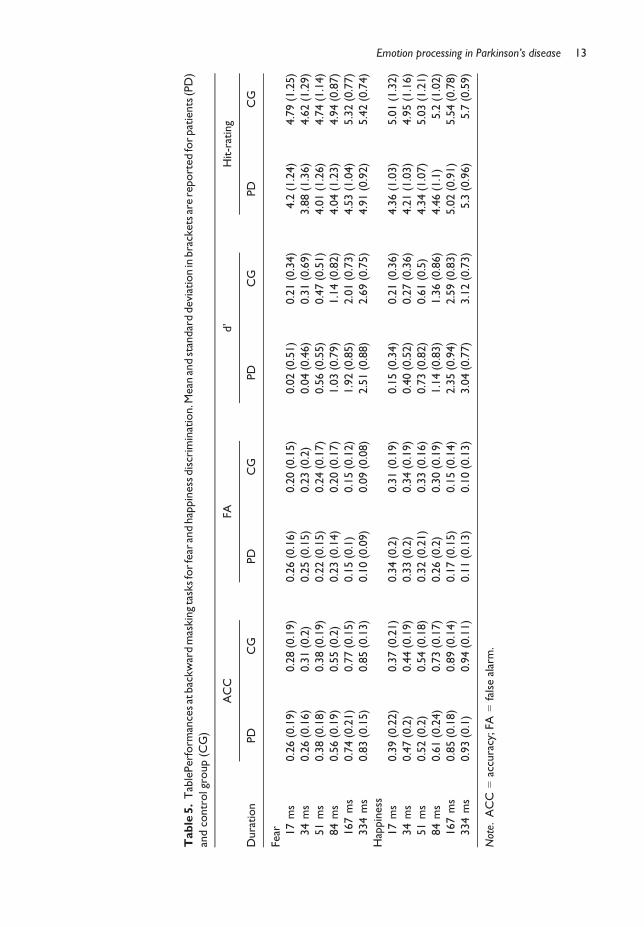
Figure 1. Timelines with stimuli examples of backward masking tasks when fear expression was the
target (left side) and when happy expression was the target (right side).
Emotion processing in Parkinson’s disease 7
Results
Ekman 60 Faces testAccuracy for each emotion separately, and the global score adjusted for demographic
variables are reported in Table 3. According to normative data for the Italian population
(Dodich et al., 2014) four (5.7%) PD patients scored below the cut-off (<37.47) and eight
(11.4%) patients had a borderline equivalent score (ES)1 of 1; whereas only one (2.2%)
healthy control scored below the cut-off. Considering cut-off scores separately for each
emotion, 24 (34.3%) PD patients were impaired in fear recognition, nine (12.8%) in anger,
seven (10%) in happiness and three (4.3%) in surprise, disgust and sadness recognition. In
the control group 3 (6.5%) participants scored below the cut-off for fear recognition, two(4.3%) for happiness and one (2.2%) for angry expressions.
The ANOVA (see Figure 2) showed significant effects of Emotion, F(3.75,
427.51) = 175.82, p < .001, partial g2 = .61, Group, F(1, 114) = 34.33, p < .001, partial
g2 = .23, and Emotion 9 Group interaction, F(3.75, 427.51) = 6.69, p < .001, partial
g2 = .05. PD patients were overall less accurate than healthy controls; post hoc tests for
themain effect of Emotion showed significant higher accuracy for happiness than all other
expressions (all ps < .001), whereas the accuracy score for fear was significantly lower
than for all other emotions (all ps < .001); moreover, surprise was significantly betterrecognized than disgust, anger and sadness (all ps < .001), and angerwas significantly less
recognized than disgust and sadness (ps < .001). Post hoc tests for the Emotion by Group
interaction revealed that PD patients’ accuracy was lower than control group in all
emotions (all ps < .01) but happiness (p = .45).
Relationship between emotion recognition and clinical variables
Bootstrap correlation analyses (Table 4) revealed that the Ekman global score wasnegatively correlated with the Hoehn and Yahr scale, TMT B, TMT BA and GDS, whereas
Table 3. Performances at the Ekman 60 Faces test expressed as adjusted global score and sub-scores for
single emotions. Mean and standard deviation in brackets are reported for patients (PD) and control
group (CG)
PD CG
Global score 47.41 (5.94) 53.09 (4.87)
Surprisea 8.63 (1.52) 9.28 (1.05)
Happiness 9.61 (0.75) 9.72 (0.69)
Feara 3.09 (2.34) 5.37 (2.53)
Disgusta 7.57 (1.88) 8.83 (1.27)
Angera 6.71 (1.88) 7.72 (1.64)
Sadnessa 7.3 (2.06) 8.76 (1.43)
Note. aIndicates significant difference in post hoc analysis on the Emotion by Group interaction.
1 Equivalent scores (ES) correspond to a five-point interval scale from0 to 4 defined on the basis of demographical adjusted scoresand non-parametric tolerance limits. An ES of 0 corresponds to performances below the fifth centile of the normal population,whereas an ES of 4 is equal or above themedian. An ES of 1 is between the outer and inner tolerance limits and the performance isconsidered borderline. ES 2 and 3 are intermediate values on a quasi-interval scale and define normal performances (Capitani &Laiacona, 1997).
8 Giulia Mattavelli et al.
positive correlations resulted with the MoCA, verbal fluency on phonological cue, line
orientation judgment test, unknown face recognition test and the IRI subscale on
empathic concern. Looking at scores for recognition of single emotions, surprise
correlated only with the unknown face recognition test; happiness with Stroop errors,
verbal fluency on phonological cue and unknown face recognition test; fear with the
Hoehn and Yahr scale, MoCA, Stroop errors, verbal fluency and line orientation judgmenttest; disgust with the MoCA, Stroop errors and time, phonological verbal fluency and line
orientation judgment test; anger with the MoCA, Stroop time and unknown face
recognition test; sadness with the MoCA, Stroop errors and time, phonological verbal
fluency, line orientation judgment test, unknown face recognition test and the IRI
subscale on empathic concern. To further investigate the possibility that emotion
recognition capacity depends on patients’ general cognitive ability we repeated the
analysis on the Ekman test, comparing healthy controls and patients, but dividing the
patients’ sample in subgroups of cognitively unimpaired patients, namely those withMoCA ES > 2 (N = 33), and patients with cognitive deterioration, namely MoCA ES < 2
(N = 23) (see Note 1 for ES definition). Thus, an ANOVA Emotion (within-subjects factor,
six levels)9Group (between-subjects factor, three levels) was performed. In cognitively
preservedpatients, one (3%) patientwas impaired in the global score of the Ekman test, 11
(33.3%) patients scored below the cut-off in fear, two (6%) in happiness and anger, one
(3%) in surprise and none in disgust and sadness recognition. In the group of patientswith
MoCAES < 2, twopatients (8.7%)were impaired in the global score, nine (39.1%)patients
scored below the cut-off in fear, four (17.4%) in anger, three (13%) in happiness, two
Figure 2. Mean accuracy for control group (CG) and patients (PD) in each emotion of the Ekman 60
Faces test. Bars represent standard error of themeans. * Indicates significant difference in post hoc analysis
(8.7%) in disgust and sadness and one (4.3%) in surprise recognition. As above, the effects
of Emotion, F(3.5, 353.7) = 173.08, p < .001, partial g2 = .64, Group, F(2, 99) = 25.05,
p < .001, partial g2 = .34, and their interaction, F(7.1, 353.7) = 4.1, p < .001, partial
g2 = .08, were significant. Post hoc tests for the Group main effect showed that allcomparisons between the three groups were significant, with the control group
performing better than cognitively preserved (p = .001) and cognitively impaired
patients (p < .001); moreover, cognitively preserved patients performed better than
cognitively impaired patients (p = .002). Post hoc analysis on Emotion by Group
interaction revealed that cognitive impaired PD patients had a significantly lower
accuracy than the control group for all emotions (ps < .01), but happiness (p = .1). In fear
recognition, also cognitively unimpaired patients had lower accuracy than the control
group (p = .005) and did not differ from cognitively impaired PD group (p = .15),whereas only in sadness recognition cognitively unimpaired patients performed
significantly better than cognitively impaired PD group (p = .002).
Backward masking paradigm
Detailed results and statistics are reported in Tables 5 and 6. The main effects of Emotion
and Duration were significant in the analysis on ACC, which was higher for happiness
(M = 0.64, SD = 0.27) than fear (M = 0.51, SD = 0.28) discrimination and was lower for17 and 34 ms presentation compared to all other presentation durations (ps < .001); ACC
significantly increased between 51, 84, 167 and 334 ms presentation (all ps < .001). The
factor Group was not significant as main effect or in interaction with other factors.
FA analysis showed significant main effects of Emotion and Duration, being FA rate
lower for fear (M = 0.19, SD = 0.15) than happiness (M = 0.26, SD = 0.2) recognition,
and lower for 334 ms and 167 ms compared to faster presentations (ps < .001).
Moreover, post hoc tests for the significant Emotion9 Duration interaction revealed that
FA ratewas higher for happy than fear emotion at 17 ms (p = .007), 34 ms (p = .002) and51 ms (p = .009) presentation, but not at longer durations (ps>.05). The effect of Groupwas not significant.
Similarly to FA, analysis on d’ showed significant effects of Emotion, Duration and
Emotion 9 Duration interaction, whereas Group was not significant as main effect or in
interaction with other factors. D’ was higher for happiness (M = 1.33, SD = 1.29) than
fear (M = 1.07, SD = 1.15) discrimination.Post hoc tests showed significantly lower d’ for
17 and 34 ms presentation compared to all other presentation durations (ps < .001), then
d’ significantly increased between 51, 84, 167 and 334 ms presentation (all ps < .001).Moreover, d’ was significantly higher for happy than fear target emotion at 167 ms and
334 ms presentation (ps < .001), but not at shorter presentation durations (ps > .05)
(Figure 3).
Responses at rating on confidence following hit or false alarm trials were also analysed.
Since some of the participants did not produce hit responses in 17 ms or 34 ms duration
conditions, hit-rating analyses included 21 PD patients and 21 controls. The main effects
were significant: Hit-rating were higher following the detection of happy (M = 4.93,
SD = 1.11) than fear (M = 4.62, SD = 1.19) expression and were higher in the control(M = 5.11, SD = 1.07) than in PD group (M = 4.44, SD = 1.16). Post hoc tests for the
main effect of Duration showed that Hit-rating was higher for 334 and 167 ms conditions
compared to all shorter durations (ps < .001). In the analysis on FA-rating the main effect
of Emotion was significant, whereas the main effect of Group or Group 9 Emotion
interaction were not significant. FA-rating was higher following FA responses in blocks
Emotion 9 Group F(1, 48) = 0.33, p = .57, partial g2 < .01
Note. *p < .05.
14 Giulia Mattavelli et al.
discrimination using a backward masking paradigm were not significantly differentbetween patients and healthy controls; (3) global score of PD patients in emotion
recognition correlated with the stage of the disease, and scores at the MoCA, TMT, verbal
fluency on phonological cue, line orientation judgment, unknown face recognition tests
and the empathic concern subscale of the IRI; (4) a weak correlation resulted with
depression whereas clinical scales on other neuropsychiatric symptoms were not
correlated.
Our results confirm the presence of emotion recognition deficit in PD and add new
evidence to the debate concerning the pattern of impairment across different emotions(Argaud et al., 2018). In line with the previous literature, by comparing a large sample of
PD patients to healthy controls, we found preserved happiness recognition and a decline
in all other emotions in PD (Argaud et al., 2018; Gray & Tickle-Degnen, 2010). On the
other hand, about one third of patients (24 out of 70) scored below the cut-off on fear
recognition suggesting a disproportional deficit in recognizing fear, at odds with studies
showing larger deficit in anger recognition (Clark et al., 2008; Lawrence et al., 2002;
Lawrence, Goerendt, & Brooks, 2007).
The Ekman global score correlated with the severity of PD motor symptoms, thegeneral cognitive abilities and tests tapping attentional, executive and visuospatial
functions. Similarly, fear recognition correlatedwith PD stage,MoCA score, executive and
visuospatial tests and resulted to be the expression with the lowest accuracy in our
sample, according to normative data (Dodich et al., 2014). Thus, it is plausible that the
Figure 3. Mean d’ for control group (CG) and patients (PD) in each duration of presentation (ms) in the
backward masking tasks on fear and happiness discrimination. Bars represents standard error of the
means.
Emotion processing in Parkinson’s disease 15
severity of PD and the general cognitive profile significantly affect the ability to recognize
the most difficult emotion. The MoCA score correlated also with anger, sadness and
disgust recognition. The comparison between the three groups (healthy controls,
patients with impaired/borderline MoCA scores and patients with higher MoCA scores)confirmed that the cognitive status affects patients’ performance at the Ekman test.
Cognitive impaired patients showed the lowest score and differed from the control group
in all expressions, but happiness. Nevertheless, also cognitive unimpaired patients (MoCA
ES > 2) performed significantly worse than controls, in particular in fear recognition. The
relationship between emotion recognition performances and neuropsychological eval-
uation has been reported in previous studies, in particular for visuospatial, attentional and
executive tasks (Assogna et al., 2010; Narme et al., 2013), however, correlationswere not
significant in other studies (Kan et al., 2002; Marneweck et al., 2014). Our data supportthe hypothesis that an emotion recognition deficit,mainly affecting fear emotion, exists in
PD even in the absence of cognitive impairments, but the magnitude of the deficit
increases with the progression of the disease and the emergence of cognitive symptoms
(Argaud et al., 2018). Notably, correlations with motor symptoms were not significant in
different previous studies (Baggio et al., 2012; Buxton,MacDonald, &Tippett, 2013; Clark
et al., 2008) and meta-analyses (Coundouris et al., 2019; Gray & Tickle-Degnen, 2010),
whereas our results, on a large sample of patients and applying bootstrap correlation
methods, suggest a relationship between the Ekman global score and motor disabilitymeasured by the Hoehn and Yahr scale. On the other hand, a weak correlation emerged
with the depression scale, whereas correlations were absent with scales on anxiety and
impulsive-compulsive disorders, in line with the literature showing that emotion
recognition deficits in PD are not related to mood disorders (Argaud et al., 2018; Gray
& Tickle-Degnen, 2010). Only the IRI subscale on empathic concern correlated with the
Ekman global score and sub-score for sadness recognition in patients. This scale measures
compassion for others, thus its relationship with emotion recognition, in particular the
expression of sadness, suggests that patients with difficulties in expression identificationsuffer also from reduced empathy. Moreover, previous studies reported that the empathic
concern subscale of the IRI was related to activity in the anterior insula and anterior
cingulate cortex in healthy participants viewing pain stimuli andwith atrophy in temporal
and frontal regions in patients with neurodegenerative diseases (Rankin et al., 2006;
Singer et al., 2004). Unfortunately, detailed brain images of our patients were not
available, but previous studies showed correlations between emotion recognition
performance and grey matter volume in the cingulate and orbitofrontal cortices in PD
(Baggio et al., 2012; Ibarretxe-Bilbao et al., 2009), thus a more speculative interpretationcould be that patients with lower scores in emotion recognition were affected by greater
neurodegeneration in cortical regions crucially related to empathy.
Together with emotion recognition, this study aimed at assessing expressions
discrimination using a backwardmasking paradigmwith different presentation durations.
This paradigm allows assessing sensitivity for stimuli processed with full awareness, but
also for stimuli automatically processed, i.e., presented below the threshold for conscious
perception (Pessoa et al., 2005). The rationale for using different types of task was to
evaluate multiple stages of emotion processing, i.e., sensory and visuo-spatial processesinvolved in forced-choice tasks as the backward masking paradigm, or identification and
labelling processes involved in recognition tasks (Haxby et al., 2000). Crucially, we did
not find significant differences between PD and control groups in the emotion
discrimination tasks. Only the confidence rating for hit trials was significantly higher in
healthy than PD participants, showing that patients were less confident in judging their
16 Giulia Mattavelli et al.
performance although thiswas not impaired as compared to controls. Two differentmeta-
analyses reported deficit both in emotion discrimination and recognition in PD
(Coundouris et al., 2019; Gray & Tickle-Degnen, 2010). In particular, previous studies
reported impaired configural processing (Narme et al., 2011) and discrimination ofdistinctiveness for faces in patients with PD, suggesting the hypothesis of a “cascade of
lower-to-higher order impairment” for facial expressions, namely a critical contribution of
visual sensory processes to deficits in emotion recognition in PD (Marneweck et al.,
2014). In contrast, different studies suggested unimpaired early visual and automatic
emotion evaluation in PD (Alonso-Recio et al, 2014; Castner et al., 2007; Wagenbreth
et al., 2016;Wieser et al., 2006). According tomodels of distributed neural system for face
and emotion perception, different stages of facial expression processing are supported by
partially distinct, albeit interactive, cortical and subcortical structures. Posterior occipito-temporal regions process perceptual information and are connected with subcortical
structures, as the amygdala and basal ganglia, and frontal regions to elaborate emotional
and social features (Haxby et al., 2000; Ishai, 2008). The hallmark of PD neurodegen-
eration is the loss of nigrostriatal dopaminergic neurons, but also the amygdala and the
prefrontal cortex are critically affected (Dauer & Przedborski, 2003; Diederich et al.,
2016). Indeed, neuroimaging studies with PD patients found that performances on the
Ekman face test correlated with grey matter volume in the orbitofrontal and dorsal
cingulate cortices (Baggio et al., 2012; Ibarretxe-Bilbao et al., 2009). On the other hand, astudy with electrophysiological recording during facial expression recognition reported
no diminished early visual processing, related to activity in posterior occipito-temporal
regions, in patients with PD (Wieser et al., 2006). Our data support these last findings,
showing that PD patients’ sensitivity for fearful and happy expressions, briefly presented
between 17 and 334 ms, was not lower than control participants.
Interestingly, ACC and FA rates were higher for happiness than fear discrimination, as
the d’ index measuring perceptual sensitivity. Previous studies used backward masking
paradigms to assess thresholds for visual awareness of fearful faces and to investigate theneural correlates of conscious and unconscious fear perception (Pessoa, 2005;
Szczepanowski & Pessoa, 2015). To the best of our knowledge, performance in detecting
fearful or happy target faceswas not directly compared before. Our results showed higher
sensitivity for happy faces, revealing no advantage for fear expression in automatic
Abbott, & Mattingley, 2004). The advantage for happiness recognition in the Ekman test
could be related to the fact that it is the only entirely positive emotion. Surprise is also
considered as positive but to a less extent,whereas the other possibilities are four negativeemotions (fear, anger, disgust, sadness). The present data suggest that happiness is easier
than fear expression also in a discrimination task, both for PD patients and control
participants, in linewith previous studies reporting unimpaired happiness discrimination
(Kan et al., 2002; Wagenbreth et al., 2016).
Some limitations of the present study should be taken into account. Neuropsycho-
logical and clinical evaluation was not carried out for the control group of healthy
participants. Thus, interpretations of correlations between emotion recognition and
cognitive and neuropsychiatric profile are specific for PD group, whereas conclusionscannot be related to healthy participants. This would be of particular interest for the IRI
scale, whose correlation with emotion recognition ability is not clearly supported by
Tucker, & Coffaro, 1989). Furthermore, we used two-dimensional static facial expres-
sions; further studies with dynamic expressions will be crucial to investigate whether the
Emotion processing in Parkinson’s disease 17
motor components of facial expressions impact onPD impairment in emotion recognition
and discrimination.
In conclusion, from a theoretical point of view, our results revealed that PD affects
emotion recognition, but not early perceptual stages of emotion processing. This suggeststhat emotional dysfunctions in PD could be related to deficit in declarative processes of
emotion categorization, with intact automatic emotion discrimination (Argaud et al.,
2018; Wieser et al., 2006). From a clinical point of view our findings highlight the
relevance of an evaluation of emotion recognition in early stage of PD. Since this ability
could be impaired even in cognitively preserved patients, a specific assessment is crucial
to identify a deficit with potential impact on patients’ social interaction in daily life
(Argaud et al., 2018). Moreover, PD is highly variable in its clinical phenotype with
patients presenting different combination of motor, non-motor and neuropsychiatricsymptoms (Erro et al., 2013). These subtypes have been related to different progression of
the disease, thus it could be relevant to include the assessment of emotion processing to
better define the cognitive profile of patients and plan therapeutic interventions oriented
to increase patients’ quality of life (Alonso-Recio, Mart�ın-Plasencia, Ruiz, & Serrano, 2018;
The data that support the findings of this study are available from the corresponding author
upon reasonable request.
References
Adolphs, R. (2002). Neural systems for recognizing emotion.CurrentOpinion inNeurobiology,12,
169–177. https://doi.org/10.1016/S0959-4388(02)00301-XAlonso-Recio, L., Mart�ın, P., Rubio, S., & Serrano, J. M. (2014). Discrimination and categorization of
emotional facial expressions and faces in Parkinson’s disease. Journal of Neuropsychology,
8(2), 269–288. https://doi.org/10.1111/jnp.12029Alonso-Recio, L., Mart�ın-Plasencia, P., Ruiz, M., & Serrano, J. M. (2018). Differences in cognitive
performance in nondemented Parkinson’s disease: A latent profile analysis of cognitive
subtypes. Journal of Clinical and Experimental Neuropsychology, 40(8), 777–789. https://doi.org/10.1080/13803395.2018.1432570
Alonso-Recio, L., Serrano, J. M., & Mart�ın, P. (2014). Selective attention and facial expression
recognition in patients with Parkinson’s disease. Archives of Clinical Neuropsychology, 29(4),
374–384. https://doi.org/10.1093/arclin/acu018Argaud, S., V�erin, M., Sauleau, P., & Grandjean, D. (2018). Facial emotion recognition in Parkinson’s
disease: A review and new hypotheses.Movement Disorders, 33(4), 554–567. https://doi.org/10.1002/mds.27305
Ariatti, A., Benuzzi, F., & Nichelli, P. (2008). Recognition of emotions from visual and prosodic cues
in Parkinson’s disease. Neurological Sciences, 29, 219–227. https://doi.org/10.1007/s10072-008-0971-9
Assogna, F., Piontieri, F. E., Cravello, L., Peppe, A., Pierantozzi,M., Stefani, A., . . . Spalletta, G. (2010).Intensity-dependent facial emotion recognition and cognitive functions in Parkinson’s disease.
Journal of the International Neuropsychological Society, 16, 867–876. https://doi.org/10.1017/s1355617710000755
Assogna, F., Pontieri, F. E., Caltagirone, C., & Spalletta, G. (2008). The recognition of facial emotion
expressions in Parkinson’s disease. European Neuropsychopharmacology, 18, 835–848.https://doi.org/10.1016/j.euroneuro.2008.07.004
Baggio, H. C., Segura, B., Ibarretxe-Bilbao, N., Marti, M. J., Valldeoriola, F., Compta, Y., . . . Tolosa, E.(2012). Structural correlates of facial emotion recognition deficits in Parkinson’s disease
Benton, A. L., Sivan, A. B., Hamsher, K. D., & Varney, N. R. (1994). Contributions to
Neuropsychological Assessment: A Clinical Manual. Oxford, UK: Oxford University Press.
Benton, A. L., & Van Allen, M. W. (1968). Impairment in facial recognition in patients with cerebral
disease. Transactions of the American Neurological Association, 93, 38–42. https://doi.org/10.1016/S0010-9452(68)80018-8
Borg, C., Bedoin, N., Bogey, S., Michael, G. A., Poujois, A., Laurent, B., &Thomas-Ant�erion, C. (2012).Implicit and explicit emotional processing in Parkinson’s disease. Journal of Clinical and
Conti, S., Bonazzi, S., Laiacona, M., Masina, M., & Coralli, M. V. (2015). Montreal Cognitive
Assessment (MoCA)-Italian version: Regression based norms and equivalent scores.
Neurological Sciences, 36(2), 209–214. https://doi.org/10.1007/s10072-014-1921-3Coundouris, S. P., Adams, A. G., Grainger, S. A., & Henry, J. D. (2019). Social perceptual function in
parkinson’s disease: Ameta-analysis.Neuroscience and Biobehavioral Reviews, 104, 255–267.https://doi.org/10.1016/j.neubiorev.2019.07.011
Dalili, M. N., Penton-Voak, I. S., Harmer, C. J., & Munaf�o, M. R. (2015). Meta-analysis of emotion
recognition deficits in major depressive disorder. Psychological Medicine, 45, 1135–1144.https://doi.org/10.1017/S0033291714002591
Dauer, W., & Przedborski, S. (2003). Parkinson’s disease: Mechanisms and models. Neuron, 39(6),
889–909. https://doi.org/10.1016/S0896-6273(03)00568-3.Davis, M. H. (1983). Measuring individual differences in empathy: Evidence for a multidimensional
approach. Journal of Personality and Social Psychology, 44, 113–126. https://doi.org/10.1037/0022-3514.44.1.113
Diederich, N. J., Goldman, J. G., Stebbins, G. T., & Goetz, C. G. (2016). Failing as doorman and disc
jockey at the same time: Amygdalar dysfunction in Parkinson’s disease.MovementDisorders,31
(1), 11–22. https://doi.org/10.1002/mds.26460
Dodich, A., Cerami, C., Canessa, N., Crespi, C., Marcone, A., Arpone, M., . . . Cappa, S. F. (2014).Emotion recognition from facial expressions: A normative study of the Ekman 60-Faces Test in
the Italian population.Neurological Sciences, 35, 1015–1021. https://doi.org/10.1007/s10072-014-1631-x
Erro, R., Vitale, C., Amboni, M., Picillo, M., Moccia, M., Longo, K., . . . Barone, P. (2013). Theheterogeneity of early Parkinson’s disease: a cluster analysis on newly diagnosed untreated
patients. PLoS ONE, 8, 1–8. https://doi.org/10.1371/journal.pone.0070244Fereshtehnejad, S. M., Romenets, S. R., Anang, J. B. M., Latreille, V., Gagnon, J. F., & Postuma, R. B.
(2015). New clinical subtypes of Parkinson disease and their longitudinal progression a
prospective cohort comparison with other phenotypes. JAMA Neurology, 72(8), 863–873.https://doi.org/10.1001/jamaneurol.2015.0703
Galeoto, G., Sansoni, J., Scuccimarri, M., Bruni, V., De Santis, R., Colucci, M., . . . Tofani, M. (2018). A
psychometric properties evaluation of the Italian version of the geriatric depression scale.
Depression Research and Treatment, 2018, 1–7. https://doi.org/10.1155/2018/1797536Giovagnoli, A. R., Del Pesce, M., Mascheroni, S., Simoncelli, M., Laiacona, M., & Capitani, E. (1996).
Trail making test: Normative values from 287 normal adult controls. The Italian Journal of
Neurological Sciences, 17(4), 305–309. https://doi.org/10.1007/BF01997792Gray, H. M., & Tickle-Degnen, L. (2010). A meta-analysis of performance on emotion recognition
tasks in Parkinson’s disease. Neuropsychology, 24(2), 176–191. https://doi.org/10.1037/
a0018104
Harding, A. J., Stimson, E., Henderson, J. M., & Halliday, G. M. (2002). Clinical correlates of selective
pathology in the amygdala of patientswith Parkinson’s disease.Brain, 125, 2431–2445. https://doi.org/10.1093/brain/awf251
Haxby, J., Hoffman, E., & Gobbini, M. (2000). The distributed human neural system for face
perception. Trends in Cognitive Sciences, 4(6), 223–233. https://doi.org/10.1016/S1364-6613(00)01482-0
Heller, J., Mirzazade, S., Romanzetti, S., Habel, U., Derntl, B., Freitag, N. M., . . . Reetz, K. (2018).Impact of gender and genetics on emotion processing in Parkinson’s disease – A multimodal
study. NeuroImage: Clinical, 18, 305–314. https://doi.org/10.1016/j.nicl.2018.01.034Herrera, E., Cuetos, F., & Rodr�ıguez-Ferreiro, J. (2011). Emotion recognition impairment in
Parkinson’s disease patientswithout dementia. Journal of theNeurological Sciences, 310(1–2),237–240. https://doi.org/10.1016/j.jns.2011.06.034
Ibarretxe-Bilbao, N., Junque, C., Tolosa, E., Marti, M. J., Valldeoriola, F., Bargallo, N., & Zarei, M.
(2009). Neuroanatomical correlates of impaired decision-making and facial emotion recognition
in early Parkinson’s disease. European Journal of Neuroscience, 30, 1162–1171. https://doi.org/10.1111/j.1460-9568.2009.06892.x
Ishai, A. (2008). Let’s face it: It’s a cortical network. NeuroImage, 40(2), 415–419. https://doi.org/10.1016/j.neuroimage.2007.10.040
Jankovic, J. (2008). Parkinson’s disease: Clinical features and diagnosis. Journal of Neurology,
Neurosurgery and Psychiatry, 79, 368–376. https://doi.org/10.1136/jnnp.2007.131045Kalampokini, S., Lyros, E., Luley, M., Sch€ope, J., Spiegel, J., B€urmann, J., . . . Unger, M. M. (2018).
Facial emotion recognition in Parkinson’s disease: Associationwith age and olfaction. Journal of
Clinical and Experimental Neuropsychology, 40(3), 274–284. https://doi.org/10.1080/
13803395.2017.1341470
Kan, Y., Kawamura, M., Hasegawa, Y., Mochizuki, S., & Nakamura, K. (2002). Recognition of
emotion from facial, prosodic and written verbal stimuli in Parkinson’s disease. Cortex, 38(4),
623–630. https://doi.org/10.1016/S0010-9452(08)70026-1Kelly, K. J., & Metcalfe, J. (2011). Metacognition of emotional face recognition. Emotion, 11, 896–
906. https://doi.org/10.1037/a0023746
Lawrence, A. D., Calder, A. J., McGowan, S. W., & Grasby, P. M. (2002). Selective disruption of the
recognition of facial expressions of anger.NeuroReport, 13, 881–884. https://doi.org/10.1097/00001756-200205070-00029
Lawrence, A. D., Goerendt, I. K., &Brooks, D. J. (2007). Impaired recognition of facial expressions of
anger in Parkinson’s disease patients acutely withdrawn from dopamine replacement therapy.
Neuropsychologia, 45(1), 65–74. https://doi.org/10.1016/j.neuropsychologia.2006.04.016Leentjens, A. F. G., Dujardin, K., Pontone, G. M., Starkstein, S. E., Weintraub, D., & Martinez-Martin,
P. (2014). TheParkinson anxiety scale (PAS): Development and validation of a newanxiety scale.
Movement Disorders, 29, 1035–1043. https://doi.org/10.1002/mds.25919
Liddell, B. J.,Williams, L.M., Rathjen, J., Shevrin, H., &Gordon, E. (2004). A Temporal dissociation of
subliminal versus supraliminal fear perception: An event-related potential study. Journal of
Cognitive Neuroscience, 16(3), 479–486. https://doi.org/10.1162/089892904322926809Marneweck, M., Palermo, R., & Hammond, G. (2014). Discrimination and recognition of facial
expressions of emotion and their links with voluntary control of facial musculature in
Parkinson’s disease. Neuropsychology, 28, 917–928. https://doi.org/10.1037/neu0000106Mattavelli, G., Pisoni, A., Casarotti, A., Comi, A., Sera, G., Riva, M., . . . Papagno, C. (2019).
Consequences of brain tumour resection on emotion recognition. Journal of Neuropsychology,
13(1), 1–21. https://doi.org/10.1111/jnp.12130Mattavelli, G., Sormaz,M., Flack, T., Asghar, A. U. R., Fan, S., Frey, J., . . .Andrews, T. J. (2014). Neural
responses to facial expressions support the role of the amygdala in processing threat. Social
Cognitive and Affective Neuroscience, 9, 1684–1689. https://doi.org/10.1093/scan/nst162Narme, P., Bonnet, A.M.,Dubois, B.,&Chaby, L. (2011).Understanding facial emotionperception in
Parkinson’s disease: The role of configural processing. Neuropsychologia, 49, 3295–3302.https://doi.org/10.1016/j.neuropsychologia.2011.08.002
Narme, P., Mouras, H., Roussel, M., Duru, C., Krystkowiak, P., &Godefroy, O. (2013). Emotional and
cognitive social processes are impaired in Parkinson’s disease and are related to behavioral
disorders. Neuropsychology, 27(2), 182–192. https://doi.org/10.1037/a0031522Papagno, C., & Trojano, L. (2018). Cognitive and behavioral disorders in Parkinson’s disease: An
Pessoa, L., Japee, S., & Ungerleider, L. G. (2005). Visual awareness and the detection of fearful faces.
Emotion, 5(2), 243–247. https://doi.org/10.1037/1528-3542.5.2.243Pietschnig, J., Moser, D., Pfl€uger, M., Pirker, W., Ratheiser, I., Pusswald, G., . . . Kryspin-Exner, I.
(2016). Facial emotion recognition and its relationship to cognition and depressive symptoms in
patients with Parkinson’s disease. International Psychogeriatrics, 28, 1165–1179. https://doi.org/10.1017/s104161021600034x
Quinlan, P. T. (2013). The visual detection of threat: A cautionary tale. Psychonomic Bulletin and
Review, 20, 1080–1101. https://doi.org/10.3758/s13423-013-0421-4Rankin, K. P., Gorno-Tempini, M. L., Allison, S. C., Stanley, C. M., Glenn, S., Weiner, M.W., &Miller,
B. L. (2006). Structural anatomy of empathy in neurodegenerative disease. Brain, 129, 2945–2956. https://doi.org/10.1093/brain/awl254
Ricciardi, L., Visco-Comandini, F., Erro, R.,Morgante, F., Bologna,M., Fasano, A., . . .Kilner, J. (2017).Facial emotion recognition and expression in Parkinson’s disease: An emotional mirror
mechanism? PLoS ONE, 12(1), 1–16. https://doi.org/10.1371/journal.pone.0169110Riggio, R. E., Tucker, J., & Coffaro, D. (1989). Social skills and empathy. Personality and Individual
Differences, 10(1), 93–99. https://doi.org/10.1016/0191-8869(89)90184-0Roesch, E. B., Sander, D., Mumenthaler, C., Kerzel, D., & Scherer, K. R. (2010). Psychophysics of
emotion: The QUEST for emotional attention. Journal of Vision, 10, 1–9. https://doi.org/10.1167/10.3.4
Sagna, A., Gallo, J. J., & Pontone, G. M. (2014). Systematic review of factors associated with
depression and anxiety disorders among older adults with Parkinson’s disease. Parkinsonism
and Related Disorders, 20(7), 708–715. https://doi.org/10.1016/j.parkreldis.2014.03.020Sheikh, J. I., & Yesavage, A. (1986). Geriatric depression scale GDS: Recent evidence and
development of a shorter version. In T. L. Brink (Ed.), Clinical gerontology: A guide to
assessment and intervention (pp. 165–173). New York, NY: Haworth.
Singer, T., Seymour, B., O’Doherty, J., Kaube, H., Dolan, R. J., & Frith, C. D. (2004). Empathy for pain
involves the affective but not sensory components of pain. Science,303, 1157–1162. https://doi.org/10.1126/science.1093535
Sprengelmeyer, R., Young, Aw, Mahn, K., Schroeder, U., Woitalla, D., B€uttner, T., Kuhn, W., &
Przuntek, H. (2003). Facial expression recognition in people with medicated and unmedicated
Stanislaw, H., & Todorov, N. (1999). Calculation of signal detection theory measures. Behavior
Research Methods, Instruments, & Computers: A Journal of the Psychonomic Society, 31(1),
137–149. https://doi.org/10.3758/BF03207704Suzuki, A., Hoshino, T., Shigemasu, K., & Kawamura, M. (2006). Disgust-specific impairment of
facial expression recognition in Parkinson’s disease. Brain, 129(3), 707–717. https://doi.org/10.1093/brain/awl011
Szczepanowski, R., & Pessoa, L. (2015). Fear perception: Can objective and subjective awareness
measures be dissociated? Journal of Vision, 7(2007), 1–17. https://doi.org/10.1167/7.4.10.Introduction
Tamietto, M., & de Gelder, B. (2010). Neural bases of the non-conscious perception of emotional
signals. Nature Reviews. Neuroscience, 11, 697–709. https://doi.org/10.1038/nrn2889Tickle-Degnen, L., & Lyons, K. D. (2004). Practitioners’ impressions of patients with Parkinson’s
disease: The social ecology of the expressive mask. Social Science and Medicine, 58, 603–614.https://doi.org/10.1016/S0277-9536(03)00213-2
Trojano, L., & Papagno, C. (2018). Cognitive and behavioral disorders in Parkinson disease: An
Williams, L. M., Liddell, B. J., Rathjen, J., Brown, K. J., Gray, J., Phillips, M., . . . Gordon, E. (2004).Mapping the time course of nonconscious and conscious perception of fear: An integration of
central and peripheral measures. Human Brain Mapping, 21(2), 64–74. https://doi.org/10.1002/hbm.10154
Williams, M. A., Morris, A. P., McGlone, F., Abbott, D. F., & Mattingley, J. B. (2004). Amygdala
responses to fearful and happy facial expressions under conditions of binocular suppression.
Journal of Neuroscience, 24, 2898–2904. https://doi.org/10.1523/jneurosci.4977-03.2004Yip, J. T. H., Lee, T. M. C., Ho, S. L., Tsang, K. L., & Li, L. S.W. (2003). Emotion recognition in patients
with idiopathic Parkinson’s disease. Movement Disorders, 18, 1115–1122. https://doi.org/10.1002/mds.10497
Young, A. W., Perrett, D., Calder, A., Sprengelmeyer, R., & Ekman, P. (2002). Facial expressions of
emotion: Stimuli and Tests (FEEST) thames. Suffolk, UK Bury St. Edmunds: Valley Test
Company.
Received 3 October 2019; revised version received 3 October 2020
Supporting Information
The following supporting informationmay be found in the online edition of the article:
Table S1. Scores at neuropsychological tests included in patients’ assessment.
Table S2. Clinical and neuropsychological data of 25 patients assessed with the