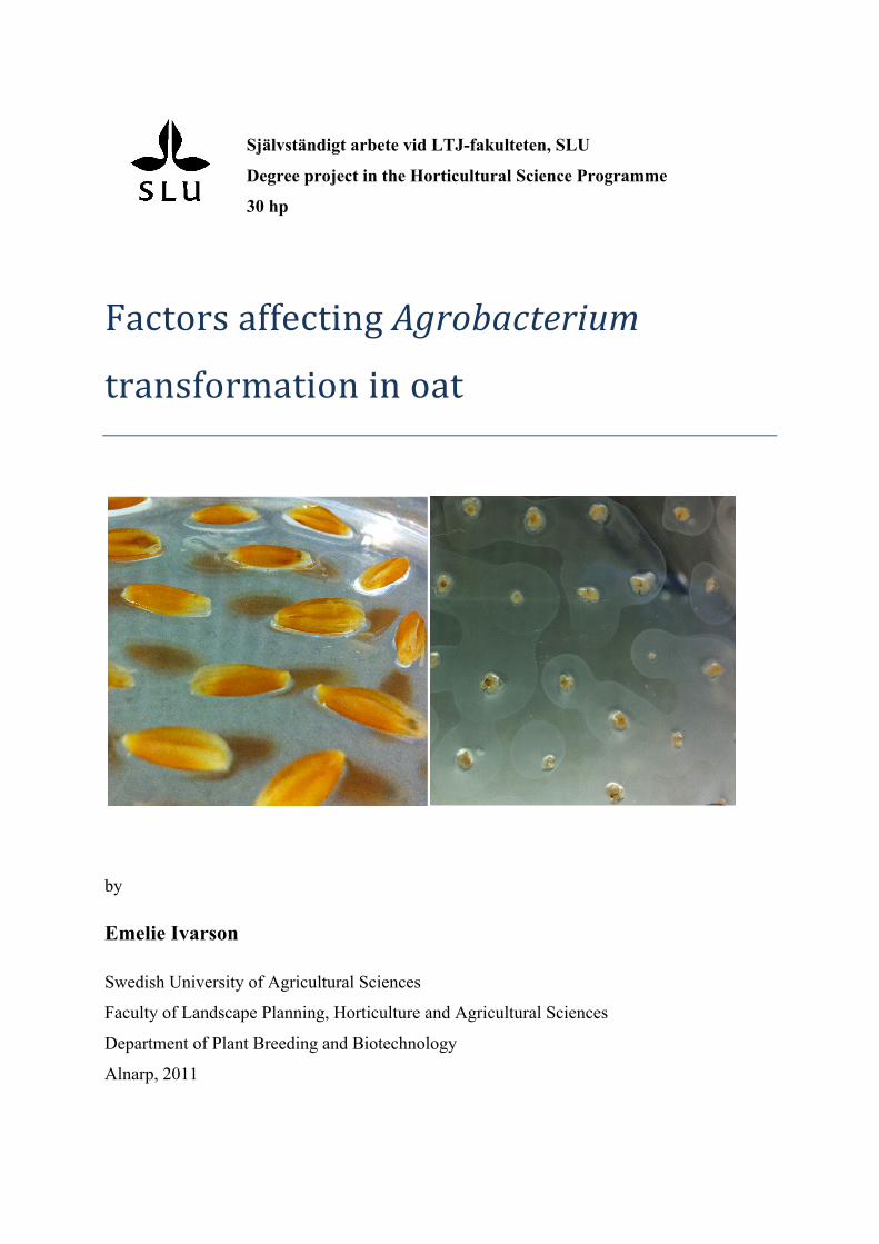
Självständigt arbete vid LTJ-fakulteten, SLU Degree project in the Horticultural Science Programme 30 hp Factors affecting Agrobacterium transformation in oat by Emelie Ivarson Swedish University of Agricultural Sciences Faculty of Landscape Planning, Horticulture and Agricultural Sciences Department of Plant Breeding and Biotechnology Alnarp, 2011
Transcript
Självständigt arbete vid LTJ-fakulteten, SLU
Degree project in the Horticultural Science Programme
30 hp
Factors affecting Agrobacterium
transformation in oat
by
Emelie Ivarson
Swedish University of Agricultural Sciences Faculty of Landscape Planning, Horticulture and Agricultural Sciences
Department of Plant Breeding and Biotechnology
Alnarp, 2011
SLU, Swedish University of Agricultural Sciences
Faculty of Landscape Planning, Horticulture and Agricultural Sciences
Department of Plant Breeding and Biotechnology
Author Emelie Ivarson
English title Factors affecting Agrobacterium transformation in oat
Swedish title Faktorer som påverkar Agrobakterium-transformering av havre
Med hjälp av transformering finns det en möjlighet att höja mängden Omega-3-
fettsyror, och därmed få en bättre Omega-6/Omega-3-balans i havre. Eftersom Omega-3-
fettsyror har visat sig vara potentiella terapeutiska agenter mot autoimmuna- och
inflammatoriska sjukdomar (Simopoulos, 2002), bör ett ökat Omega-3-fettsyreinnehåll leda
till ett större intresse av att använda havre som föda.
Transformering möjliggör en ökning av Omega-3-fettsyror i havre, men tidigare
studier gällande transformering av Avena sativa L. har resulterat i låga
transformeringsfrekvenser. Monokotyledoner är inte naturliga värdar för Agrobacterium,
varför dessa är svårare än dikotyledoner att infektera. Förhållandena måste vara optimala för
att en transformering ska ge ett lyckat resultat.
Målet med detta arbete har varit att ta reda på vilka faktorer som påverkar
havretransformering, samt att arbeta fram ett transformeringsprotokoll som ger höga
transformeringsfrekvenser.
Hypokotylexplantat från havresorten Matilda transformerades med hjälp av
Agrobacterium. I försöket utfördes 15 transformeringsomgångar, där olika kombinationer av
bakteriestammar, vektorer och medier testades. GUS- och GFP-analyser genomfördes för att
bekräfta att transformeringarna lyckats.
Vid analyserna av GUS-uttryck uppvisade inget av explantaten något GUS-
uttryck, men det går inte att dra någon säker slutsats av resultatet. Låga pH-värden triggar
uttryck av endogen GUS-liknande aktivitet, men ett ökat pH-värde kanske inte bara
undantrycker uttryck av endogent GUS utan även uttryck av riktigt GUS.
Explantaten som analyserades för uttryck av GFP uppvisade vitaktiga fläckar,
men ytterligare odling och analyser av explantaten krävs för att kunna bekräfta uttryck av
GFP.
Fler försök krävs för att hitta ett välfungerande havretransformeringsprotokoll.
Introduction
Oat Oat is a member of the Poaceae family (Kellogg, 1998). The cultivars used in cultivation are
hexaploids (Bennet & Smith, 1991). Oat has a long history in cultivation, being one of the
crops cultivated by mankind for longest time (Lásztity, 1998). In 2009 the world production
of oat was estimated to 23 millions of tonnes, with Russia being the greatest producer
(FAOSTAT, 2011).
In Northern Europe oat is an important and traditional agricultural crop
(Bräutigam et al., 2005). In Sweden most of the oat is used as animal feed (Bräutigam et al.,
2005), but the recent high oil prices and the low oat prices have led to an increase in using oat
in combustion. A minor part of the oat production (approximately 5 %) is used as food. This
part is important though, since several parts of the food chain are affected. The greatest part of
the production is based on contracts, where both traders and producers are involved (Carlsson,
Personal conversation).
The nutritional advantage of oat is being more and more emphasized, which has
led to an increase rather than a decrease in using oat as food (Carlsson, Personal
conversation). In comparison to proteins of other cereals, the amino acid composition of the
oat proteins is nutritionally superior (Lásztity, 1998). Oat also has a high content of desirable
soluble fibers (β-glucans), important vitamins and minerals (Sadiq Butt et al., 2008) and
antioxidants (Ryan et al., 2007).
Compared to other cereals, oat has a much higher content of lipids; reaching
from 2-15.5 %, depending on environmental and genetic factors as well as the method chosen
for determination (Zhou et al., 1999). The oil is interesting since it has high energy content
and a relatively low portion of saturated fatty acids. A drawback, which may hinder a future
increase in using oat as feed and food, is its imbalance in Omega-6/Omega-3 fatty acids. The
level of the unsaturated fatty acid 18:2 (ω-6) is much higher (36-47 %:1-2 %) than the level of
the unsaturated fatty acid 18:3 (ω-3) (Welch & Legett, 1997). Studies have shown that an
imbalance in the fatty acid composition is a possible factor behind the increasingly frequent
cases of cardiovascular disease, cancer, diabetes, asthma, depression, obesity, autoimmune
diseases and rheumatism in the Western countries (Simopoulos, 2004).
Omega-3- & Omega-6 Fatty Acids Polyunsaturated fatty acids (PUFAs) are an important part of our diet. On basis of the location
of their first double bond, they are divided into two subcategories: omega-3 (n-3) and omega-
6 (n-6) fatty acids. The first double bond is found on the third carbon molecule on the omega-
3 fatty acids, while it is situated on the sixth carbon molecule on the omega-6 fatty acids (Lee
& Lip, 2003).
Studies have shown that omega-3 fatty acids are capable of modifying
inflammatory and immune reactions, which makes them potential therapeutic agents for
autoimmune and inflammatory diseases (Simopoulos, 2002).
PUFAs are classified as essential nutrients for human health since mammals
lack the compounds involved in the synthesis of PUFAs. The western diet contains a
sufficient amount of omega-6 fatty acids, but the level of omega-3 fatty acids is generally
much lower. Today, plant oils constitute the main source of omega-6 fatty acids while fish-
and algal oils are the sources richest in omega-3 fatty acids. Since fish- and algal oils are not
always suitable or economical for human use, a more economically dietary source of omega-3
fatty acids is of commercial interest (Pereira et al., 2004).
Genetic improvement
Oat improvement by conventional breeding Traditional breeding by crossing is a common way for breeding new cultivars, in which one
donor plant with the trait of interest is crossed with a recipient plant with only one or few
drawbacks. The problem with crossing is that the whole genomes of both plants are mixed
and recombinant, leading to an incorporation of both wanted and unwanted genes. In order to
achieve an improved variety, several backcrosses are necessary to get rid of the unwanted
genes/traits (Roberts, 1984).
Genetic transformation Genetic modification is an efficient and straightforward method for directly introducing novel
genes, conferring desirable traits, into the target plant genome. In combination with
conventional breeding programs, transformation enables insertion of transgenes encoding
useful traits into crops within a workable time frame. By genetic manipulation, the
productivity of crops can be greatly improved through increased resistance against diseases,
pests and environmental stress factors together with a qualitative change of the seed
composition. By designing plants that produce high volumes of pharmaceuticals,
nutraceuticals and other beneficial substances the nutritional value of crops can be improved.
In addition to the possibility of improving crops, transformation also enables study of gene
function and the regulation of physiological and developmental processes (Hansen & Wright,
1999). Genetic transformation also enables analysis and understanding of the underlying
mechanisms behind expression of transgenes or endogenous genes (Gasparis et al., 2008).
Plant genetic transformation consists mainly of two methods: biolistic and
Agrobacterium transformation. In most applications, the Agrobacterium-mediated method is
the most reasonable one (Gasparis et al., 2008).
Agrobacterium-mediated transformation
Binns & Thomashow (1988) discovered that the tumor-inducing (Ti) plasmid of
Agrobacterium is capable of transferring a DNA segment (T-DNA) into the nucleus of the
host plant cell. The T-DNA transfer is controlled by border sequences on the T-DNA. The
finding made plant genetic transformation via A. tumefaciens possible.
A transfer of the T-DNA is not possible without expression of the vir genes
located on the Ti-plasmid in the bacterium. Only a few vir genes are expressed under normal
bacterial growth conditions, while most of them are induced by plant cell exudates. In
uninjured plants the plant cell exudates (phenolic compounds such as acetosyringone ) that
trigger the expression of vir genes are present only at very low levels. In order to increase the
level of such compounds, the tissue has to be wounded. The increased level of cell exudates
triggers A. tumefaciens to initiate vir gene expression and related responses that are necessary
for a successful plant cell transformation (As reviewed by Binns & Thomashow, 1988).
The capacity that A. tumefaciens is capable of sensing the phenolics probably
results in a recruitment of the bacterium to the regions of wounded tissues (As reviewed by
Binns & Thomashow, 1988).
The T-DNA contains two types of genes; the oncogenes and the opine
biosynthetic genes (Binns & Thomashow, 1988). The oncogenes are encoding enzymes that
are involved in the production of auxins and cytokinins, resulting in the tumor structures
(Opabode, 2006). The opine biosynthetic genes catalyze the production of unusual amino
acids and sugar derivatives (opines), which are used by the bacteria either as a carbon and
nitrogen source or as an inducer of plasmid transfer between bacteria (Binns & Thomashow,
1988).
Biolistic transformation – Particle bombardment
Particle bombardment is a technique enabling a direct transfer of genetic material into plant
tissues. The principal of this method is that DNA or RNA is coated to particles of gold or
tungsten and shooted into the target tissue with help of streams of pressurized helium,
(Ziolkowski, 2007).
Particle bombardment is one of the techniques that has made it possible to
introduce traits that are of agricultural value to crops, such as insect resistance, leading to an
increase in both productivity and efficiency of the crop (Ziolkowski, 2007). However, this
method often results in low transformation efficiency and multiple copies of transgene
integration.
Factors affecting Agrobacterium transformation of monocots Monocotyledons are not natural hosts of Agrobacterium (De Cleene & De Ley, 1976), which
is why Agrobacterium-mediated transformation of monocots has been very difficult and
unreliable (Sood et al., 2011). A great number of factors affect Agrobacterium transformation,
which often makes the development of a new transformation protocol for a given species a
difficult and time-consuming process, especially for monocotyledonous species.
Genotype, age & physiological state of explants Genotype variation in regeneration has been reported in many plant species and this has also
been found to be true for oat (Gasparis et al., 2008). The age and the physiological state of the
explant also greatly affect the transformation result. The cells that are receiving the transgene
need to recover quickly from the shock that the transformation event brings about. Apart from
a fast recovery, the cells have to be competent for regeneration and be able to grow into a
complete plant. The recovery of the infected cells has shown to be very difficult in monocots,
why the focus of earlier trials to a great extent has been on optimization of the factors
influencing the plant regeneration capacity (as reviewed by Sood et al., 2011).
Agrobacterium strains & vectors The choice of bacterium strains and vectors has been show to be of great importance in
transformation of monocotyledons. Only a few Agrobacterium strains have resulted in
successful transformations of monocotyledons. The Agrobacterium strain A281 is a so-called
supervirulent strain with a wide host-range and an inducement of large tumors (Wei et al.
2000) due to its additional vir genes (Jones et al. 2005).
Agrobacterium attachment A surfactant is a type of wetting agent that has shown to increase the efficiency of the T-DNA
delivery in immature embryos of wheat (Cheng et al., 1997). The surfactant facilitates the
attachment of Agrobacterium to the surface and/or eliminates substances that inhibit
attachment of the bacterium (as reviewed in Opabode, 2006). Apart from chemical agents and
surfactants such as Tween 20, Silwet L77 and Pluronic acid F68, (as reviewed in Opabode,
2006), an optimal density of Agrobacterium can also facilitate the attachment of the bacterium
(as reviewed in Sood et al., 2011).
Co-cultivation The duration, temperature, irradiance, medium composition and pH need to be optimal during
co-cultivation, since this step comprises the delivery and the integration of the T-DNA.
Optimization of parameters as medium strength, sugars, vir inducing chemicals and plant
growth regulators have resulted in successful transformations of monocots. A reduce in the
salt strength of inoculation- and co-culture media has proven to result in a more successful
transformation of wheat (as reviewed in Sood et al., 2011).
Elimination of residual Agrobacterium High levels of Agrobacterium can lead to necrosis and bacterial overgrowth of the
transformants. To obtain a good recovery of the transformants and a higher efficiency of the
transformation, it is important to get rid of residual Agrobacterium (as reviewed in Sood et al.,
2011).
Current state of oat transformation
Agrobacterium-mediated transformation of oat (Avena sativa L.) cultivars via immature embryo and leaf explants In a trial, Gasparis et al. (2008) transformed oat using Agrobacterium with immature embryos
or leaf base segments as explants of three different cultivars and three combinations of
strain/vector in combination with different selection genes. Among all different combinations,
only one of the strain/vector combinations resulted in transgenic plants. The highest
transformation rate generated by one of the three cultivars was 12.3 % for the immature
embryo explants and 8.2 % for the leaf base segment explants. For the other two cultivars, the
transformation rates were 1.1 and 3.4 % respectively and transgenic plants were only
recovered from the immature embryos.
In a second step of the trial, Gasparis et al. (2008) evaluated the suitability of the
pGreen binary vector in oat transformation. The vector was combined with four different
selection cassettes: nos::nptII, 35S::nptII, nos::bar and 35S::bar. All cassettes except one
(35S::bar) generated putative transgenic plants. The highest transformation efficiency
achieved was 5.3 %.
Project aim Earlier trials concerning transformation of oat have resulted in low transformation frequencies
or no success at all. In order to enable efficient modification of important traits, such as
omega-3 fatty acid in oat, a well-functional transformation protocol must be first developed.
The aim of this project was to evaluate several factors affecting Agrobacterium-
mediated transformation of oat. Furthermore, the aim was also to obtain a functional
transformation protocol.
Material & Method
Plant material The plant material was seeds of the oat cultivar Matilda, which were kindly provided by
Svalöv Weibull.
Strains and Vectors Different combinations of strain/vector were used to find the combination optimal for a
successful transformation. Among them, most combinations (EHA101/pSCV1.6,
M K3(Fe(CN)6), 0.1 M K4(Fe(CN)6*6H20, 0.5 M Na2EDTA and X-Gluc) and allowed to
incubate at 37°C over night. The following day, in cases necessary, the explants were rinsed
with ethanol (absolute) to get rid of chlorophyll (Jeffersson, 1987).
Since a low pH value can trigger the expression of endogenous GUS-like
activity (Solís-Ramos et. al., 2010), tests with different pH were conducted using leaf
segments from transgenic Lepidium campestre in comparison with non-transgenic ones. After
having obtained reliable results on Lepidium, similar tests were carried out in oat.
GFP analysis
To analyze the presence of green fluorescent protein (GFP), the explants infected with
Agrobacterium carrying the vector pCW498GFP were analyzed in Bio-Rad’s Versa Doc
Imaging System.
Results & Discussion
Transformation of Agrobacterium by electroporation No colonies appeared after incubation of LBA4404/pUC52AtWRI1 and
LBA4404/pCW498GFP, which must be due to failure in the electroporation event.
Transformation of plasmids into Agrobacterium After incubation, colonies appeared on the plate containing LBA4404/pCW498GFP, while no
colonies appeared on the plates containing AGL1/pUC52AtWRI1 and AGL1/pCW498GFP.
The bacteria’s inability to grow indicates sensitivity to antibitotics, which means that the
plasmids were not successfully transformed into the competent cells of Agrobacterium. A
possible explanation to the transformation failure is that the competent cells might not have
been competent, and thus not able to be transformed.
Small scale preparation of plasmid DNA The DNA-concentration of the Agrobacterium sample was low (260/280 = 2,01 and 260/230
= 1,36) so the sample was divided into two, with one part of the sample undiluted and one
part of the sample diluted by 10. The reason behind dilution of DNA is that a too high DNA
concentration can hinder amplification of the DNA in the PCR analysis.
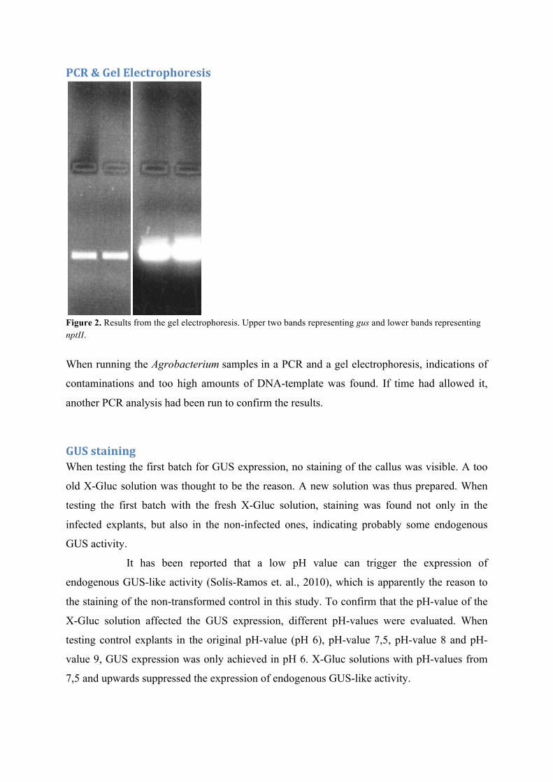
PCR & Gel Electrophoresis
Figure 2. Results from the gel electrophoresis. Upper two bands representing gus and lower bands representing nptII.
When running the Agrobacterium samples in a PCR and a gel electrophoresis, indications of
contaminations and too high amounts of DNA-template was found. If time had allowed it,
another PCR analysis had been run to confirm the results.
GUS staining When testing the first batch for GUS expression, no staining of the callus was visible. A too
old X-Gluc solution was thought to be the reason. A new solution was thus prepared. When
testing the first batch with the fresh X-Gluc solution, staining was found not only in the
infected explants, but also in the non-infected ones, indicating probably some endogenous
GUS activity.
It has been reported that a low pH value can trigger the expression of
endogenous GUS-like activity (Solís-Ramos et. al., 2010), which is apparently the reason to
the staining of the non-transformed control in this study. To confirm that the pH-value of the
X-Gluc solution affected the GUS expression, different pH-values were evaluated. When
testing control explants in the original pH-value (pH 6), pH-value 7,5, pH-value 8 and pH-
value 9, GUS expression was only achieved in pH 6. X-Gluc solutions with pH-values from
7,5 and upwards suppressed the expression of endogenous GUS-like activity.
The pH-value was kept at 7.5 in analysis of all of the following oat batches. No
staining was achieved in any explants of the batches of transformation. A lack of GUS
expression indicates a failure in transformation, but that conclusion cannot be drawn with
certainty in this trial. Maybe the histological GUS staining method is not a trustworthy or
suitable method to confirm a successful transformation of oat. The increased pH-value might
not only suppress the expression of endogenous GUS-like activity, but also hinder a working
enzyme activity of true GUS. To confirm transformation of oat, the finding of methods other
than histological GUS staining is of interest.
Figure 3. Results from GUS-assays; left picture showing the absence of GUS-expression in the leaf fragments of the non-transgenic Leipium (left tube) and GUS-expression in the leaf fragments of the transgenic Lepidium (right tube). Right picture shows no GUS-expression in any of the oat calli (first and second tube) or in the leaf fragments (third tube).
GFP analysis
Figure 4. Result from a GFP analysis. Whitish-colored spots indicate a possible GFP expression.
No absolute conclusion can be drawn from the analysis of explants transformed with
constructs harboring the green fluorescent protein (GFP) in this trial. The non-transformed
explants exhibited an even dark color, but the transformed explants exhibited small spots that
were whitish-colored. The spots might have been GFP expression, but the time limitations in
this trial did not allow further analysis to confirm that the spots actually expressed GFP.
In future trials, the GFP analysis will be repeated in the explants transformed in
this trial. The additional cultivation of the explants may increase the possible GFP expression.
Regeneration Both the transformed explants and the non-transformed controls exhibited a poor
regeneration. Regeneration of transformed monocots has showed to be difficult, but good
regeneration has been achieved in both transformants and controls in trials by Leonova
(Leonova, Personal conversation). The poor regeneration generated in this trial indicates that
the seeds are too old.
Perspective The aim to achieve a well-functional transformation protocol will not end with this master
project, but will continue for a further trial. The first parameter that will be altered is the plant
material. New and fresh oat seeds will be used, which hopefully will result in a better
regeneration. In some batches of transformation, acetosyringone will be added not only during
co-cultivation, but also in the selection medium to see if a better infection is achieved. A
surfactant will be added to the inoculation medium in some of the batches to see if a better
attachment (and thereby a more effective infection) is generated. An addition of silver nitrate
in the co-culture medium can suppress Agrobacterium growth and lead to a more stable
transformation (as reviewed in Sood et al., 2011), and thus will be utilized in some of the
future transformation batches. Hopefully these alterations in the transformation method will
result in a well-functional oat transformation protocol.
References
Bennet M. D. & Smith J.B. (1991). Nuclear DNA amounts in angiosperms. Philosophical
Transactions of the Royal Society: B 334, 309-345.
Binns A. N. & Thomashow M. F. (1988). Cell Biology of Agrobacterium Infection and
Transformation of Plants. Annual Review of Microbiology 42, 575-606.
Bräutigam M., Lindlöf A., Zakhrabekova S., Gharti-Chhetri G., Olsson B. and Olsson O.
(2005). Generation and analysis of 9792 EST sequences from cold acclimated oat, Avena
sativa. BMC Plant Biology 5:18.
Cheng M., Fry J. E., Pang S., Zhou H., Hironaka C. M., Duncan D. R., Conner T. W. & Wan
Y. (1997). Genetic Transformation of Wheat Mediated by Agrobacterium tumefaciens. Plant
Physiology 115, 971-980.
De Cleene M. & De Ley J. (1976). The Host Range of Crown Gall. The Botanical Review 42,
389-466.
FAOSTAT, Food and Agriculture Organization of the United Nations. Homepage [online]
Chiu S-H. (2006). Proteomic analysis of Agrobacterium tumefaciens response to the vir gene
inducer acetosyringone. Proteomics 6, 4130–4136.
Lásztity R. (1998). Oat grain – a wonderful reservoir of natural nutrients and biologically
active substances. Food Reviews International 14:1, 99-119.
Lee K. W. & Lip G. Y. H. (2003). The role of -3 fatty acids in the secondary prevention of
cardiovascular disease. Quarterly journal of medicine 96, 465-480.
Leonova S. Postgraduate student, Department of Plant Breeding and Biotechnology, SLU,
Alnarp. Modification of protocol by Gasparis et al. (2008).
Mihaljević S., Perić M & Jelaska S. (2001). The sensitivity of embryogenic tissue of Picea
omorika (Pancˇ.) Purk. to antibiotics. Plant Cell, Tissue and Organ Culture 67, 287–293.
Murashige T. & Skoog F. (1962). A revised medium for rapid growth and bioassays with
tobacco tissue cultures. Physiologia Plantarum 15, 473-497.
Nirwan R. S. & Kothari S. L. (2003). High copper levels improve callus induction and plant regeneration in Sorghum Bicolor (L.) Moench. In Vitro Cellular & Developmental Biology – Plant 39, 161-164.
Opabode J. T. (2006). Agrobacterium-mediated transformation of plants: emerging factors
that influence efficiency. Biotechnology and Molecular Biology Review 1:1, 12-20.
Pereira S. L., Huang Y-S., Bobik E. G., Kinney A. J., Stecca K. L., Packer J. C. L. & Mukerji
P. (2004). A novel ω3-fatty acid desaturase involved in the biosynthesis of eicosapentaenoic
acid. Biochemical Journal 378, 665–671.
Roberts L. (1984) Genetic Engineering of Plants: Agricultural Research Opportunities and
Policy Concerns. Washington, D.C. : National Academy Press.
Ryan D., Kendall M., & Robards K. (2007). Bioactivity of oats as it relates to cardiovascular
disease. Nutrition Research Reviews 20, 147-162.
Sadiq Butt M., Tahir-Nadeem M., Khan M. K. I., Shabir R., Butt M. S. (2008). Oat: unique
among the cereals. European Journal of Nutrition 47, 68-79.
Sambrook J. & Russell W. (2001). Molecular cloning. 3rd edition. Cold Spring Laboratory
Press.
Simopoulos A. P. (2002) Omega-3 Fatty Acids in Inflammation and Autoimmune Diseases
Journal of the American College of Nutrition 21:6, 495–505.
Simopoulos A. P. (2004). Omega-6/Omega-3 Essential Fatty Acid Ratio and Chronic
Diseases. Food Reviews International 20:1, 77-90.
Solís-Ramos L. Y., González-Estrada T., Andrade-Torres A., Godoy-Hernández G., Castaño
de la Serna E. (2010). Endogenous GUS-like activity in Capsicum chinense Jacq. Electronic
Journal of Biotechnology 13:4.
Sood P., Bhattacharya A. & Sood A., (2011). Problems and possibilities of monocot
transformation. Biologia Plantarum 55, 1-15. Wei L., Guangqin G. & Guochang Z. (2000). Agrobacterium-mediated transformation: state
of the art and future prospect. Chinese Science Bulletin 45, 1537-1546.
Welch R. W. & Leggett J. M. (1997). Nitrogen Content, Oil Content and Oil Composition of
Oat Cultivars (A. sativa) and Wild Avena Species in Relation to Nitrogen Fertility, Yield and
Partitioning of Assimilates. Journal of Cereal Science 26, 105-120.
Zhou M., Robards K., Glennie-Holmes M. & Helliwell S. (1999). Oat lipids. Journal of the
American Oil Chemists’ Society 76:2, 159-169.
Ziolkowski M. J. (2007). Advancements in Biolistics and Applications for Agriculturally