Page 1
FACTORS AND COMMON BACTERIAL PATHOGENS ASSOCIATED
WITH POST-CAESAREAN WOUND SEPSIS AT HOIMA REGIONAL
REFERRAL HOSPITAL,
UGANDA
MUHUMUZA ISMAEL (MBChB, KIU, 2014)
MMED/3347/153/DU
A DISSERTATION SUBMITTED IN PARTIAL FULFILMENT OF
THE REQUIREMENTS FOR THE A WARD OF THE DEGREE OF
MASTER OF MEDICINE IN OBSTETRICS AND
GYNECOLOGY OF KAMPALA
INTERNATIONAL
UNIVERSITY
MARCH2019
Page 2
DECLARATION
I, Muhumuza Ismael, hereby declare that this dissettation was a result of my own original
work and that it has never been submitted to any other institution of higher learning locally or
internationally for any award.
. t'DI Signature: ......................................... . Date: ... .( .'!! :?:!?.~':: .! .................. . Dr. Muhumuza Ismael
Reg. No: MMED/3347/153/DU
Department of Obstetrics and Gynecology
Kampala lntemational University Teaching Hospital, Uganda
Page 3
APPROVAL
I have supervised Dr. Muhumuza Jsmael in the process of developing this dissertation titled
"Factors and common bacterial pathogens associated with post-caesarean wound sepsis
at Hoima Regional Referral Hospital" and I have approved and forwarded this work for
examination.
Supervisors
1. Signature: ... ,tY~ ................................... Date: ...... ~ .'f) .:?.} .~. j Dr. Nzabandora Emmanuel (MBChB, MMED Obs & Gyn.),
Lecturer
Department of Obstetrics and Gynaecology
Kampala Intemational University Westem Campus
2. Signature: . /.':~ .. ........ . ............ . ................ ... Date ...... 1. f.").;<.\ .~ .. ~ Prof. Ubamel Almenarez (MBChB, MMED Obs & Gyn.),
Lecturer
Depattment of Obstetrics and Gynaecology,
Kampala International University Western Campus
ii
Page 4
DEDICATION
A great deal of time and effort has been incurr-ed in the course of developing this book. I
dedicate this work to my Father Mohammed Bituura Agaba, my dear Uncle Reverend
Cmmon Benon Byamugisha, my mother Kyampaire Rehema and to my supervisors, all
lecturers and colleagues who serve in Obstetrics and Gynaecology department at Kampala
Intemational University Teaching Hospital.
iii
Page 5
ACKNOWLEDGEMENT
This research is the result of support from several sources and I wish to acknowledge them
all.
I extend my sincere gratitude to my supervisors Dr. Nzabandora Emmanuel and Professor
Ubamel Almenares for their invaluable advice, guidance, patience and encouragement
throughout the research period.
I acknowledge head of Obstetrics and Gynaecology department, Kampala Intemational
University Teaching Hospital (KlUTH) Dr. Nzabandora Emmanuel and my mentor Professor
Ivan Bonet Fonseca for the continuous mentorship and professional guidance while building
this work.
My lecturers m the department (Professor Emilio Sanchezi, Dr. Nyongozi Baltazar, Dr.
Saima Husnain, Dr. Muhumuza Joy, Dr.Kajabwangu Rogers ,Dr.Mulumba Richard and Dr.
Damulira Adam)for their critique and input in the development of the research
A special recognition to my MMED OBS/GYN colleagues for all the support and
encouragement that you granted me
I appreciate the postnatal and the laboratory staff of Hoima Regional Referral Hospital
A special tribute goes out to my family for encouragement, emotional and financial support
during the entire period of my studies.
I also acknowledge the contribution of the research participants who made this study
possible.
In a special way, I thank my wife Akankunda Sayuni for the continued support in ways that I
can't exhaust here; above all I thank the almighty God who has enabled me to make it this far
and to produce this work.
iv
Page 6
TABLE OF CONTENTS
DECLARATION ............................................................................................................................. i
APPROVAL ................................................................................................................................... ii
DEDICATION ............................................................................................................................... iii
ACKNOWLEDGEMENT ............................................................................................................. iv
TABLE OF CONTENTS ................................................................................................................ v
LIST OF TABLES ......................................................................................................................... ix
LIST OF FIGURES ························································································································ X
LIST OF ABBREVIATIONS AND ACRONYMS ...................................................................... xi
OPERATIONAL DEFINITIONS ................................................................................................. xii
ABSTRACT ................................................................................................................................. xiii
CHAPTER ONE ............................................................................................................................. 1
1.0 INTRODUCTION .................................................................................................................... 1
1.1 Background to the study ........................................................................................................... 1
1.2 Problem statement.. ................................................................................................................... 3
1.3 Objectives ................................................................................................................................. 4
1.3 .1 Purpose of the study ............................................................................................................... 4
1.3.2 Specific objectives ................................................................................................................. 4
1.4 Research questions .................................................................................................................... 4
1.5 Justification ............................................................................................................................... 4
1.6 Significance of the study ........................................................................................................... 5
1.7 Conceptual framework .............................................................................................................. 6
1. 7.1 Description of Conceptual Framework .................................................................................. 6
1. 8 Study scope ............................................................................................................................... 7
1.8.1 Content Scope ........................................................................................................................ 7.
1.8.2 Geographical Scope ............................................................................................................... 8
1.8.3 Time Scope ............................................................................................................................ 8
v
Page 7
CHAPTER TWO ............................................................................................................................ 9
2.0 LITERATURE REVIEW ......................................................................................................... 9
2.1 Prevalence of post-caesarean wound sepsis .............................................................................. 9
2.2 Factors associated with post-caesarean wound sepsis ............................................................ 10
2.3 Common bacterial pathogens in post-caesarean wonnd sepsis ............................................... 12
2.4 Antibacterial drug Susceptibility Pattems of Bacterial Isolates ............................................. 13
2.4.1 Methods for antimicrobial susceptibility testing .................................................................. 14
2.4.2 Pe1forming MIC vs disk diffusion tests ............................................................................... 15
CHAPTER THREE ...................................................................................................................... 16
3.0 RESEARCH METHODOLOGY ............................................................................................ 16
3.1 Study design ............................................................................................................................ 16
3.3 Stndypopulation ..................................................................................................................... 17
3.4 Selection criteria ..................................................................................................................... 17
3.4.1 Inclusion Criteria ................................................................................................................. 17
3.4.2 Exclusion Criteria ................................................................................................................ 17
3.5 Sample size ............................................................................................................................. 17
3.6 Sample size determination ...................................................................................................... 17
3.7 Sampling technique ................................................................................................................. 19
3.8 Data collection instruments ..................................................................................................... 20
3.9 Sample collection and transportation ...................................................................................... 21
3.10 Validity of data collection instruments ................................................................................. 21
3.11 Reliability of data collection instruments ............................................................................. 21
3.12 Sample processing and analysis ............................................................................................ 22
3.12.1 Isolation .............................................................................................................................. 22
3.12.2 Direct Gram Microscopy ................................................................................................... 22
3.12.3 Identification of bacterial isolates ...................................................................................... 22
3.12.3.1 Cultural characteristics .................................................................................................... 22
vi
Page 8
3.12.3.2 Biochemical tests ............................................................................................................ 23
3.12.4 Susceptibility Pattern Determination (Kirby-Bauer disc diffusion technique) .................. 25
3.13 Quality control ...................................................................................................................... 26
3.14 Data analysis ......................................................................................................................... 26
3.15 Ethical considerations ........................................................................................................... 27
3.15.1 Informed consent and respect for participants ................................................................... 27
3.15.2 Risks and adverse events to study participants .................................................................. 27
3.15.3 Benefits oftbe research ...................................................................................................... 27
3.15.4 Privacy and Confidentiality ............................................................................................... 27
3.15.5 Selection of Participants .................................................................................................... 27
3.15 .6 Incentives and Reimbursement .......................................................................................... 28
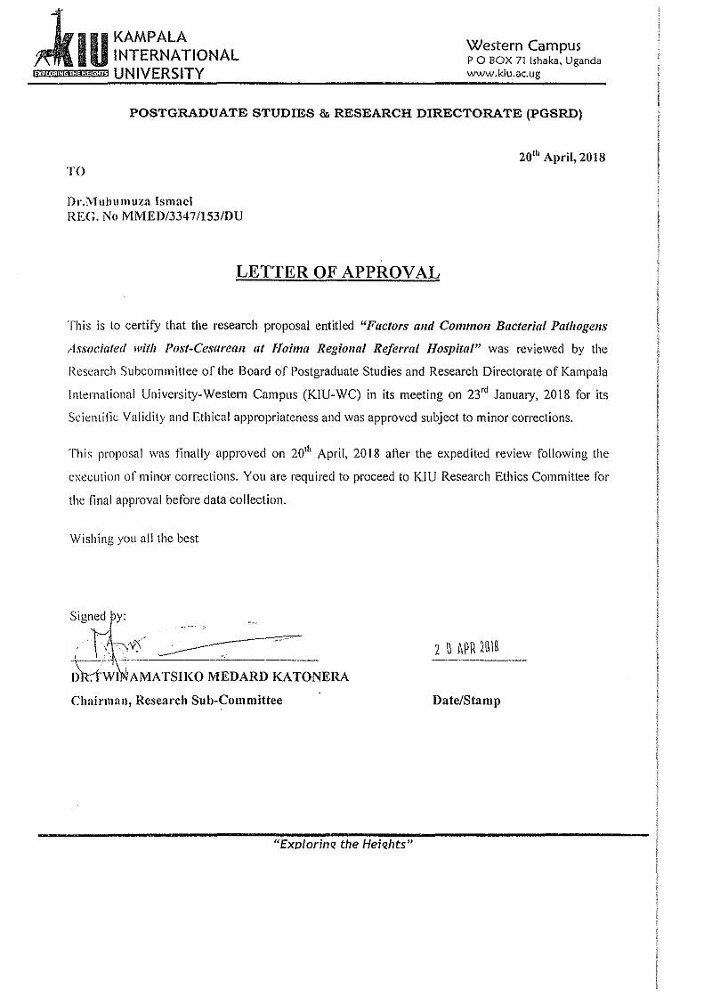
3.15.7 Approval Procedure ........................................................................................................... 28
3.15.8 Respect for community ...................................................................................................... 28
3.16 Dissemination of results ........................................................................................................ 28
CHAPTER FOUR ......................................................................................................................... 29
4.0 RESULTS ............................................................................................................................... 29
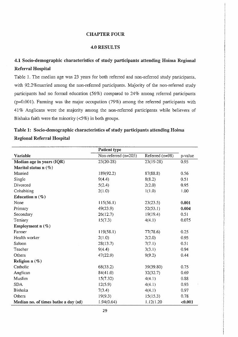
4.1 Socio-demographic characteristics of study participants attending Hoima Regional
Refenal Hospital ........................................................................................................................... 29
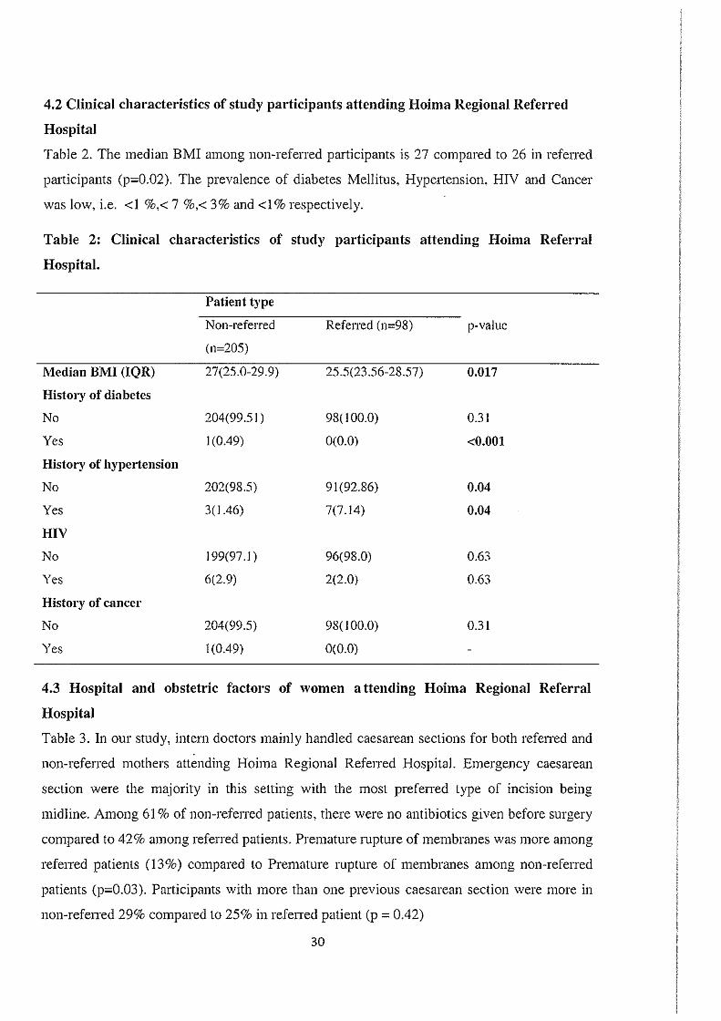
4.2 Clinical characteristics of study pm1icipants attending Hoima Regional Referred Hospital.. 30
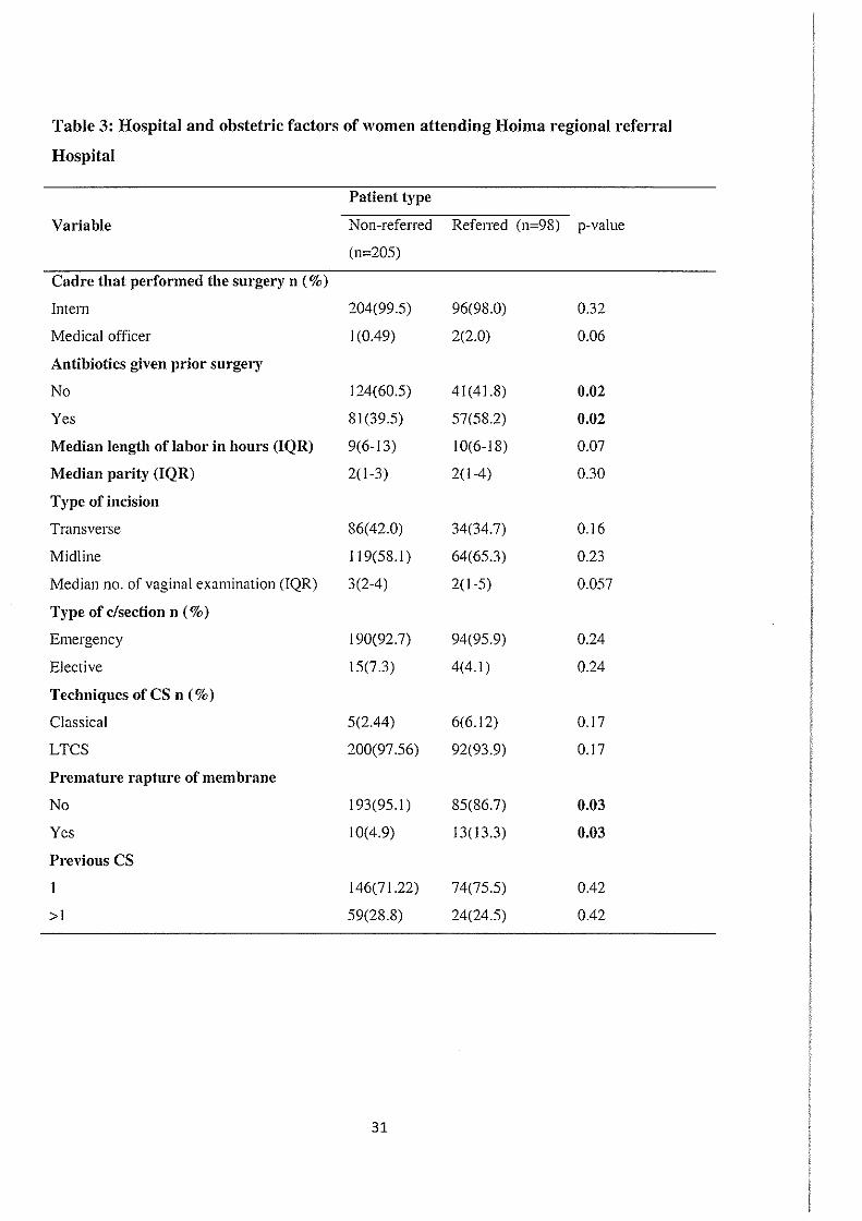
4.3 Hospital and obstetric factors of women attending Hoima Regional Referral Hospital... ...... 30
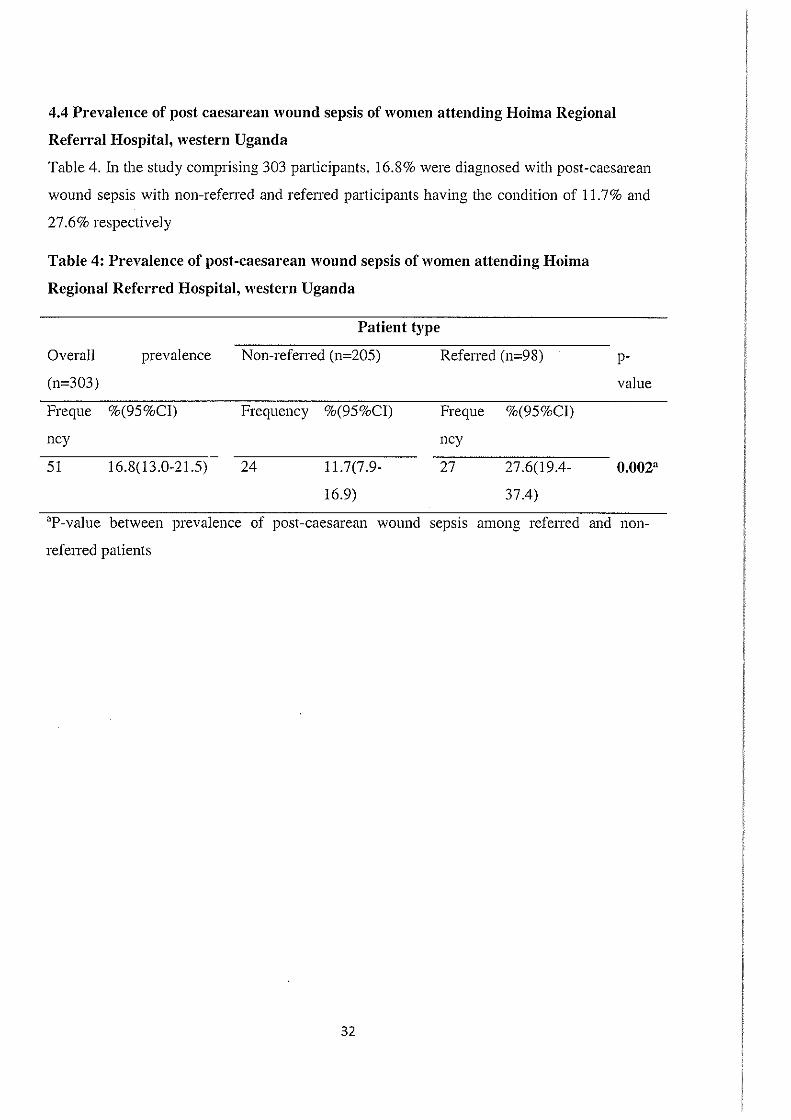
4.4 Prevalence of post caesarean wound sepsis of women attending Hoima Regional Referral
Hospital, westem Uganda ............................................................................................................. 32
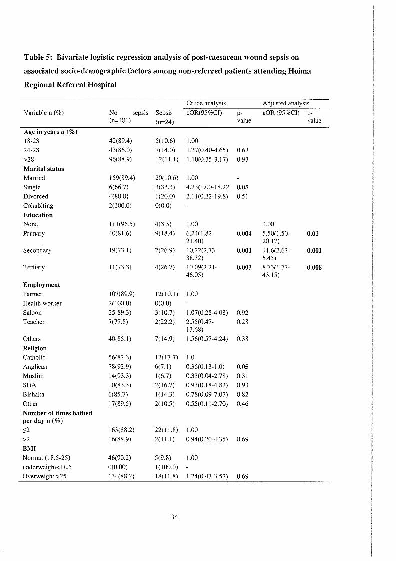
4.5 Bivariate and multivariate logistic regression analysis of post-caesarean wound sepsis on
associated socio-demographic factors among non-referred patients attending Hoima Regional
Refenal Hospital ........................................................................................................................... 33
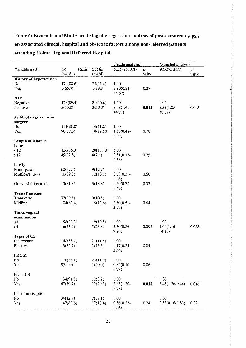
4.6 Bivariate and Multivariate logistic regression analysis of post-caesarean wound sepsis on
associated clinical, hospital and obstetric factors among non-referred patients attending
Hoima Regional Refen·al Hospital. ............................................................................................... 35
vii
Page 9
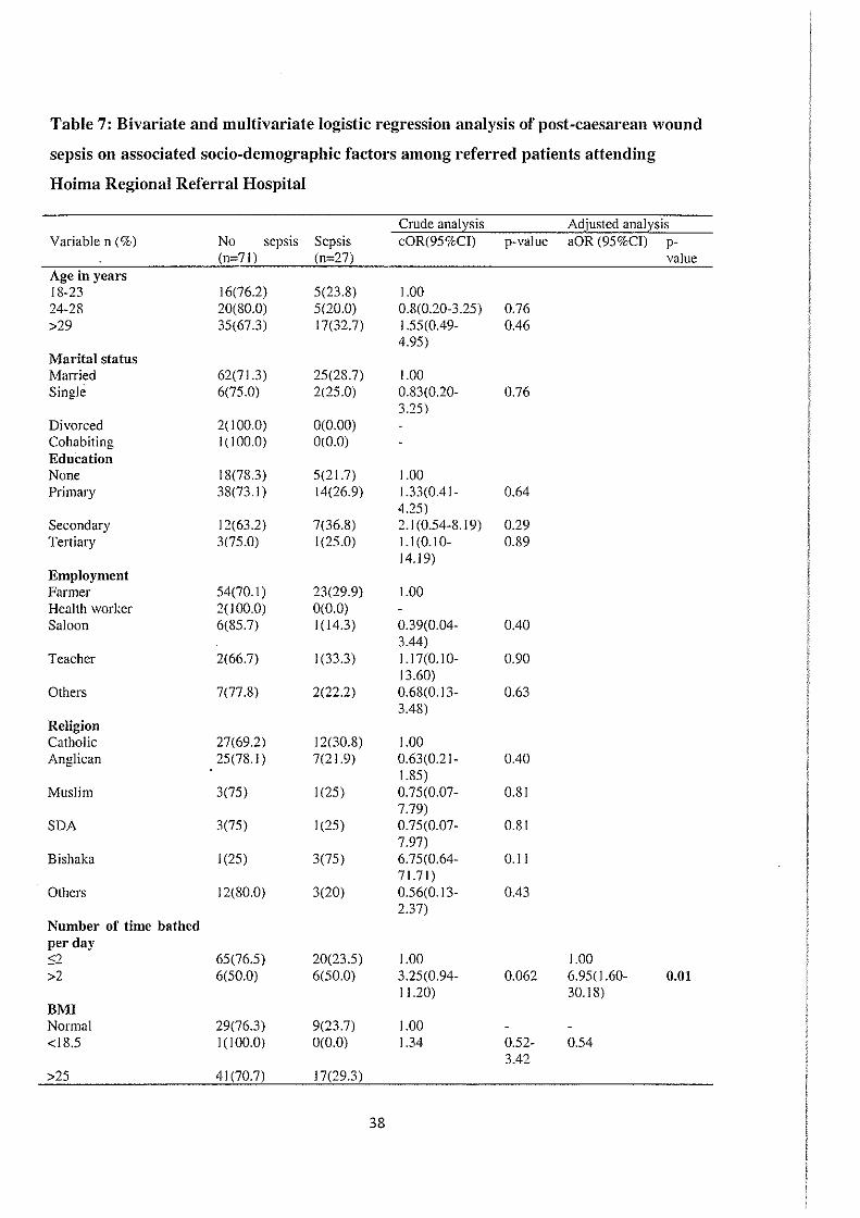
4.7 Bivariate and multivariate logistic regression analysis of post-caesarean wound sepsis on
associated socio-demographic factors among refened patients attending Hoima Regional
Refenal Hospital ........................................................................................................................... 37
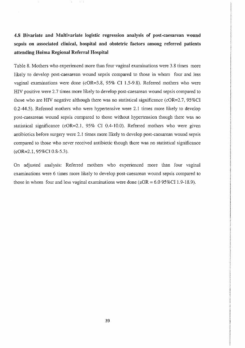
4.8 Bivariate and Multivariate logistic regression analysis of post-caesarean wound sepsis on
associated clinical, hospital and obstetric factors among refened patients attending Hoima
Regional Refenal Hospital ........................................................................................................... 39
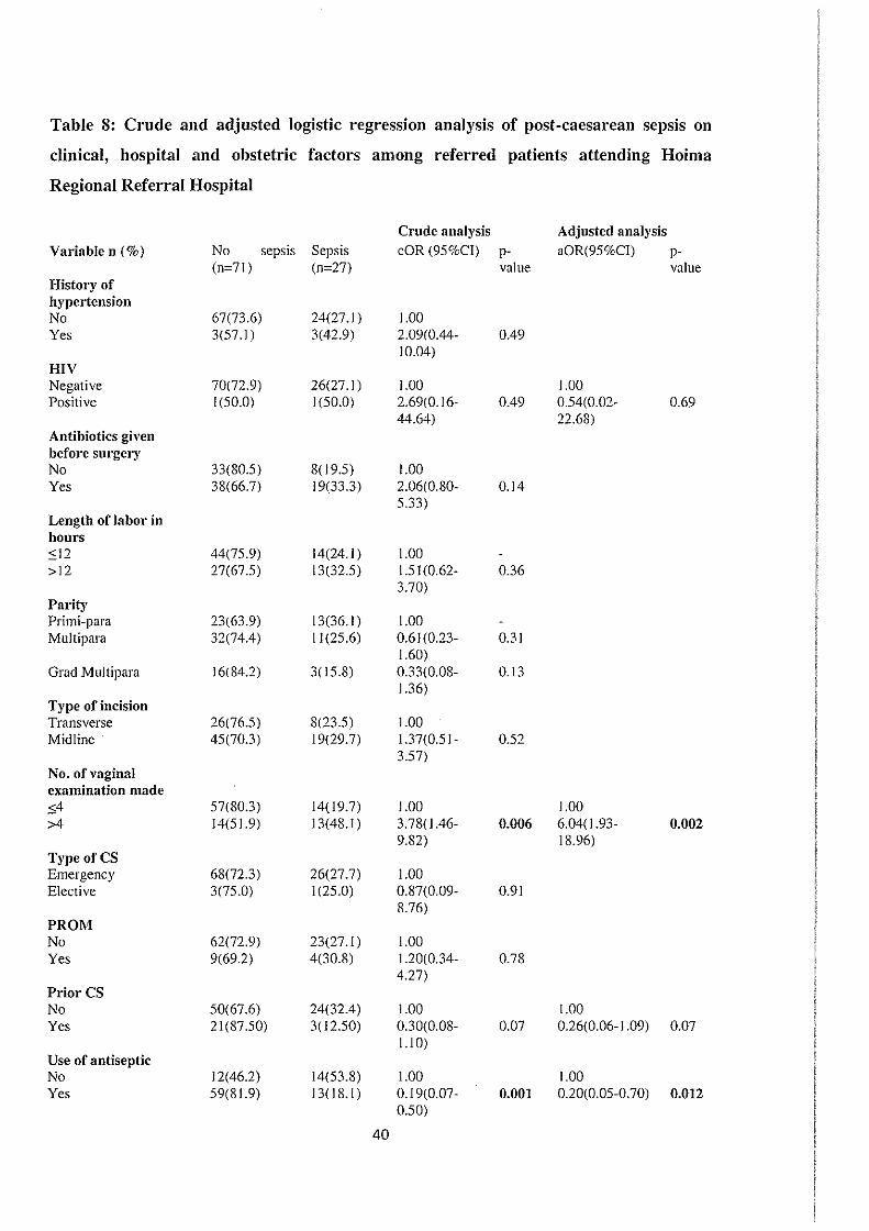
4.9 Common bacterial pathogens that were isolated among study participants attending
Hoima Regional Referral Hospital.. .............................................................................................. 41
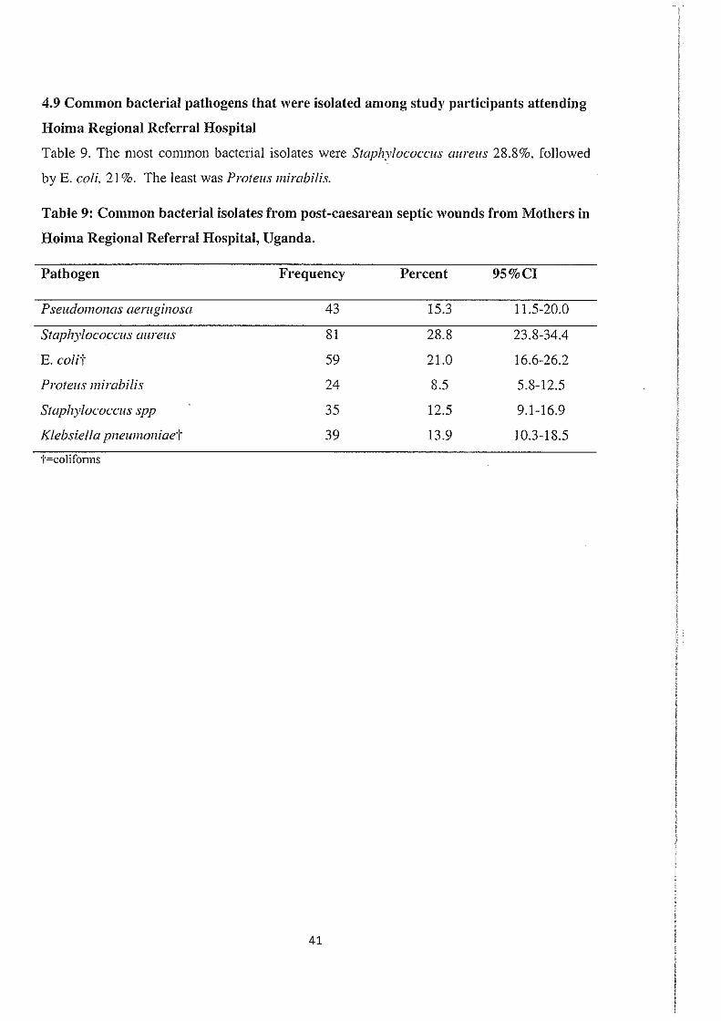
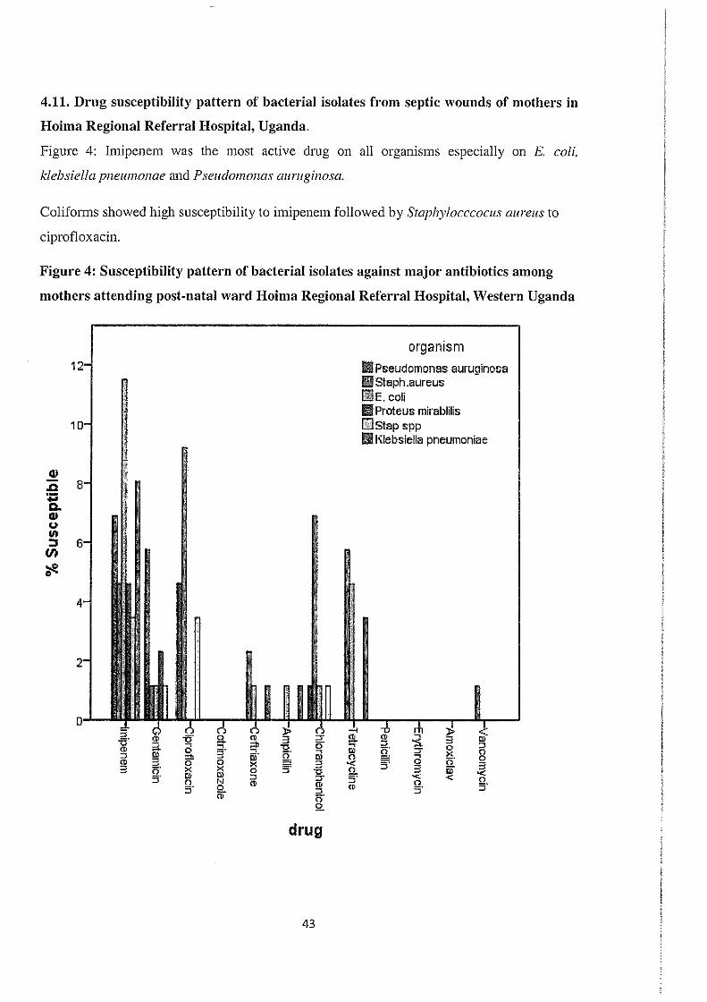
4.1 0. Drug susceptibility pattem of bacterial isolates from septic wounds of mothers in
Hoima Regional Referral Hospital, Uganda ................................................................................. 42
4.11. Drug susceptibility pattern of bacterial isolates from septic wounds of mothers in
Hoima Regional Refenal Hospital, Uganda ................................................................................. 43
CHAPTER FIVE .......................................................................................................................... 44
5.0 DISCUSSION, CONCLUSION, RECOMMENDATIONS AND LIMITATIONS .............. 44
5.1 DISCUSSION ......................................................................................................................... 44
5.1.1 Prevalence of post caesarean wound sepsis among mothers attending post-natal ward at
Hoima Regional Referral Hospital, Westem Uganda ................................................................... 44
5.1.2 Risk factors of post caesarean wound sepsis among mothers attending Hoima Regional
Refen·al Hospital, Western Uganda .............................................................................................. 44
5.1.3 Common bacterial isolates ................................................................................................... 46
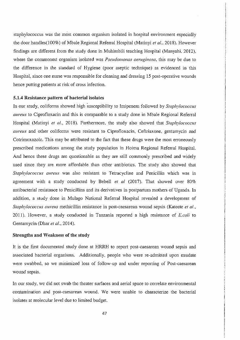
5.1.4 Resistance pattem of bacterial isolates ................................................................................ 47
5.2 CONCLUSIONS ..................................................................................................................... 48
5.3 RECOMMENDATIONS ........................................................................................................ 48
5.4 LIMITATIONS ....................................................................................................................... 48
REFERENCES ............................................................................................................................. 49
Appendix I: INFORMED CONSENT .......................................................................................... 57
Appendix II: TRANSLATED CONSENT FORM: (RUNYOORO-RUTOORO) ....................... 61
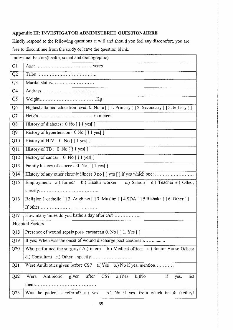
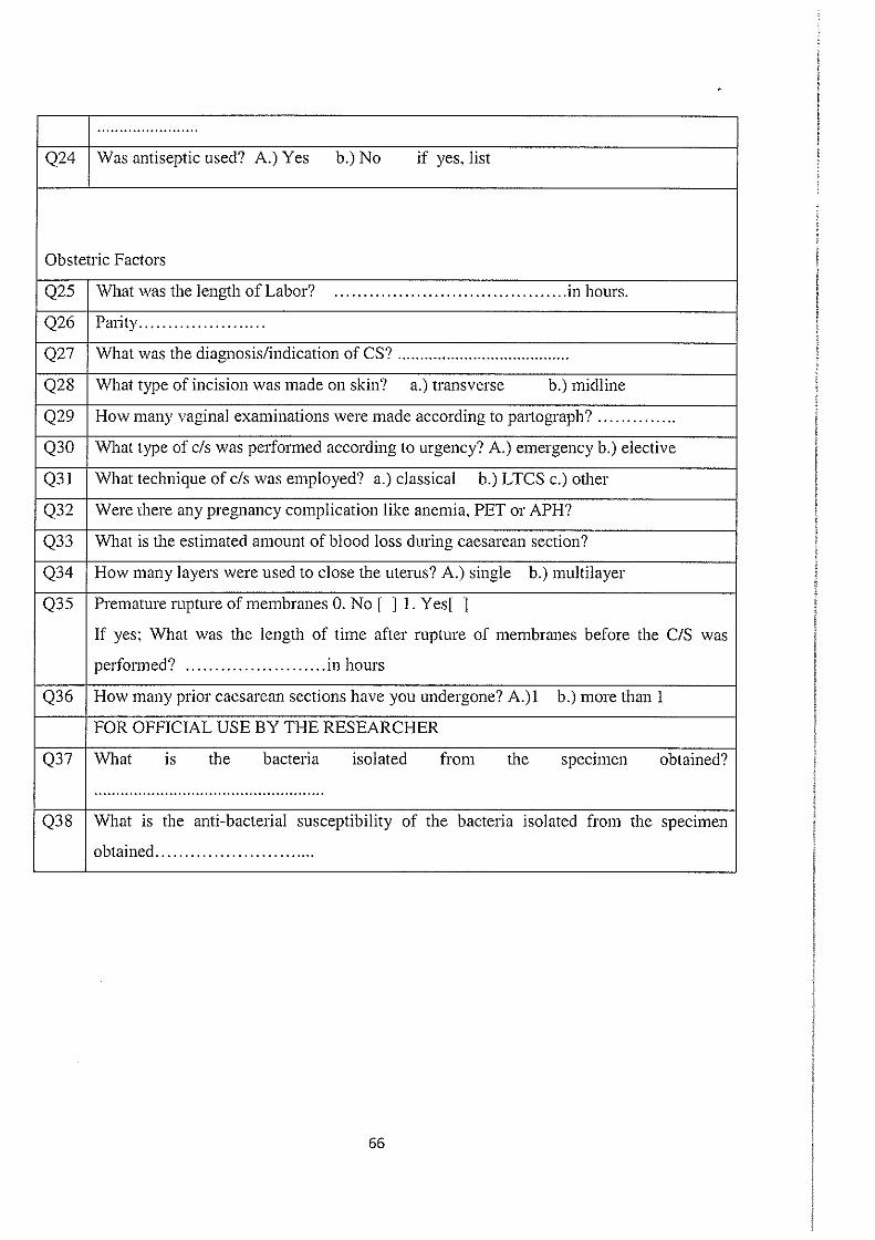
Appendix III: INVESTIGATOR ADMINISTERED QUESTIONAIRRE .................................. 65
viii
Page 10
LIST OF TABLES
. Table 1: Socio-demographic characteristics of study participants attending Hoima Regional
Refenal Hospital ...................................................................................................................... 29
Table 2: Clinical characteristics of study participants attending Hoima Refenal Hospital.. ... 30
Table 3: Hospital and obstetric factors of women attending Hoima regional refenal Hospital
.................................................................................................................................................. 31
Table 4: Prevalence of post-caesarean wound sepsis of women attending Hoima Regional
Refened Hospital, westem Uganda ......................................................................................... 32
Table 5: Bivariate logistic regression analysis of post-caesarean wound sepsis on associated
socio-demographic factors among non-refened patients attending Hoima Regional Referral
Hospital .................................................................................................................................... 34
Table 6: Bivariate and Multivariate logistic regression analysis of post-caesarean sepsis on
associated clinical, hospital and obstetric factors among non-refened patients attending
Hoima Regional Refened Hospital. ......................................................................................... 36
Table 7: Bivariate and multivariate logistic regression analysis of post-caesarean wound
sepsis on associated socio-demographic factors among refeiTed patients attending Hoima
Regional Refenal Hospital ...................................................................................................... 38
Table 8: Crude and adjusted logistic regression analysis of post-caesarean sepsis on clinical,
hospital and obstetric factors among referred patients attending Hoima Regional Referral
Hospital .................................................................................................................................... 40
Table 9: Common bacterial isolates from post-caesarean septic wounds from Mothers in
Hoima Regional Referral Hospital, Uganda ........................................................................... .41
ix
Page 11
LIST OF FIGURES
Figure I: Conceptual framework ............................................................................................... 6
Figure 2: Data collection Scheme ............................................................................................ 20
Figure 3. Resistance pattern of bacterial isolates against major antibiotics among mothers
attending post-natal ward Hoima Regional Refenal Hospital, Westem Uganda ................... .42
Figure 4: Susceptibility pattem of bacterial isolates against major antibiotics among mothers
attending post-natal ward HoinJa Regional Refenal Hospital, Westem Uganda ................... .43
X
Page 12
APH
E. coli
HRRH
KIU-REC
KIU-WC
MRSA
PCT
PET
RCOG
BMC
BMI
LTCS
KNH
cs
cOR
a OR
CI
Sd
IQR
LIST OF ABBREVIATIONS AND ACRONYMS
Antepartum Hemorrhage
Escherichia coli
Hoima Regional Referral Hospital
Kampala Intemational University Research Ethics Committee
Kampala Intemational University- Westem Campus
Methicillin-Resistant Staphylococcus au reus
Procalcitonin
Pre-eclamptic Toxemia (Preeclampsia)
Royal College of Obstetrics and Gynecology
BioMed Central
Body Mass Index
Lower Transverse Caesarean Section
Kenyatta National Hospital
Caesarean Section
crude Odds Ratio
adjusted Odds Ratio
Confidence Interval
Standard Deviation
Interquanile Range
xi
Page 13
Antibacterial agent:
Antibiotic prophylaxis:
Caesarean section:
Immediate puerpel"ium:
Puerperium:
OPERATIONAL DEFINITIONS
A drug that kills bacteria or stops their growth (Matinyi et a!.,
2018).
Antimicrobial drug administered in absence of any signs or
symptoms of sepsis to prevent occurrence of sepsis (Dhar et a/.,
2014).
The delivery of a baby, placenta and membranes through a
surgical incision in the mother's abdominal wall and uterine
after 28 weeks of amenorrhea (Chu eta/., 2012).
The first twenty four hours following tennination of pregnancy.
Period from the termination of labor to complete involution of
the uterus, usually defined as forty two days (RCOG, 2012).
Post-caesarean wound sepsis: Infection that develops on the incision site following
Prolonged mpture of
Membranes:
Premature rupture of
Membranes:
Sepsis:
Wound:
caesarean delivery and is diagnosed by clinician (Gelaw eta/.,
2017).
The rupture of membranes for more than 24 hours before
onset of labor (Banos eta/., 2010).
Rupture of membranes at least one hour before onset of labor
A condition that is life-threatening which occurs when the
body's response to infection causes injury to its own organs and
tissues (Kabau. 2014).
Trauma to living tissue caused by a blow or cut resulting into a
cut or breakage in skin (Gelaw eta/., 2017).
xii
Page 14
ABSTRACT
Background: Post-caesarean wound sepsis is among the most common problem for patients
who undergo caesarean section. It remains a common and widespread problem contributing
to morbidity and mortality; this could be due to an increase in antimicrobial resistant bacterial
pathogens. Therefore, a study to identify and document the factors associated with wound
sepsis and common bacterial pathogens can provide solution to prevent incidence and
establish microbiological mapping, and this is the intension for this research.
Objectives: To determine prevalence, identify factors, common bacterial pathogens from
post-caesarean wounds and antibacterial susceptibility pattem at Hoima Regional Referral
Hospital.
Research methods: This was a cross-sectional study conducted among patients with post
caesarean wound sepsis in the post-natal wards at Hoima Regional Referral Hospital.
Consecutive enrolment of 303 participants who consented to participate was done daily until
a required sample size was realized from July to September, 2018. Structured questionnaires
were used to collect data on associated factors and wound swabs were done. Culturing for
colony characteristics followed by Gram stain was used for provisional identity of pathogenic
bacteria. Further identification was done by a set of biochemical tests. Antibacterial
susceptibility pattem of isolated bacterial pathogens was determined by Kirby Bauer disc
diffusion method. Data was analyzed using STAT A VERSION 14.2.
Results: The wound sepsis rate was 16.8%. Being educated, multiple vaginal examination,
hygiene, previous caesarean sections and HIV seropositivity were all significantly associated
with wound sepsis (P value <0.05). Majority of the wound swab specimen yielded
Staphylococcus aureus, and the least-prevalent pathogen was Proteus mirabilis. Coliforms
showed high susceptibility to Imipenem followed by Staphylocccocus aureus to
ciprofloxacin. Resistance was highest for coliforms and Staphylococcus aureus against,
ciprofloxacin, gentamycin, ceftriaxone and cotrimoxazole.
Conclusions and recommendations: The rate of caesarean wound sepsis is high at Hoima
Regional Refenal Hospital. Being educated, multiple vaginal examination, hygiene, previous
caesarean sections and HIV sero-positivity are significantly associated with wound sepsis.
Staphylococcus au reus is the commonest organism isolated from exudates of septic wounds
after caesarean section. Health workers should be informed about the high prevalence, the
identified associated factors and common pathogens for proper management and also rational
use of antibiotics to combat resistance.
xiii
Page 15
1.1 Backgr·ound to the study
CHAPTER ONE
1.0 INTRODUCTION
Caesarean section is delivery of a baby through a surgical incision in the mother's abdomen
and uterus after 28 weeks of amenonhea (Chu eta/., 2012). Caesarean section is done when
vaginal delivery may cause a risk to the mother or baby such as when there is fetal distress, or
when the baby is abnormally positioned or there is prolonged labor or a case of maternal
factors such as pre-eclampsia or contracted pelvis.
Sepsis is a life-threatening illness caused by the body's response to an infection and develops
when mediators of inflammation are released in the general body circulation (Prucha,
Bellingan, & Zazula, 2015; Singer et a/., 2016). Modern understanding of sepsis is in
reference to human response to infection and this is mediated by several inflammatory
mediators (Vincent et al., 2013). This shows that sepsis is a complex physiological and
metabolic response of the body and is cunently a major reason for admission of patients to
the intensive care unit (Angus & van der Poll, 2013).
In medical practice, post-operative sepsis has been recognized as a major cause of mortality
and morbidity in patients as a result of dysregulation of host immune response to infection.
This leads to production of cytokines, prostanoids and nitric oxide, which suppress immunity
of the body further (Monkhouse, 2006). In addition, the leading cause of severe sepsis has
been shown to be related primarily to Gram-negative bacterial infections and the prognosis is
grave in older people, black race and those with a pre-existing illness (Mayr eta/,. 2014). In
addition, gram positive bacteria, in particular Streptococci species have also been isolated as
major pathogens in sepsis (Acosta & Knight, 2013).
Post-caesarean wound sepsis is co1mnon among women following caesarean section and this
can easily progress into septic shock if poorly managed (Kalisa, Rulisa, van den Akker, &
van Roosmalen, 2016; Morgan & Roberts, 2013; Sagy, Al-Qaqaa, & Kim, 2013). Early
diagnosis of post-caesarean wound sepsis using sepsis biomarkers (Omar, 2010) such as pro
calcitonin (PCT) is an important medical practice. These biomarkers are hardly available in
several health care units in developing countries and this has led to the development of a
global campaign on development of appropriate guidelines for successful management of
sepsis (Bloos & Reinhmt, 2014; Dellinger eta/., 2013). Successful management is crucial to
1
Page 16
ensure that unnecessary prolonged activation of the immune response is inhibited (Prucha et
a/., 2015; Stearns-Kurosawa eta/., 2011; Yealy eta/., 2015).
Though caesarean sections are done under aseptic conditions, the risk of post-caesarean
wound sepsis always exists (Dlamini eta/., 2015), which puts post-caesarean wound sepsis
among the most cmmnon nosocomial infections. Globally, the rate of caesarean section is
increasing rapidly (Ostovar eta/., 2010) and the most common complication after caesarean
section is wound sepsis, with an incidence of 3-15% (Zuarez-Easton et a/,. 2017a). Post
caesarean wound sepsis is also associated with matemal mortality rate of up to 3% especially
in health units that have no facilities to canyout safe caesarean sections or treat post
caesarean complications ( Gibbons eta/., 2012, Zuarez-Easton eta/,. 2017b).
In sub-Saharan Africa, post-caesarean wound sepsis has been shown to be in the range of
1.7% to 10.4% showing that the condition is significant in the continent (Chu eta/,. 2015;
HmTison & Goldenberg, 2016). This has been attributed to poor accessibility to health care
services which is below 3% and thus resulting in poor postnatal follow up and wonnd care
(Chu eta/., 2012; Irani & Deering, 2015). In addition, post-caesarean wound sepsis in sub
Saharan Africa has been associated with poverty, environmental pollution, poor preoperative
care, malnutrition, anemia, wound contamination, poor antibiotic selection and poor
immunity (Gelaw eta/., 2017).
In East Africa, the rate of caesarean sections is below 40%, which is a higher rate (Wmjoloh
et al., 2012) compared to the recommended rate of caesarean section as per the WHO which
considers that the best caesarean section rate is between 10-15% (Uriel, 20 18). This increases
the risk of post-caesarean wound sepsis.
In Rwanda the prevalence of post-caesarean wound sepsis has been shown to be at 4.9%
(Bizimana eta/., 2016) and in a parallel study from Zanzibar, post-caesarean wound sepsis
was shown to be caused primarily by Staphylococcus aureus and Escherichia coli and these
had led to increased hospital stay (Omar, 2010).
In Uganda, a study conducted in Jinja (Anguzu, 2007) showed that the major pathogenic
bacteria associated with post-operative sepsis are; Staphylococcus aureus, colifom1s, Proteus
mirabilis, Pseudomonas aeruginosa, Klebsiella pneumoniae and Enterobacter spp. In
addition, the study showed that these pathogens were highly resistant to ampicillin,
amoxicillin and chloramphenicol. The threat of post-operative bacterial sepsis in HIV
2
Page 17
infected patients has been established at 0.9% and majority of bacterial isolates have been
reported to be resistant to common antibiotics used (Anguzu & Olila, 2007: Kateete et
a/,.2011; Sekirime & Lule, 2009).
A study conducted at Mulago National Referral Hospital in Uganda showed that majority of
these septic wounds have pathogens that are methicillin resistant thus posing a major
healthcare challenge due to the limited class of dmgs available in several healthcare centers
in Uganda (Kateete eta/., 2011). A more recent study has shown antimicrobial resistance of
in 80% of post-partum mothers in Uganda (Bebell eta/,. 2017).
At Hoima Regional RefeJTal Hospital (HRRH) there was a high turnover of patients with
eight to ten caesarean sections done in a day (maternity theatre register). It was also noted
that theatres were being shared by other surgical teams and that the patients with post
caesarean wound sepsis were not isolated from the rest of the patients in ward. according to
the findings during an onsite visit to the Hospital.
Several associated factors including patient related factors, hospital factors and obstetric
factors have made management of post-caesarean wound sepsis challenging, leading to an
increase in hospital stays (Dhar eta/., 2014). Hence, thorough identification of the bacterial
pathogens and the associated factors of post-caesarean wound sepsis is important for
developing proper protocols to reduce its incidence and complications.
1.2 Problem statement
According to semi-mmual rep011 of 2017, Hoima Regional Referral Hospital (HHRH)
receives 20-30 patients in labor per day including 7-10 referrals, with a minimum of ten
caesarean deliveries being done per day (HRRH semi-annual maternal rep011, 2017). Post
caesarean wound sepsis in HRRH is high despite using aseptic technique and depanment
protocol of intravenous triple antibiotics (Ceftriaxone, Metronidazole and Gentamycin) after
caesarean section.
According to HRRH maternal report, in the month of September, 2017, there were 33 cases
of wound sepsis out of 199 caesarean sections done, which translates to a rate of 16.6%. This
leads to prolonged hospital stays and increased risk of complications like burst abdomen,
hysterectomy, repeat operations and even maternal death. This not only burdens the health
system greatly but it also financially depletes the patients and their families. The factors
3
Page 18
behind this high rate of caesarean wound sepsis, associated pathogens and their susceptibility
pattems have not been studied at HRRH.
1.3 Objectives
1.3.1 Purpose of the study
To assess the factors and to identify common bacterial pathogens associated with post
caesarean wound sepsis at Hoima Regional RefetTal Hospital.
1.3.2 Specific objectives
1. To determine the prevalence of post-caesarean wound sepsis at Hoima Regional
RefetTal Hospital.
2. To identify the factors associated with post-caesarean wound sepsis at Hoima
Regional RefetTal Hospital.
3. To identify the common bacterial pathogens among patients with post-caesarean
wound sepsis patients at Hoima Regional Refenal Hospital.
4. To assess the antibacterial drug susceptibility pattem of bacterial isolates among
patients with post-caesarean wound sepsis at Hoima Regional Referral Hospital.
1.4 Research questions
1. What is the prevalence of post-caesarean wound sepsis at Hoima Regional Referral
Hospital?
2. What are the factors associated with post-caesarean wound sepsis at Hoima Regional
RefetTal Hospital?
3. What are the common bacterial pathogens among patients with post-caesarean wound
sepsis at Hoima Regional Refenal Hospital?
4. What is the antibacterial drug susceptibility pattem of bacterial isolates among
patients with post-caesarean wound sepsis at Hoima Regional Refetnl Hospital.?
1.5 Justification
In Uganda, a couple of studies have been conducted on bums and post-operative sepsis
(Anguzu, 2007; Kateete eta/., 2011), while limited studies have been conducted primarily on
caesarean section patients. In addition, a high percentage (80%) of antimicrobial resistance
4
Page 19
has been reported (Bebell eta/., 2017) which implies that the management of post-caesarean
section mothers may be complicated. Information gathered from this study would help to
reduce the incidence of post-caesarean wound sepsis, improve prognosis in affected patients,
and develop effective strategies for a reduction in antimicrobial resistance through effective
therapy administration. This would subsequently lead to establishment of microbiological
mapping at Hoima Regional Referral Hospital.
1.6 Significance of the study
The ministry of health and research institutions will gam information on the common
bacterial isolates in post-caesarean wound sepsis (Omar, 2010; Royal College of Obstericians
& Gynaecologists, 2012), thus helping in formulating appropriate policies for improved
health service provision in the region and increase availability of the drugs sensitive to the
common bacteria. This may subsequently lead to improved livelihoods in the community and
save the govemment and patients from costs that would be spent treating resistant microbial
agents (Dellinger et al., 2013; Morgan & Roberts, 2013).
The study findings established the cmTent rates of post-caesarean wound sepsis at Hoima
Regional Referral Hospital. Since the study assessed the various factors associated with post
caesarean wound sepsis, the results may fonn a basis for future measures to reduce post
caesarean wound sepsis at the Hoima Regional Refe1Tal Hospital in addition to serving as a
benchmark for future references for post-caesarean wound sepsis.
5
Page 20
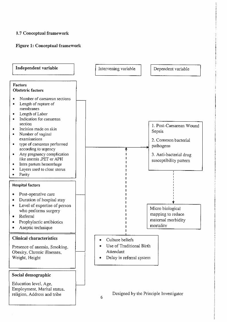
1.7 Conceptual framework
Figure 1: Conceptual framework
Independent variable Intervening variable Dependent variable
Factors Obstetric factors
• Number of caesarean sections I--• Length of rupture of
membranes • Length of Labor • Indication for caesarean
section I. Post -Caesarean Wound • Incision made on skin Sepsis • Number of vaginal
examinations 2. Common bacterial • type of caesarean pe1formed pathogens
according to urgency t • Any pregnancy complication 3. Anti-bacterial drug
like anemia ,PET or APH susceptibility pattern • Intra pmtum hemon-hage • Layers used to close uterus
I
• Parity I I I I
Hospital factors I I I
Post -operative care I • I-- I
Duration of hospital stay I • t • Level of expenise of person
Micro biological who performs surgery mapping to reduce • Refen-al
0 Prophylactic antibiotics matemal morbidity mortality • Aseptic technique
• Clinical characteristics • Culture beliefs
Presence of anemia, Smoking, • Use of Traditional Birth
Obesity, Chronic illnesses, I-- Attendant
Weight, Height • Delay in referral system
Social demographic
Education level, Age, -Employment, Marital status. religion, Address and tribe
6 Designed by the Principle Investigator
Page 21
1.7.1 Description of Conceptual Framework
The diagram above shows the interaction between the independent (obstetric, hospital factors,
clinical characteristics and social demographic), intervening (use of traditional birth
attendants and delay in referral system) and dependent variables (Post-ceasarean wound
Sepsis, Common bacterial pathogens and Anti-bacterial drug susceptibility pattem) and in
tum reduce matemalmorbidity and mortality.
Factors that might affect the woman following caesarean section to get post-caesarean wound
sepsis include: Obstetric factors for example parity; prolonged labor; indication for caesarean
section like obstructed labor; length of labor; number of caesarean sections; wrong technique
of caesarean section; type of caesarean section performed according to urgency (emergency
or elective caesarean section); incision made on the skin; layers used to close the uterus; any
pregnancy complication like APH; Post-partum hemorrhage; PET; and number of vaginal
examinations, might increase the chance of introducing bacteria into the uterus through the
vagina.
Hospital factors for example post-operative care, duration of hospital stay, level of expertise
of person who performs the surgery, refernl, prophylactic antibiotics, aseptic technique
used, and clinical characteristics for example presence of anemia, smoking, obesity, and
chronic illnesses like diabetes, hypertension among others, may contribute to the
development of wound sepsis after caesarean section. Others that contribute to this may be
socio-demographic factors for example low socioeconomic status, hygiene, marital status,
religion, education and age.
1.8 Study scope
1.8.1 Content Scope
The study placed emphasis on factors and common bacterial pathogens associated with
prevalence of post-caesarean wound sepsis. Women with eligibility criteria were recruited
and consented. Exudate swabs were collected to determine bacterial causes of post-caesarean
wound sepsis. In particular, emphasis was placed on Gram positive and Gram negative
bacterial pathogens and also their drug susceptibility testing antibiotic discs: Gentamycin,
Ceftriaxone, ciprofloxacin, ampicillin, amoxiclav, cotrimoxazole, chloramphenicol,
tetracycline, imipenem, vancomycin and penicillin. A questiom1aire was filled to obtain the
factors associated with post -caesarean wound sepsis.
7
Page 22
1.8.2 Geographical Scope
The study was done at Hoima Regional Referral Hospital in the maternity postnatal ward.
Mothers who meet eligibility criteria were recruited from the postnatal ward and a private
side room allocated for the study was used. The study participants came from catchment
areas of Hoima Regional Referral Hospital such as Kiboga, Mubende, Masindi, Kiryandongo
and other neighboring districts.
1.8.3 Time Scope
The data collection period was limited to a period of July 2018 to September 2018. This was
adequate duration to achieve the study sample size basing on the reports of admissions at
HRRH.
8
Page 23
CHAPTER TWO
2.0 LITERATURE REVIEW
2.1 Prevalence of post-caesarean wound sepsis
Compared to spontaneous vaginal delivery, caesarean section is associated with increased
neonatal and matemal morbidity and mortality (Oboro eta/., 2010). Post-caesarean delivery
complications include wound separation and once complicated by infections may develop
into sepsis (Quinlan & Murphy, 2015), thus showing a need to identify the antibacterial
sensitivity profile in the bacterial isolates for effective patient management. Post-caesarean
wound sepsis represent a significant health and economic challenge and identifying the
organisms and techniques to manage caesarean wounds is essential for obstetricians
(Fitzwater & Tita. 2014).
Following invasion of the wound by pathogenic bacteria and mismanagement, wound sepsis
has been found to be an inevitable outcome (Singer eta/., 2016). Sepsis is a condition that is
life-threatening and it occurs when the body's response to infection causes injury to its own
organs and tissues. Since this high prevalence is in spite of the advances in medical
knowledge and treatment (Royal College of Obstericians & Gynaecologists, 2012), it
demonstrates a need to understand it better for its effective management. Moreover caesarean
delivery remains one of the most important factors of puerperal sepsis of which post
caesarean wound sepsis is among (Conroy eta/., 2012) since mothers undergoing caesarean
section have a 5 to 20-fold higher chance of getting puerperal sepsis compared with mothers
who give birth vaginally (Kabau, 2014).
Post-caesarean wound sepsis remains a major source of morbidity and mortality in
postpmtum mothers especially in the developing countries like Uganda (Kabau. 2014). Post
caesarean wound sepsis is also associated with long duration of hospital stay, increased cost
of care and increased morbidity and mortality (Dhar eta/., 2014).
There are several factors that exacerbate post-caesarean wound sepsis including patient
related factors, hospital factors and obstetric factors. It is these factors that have made
management of post-caesarean wound sepsis challenging and increase in hospital stays (Dhar
et al., 2014) Hence, thorough identification of the bacterial pathogens and factors that are
associated with post-caesarean wound sepsis is important for developing proper protocols to
reduce its incidence and complications.
9
Page 24
In one study by Ngowe et a/., (2014), tbey linked the high prevalence of post-operative
infection (20.6%) to the fact that most of their participants were post-caesarean section
patients. They reported that the surgical site after a caesarean section is more prone to
infection given that there is a direct connection between tbe site and the bacteria flora
environment of tbe vagina. Besides, before a caesarean section, the patient is subjected to
multiple vagina/cervical examinations which cmTy bacteria from the vagina into the bacteria
free uterine cavity. The rate of 19.4% of post-caesarean wound sepsis was similar to 7- 20%
repo11ed by (Ngowe el a!., 2014).
In another study, one in ten (11 %) of all caesarean sections had developed wound sepsis. The
figure might have been largely underestimated as tbe study was exclusively reliant on
medical records review and it did not involve post -discharge follow-up. Further studies
conducted in Norway and Scotland reported tbat 86% (15) and 71% (16) of the sepsis
occmTed after discharge and also tbe most of tbe diagnosis was based on clinical basis and
could have missed the patients who may not present with the classical signs of inflammation
(Wodajo, Belayneh, & Gebremedhin, 2017).
2.2 Factm·s associated with post-caesarean wound sepsis
In the development of post-caesarean wound sepsis, increased number of parity, presence of
non-communicable diseases and infectious diseases , anaemia, pre-eclampsia and being obese
are the major factors that have been associated with post-caesarean wound sepsis (Dhar eta/.,
2014). h1 East Asia, the main factors for surgical site sepsis amongst post-operative mothers
have been found to be independently related and these include pre-operative remote infection,
chorioa11111ionitis, matemal preoperative condition especially among those witb an ongoing
infection, pre-eclampsia, higher body mass index (obesity) and increased blood loss during
surgery (Dagshinjav eta/,. 2017).
In addition, a study conducted at the Washington University Teaching Hospital by Temming
et al., (2017), showed that post-operative sepsis was common amongst mothers who had been
subjected to a low transverse caesarean section technique and prophylactic treatment using
cephalosporins was associated with improved outcomes. Also, according to Chu et a!.,
(2015), premature rupture of membrm1es and increased number of vaginal examinations were
associated witb post-caesarean wound sepsis. This has subsequently led to the need for
making adjustments while dealing witb patients who are at high risk of developing post-
10
Page 25
caesarean wound sepsis. Consequently, the Cohen's incision for entry, single closure of the
uterus and non-closure of both layers of the peritoneum have been recommended for effective
management of high risk patients (Hema & Johanson, 2002).
Caesarean sections wound sepsis classification involves four classes, namely:- Class I which
involves a clean wound in which no inflammation is encountered on entry; Class II which is
a clean-contaminated wound in which entry is done under controlled conditions and basic
level of contamination may be observed as per routine caesarean sections; Class III caesarean
section which involves contamination in which there is an accidental major breakage in
sterile technique or spillage from the uterine contents; and Class IV caesarean section wound
sepsis which is common in old traumatic wounds with retained devitalized tissue. and which
involves dirt and severe infection of the surgical wound. This is common among patients who
are undertaking multiple caesarean sections and is complicated by pre-existing clinical
infection (Conroy eta/., 2012).
In addition, more recent evidence has shown that using a plastic retractor instead of the
traditional Collins metal self-retaining wound retractor, reduces the risk for the development
of post-caesarean wound sepsis in mothers, showing a need to revise the routine materials
used during surgery (Hinkson et al,. 2016). In multiparous mothers, development of wound
sepsis after caesarean section has been shown to be associated with uterine wound dehiscence
and this wan-ants further investigations to be conducted in affected mothers to improve on
their prognosis (Bharatam eta/., 20 15).
Cunent evidence has also shown that women who received both chlorhexidine-alcohol and
iodine-alcohol for skin antisepsis at caesarean section compared to those who received one
had a lower risk for the development of post-caesarean wound sepsis (Temming eta/., 2017).
The same study also showed that obesity, smoking and presence of non-communicable
diseases, obstetrician experience and skin incision type, were not major factors in patients
who received prophylactic antibiotics within 60 minutes of caesarean section and prior to
skin incision (Dlamini et al., 2015). Application of chlorhexidine-alcohol skin antiseptics
within 3 minutes to skin incision and closure of the subcutaneous layer when it was greater
than two centimetres deep reduced the risk of post-caesarean wound sepsis (Temming et al.,
2017).
11
Page 26
Caesarean section is often indicated following fetal distress, prolonged second stage labor,
breech, and cephalo-pelvic dispropm1ion. However, the development of sepsis means that
patients would stay longer in the hospital than necessary probably as a result of severe
hemonhage during par1urition (Mylonas & Friese, 2015). Mismanagement of sepsis of
bacterial origin as a result of poor post-operative care has been shown to be associated with
high matemal mo11alities the prevalence of which is highest in developing countries. thus
leading to the surviving sepsis campaign which advocates for improved management of
patients (Acosta et al., 2013). This is impm1ant since poor post-operative care would lead to
increased matemal and neonatal morbidity (Oboro et al., 2010) which would lead to
decreased incidence of wound infections, thus reducing the risk posed by sepsis to post
operative in the community.
In Norway, low level of education of patients has been associated with a higher risk for
caesarean sections wound seps;s as compared to the highly educated group (Tollane et a/,
2007). This has led to a lot of controversy as to whether caesarean sections wound sepsis are
basically for the poor in developed countries. In addition a study by Cesaroni et a/., (2008)
showed that women with a primary level of education had an over 20% risk for caesarean
sections wound sepsis than those who had attended university.
Hygiene has also been associated with the ability to influence microbial colonization of the
wound following caesarean sections thus affecting the immune status of the patient (Neu &
Rushing, 2011 ). This shows that improved hygiene leads to improved patient outcomes. In
addition. caesarean sections wound sepsis have been associated with socio-economic status
that is to say, low in poor communities (employment), high in highly educated patients, being
manied and age (Faisal eta/,. 2017). According to a recent study done in Rwanda patients
who are referred from lower health units usually present in critical conditions and their
prognosis are poorer compared to those who are not referred (Kalisa eta/,. 2016).
2.3 Common bactel"ial pathogens in post-caesarean wonnd sepsis
The common bacterial pathogens causing sepsis in the hospital (Royal College of
Obstericians & Gynaecologists. 2012) include Streptococcus pyogenes, Staphylococcus
azaeus, colifonns, Streptococcus pneumonia, Clostridium septicum, Methicillin-resistant
Staphylococcus aureus (MRSA), Escherichia coli and Morganella morganii. In East Africa,
the prevalent pathogens are .Staphylococcus a111·eus, coliforms, Pseudomonas aeruginosa,
Proteus mirabilis, Klebsiella pneumoniae, Escherichia coli, and Enterobacter Spp (Anguzu 12
Page 27
& Olila, 2007; Sekirime & Lule, 2009) which shows an interplay of both Gram-negative and
Gram-positive bacteria. In addition, Cordioli eta/., (2013) has shown that the major Gram
negative bacteria in post-caesarean sepsis are Escherichia coli, Hemophilus il?f/uenza,
Klebisiel/a spp., Enterobacter spp., Proteus spp.. Pseudomonas spp.. Serratia spp.
Furthermore, the major Gram-positive bacteria (Cordioli et al., 2013) have been reported to
include Pneumococus, Streptococcus groups A, B and D., Enterococcus, Staphylococcus
aureus, Listeria monocytogenes, while the major anaerobic bacteria have been shown to be
Bacteriodes species, Clostridium peifringens, Fusobacterium species, Peptococcus and
Peptostreptococcus. These observations show that aerobic bacteria are a major concem in the
development of post -caesarean wound sepsis.
2.4 Antibacterial drug Susceptibility Patterns of Bacterial Isolates
In the management of post-caesarean wonnd sepsis, early identification of the problem and
constitution of the appropriate therapy is important in the improvement of patient's prognosis
(Moores, 2013). However, with the increasing burden of antibiotic resistance, offering the
right treatment is cunently a challenge, especially in developing countries were laboratory
costs are highly exaggerated (Ezeonwumelu et al., 2016). This is important since bacterial
culture is important in identifying major pathogens and making appropriate and effective
diagnoses (Bonham, 2009; Cheesbrough, 2006). This is highly important since prophylactic
treatment has been shown to improve on post-operative wound healing in a matemal
population (Dlamini eta/., 2015).
Antibacterial sensitivity is important for effective management of post-caesarean wound
sepsis of bacterial origin since treatment failures lead to a poor prognosis in affected patients
(Anguzu., 2007). Under normal conditions, large numbers of the peripheral blood neutrophils
enter sites of bacterial infection by first adhering to activated endothelial cells and then
migrating along a gradient of chemotactic factors. In contrast, neutrophils from septic patients
have increased expression of surface integrins which promote firm adhesion to endothelial
cells. As a sequence, the neutrophils remain bound more tightly to the endothelial cells and
fail to migrate appropriately into the site of bacterial infection (Jacobi, 2002).
At Mbarara Regional Referral Hospitai(U ganda), antibacterial resistance has been shown to
be highest among the penicillins and their derivatives (Bebell eta/., 2017), highlighting the
need to identify the sensitivity profile in post-partum women after caesarean section. A study
conducted at Mulago Hospital, in Central Uganda, has also shown that antimicrobial 13
Page 28
resistance to various antimicrobial agents is a real threat (Kateete eta! .. 2011). In Tanzania,
general resistance to antimicrobial agents has also been found to be high with gentamicin
being found to be the only effective antibacterial agent against the isolates (Dhar eta/., 2014).
This implies that without an updated susceptibility profile in a given population, it would be
challenging for clinicians to effectively manage sepsis in post-caesarean mothers (Royal
College of Obstericians & Gynaecologists, 2012).
2.4.1 Methods for antimicrobial susceptibility testing
Due to the high burden of bacterial resistance to antimicrobials, there has been interest in
dmg susceptibility testing to ensure good treatment outcomes. Two main methods are used
for susceptibility testing which are the disc diffusion and minimum inhibitory concentration
(MIC) tests for aerobic bacteria (Tenover, 2009). Selection of the most appropriate
antimicrobial agents to test and to report is a decision best made by each laboratory in
consultation with the infectious diseases practitioners and the pharmacy, as well as the
pharmacy and therapeutics and infection control committees of the medical staff (Cavalieri,
Rankin, Harbeck, & Sautter, 2005).
The disk diffusion method has several steps. Once isolated colonies are available from an
organism that has been identified as a potential pathogen, it is necessary to proceed to
pe1form the susceptibility test as follows: The colonies are selected first and then the
inoculum suspension is prepared and standardized. h1oculation of the plate is then done. The
antimicrobial disks are added and then the plates are incubated. The diameters of the zones
inhibition are then measured and the results interpreted using the criteria by Clinical and
Laboratory Standards Institute (CLSI), fonnerly known as the National Committee for
Clinical Laboratory Standards (NCCLS) (CLSI, 2017; Cavalieri eta/., 2005).
The other common method is the minimum inhibitory concentration test. The minimal
inhibitory concentration (MIC) of an antimicrobial agent is the lowest concentration of the
antimicrobial agent that inhibits a given bacterial isolate from multiplying and producing
visible growth in the test system. The concentration in the laboratory is determined by
incubating a known quantity of bacteria with specified dilutions of the antimicrobial agent.
The results are interpreted as susceptible, intennediate, or resistant using the criteria for the
Clinical Laboratories (CLSI, 2017). The MIC tests can be done by either broth or agar media,
but broth microdilution is the most widely used method in clinical laboratories. MIC panels
14
Page 29
that contain dilutions of one or multiple antimicrobial agents in a broth microdilution format
are on market and are cleared before use by Food and Drug Authority.
2.4.2 Performing MIC vs disk diffusion tests
MIC tests are required for some organisms/antimicrobial combinations for which disk
diffusion testing has proven to be unreliable (Cavalieri et a!., 2005; Tenover, 2009). These
include:
1. Streptococcus pneumoniae which requires that MIC test be performed for penicillin
when isolates show zones of inhibition <20 mm around oxacillin disks (a screening
test for penicillin resistance), and also MIC tests for cefotaxime or ceftriaxone
because breakpoints for disk diffusion testing have not been established for these
agents.
ii. Viridans streptococci which also require detennination of M!Cs when isolates are
from normally sterile body sites.
iii. Staphylococcus species which require that MIC tests be pelformed to detect decreased
susceptibility to vancomycin since this cmmot be determined using the disk diffusion
test. These specific methods are mainly recommended for use when the isolates are
resistant to the common antibiotics. This was in one of the studies in Uganda with
commendable outcomes (Najjuka eta/., 2016). Antimicrobial susceptibility pattem of
isolated bacterial pathogens will be performed by Kirby Bauer disc diffusion method
according to the guidelines of the Clinical and Laboratory Standards Institute. We
chose this method as it is the one recommended by the CLSI (CLSI, 2017).
15
Page 30
CHAPTER THREE
3.0 RESEARCH METHODOLOGY
3.1 Study design
This was a cross-sectional study because the purpose of the study was to determine
prevalence, identify factors, common bacterial pathogens from post -caesarean wounds and
antibacterial susceptibility pattern at Hoima Regional RefeJTal Hospital.
3.2 Study site and setting
The study was conducted in the postnatal ward at Hoima Regional Referral Hospital which is
a public hospital. Hoima District has GPS coordinates 0 I 24N, 31 18E and is approximately
230 km by road from Kampala, which is the capital city of Uganda. The major tribe is
Banyoro and the main religions are Islam and Christianity; and the majority of the population
are cultivators and animal keepers.
Hoima Regional RefeJTal Hospital is a well-established hospital and it offers both in-patient
and out-patient services with a range of departments and clinics, including General Surgery,
Obstetrics and Gynecology and Internal Medicine. The hospital is well equipped with a bed
capacity of 400. The Obstetrics and Gynecology Department of Hoima Regional RefeJTal
Hospital has four specialists, one Resident doctor, five intern doctors and 13 midwives. The
obstetrics and gynecology department has I 10 beds.
According to the hospital records (HRRH semi-annual maternal report, 20 17), Hoima
Regional RefeiTal Hospital performs approximately a minimum of 10 caesarean sections per
day. These are done in two theatres that are shared by other surgical teams. The hospital had
a range of 10-20 vaginal deliveries per day and also offers antenatal and postnatal services.
The main laboratory of Hoima Regional Refernl Hospital consists of the following sections:
hematology and blood bank. chemistry, parasitology and microbiology. It was composed of
20 staff members and these include three specialists, two laboratory technologists, eight
laboratory technicians, six laboratory assistants, and one laboratory attendant. The exudate
swab samples were processed in the microbiology laboratory which was operated by one
laboratory technologist, one laboratory technician, one laboratory assistant and one laboratory
attendant. It was well equipped to can·y out culture and sensitivity and other microbiological
tests. Some of the equipment found in this laboratory were; autoclave, incubator, microscope,
hot air oven, refrigerator, safety cabinet and gas cylinder. It also had enough stains which
16
Page 31
were used in the processing of samples. These include crystal violet solution. Lugol's iodine,
neutral red solution and 50% acetone alcohol.
3.3 Study population
The study population were mothers who have delivered by caesarean section at Hoima
Regional Refenal Hospital during the period of the study.
3.4 Selection criteria
3.4.1 Inclusion Critel'ia
All adult and emancipated minor mothers (on ward or re-admitted) who would have delivered
by caesarean section at Hoima Regional Referral Hospital with or without post-caesarean
wound sepsis.
3.4.2 Exclusion Criteria
Those done caesarean section from other health units and then referred to Hoima regional
refenal hospital were excluded due to limited access to their medical records. Mothers who
would have had a re-exploration due to caesarean section complications other than suspected
sepsis. as well as those who were in their early puerperium and those who reported after six
weeks, were also excluded.
3.5 Sample size
The minimum sample size for this study was 271.
3.6 Sample size determination
Specific objective one: The prevalence of post caesarean wound sepsis in Uganda was found
to be 22.2% (Hassan & Alegbeleye, 2018).
Using formula (Daniel, 1999):
(zaFx px (1- p) n=
Where:
Z =Standard normal deviate at 95% level of confidence; z= 1.96
Z.,= z-statistic at a=l.96
17
Page 32
p =prevalence of post-caesarean wound sepsis in Uganda, p=22.2%
e = level of precision (in proportion of one, if 5% e=0.05)
n = Desired sample size
(1.96) 2 x 0.222x (1- 0.222) 11
= 0.052 266
Objective two: The associated factors of post-caesarean wound sepsis; the sample size was
detennined according to modified Daniel's formula (Daniel, 1999);
1 (z., + zp)2 xR px (1- p)
n= 2 e
Where:
Z =Standard normal deviate ~.t 95% level of confidence; z= 1.96
Z,.= z-statistic at a=l.96. z11 = z-statistic at B=0.84
p =prevalence of post-caesarean wound sepsis in Uganda, p=22.2%
e =level of precision (in proportion of one, if 5% e=0.05)
n =Desired sample size
R = Odds ratio=2.0
1 (1.96 + 0.84) 2 x 2 x 0.222x (1- 0.222)
11 = 0.05 2 271
Objective three & four: The common bacterial cause of post-caesarean wound seps1s.
Pseudomonas aeruginosa. 21% in Muhimbili University teaching hospital (Manyahi, 2012);
Using formula; (Daniel, 1999)
Where;
n = Desired sample size
z = Standard normal deviate at 95% level of confidence; z= 1.96
p =expected prevalence of post-caesarean wound sepsis, p=0.21
18
Page 33
d = level of precision (in propmtion of one, if 5% d=0.05)
(Z,.)2xp(1- p) n = (d)z
(1.96) 2 X 0.21x (1- 0.21) n = 0.052 = 255
Therefore the overall minimum sample size for this study was 303 participants.
3.7 Sampling technique
Consecutive emollment of participants who consent to participate in the study. This was
cmTied out on a daily basis until required sample size.
19
Page 34
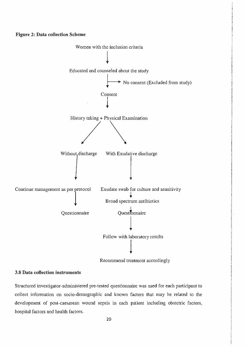
Figure 2: Data collection Scheme
Women with the inclusion criteria
1 Educated and counseled about the study
Continue management as per rotocol
Questionnaire
3.8 Data collection instruments
~ No consent (Excluded from study)
Consent
l
Exudate swab for culture and sensitivity
+ Broad spectrum antibiotics
Questronnaire
l Follow with 1boratory results
Recommend treatment according! y
Structured investigator-administered pre-tested questi01maire was used for each participant to
collect information on socio-demographic and known factors that may be related to the
development of post-caesarean wound sepsis in each patient including obstetric factors,
hospital factors and health factors.
20
Page 35
A detailed history was elicited (English), translated where necessary for women who did not
understand English; and physical examination was performed. Presence or absence of post
caesarean wound discharge (exudate) was noted. Swabs for mothers with discharge were
taken and cultured in the laboratory according to standard clinical laboratory guidelines.
Susceptibility testing was carried out according to Kirby Baur diffusion methods.
3.9 Sample collection and transportation
Patients with post-caesarean wound sepsis who met the inclusion criteria of the study were
educated and counseled about the study and those who consented to participate, were
recruited for the study. They were requested to allow history taking and physical examination
and when a patient had post-caesarean wound discharge (Exudate), a sample was taken for
microbiological analysis. Using sterile swab sticks, two samples from each participant were
collected by gently rubbing the sterile swab sticks in the infected site (wound depth) using
aseptic technique and immediately replaced inside the swab sticks case. The sterile swab stick
was labeled with each pmiicipant's study number and transpoi1ed to Microbiology laboratory
immediately for processing, and in any delay, the sample was stored aerobically in the
refrigerator at 4-8°C.
3.10 Validity of data collection instruments
The data collection instruments was pretested m an independent laboratory (Kampala
Intemational University Teaching Hospital) to identify possible sources of enors that may
arise during data collection. To establish the Content Validity Index, 15 respondents who
were not part of the sample population were administered a questimmaire to measure the
inter-respondent agreement. The agreement of more than 78% was a measure that the items
of the questionnaire could provide a picture of factors associated with post-caesarean wound
sepSIS.
3.11 Reliability of data collection instruments
Data was obtained by a pre-determined questionnaire and by using the Cronbach' s coefficient
alpha of more than 0.8, the items of the questimmaire are checked for reproducible and
consistent. The specimen was collected while ensuring sterile conditions so that reliability
was ensured.
21
Page 36
3.12 Sample pmcessing and analysis
3.12.1 Isolation
The collected samples were inoculated on blood agar, chocolate agar, MacConkey agar and
mam1itol salt agar. They were then incubated both aerobically and anaerobically at 37oc for
24-48hrs.
3.12.2 Direct Gram Microscopy
A direct smear was made for Gram stain; a drop of sterile normal saline was added at the
center of a clean dried glass slide and the swab containing the sample rolled in the drop of
nonnal saline spreading it on the glass slide in a circular motion to make a thin smear of the
size of a fifty shilling coin. The smear was allowed to air-dry and then heat-fixed by passing
it at least three times over a Bunsen flame. The slide was placed on the staining rack and
flooded with crystal violet solution for 60 seconds, washed with clean water and covered with
Lugol's Iodine (a mordant) and then allowed to act for a minute.
The slide was again washed in clean water and then decolorized with 50% acetone- alcohol
under slow rmming tap water until a faint pink color was observed or no more color tend to
flow from the smear. The process of decolorizing did not exceed 30 seconds. After
decolorizing, the slide was washed in clean water and counterstained with neutral red
solution. The slide was then washed in clean water; air-dried and observed under the
microscope with xlOO objective lens (oil immersion lens). Gram-positive bacteria was
observed as blue or purple color and Gram-negative as red or pink color. Also, the
morphology and shape of the bacteria was used to identify whether they are cocci,
diplococcic, cocci in chains, clusters, and whether they are rods in appearance. Pus cells were
also observed in the direct Gram-stained slide.
3.12.3 Identification of bacterial isolates
3.12.3.1 Cultural characteristics
The colony morphological characteristics of the bacterial isolates were observed as follows;
color, margin, mucoid, texture, and hemolysis on blood agar medium, among others. This
helped in determining the characteristics of the colonies of the bacteria on culture media such
as Lactose or non-lactose fermenters on MacConkey agar and type of hemolysis (alpha, beta,
and gamma hemolysis) on blood agar.
22
Page 37
3.12.3.2 Biochemical tests
The isolates were identified using the biochemical tests that included catalase. optochin,
bacitracin, coagulase, indole, citrate utilization, urea utilization, triple sugar iron agar
fermentation, MR-VP test and oxidase as described below:
i. Catalase test
The Catalase Test was can·ied out to differentiate between Streptococcus and Staphylococcus
species and this was done according to the method described by Cheesbrough, (2006), to
determine the ability of the isolate to produce the enzyme, catalase. A drop of 3% hydrogen
peroxide was added to a loop full of the test organisms. Presence of bubbles indicated
catalase activity. Streptococcus species was catalase positive while Staphylococcus species
was catalase negative.
ii. Indole test
The Indole Test was canied out according to the method described by Cheesbrough, (2006)
to determine the ability of the isolate to degrade amino acid tryptophan and produce
tryptophanase enzyme. A I% tryptophan broth in a test tube was inoculated with 7 days
isolate and incubated at 37°C for 48 hours. After 48 hours, I ml of chloroform was added to
the broth. The test tube was shaken gently, and 2.1 ml of Kovac's reagent was added and
again shaken gently. This was allowed to stand for 20 minutes. The formation of red
coloration at the top layer, indicated a positive test, while a yellow coloration indicated
negative result. Escherichia coli and Proteus are indole-positive.
iii. Urease test
The Urease test was canied out according to the method described by Cheesbrough, (2006) to
detennine the ability of the bacteria to hydrolyse urea and produce ammonia and carbon
dioxide. The test organism were inoculated into urease broth and incubated at 30°C for 72
hours. Purplish pink coloration of the medium indicated a positive reaction for Proteus and
negative for other enterobacteria like Klebsiella and E. Coli.
iv. Citrate utilization
This was carried out by inoculating the test organism in test tube containing Simon's citrate
medium and incubated for 24 to 72 hours. The development of deep-blue color after
incubation was indicate a positive result (Cheesbrough, 2006). Klebsiella species are citrate
positive.
23
Page 38
v. Triple sugar- iron test
Triple sugar iron test was carried out according to the method described by Cheesbrough,
(2006); the test determined the ability of the organism to ferment the three sugar component
of the medium: glucose, lactose and sucrose. The medium contains a pH indicator (phenol
red) and a detection system (thiosulphate and ferrous sulphate) for hydrogen sulphide (H2S).
The medium was prepared as an agar slant. The test organism was inoculated by stabbing the
medium using sterilized straight wire loop and the surface of the slope was also streaked with
the test organism. The test was incubated at 37°C for 3 days. After incubation, gas production
was determined by observing the cracking of the medium, and production of H2S was
observed by the blackening of the butt (bottom) of the medium. The triple-sugar iron-agar
aided in identification of Escherichia coli which ferments all three sugars and produce acid,
tuming the media into yellow color. Proteus species produces 1:-hS which is indicated by
black coloration of the media and fermentation at the butt of the tube.
vi. Methyl red -Voges- Proskauer test (MR-VP)
Methyl red- Voges - Proskauer test (MR-VP) was can·ied out according to the method
described by Cheesbrough, (2006). It was used to determine the ability of the organisms to
ferment glucose with production of acid. Five milliliters (5 ml) of MR-VP broth were
inoculated with the test organism and incubated for 48 to 72 hours at 37°C. After incubation,
2 to 3 drops of methyl red test were added to lml of the broth. A red color signified a positive
methyl red test, while yellow color signified a negative test. To what remained, five drops of
4% potassium hydroxide (KOH) were added followed by fifteen drops of 5% a -naphthol in
ethanol. The development of red color within I hour indicates VP positive test while no color
change indicated VP negative test. Escherichia coli is methyl red positive and voges
proskauer negative.
vii. Coagulase test
This test was canied out according to the method described by Cheesbrough, (2006). It was
used to identify Stapylococcus au reus which produces the enzyme coagulase. The rapid slide
test was done by placing a drop of distilled water on each end of slide. Then a colony of the
test organism (previously checked by Gram-staining) was emulsified in each of the drops to
make two thick suspensions. A loopful of plasma was added to one of the suspensions (no
plasma was added to the second suspension), and mixed gently. Fmmation of clumps of the
organisms within I 0 seconds was indicative of a positive test while absence of these clumps
was indicative of negative results. 24
Page 39
For suspected Staphylococcus au reus isolates which tum negative for the rapid slide test, the
test was done by emulsifying several isolated colonies of test organism in 1 ml of diluted
rabbit plasma (1 :5) dilution to give a milky suspension. The tubes were then incubated at
35°C in water bath for 4 hours. These were then examined at intervals of 1. 2 and 4 hours for
clot formation by tilting the tube through 90°. If the test was still negative, the tube was left at
room temperature overnight and examined again for Staphylococcus aureus that produced a
delayed clot.
viii. Oxidase test
The test was used m identification of organisms which produce the enzyme
cytochrome oxidase. A filter paper soaked with the substrate tetramethyl-p-phenylenediamine
dihydrochloride was moistened with sterile distilled water. Using a glass rod, a colony of the
test organism was smeared on the filter paper. The development of a blue-purple color within
a I 0 seconds was indicative of positive test while absence or formation of a blue-purple color
after I 0 seconds was considered negative (Cheesbrough, 2006). Pseudomonas species and
Neisseria species are oxidase positive.
3.12.4 Susceptibility Pattern Determination (Kirby-Bauer disc diffusion technique)
The susceptibility pattem of the identified pathogens was determined by agar disc diffusion
technique by Kirby-Bauer using Muller Hinton agar. The Muller Hinton agar was prepared
according to the manufacturer's instructions under sterile conditions to avoid any
contamination that may result. About 4-5 colonies of the organism were diluted in sterile
peptone water and mixed and incubated for 4-5 minutes and its turbidity compared to a
McFarland standard 0.5%.
A prepared Muller Hinton agar was dried in an incubator for at least 15 minutes and using a
sterile glass rod or swab stick, the surface were smeared with the diluted organism of the
peptone water, and using sterile forceps. different antibiotic containing discs were placed on
the dried smface of the Muller Hinton agar containing the organism and then placed in the
incubator at 3 7°C for 24 hours.
The diameter of a clear zone sunounding the antibiotic disc was measured in millimeters and
compared to a standard antibiotic chart for measuring zones of inhibition. Zones of inhibition
measured were recorded as susceptible (S), Intermediate (I), and Resistant (R) according to
the standard chart (Cheesbrough, 2006; Dlamini eta/., 2015).
25
Page 40
3.13 Quality control
Inclusion and exclusion criteria were strictly adhered to. A common pretested questionnaire
which was edited prior to their use was used. The questionnaires was checked for
completeness before collection to ensure valid data is obtained. Under the guide of two
independent microbiologists from different laboratories characterization of the bacterial
isolates was conducted to a maximum. Antibiotic discs selected for the study reflected the
antibiotics easily accessible on the Ugandan market to achieve the susceptibility patterns.
Samples were labelled according to the numerical code of the participants for easy
identification and follow up. The Runyoro consent was back translated to English to see if it
was similar.
3.14 Data analysis
Data on questionnaires was entered in Microsoft Excel version 2010. and then data from
Excel was exported to IBM SPSS statistics version 23 as well as STAT A 14.2 (Statacorp,
USA Texas). Socio-demographic and clinical factors were summarized as means and
medians, standard deviations and interquartile range (for continuous variables). Proportions,
percentages and frequencies were used for categorical variables using STAT A 14.2.
Objective one: Prevalence of post caesarean wound sepsis was summarized as frequencies
and percentages and at 95% CI.
Objective two: The factors associated with post-caesarean wound sepsis were assessed using
logistic regression. Binary logistic Regression was carried out using STAT A version 14.2 to
obtain Odds Ratio. For factors with p-value :::: 0.2 at bivariate and those with biological
plausibility were carried for multivariate analysis.
Objective three: Percentages were computed for each of the individual bacteria isolates.
Objective four: Percentages were computed for each of the individual bacteria isolates which
are; Susceptible (S), Intermediate (I) and Resistant (R). Information was presented in bar
graphs.
26
Page 41
3.15 Ethical considerations
3.15.1 Informed consent and respect for participants
Voluntary recruitment was done and an infonned consent was signed. Informed consent from
participants was obtained after fnlly explaining the details of the study to them in English and
local languages (copy attached at Appendices I and II). Emancipated minors required
presence of their guardians to consent. Pa11icipants were not forced to enroll themselves if
they did not want to. Participant were free to withdraw from the study at any time as they
wish without coercion or compromise of care they are entitled to.
3.15.2 Risks and adverse events to study participants
Post-caesarean mothers may undergo pain during swabbing. However. the process of
obtaining a swab was done gently and professionally to minimize risk of pain and minimize
re-infection as much as possible. Additionally, culture and sensitivity tests are the
recommended guidelines prior to antibiotic therapy to minimize the risk of antibiotic
resistance.
3.15.3 Benefits of the research
The study did not provide direct benefits to the participants other than ensuring that routine
care for wound sepsis was provided. The community benefited as through disseminate major
of research findings on the most active antibiotics that should be prescribed to post- caesarean
mothers at Hoima Regional Referral Hospital. These findings have significantly played a key
role in controlling sepsis at the study site.
3.15.4 Privacy and Confidentiality
Identification of pal1icipants was done by means of numerical codes. Details of respondents
was kept under Jock and key for privacy and confidentiality purposes throughout the course
of research. Respect of the respondents' rights and fair treatment were strictly adhered to thus
minimizing harm and discomfol1 to them. There was no disclosure of participants' names to
the public and all identities were removed from the results before publication.
3.15.5 Selection of Participants
Consecutive sampling method was used to select pa11icipants to ensure equal chance of being
selected for the study. Eligibility criteria was strictly adhered to and sample size was
27
Page 42
sufficient enough since an observed power of 80% was used in computation. No bias was
given in terms of tribe, interest group, race or religion.
3.15.6 Incentives and Reimbursement
No monetary or any other form of incentives was offered to the participants but compensation
and reimbursement of funds used by participants for the purpose of being in the study was
offered where applicable.
3.15.7 Approval Procedure
Approval to cany out the study was acquired from the department of obstetrics and
gynecology, the faculty and post graduate directorate and finally the Kampala International
University Research Ethics Committee (KIU-REC). After approval by the KIU-REC, the
study was registered with the Uganda National Council for Science and Technology
(UNCST). The UNCST certificate was presented to the hospital administration of Hoima
Regional Refenal Hospital prior to data collection to acquire permission to proceed with the
study at the study area.
3.15.8 Respect for· community
The procedures involved in this study did not go against the local community beliefs,
traditions and culture. Findings from the study were communicated to the head of Obstetrics
and Gynecology department of Hoima Regional Refenal Hospital as a fonnal feedback as
well as office of District Health Officer, Hoima district so that the community can gain from
it.
3.16 Dissemination of results
A copy of study findings/results obtained will be sent to the Post-graduate directorate records
department, to the department of obstetrics and gynaecology KlU-TH and HRRH, as well as
office of District Health Officer, Hoima district. This work will be published in journals.
28
Page 43
CHAPTER FOUR
4.0RESULTS
4.1 Socio-demographic characteristics of study participants attending Hoima Regional
Referral Hospital
Table 1. The median age was 23 years for both referred and non-referred study pm1icipants.
with 92.2%manied among tbe non-referred participants. Majority of the non-refened study
participants had no formal education (56%) compared to 24% among referred pm1icipants
(p=O.OOl). Farming was the major occupation (79%) among the refeiTed pmiicipants with
41% Anglicans were tbe majority among the non-refened participants while believers of
Bishaka faith were the minority (<5%) in both groups.
Table 1: Socio-demographic characteristics of study participants attending Hoima
Regional Referral Hospital
Patient type Variable Non-referred (n=205) Referred (n=98) p-value Median age in years (IQR) 23(20-28) 23(19-28) 0.95 Marital status n (%) Manied 189(92.2) 87(88.8) 0.56 Single 9(4.4) 8(8.2) 0.51 Divorced 5(2.4) 2(2.0) 0.95 Cohabiting 2( 1.0) I (1.0) 1.00 Educationn (%)
None 115(56.1) 23(23.5) 0.001 Primary 49(23.9) 52(53.1) 0.004 Secondary 26(12.7) 19(19.4) 0.51 Tertiary 15(7.3) 4(4.1) 0.075 Employment n (%)
Farmer 119(58.1) 77(78.6) 0.25 Health worker 2( 1.0) 2(2.0) 0.95 Saloon 28(13.7) 7(7.1) 0.51 Teacher 9(4.4) 3(3.1) 0.94 Others 47(22.9) 9(9.2) 0.44 Religion n (%) Catholic 68(33.2) 39(39.80) 0.75 Anglican 84(41.0) 32(32.7) 0.69 Muslim 15(7.32) 4(4.1) 0.88 SDA 12(5.9) 4(4.1) 0.93 Bishaka 7(3.4) 4(4.1) 0.97 Others 19(9.3) 15(15.3) 0.78 Median no. of times bathe a day (sd) 1.94(0.64) 1.12( 1.20 <0.001
29
Page 44
4.2 Clinical characteristics of study participants attending Hoima Regional Referred
Hospital
Table 2. The median BMI among non-referred participants is 27 compared to 26 in referred
pa!1icipants (p=0.02). The prevalence of diabetes Mellitus, Hypei1ension, HIV and Cancer
was low, i.e. <I %,< 7 %,< 3% and <1% respectively.
Table 2: Clinical characteristics of study participants attending Hoima Referral
Hospital.
Patient type
Non-referred Refened (n=98) p-value
(11=205)
Median BMI (IQR) 27(25.0-29.9) 25.5(23.56-28.57) 0.017
History of diabetes
No 204(99.51) 98( 100.0) 0.31
Yes I (0.49) 0(0.0) <0.001
History of hypertension
No 202(98.5) 91(92.86) 0.04
Yes 3(1.46) 7(7.14) 0.04
HIV
No 199(97.1) 96(98.0) 0.63
Yes 6(2.9) 2(2.0) 0.63
History of cancer
No 204(99.5) 98(100.0) 0.31
Yes I (0.49) 0(0.0)
4.3 Hospital and obstetric factors of women attending Hoima Regional Referral
Hospital
Table 3. In our study, intern doctors mainly handled caesarean sections for both refeJTed and
non-refened mothers attending Hoima Regional Refened Hospital. Emergency caesarean
section were the majority in this setting with the most preferred type of incision being
midline. Among 61% of non-referred patients, there were no antibiotics given before surgery
compared to 42% among referred patients. Premature mpture of membranes was more among
refened patients (13%) compared to Premature rupture of membranes among non-referred
patients (p=0.03). Participants with more than one previous caesarean section were more in
non-referred 29% compared to 25% in refened patient (p = 0.42)
30
Page 45
Table 3: Hospital and obstetric factors of women attending Hoima regional referral
Hospital
Patient type
Variable Non-referred Referred (n=98) p-value
(11=205)
Cadre that performed the surgery n (%)
lntem 204(99.5) 96(98.0) 0.32
Medical officer I (0.49) 2(2.0) 0.06
Antibiotics given prior surgery
No 124(60.5) 41 (41.8) 0,02
Yes 81(39.5) 57(58.2) 0,02
Median length of labor in hours (IQR) 9(6-13) 10(6-18) 0.07
Median parity (IQR) 2(1-3) 2(1-4) 0.30
Type of incision
Transverse 86(42.0) 34(34.7) 0.16
Midline 119(58.1) 64(65.3) 0.23
Median no. of vaginal examination (IQR) 3(2-4) 2(1-5) 0.057
Type of dsection n (%)
Emergency 190(92.7) 94(95.9) 0.24
Elective 15(7.3) 4(4.1) 0.24
Techniques of CS n (%)
Classical 5(2.44) 6(6.12) 0.17
LTCS 200(97.56) 92(93.9) 0.17
Premature rapture of membrane
No 193(95.1) 85(86.7) 0.03
Yes I 0(4.9) 13(13.3) 0-03
Previous CS
146(71.22) 74(75.5) 0.42
>I 59(28.8) 24(24.5) 0.42
31
Page 46
4.4 Prevalence of post caesarean wonnd sepsis of women attending Hoima Regional
Referral Hospital, western Uganda
Table 4. In the study comprising 303 participants, 16.8% were diagnosed with post-caesarean
wound sepsis with non-refetTed and refened participants having the condition of 11.7% and
27.6% respectively
Table 4: Prevalence of post-caesarean wonnd sepsis of women attending Hoima
Regional Referred Hospital, western Uganda
Overall
(n=303)
prevalence
Freque %(95%CI)
ncy
51 16.8(13.0-21.5)
Patient type
Non-referred (n=205) Referred (n=98)
Frequency %(95%CI)
24 11.7(7.9-
16.9)
Freque %(95%CI)
ncy
27 27.6(19.4-
37.4)
p
value
0.002a
•P-value between prevalence of post-caesarean wound sepsis among refetTed and non
refened patients
32
Page 47
4.5 Bivadate and mnltivariate logistic regression analysis of post-caesarean wound
sepsis on associated socio-demographic factors among non-referred patients attending
Hoima Regional Referral Hospital
Table 5. In the study single mothers were four times more likely to develop post-caesarean
wound sepsis compared to manied mothers (c0R=4.2, 95% CI 1.0- 18.2). Mothers who
completed primary education were six times more likely to develop post-caesarean wound
sepsis compared to those who didn't attain formal education (cOR=6.2, 95%CI 1.8-21.4).
Mothers who completed secondary level were ten times more likely to develop post
caesarean wound sepsis than those who didn't attain fon11al education (cOR=10.2. 95% CI
2.7-38.3). Mothers who completed te1tiary education were ten times more likely to develop
post-caesarean wound sepsis compared to those who didn't attain formal education (cOR=
10.1, 95% CI 2.2-46.1). Anglican mothers were 60% times less likely to develop post
caesarean wound sepsis than Catholics (cOR= 0.4, 95% CI 0.1-0.2).
On adjusted analysis: Non-referred mothers who completed primary education were 5.5 times
more likely to develop post-caesarean wound sepsis compared to those who never attained
formal education (a0R=5.5, 95%CI 1.5-20.2). Non-referred mothers who completed
secondary level were 11.6 times more likely to develop post-caesarean wound sepsis
compared to those who never attained formal education (a0R=11.6, 95%CI 2.6-51.5). Non
refeiTed mothers who completed tertiary level were 8.7 times more likely to develop post
caesarean wound sepsis compared to those who never attained formal education (a0R=8.7,
95%CI 1.8-43.1).
33
Page 48
Table 5: Bivariate logistic regression analysis of post-caesarean wound sepsis on
associated socio-demographic factors among non-referred patients attending Hoima
Regional Referral Hospital
Crude analysis Adjusted analysis
Variable n (%) No sepsis Sepsis c0R(95%CI) p- aOR (95%CI) p-(n=l8l) (n=24) value value
Age in years n (%)
18-23 42(89.4) 5( I 0.6) 1.00
24-28 43(86.0) 7(14.0) 1.37(0.40-4.65) 0.62
>28 96(88.9) 12(11.1) 1.1 0(0.35-3.17) 0.93
Marital status
Married 169(89.4) 20(10.6) 1.00
Single 6(66.7) 3(33.3) 4.23( 1.00- I 8.22 0.05
Divorced 4(80.0) 1(20.0) 2.11 (0.22-19.8) 0.51
Cohabiting 2( 100.0) 0(0.0)
Education
None Ill (96.5) 4(3.5) 1.00 1.00
Primary 40(81.6) 9(18.4) 6.24( 1.82- 0.004 5.50( 1.50- 0.01 21.40) 20.17)
Secondary 19(73.1) 7(26.9) I 0.22(2.73- 0.001 11.6(2.62- 0.001 38.32) 5.45)
Tertiary II (73.3) 4(26.7) 10.09(2.21- 0.003 8.73(1.77- 0.008 46.05) 43.15)
Employment
Farmer I 07(89.9) 12(10.1) 1.00
Health worker 2(1 00.0) 0(0.0)
Saloon 25(89.3) 3(1 0.7) 1.07(0.28-4.08) 0.92
Teacher 7(77.8) 2(22.2) 2.55(0.47- 0.28 13.68)
Others 40(85.1) 7(14.9) 1.56(0.57-4.24) 0.38
Religion
Catholic 56(82.3) 12(17.7) 1.0
Anglican 78(92.9) 6(7.1) 0.36(0.13-1.0) 0.05
Muslim 14(93.3) 1(6.7) 0.33(0.04-2.78) 0.31
SDA 10(83.3) 2(16.7) 0.93(0.18-4.82) 0.93
Bishaka 6(85.7) 1(14.3) 0. 78(0.09-7 .07) 0.82
Other 17(89.5) 2(1 0.5) 0.55(0.1 1-2.70) 0.46
Number of times bathed per day n (%)
:::;2 165(88.2) 22(11.8) 1.00
>2 16(88.9) 2(11.1) 0.94(0.20-4.35) 0.69
BMI Normal (18.5-25) 46(90.2) 5(9.8) 1.00
underweight<l8.5 0(0.00) I( I 00.0)
Overweight >25 134(88.2) 18(11.8) 1.24(0.43-3.52) 0.69
34
Page 49
4.6 Bivariate and Multivariate logistic reg1·ession analysis of post-caesm·ean wound
sepsis on associated clinical, hospital and obstetric factors among nm1-1·eferred patients
attending Hoima Regional Refen-al Hospital
Table 6. Mothers who are HIV positive were 8.5 times more likely to develop post-caesarean
wound sepsis than those who are HIV negative (c0R=8.5, 95%CI 1.6-44.7). Mothers who
had prior caesarean section were 2.9 time more likely to develop post-caesarean wound sepsis
than those without prior caesarean section (c0R=2.9, 95% CI 1.2-6.8).
On adjusted analysis: Non-refeiTed mothers who were HIV positive were 6.4 times more
likely to develop post-caesarean wound sepsis compared to those who were HIV negative
(a0R=6.4, 95%CI 1.1-38.6). Non-referred mothers who experienced more than 4 vaginal
examinations were four times more likely to develop post-caesarean wound sepsis compared
to those done four times or less (a0R=4.0, 95% CI 1.1-14.3). Mothers with prior caesarean
section were 3.5 time more likely to 'develop post-caesarean wound sepsis compared to those
without (a0R=3.5, 95%CI 1.3-9.5)
35
Page 50
Table 6: Bivariate and Multivariate logistic reg1·ession analysis of post-caesarean sepsis
on associated clinical, hospital and obstetric factors among non-referred patients
attending Hoima Regional Referred Hospital.
Crude analysis Adjusted analysis Variable n (%) No sepsis Sepsis cOR (95%CI) p- a0R(95%CI) p-
(n=lSl) (n=24) value value History of hypertension No 179(88.6) 23(11.4) 1.00 Yes 2(66.7) 1(33.3) 3.89(0.34- 0.28
44.62) HIV Negative 178(89.4) 21 (10.6) 1.00 1.00 Positive 3(50.0) 3(50.0) 8.48(1.61- 0.012 6.35( 1.05- 0.045
44.71) 38.62) Antibiotics given prior surgery No Ill (88.0) 14(11.2) 1.00 Yes 70(87.5) 1 0(12.50) 1.13(0.48- 0.78
2.69) Length of labor in hours <12 126(86.3) 20(13.70) 1.00 >12 49(92.5) 4(7.6) 0.51(0.17- 0.25
1.58) Parity Primi-para 1 62(87.3) 9(12.7) 1.00 Multipara (2-4) 1 0(89.8) 12(10.2) 0.78(0.31- 0.60
1.96) Grand Multipara >4 13(81.3) 3( 18.8) 1.59(0.38- 0.53
6.69) Type of incision Transverse 77(89.5) 9(10.5) 1.00 Midline 104(87.4) 15(12.6) 2.60(0.51- 0.64
2.97) Times vaginal examination ~4 158(89.3) 19(10.5) 1.00 1.00 >4 16(76.2) 5(23.8) 2.60(0.86- 0.092 4.00( 1.10- O.o35
7.90) 14.28) Types ofCS Emergency 168(88.4) 22(11.6) 1.00 Elective 13(86.7) 2(13.3) 1.17(0.25- 0.84
5.56) PROM No 170(88.1) 23(11.9) 1.00 Yes 9(90.0) 1(10.0) 0.82(0.1 0- 0.86
6.78) Prior CS No 134(91.8) 12(8.2) 1.00 1.00 Yes 47(79.7) 12(20.3) 2.85(1.20- O.Q18 3.46(1.26-9.46) 0.016
6.78) Use of antiseptic No 34(82.9) 7(17.1) 1.00 1.00 Yes 147(89.6) 17(10.4) 0.56(0.22- 0.24 0.53(0.16-1.83) 0.32
1.46)
36
Page 51
4.7 Bivariate and multivariate logistic reg1·ession analysis of post-caesarean wound
sepsis on associated socio-demographic factors among referred patients attending
Hoima Regional Referral Hospital
Table 7. Mothers who bathed more than two time were seven times more likely to develop
post-caesarean wound sepsis compared to those who bathed two time or less (aOR = 7.0
,95% CI 1.6-30.2). Referred Bishaka faith mothers were 6.8 times more likely to develop
post-caesarean sepsis compared to catholics though it was statistically not significant
(c0R=0.6, 95%CI 0.6-71.7). Referred mothers who bathed more than two times were 3.3
times more likely to develop post-caesarean wound sepsis compared to those who bathed two
times or less though it was statistically not significant (c0R=3.5, 95%CI 0.9-11.2). Referred
mothers who were underweight were 1.3 times more likely to develop post-caesarean wound
sepsis compared to those with normal BMI though it was statistically not significant
(c0R=l.3, 95% CI 0.5-3.4)
37
Page 52
Table 7: Bivariate and multivariate logistic regression analysis of post-caesarean wound
sepsis on associated socio-demog1·aphic factors among referred patients attending
Hoima Regional Referral Hospital
Crude analysis Adjusted analysis Variable n (%) No sepsis Sepsis c0R(95%CI) p-value aOR (95%CI) p-
(n=71) (n=27) value Age in years 18-23 16(76.2) 5(23.8) 1.00 24-28 20(80.0) 5(20.0) 0.8(0.20-3.25) 0.76 >29 35(67.3) 17(32.7) 1.55(0.49- 0.46
4.95) Marital status Manied 62(71.3) 25(28.7) 1.00 Singh! 6(75.0) 2(25.0) 0.83(0.20- 0.76
3.25) Divorced 2( 1 00.0) 0(0.00) Cohabiting I (I 00.0) 0(0.0) Education None 18(78.3) 5(21 .7) 1.00 Primary 38(73.1) 14(26.9) 1.33(0.41. 0.64
4.25) Secondary I 2(63.2) 7(36.8) 2.1(0.54-8.19) 0.29 Tertiary 3(75.0) I (25.0) I. I (0. I 0- 0.89
14.19) Employment Farmer 54(70.1) 23(29.9) 1.00 Health worker 2(100.0) 0(0.0) Saloon 6(85.7) I (14.3) 0.39(0.04- 0.40
3.44) Teacher 2(66.7) I (33.3) 1.17(0.10- 0.90
13.60) Others 7(77.8) 2(22.2) 0.68(0.13- 0.63
3.48) Religion Catholic 27(69.2) 12(30.8) 1.00 Anglican 25(78.1) 7(21.9) 0.63(0.21- 0.40
1.85) Muslim 3(75) 1(25) 0.75(0.07- 0.81
7.79) SDA 3(75) I (25) 0.75(0.07- 0.81
7.97) Bishaka I (25) 3(75) 6.75(0.64- 0.11
71.71) Others 12(80.0) 3(20) 0.56(0.13- 0.43
2.37) Number of time bathed per day ::;2 65(76.5) 20(23.5) 1.00 1.00 >2 6(50.0) 6(50.0) 3.25(0.94- 0.062 6.95( 1.60- 0.01
11.20) 30.18) BMI Normal 29(76.3) 9(23.7) 1.00 <18.5 I (I 00.0) 0(0.0) 1.34 0.52- 0.54
3.42 >25 41 (70.7) 17(29.3)
38
Page 53
4.8 Bivariate and Multivariate logistic regression analysis of post-caesarean wound
sepsis on associated clinical, hospital and obstetric factors among referred patients
attending Hoima Regional Referral Hospital
Table 8. Mothers who experienced more than four vaginal examinations were 3.8 times more
likely to develop post-caesarean wound sepsis compared to those in whom four and Jess
vaginal examinations were done (c0R=3.8, 95% CI 1.5-9.8). Referred mothers who were
HIV positive were 2.7 times more likely to develop post-caesarean wound sepsis compared to
those who are HIV negative although there was no statistical significance (c0R=2.7, 95%CI
0.2-44.5). RefeJTed mothers who were hype1tensive were 2.1 times more likely to develop
post-caesarean wound sepsis compared to those without hypertension though there was no
statistical significance ( c0R=2.1, 95% CI 0.4-10.0). RefeJTed mothers who were given
antibiotics before surgery were 2.1 times more likely to develop post -caesarean wound sepsis
compared to those who never received antibiotic though there was no statistical significance
(c0R=2.1, 95%CI 0.8-5.3).
On adjusted analysis: Referred mothers who experienced more than four vaginal
examinations were 6 times more likely to develop post-caesarean wound sepsis compared to
those in whom four and less vaginal examinations were done (aOR = 6.0 95%CI 1.9-18.9).
39
Page 54
Table 8: Crude and adjusted logistic regression analysis of post-caesarean sepsis on
clinical, hospital and obstetric factors among referred patients attending Hoima
Regional Referral Hospital
Crude anal~~sis Adjusted analysis
Variable n (%) No sepsis Sepsis cOR (95%Cl) p- a0R(95%CI) p-(n=71) (n=27) value value
History of hypertension No 67(73.6) 24(27.1) 1.00 Yes 3(57. I) 3(42.9) 2.09(0.44- 0.49
10.04) HIV Negative 70(72.9) 26(27. I) 1.00 1.00 Positive I (50.0) I (50.0) 2.69(0.16- 0.49 0.54(0.02- 0.69
44.64) 22.68) Antibiotics given before surgery No 33(80.5) 8(19.5) 1.00 Yes 38(66.7) I 9(33.3) 2.06(0.80- 0.14
5.33) Length of labor in hours $12 44(75.9) 14(24.1) 1.00 >12 27(67.5) 13(32.5) 1.5 I (0.62- 0.36
3.70) Parity Primi-para 23(63.9) 13(36.1) 1.00 Multipara 32(74.4) I l (25.6) 0.61 (0.23- 0.31
1.60) Grad Multipara 16(84.2) 3(15.8) 0.33(0.08- 0.13
1.36) Type of incision Transverse 26(76.5) 8(23.5) 1.00 Midline 45(70.3) 19(29.7) 1.37(0.51- 0.52
3.57) No. of vaginal examination made ::;4 57(80.3) 14(19.7) 1.00 1.00 >4 14(51.9) 13(48.1) 3.78( 1.46- 0.006 6.04( 1.93- 0.002
9.82) 18.96) Type ofCS Emergency 68(72.3) 26(27.7) 1.00 Elective 3(75.0) 1(25.0) 0.87(0.09- 0.91
8.76) PROM No 62(72.9) 23(27. I) 1.00 Yes 9(69.2) 4(30.8) I .20(0.34- 0.78
4.27) Prior CS No 50(67.6) 24(32.4) 1.00 1.00 Yes 21(87.50) 3(12.50) 0.30(0.08- 0.07 0.26(0.06- 1.09) 0.07
1.10) Use of antiseptic No 12(46.2) 14(53.8) 1.00 1.00 Yes 59(8 1.9) I 3( 18. I) 0.19(0.07- 0.001 0.20(0.05-0.70) 0.012
0.50)
40
Page 55
4.9 Common bacterial pathogens that were isolated among study participants attending
Hoima Regional Referral Hospital
Table 9. The most common bacterial isolates were Staphylococcus aureus 28.8%, followed
by E. coli, 21%. The least was Proteus mirabilis.
Table 9: Common bacterial isolates f1·om post-caesarean septic wounds from Mothers in
Hoima Regional Referral Hospital, Uganda.
Pathogen Frequency Percent 95%CI
Pseudomonas aeruginosa 43 15.3 11.5-20.0
Staphylococcus au reus 81 28.8 23.8-34.4
E. com 59 21.0 16.6-26.2
Proteus mirabilis 24 8.5 5.8-12.5
Staphylococcus spp 35 12.5 9.1-16.9
Klebsiella pneumoniae·i· 39 13.9 10.3-18.5
t-coliforms
41
Page 56
4.10. Drug susceptibility pattern of bactel'ial isolates from septic wounds of mothers in
Hoima Regional Referral Hospital, Uganda
Figure 3: Ciprofloxacin, gentamicin, ceftriaxone,penicillin and cotrimoxazole were the most
resisted drugs by Staphylococcus au reus, E. coli and Klebsiella Pneumoniae.
Figure 3. Resistance patteru of bacterial isolates against major antibiotics among
mothers attending post-natal ward Hoima Regional Referral Hospital, Westeru Uganda
Q.l u c: Ill .... Ill
'iii Q.l a: ~
drug
42
organism
II Pseudomonas auruginosa II staph .aureus f!!i!E. coli II Proteus mirablilis 1:3stap spp II Klebsiella pneumoniae
Page 57
4.11. Drug susceptibility pattern of bacterial isolates from septic wounds of mothers in
Hoima Regional Referral Hospital, Uganda.
Figure 4: Imipenem was the most active drug on all organisms especially on E. coli,
klebsiella pneumonae and Pseudomonas auruginosa.
Colifonns showed high susceptibility to imipenem followed by Staphylocccocus au reus to
ciprofloxacin.
Figure 4: Susceptibility pattern of bacterial isolates against major antibiotics among
mothers attending post-natal ward Hoima Regional Referral Hospital, Westem Uganda
drug
43
organ1sm
Ill Pseudomonas auruginosa lllstaph.aureus Iii E. coli Ill Proteus mirablilis !Bstap spp Ill Klebsiella pneumoniae
Page 58
CHAPTER FIVE
5.0 DISCUSSION, CONCLUSION, RECOMMENDATIONS AND LIMITATIONS
5.1 DISCUSSION
5.1.1 Prevalence of post caesarean wound sepsis among mothers attending post-natal
ward at Hoima Regional Referral Hospital, Western Uganda
In our study the overall prevalence of post-caesarean wound sepsis was 16.8%. This was
much higher than the reported Sub-saharan Africa prevalence of 7.3% by Chu eta/., (2012).
The discrepancy could be ·due to small area of coverage within the population by the
researcher as he only considered Burundi, DRC and Sien·a Leone and generalized it to all
sub-saharan Africa. Though Chu et al., had a bigger sample size compared to this study, this
study considered only one referral hospital. In addition, the CUJTent prevalence is also lower
than that of 22.2% done in Kenyatta National Hospital. (Hassan & Alegbeleye, 2018). This
may be due to fact that KNH is the biggest national refenal Hospital in East Africa handling
more cases than HRRH.
Our study findings show that post-caesarean wound sepsis was higher among refeJTed
patients than non-refe1Ted patients. This result could be due to complications such as
obstructed labour that was higher among the refened patients (48%, 47/98) and this may be
attributed to the multiple vaginal examinations before caesarean section and delayed
ambulation after surgery. Our findings agree with those by Sai eta/ where 70% of the cases
in Govemment Hospital, Tirupati, Chitoor district, India were due to obstructed labor (Sai et
al., 2016).
5.1.2 Risk factors of post caesarean wound sepsis among mothers attending Hoima
Regional Referral Hospital, Western Uganda
High level of education (primary and post primary) was a significant predictor of post
caesarean wound sepsis among non-refened patients. This is contrary to the findings by
Paschal et al from Northem Ghana where mothers with higher level education were less
likely to experience post caesarean sepsis compared to those with primary and lower
education (Apanga et a/., 2018). Our findings could be explained by non-compliance of
educated mothers towards health education offered by health workers; in our setting there is
44
Page 59
the "we know it all" among the educated mothers (especially secondary school education) as
compared to mothers with no fonnal education. Also it may be due to increased maternal
requests for caesarean sections which is a risk factor for post-caesarean wound sepsis among
educated mothers (Abebe et a/., 2015). However, our findings are consistent with those
obtained by Rajab et al in South Westem Iran where mothers that had attained high school
education were 1.4 times likely to have post-caesarean wound sepsis than those with lower
school education (Rajabi eta/., 2015), and this suggests higher risk of post-caesarean wound
sepsis among this group.
Behaviourally, mothers (non-refened) who reported having over 2 baths per day were more
likely to experience post-caesarean wound sepsis compared to their counterparts who
reported to have two or less number baths per day. This is against biological plausibility and
it is possible that baths taken may be unhygienic as our participants were culturally oriented
who incorporate herbs in their baths and this increases chances of microbial contamination of
the wound post-caesarean section (Neu & Rushing, 2011). Additionally, the study
participants were from low income background and possibility of having no recommended
bathing soap is likely. There is need to sensitise such mothers against use of herbal
concoctions in their baths.
Mothers who experienced more than four vaginal examinations had increased risk of post
caesarean sepsis compared to mothers who experienced less than 4 vaginal examinations. Our
findings are consistent with findings by Hassan et al obtained among mothers attending P01t
Harcourt Teaching Hospital, Southem Nigeria (Hassan & Alegbeleye, 20 18). The similarity
is the highest delivery rate between the two hospitals; Hoima Regional refenal Hospital and
P01t Harcourt teaching Hospital. Frequent vaginal examinations are likely to introduce
microbial pathogens during examination into uterus through the vagina.
In our study, mothers who were HIV positive were more likely to develop post-caesarean
wound sepsis compared to HIV sero-negative mothers. Our findings are similar to those
obtained by Marse! in Tygerberg Hospital, South Africa where HIV ART naive patients were
about 6 times more likely to experience post-caesarean sepsis (Coetzer, 2017). This can be
explained by immunosuppression associated with HIV positivity that facilitate bacterial
infection in such mothers.
45
Page 60
In our study, mothers who had a prior caesarean section were more likely to have post-
caesarean wound sepsis compared to those with no prior caesarean section. The explanation
could be explained by several reasons: There are high chances of prolonged caesarean section
for mothers with prior Caesarean section due to fibrosis and adhesion. Also, prolonged CS is
likely to result into severe bleeding leading to anemia, delayed or no ambulation that have a
significant role on risk of Post-caesarean wound sepsis.
We found out that antiseptic use (Savlon) was protective against post--caesarean wound
sepsis among refened mothers and this is so because most of these mothers are from refugee
camps (Kyangwali refugee resettlement) where they are supported with supplies like
antiseptics. Our findings are consistent with those found by Kawakita and Landy in their
review paper where use of Chlorhexidine (similar ingredient for savlon) as antiseptic was
protective against post-caesarean wound sepsis (Kawakita, 2017).
We found that single mothers were more likely to have post-caesarean wound seps1s
compared to the manied mothers. This is possible because such mothers have got reduced
care and support as compared to married ones.
Mothers of Anglican Faith were 64% .less likely to have post-caesarean wound sepsis
compared to Catholics. In Anglican settings, there are formed groups such as Mother's Union
which hold regular discussions /guidance about matemal health and hygiene among other
activities. This has a bearing on reducing post-caesarean sepsis. Conversely catholic faith is
liberal about alcohol consumption and this may have an implication on the risk of post
caesarean wound sepsis among the catholic mothers.
5.1.3 Common bacterial isolates
The study showed that the most common organisms were Staphylococcus au reus followed by
E. coli. Findings in the current study are in agreement with findings in central Uganda in
which Staphylococcus aureus and colifom1s have been shown to be common pathogens in
post-caesarean wound sepsis (Anguzu, 2007). In addition, in Zanzibar, Staphylococcus
au reus and E. coli were found to be common pathogens in sepsis (Omar, 20 I 0), showing
similarities in the common pathogens of Tanzania and Uganda, this may be attributed to the
fact that staphylococcus is a normal skin flora, and may have contaminated the wound during
caesarean section since most of the work was handled by intem doctors who possibly had
poor surgical techniques .In addition, a study done by Matinyi et al.,(2018) revealed that
46
Page 61
staphylococcus was the most common organism isolated in hospital environment especially
the door handles(lOO%) of Mbale Regional Referral Hospital (Matinyi eta/., 2018). However
findings are different from the study done in Muhimbili teaching Hospital (Manyahi. 2012),
where the commonest organism isolated was Pseudomonas aeruginosa, this may be due to
the difference in the standard of Hygiene (poor aseptic technique) as evidenced in this
Hospital, since one nurse was responsible for cleaning and dressing 15 post-operative wounds
hence putting patients at risk of cross infection.
5.1.4 Resistance pattern of bacterial isolates
In our study, coliforms showed high susceptibility to Imipenem followed by Staphylococcus
aureus to Ciprofloxacin and this is comparable to a study done in Mbale Regional Referral
Hospital (Matinyi et a/., 2018). Furthermore, the study also showed that Staphylococcus
aureus and other coliforms were resistant to Ciprofloxacin, Ceftriaxone, gentamycin and
Cotrimoxazole. This may be attributed to the fact that these drugs were the most enoneously
prescribed medications among the study population in Hoima Regional Refen·al Hospital.
And hence these drugs are questionable as they are still commonly prescribed and widely
used since they are more affordable than other antibiotics. The study also showed that
Staphylococcus aureus was also resistant to Tetracycline and Penicillin which was in
agreement with a study conducted by Bebell et a/ (20 17). That showed over 80%
antibacterial resistance to Penicillins and its derivatives in postpartum mothers of Uganda. In
addition, a study done in Mulago National Referral Hospital revealed a development of
Staphylococcus aureus methicillin resistance in post-caesarean wound sepsis (Kateete et al.,
2011 ). However, a study conducted in Tanzania reported a high resistance of E. coli to
Gentamycin (Dhar eta/., 2014).
Strengths and Weakness of the study
It is the first documented study done at HRRH to report post-caesarean wound sepsis and
associated bacterial organisms. Additionally, people who were re-admitted upon exudate
were swabbed, so we minimized loss of follow-up and under reporting of Post-caesarean
wound sepsis.
In our study, we did not swab the theater surfaces and aerial space to correlate environmental
contamination and post-caesarean wound. We were unable to characterize the bacterial
isolates at molecular level due to limited budget.
47
Page 62
5.2 CONCLUSIONS
I. The prevalence of post-caesarean wound sepsis at Hoima Regional Referral Hospital
is 16.8%.
2. The major significant risk factors of post-caesarean wound sepsis were being
educated, history of previous caesarean section, hygiene, obstructed labor, HIV
seropositivity and multiple vaginal examinations prior to surgery.
3. The most common bacterial pathogens were Staphylococcus aureus and the least was
Proteus mirabilis.
4. Resistance was highest for colifonns and Staphylococcus aureus against,
ciprofloxacin, gentamycin, penicillin. ceftriaxone and cotrimoxazole.
5.3 RECOMMENDATIONS
1. The hospital management should alert the health workers about the high prevalence of
post-caesarean sepsis at this hospital to devise means of controlling it.
2. Enhanced awareness to the health workers and the patients on the major risk factors of
caesarean wound· sepsis and attending to them during management.
3. Health workers should enhance hygiene amongst mothers before, during and after
caesarean section.
4. Educating Health worker> on rational use of antibiotics especially with antibiotics that
target Staphylococcus au reus.
5. I recommend a study that will involve swabbing theater and aerial space to correlate
environmental contamination and post-caesarean wound sepsis.
5.4 LIMITATIONS
The study didn't follow up the mothers for a long period since this was a prevalence study in
comparison to a cohor1 or case-control study which would help identify key variations in the
risk factors. In addition, study didn't establish the resistance genes of staphylococcus.
48
Page 63
REFERENCES
Abebe, E E,, Gebeyehu, A. W., Kidane, AN., & Eyassu, G. A (2015). Factors leading to
caesarean section delivery at Felegehiwot referral hospital, Northwest Ethiopia: a
retrospective record review. Reproductive Health, 13(1), 6.
https://doi.org/10.1 186/s12978-015-0114-8
Acosta, C. D., & Knight, M. (2013). Sepsis and matemal mortality. Current Opinion in
Obstetrics & Gynecology, 25, 109-116.
https:/ /doi.org/1 0.1097 !GCO .ObO 1 3e32835e0e82
Acosta, C. D., Knight, M., Lee, H. C., Kurinczuk, J. J., Gould, J. B., & Lyndon. A. (2013).
The Continuum of Maternal Sepsis Severity: Incidence and Risk Factors in a
Population-Based Cohort Study. PLoS ONE, 8(7), 1-8.
https://doi.org/1 0.137 l/joumal.pone.00671 75
Angus, D. C., & van der Poll, T. (2013). Severe Sepsis and Septic Shock. New England
lou mal of Medicine, 369(9), 840-851. https://doi.org/10.1056/NEJMra1208623
Anguzu J.R., 0. D. (2007). Drug sensitivity patterns of bacterial isolates from septic post
operative wounds in a regional referal hospital in Uganda. African Health Sciences, 7,
148-154. https://doi.org/1 0.5555/afhs.2007 .7 .3.148
Apanga, P. A, & Awoonor-Williams, J. K. (2018). Predictors of caesarean section in
Northern Ghana: a case-control study, 8688, 1-11.
https://doi.org/1 0.11604/pamj.2018.29.20.1391 7
Barros, F. C., Bhutta, Z. a. Batra, M., Hansen, T. N., Victora, C. G., & Rubens, C. E. (2010).
Global report on preterm birth and stillbirth (3 of 7): evidence for effectiveness of
interventions. BMC Pregnancy Childbirth, 10 Supp/1(Suppl 1), S3.
https://doi.org/10.1186/1471-2393-1 O-s1-s3
Bebell, L., Ngonzi, J., Bazira, J., & Fajardo, Y. (2017). Antimicrobial-Resistant h1fections
Among Postpmtum Women at a Ugandan Referral Hospital MSF Field Research. PLoS
ONE, 174, 1-13. https://doi.org/10.1371/joumal.pone.OJ75456
Bharatam, K. K., Sivaraja, P. K., Abineshwar, N.J., Thiagarajan, V., Thiagarajan, D. A.,
49
Page 64
Bodduluri, S., ... Priya, S. (20 15). The tip of the iceberg: Post caesarean wound
dehiscence presenting as abdominal wound sepsis. International Journal of Surgery
Case Reports, 9(1), 69-71. https://doi.org/10.1016/j.ijscr.2015.02.013
Bizimana, K., Ndoli, J., Bayingana, C., Baluhe, I., Gilson, G. J., & Habimana, E. (2016).
Prevalence and Risk Factors for Post Caesarean Delivery Surgical Site Infection in a
Teaching Hospital Setting in Rural Rwanda: A Prospective Cross Sectional Study.
International Journal o.f Current Microbiology and Applied Sciences, 5(6), 631-641.
https:/ /doi.org/1 0.20546/ijcmas.20 16.506.069
Bloos, F., & Reinhart, K. (2014). Rapid diagnosis of sepsis. Virulence, 5(1). 154-160.
https://doi.org/1 0.4161/vim.27393
Bonham, P. a. (2009). Identifying and treating wound infection. Journal a_{ Gerontological
Nursing, 35(10), 12-16. https://doi.org/10.3928/00989134-20090903-03
Cavalieri, J. S., Rankin, I. D., Harbeck, R. J., & Sautter, R. L. (2005). Manual of
Antimicrobial Susceptibility Testing. American Society for Microbiology, 189( 1 ), 68-
176.
Cesaroni, G., Forastiere, F., & Perucci, C. A. (2008). Are caesarean deliveries more likely for
poorly educated parents? A brief report from Italy. Birth, 35(3), 241-244.
https://doi.org/10.1111/j.1523-536X.2008.00245.x
Cheesbrough, M. (2006). District laboratory practice in tropical countries. District
laboratory practice in tropical countries Part 2 (Vol. Part 1).
https://doi.org/1 0.1017 /CB09780511581304
Chu, K., Cortier, H., Maldonado, F., Mashant, T., Ford, N., & Trelles, M. (2012). Caesarean
Section Rates and Indications in Sub-Saharan Africa: A Multi-Country Study from
Medecins sans Frontieres. ?LoS ONE, 7(9).
https:/ /doi.org/1 0.1371/joumal. pone.0044484
Chu, K., Maine, R., & Trelles, M. (2015). Caesarean section surgical site infections in sub
Saharan Africa: A multi-country study from Medecins sans Frontieres. World Journal of
Surgery, 39(2), 350-355. https://doi.org/1 0.1007 /s00268-0 14-2840-4
50
Page 65
CLSI. (2017). Performance Standards for Antimicrobial Susceptibility Testing. CLSI
supplement. In CLSI supplement MIOO (27th ed). Wayne, PA: Clinical and Laboratory
Standards Institute.
Coetzer, M. (2017). A Retrospective Audit of Post-Caesarean Sepsis at Tygerberg Hospital
By Dr Marse! Coetzer. Stellenbosch University, (December).
Conroy. K., Koenig, A. F., Yu, Y.-H., Courtney, A., Lee, H. J., & Norwitz, E. R. (2012).
Infectious morbidity after caesarean delivery: 10 strategies to reduce risk. Reviews in
Obstetrics & Gynecology, 5(2), 69-77. https://doi.org/10.3909/riog0188
Cordioli, R. L., Cordioli, E., Negrini, R., & Silva, E. (20 13 ). Sepsis and pregnancy: do we
know how to treat this situation? Revista Brasileira de Terapia Intensiva, 25(4), 334-
344. https://doi.org/1 0.5935/01 03-507X.20130056
Dagshinjav, N., Tudevdorj, E., Davaasuren, M., & Gurjav, N. (2017). Risk Factors for Sepsis
Following Caesarean Section in Ulaanbaatar: A Case-Control Study. Cent Asian J Med
Sci., 3(1 ), 81-87. https://doi.org/https://doi.org/1 0.3909/riog0188
Daniel WW. (1999). Biostatistics: A Foundation for Analysis in the Health Sciences. 7th
edition. New York: John Wiley & Sons.
Dellinger, R. P., Levy, M. M., Rhodes, A., Annane, D., Gerlach, H., Opal, S.M., ... Moreno,
R. (2013). Surviving Sepsis Campaign. Critical Care Medicine, 41(2), 580-637.
https://doi.org/1 0.1097 /CCM.Ob013e31827e83af
Dhar, H., Al-Busaidi, I., Rathi. B., Nimre, E. A., Sachdeva, V., & Hamdi, I. (2014). A study
of post-caesarean section wound infections in a regional referral hospital, Oman. Sultan
Qaboos University Medical Journal, 14(2), 211-217.
Dlamini, L. D., Sekikubo, M., Tumukunde, J., Kojjo, C., Ocen, D., Wabule, A., & Kwizera,
A. (2015). Antibiotic prophylaxis for caesarean section at a Ugandan hospital: a
randomised clinical trial evaluating the effect of administration time on the incidence of
postoperative infections. BMC Pregnancy and Childbirth, 15, 91.
https://doi.org/1 0.1186/s 12884-015-0514-3
Everett, J., Tumer, K., Cai, Q., Gordon, V., Whiteley, M., & Rumbaugh, K. (2017). Arginine
51
Page 66
Is a Critical Substrate for the Pathogenesis of Pseudomonas aeruginosa in Bum Wound
Infections. MBio, 8(2). e02160-16. https://doi.org/10.1128/mBio.02160-16
Ezeonwumelu, 0. J., Muhammad, N., Keneth, I. K., Steve, 0. 0., Julius, K. T., Ezera, A., ...
Frederick, B. (2016). Resistance, Minimum Inhibitory and Bactericidal Concentration
Profiles of Oral Bacteria from HIV I AIDS Patients in South Westem Uganda. British
Journal oF Medicine and Medical Research, 18(11), 68-79.
https:/ /doi.org/1 0. 9734/BJMMR/20 16/28491
Faisal-Cury, A., Menezes, P.R., Quayle, J., Santiago, K., & Matijasevich, A. (2017). The
relationship between indicators of socioeconomic status and caesarean section in public
hospitals. Revista de Smide Priblica, 51 (I). 122-138. https://doi.org/1 0.1590/s 1518-
8787.2017051006134
Fitzwater, J. L., & Tita, A. T. N. (2014). Prevention and management of caesarean wound
infection. Obstetrics and Gynecology Clinics of North America, 41 ( 4 ), 671-689.
https:/ /doi.org/1 0.1 0 16/j .ogc.20 14.08.008
Gelaw, K. A., Aweke, A.M., & Astawesegn, F. H. (2017). Surgical site infection and its
associated factors following caesarean section : a cross sectional study from a public
hospital in Ethiopia, 1-7. https://doi.org/10.1186/s13037-017-0131-3
Hassan. K. 0., & Alegbeleye, J. 0. (2018). Post Caesarean Section Wound Infection and
Microbiological Pattern at the University of Port, 6{1), 1-8.
https://doi.org/1 0.5923/j.rog.20180601.01
Hema, K. R., & Johanson, R. (2002). Caesarean section: techniques and complications.
Curre11f Obstetrics and Gynaecology, 12, 65-72.
https://doi.org/1 0.1 054/cuog.2001.0237
Hinkson, L., Siedentopf, J., Weichert, A., & Hemich, W. (2016). Surgical site infection in
caesarean sections with the use of a plastic sheath wound retractor compared to the
traditional self-retaining metal retractor. European Journal of Obstetrics and
Gynecology, 203, 232-238. https://doi.org/10.1016/j.ejogrb.2016.06.003
Hunter, J.D., & Doddi, M. (2010). Sepsis and the heart. British Journal of Anaesthesia,
104(1), 3-11. https://doi.org/10.1093/bja/aep339 52
Page 67
Irani, M., & Deering, S. (2015). Challenges affecting access to caesarean delivery and
strategies to overcome them in low-income countries. International Journal of
Gynecology and Obstetrics, 131(1 ), 30-34. https://doi.org/10.1016/j.ijgo.2015.04.036
Jacobi, J. (2002). Pathophysiology of sepsis. American Journal of Health-System Pharmacy,
59(SUPPL. I), 1435-1444. https://doi.org/1 0.2353/ajpath.2007 .060872
Kabau, D. M. (2014). Incidence and Determinants of Surgical Site Infection After Caesarean
Delivery at Kenyatta National Hospital. East African Medical Journal, 165, 65-74.
https:/ /doi.org/https:/ /doi.org/1 0.10 16/j .ijscr.20 15.02.013
Kalisa, R., Rulisa, S., van den Akker, T., & van Roosmalen, J. (2016). Matemal Near Miss
and quality of care in a rural Rwandan hospital. BMC Pregnancy and Childbirth, 16(1),
324. https://doi.org/10.1!86/s12884-0 16-1119-1
Kateete, D., Kateete, D. P .. Namazzi, S., Okee, M., Okeng, A., Baluku, H., & Musisi, N. L.
(2011 ). High prevalence of Methicillin resistant Staphylococcus aureus in the surgical
units of Mulago hospital in Kampala, Uganda. BMC Research Notes, 4, 326.
https://doi.org/1 0.1186/1756-0500-4-326
Kawakita, T. (2017). Surgical site infections after caesarean delivery: epidemiology,
prevention and treatment, 1-9. https://doi.org/10.1!86/s40748-017-0051-3
Manyahi, J. (2012). Bacteriological Spectrum Of Post Operative Wound Infections And Their
Antibiogram In A Tertiary Hospital , DarEs Salaam , Tanzania Bacteriological
Spectrum Of Post Operative Wound Infections And Their Antibiogram In A Te11iary
Hospital , DarEs Salaam,. Muhimbili University of Health and Allied Sciences, 5(1 ),
15-84. https://doi.org/10.5923/j .rog.20180601.01
Matinyi, S., Enoch, M., Akia, D., Byaruhanga, V., Masereka, E., Ekeu, 1., & Atuheire, C.
(2018). Contamination of microbial pathogens and their antimicrobial pattern in
operating theatres of peri-urban eastem Uganda: a cross-sectional study. BMC b!fectious
Diseases, 18(1), 460. https://doi.org/10.1186/sl2879-0!8-3374-4
Mayr, F. B., Yende, S., & Angus, D. C. (2014). Epidemiology of severe sepsis. Virulence,
5(1 ), 4-11. https://doi.org/1 0.4161/viru.27372
53
Page 68
Monkhouse, D. (2006). Postoperative sepsis. Current Anaesthesia and Critical Care. 17(1-
2), 65-70. https://doi.org/1 0.10 16/j.cacc.2006.05.005
Moores, J. (2013). Vitamin C: a wound healing perspective. British Journal of Community
Nursing, Suppl, S6, SS-11. https://doi.org/10.12968/bjcn.18.Supl2.S6
Morgan, J., & Roberts, S. (2013). Matemal Sepsis. Obstetrics and Gynecology Clinics of
North America, 40(1), 69-87. https://doi.org/10.1016/j.ogc.2012.11.007
Mylonas, L & Friese, K. (2015). Indications for and Risks of Elective Caesarean Section.
Deutsches A.i·zteblatt International, I 12(29-30), 489-495.
https://doi.org/1 0.3238/arztebl.20 15.0489
Najjuka, C. F., Kateete, D.P., Kajumbula, H. M., Joloba, M. L., & Essack. S. Y. (2016).
Antimicrobial susceptibility profiles of Escherichia coli and Klebsiella pneumoniae
isolated from outpatients in urban and rural districts of Uganda. BMC Research Notes,
1-14. https://doi.org/10.1!86/sl3104-016-2049-8
Neu, J., & Rushing, J. (2011). Caesarean Versus Vaginal Delivery: Long-term Infant
Outcomes and the Hygiene Hypothesis. Clinics in Perinatology, 38(2), 321-331.
https://doi.org/1 0.10 16/j.clp.2011.03.008
Ngowe, M., Toure, A., Ff, M. T., Chichom, A., Tchounzou, R., & Ma, S. (2014). Prevalence
and Risk Factors Associated with Post Operative Infections in the Limbe Regional
Hospital of Cameroon. The Open Surgery Journal, 8(1), 1-8.
https://doi.org/1 0.2174/1874300501408010001
Oboro, V., Adewunmi, A., Ande, A., Olagbuji, B., Ezeanochie, M., & Oyeniran, A. (2010).
Morbidity associated with failed vaginal birth after caesarean section. Acta Obstetricia
et Gynecologica Scandinavica, 89(1), 1229-1232.
https://doi.org/1 0.3109/00016349.2010.499448
Omar, I. M. (2010). Post Caeserean Section Wound Sepsis at Minazi Mmoja Hospital
(Zanzibar). University of Nairobi, 16( I), 0-46.
https://doi.org/https://doi.org/1 0.1 0!6/j.clp.2011.03.008
Ostovar, R., Rashidian, A., Pourreza, A., Rashidi, B. H., Hantooshzadeh, S., Ardebili. H. E.,
54
Page 69
& Mahmoudi. M. (20 I 0). Developing criteria for Caesarean Section using the RAND
appropriateness method. BMC Pregnancy & Childbirth. 10(52), 1-8.
https://doi.org/10.1186/1471-2393-1 0-52
Prucha, M., Bellingan, G., & Zazula, R. (2015). Sepsis biomarkers. Clinica Chimica Acta.
440(2), 97-103. https://doi.org/10.1016/j.cca.2014.11.012
Quinlan, J.D., & Murphy, N.J. (2015). Caesarean delivery: Counseling issues and
complication management. American Family Physician, 91(3). 178-184.
Rajabi, A.. Maharlouei, N., & Rezaianzadeh, A. (2015). Risk factors for C-section delivery
and population attributable risk for C-section risk factors in Southwest of Iran : a
prospective coh01t study.
RCOG. (2012). Bacterial Sepsis following Pregnancy. Green-Top Guideline No. 64b,
April(64), 1-21.
Rogerson, S. J. (2011). The immune response to infection. The Lancet lilfectious Diseases,
11(8), 594. https://doi.org/10.1016/Sl473-3099(11)70211-0
Royal College of Obstericians & Gynaecologists. (2012). Bacterial Sepsis following
Pregnancy. Green-Top Guideline No. 64b, (64), 1-21.
Sai, T. S .. Seshasai, S., & Vasundhara, M. K. (2016). A study of sh01t term ( 6weeks) wound
complications after caesarean section, 2(5), 567-571.
Sekirime, W. K., & Lule, J. C. (2009). Outcome of caesarean section in asymptomatic HIV -I
infection in Kampala, Uganda. Journal of Obstetrics and Gynaecology Research, 35(4).
679-688. https://doi.org/IO.lllllj.l447-0756.2008.0!002.x
Singer, M., Deutschman, C. S .. Seymour, C. W., Shankar-Hari, M., Annane, D., Bauer, M.,
... Angus, D. C. (2016). The Third Intemational Consensus Definitions for Sepsis and
Septic Shock (Sepsis-3). lama, 315(8), 801-810.
https://doi.org/1 0.1 001/jama.20 16.0287
Temming, L.A., Raghuraman, N., Carter, E. B., Stout, M. J., Rampersad, R. M., Macones, G.
A., ... Tuuli, M.G. (2017). Impact of evidence-based interventions on wound
55
Page 70
complications after caesarean delivery. American Journal of Obstetrics and Gynecology,
217(4), 449.e1-449.e9. https://doi.org/10.1016/j.ajog.2017.05.070
Tenover. F. C. (2009). Antimicrobial Susceptibility Testing Methods for Bacterial Pathogens
(pp. 1151-1152). https://doi.org/10.1 007/978-1-60327-595-8
Tollanes, M. C., Thompson, J. M.D., Daltveit, A. K., & Irgens, L. M. (2007). Caesarean
section and maternal education; Secular trends in Norway, 1967-2004. Acta Obstetricia
et Gynecologica Scandinavica, 86(7), 840-848.
https://doi.org/1 0.1080/00016340701417422
Uriel, M. (2018). The Rise in Caesarean Section Rate, 3(1 ), 3-4.
Vincent, J. L., Opal, S.M., Marshall, J. C., & Tracey. K. J. (2013). Sepsis definitions: Time
for change. The Lancet, 381(9868), 774-775. https://doi.org/10.1016/S0140-
6736(12)61815-7
Wodajo, S., Belayneh, M., & Gebremedhin, S. (2017). Magnitude and Factors Associated
With Post-Caesarean Surgical Site Infection at Hawassa University Teaching and
Refenal Hospital , Southem Ethiopia: A Cross-sectional Study. J Health Sci, 27(3),
283. https://doi.org/DOI: http://dx.doi.org/l 0.4314/ejhs.v27i3.1 0
W01joloh, A., Manongi, R .. Oneko, 0., Hoyo, C., Daltveit, A. K., & Westreich, D. (2012).
Trends in caesarean section rates at a large East African referral hospital from 2005-
2010. Open Journal of Obstetrics and Gynecology, 2, 255-261.
https://doi.org/1 0.4236/ojog.20 12.23053
Zuarez-Easton, S., Zafran, N., Garmi, G., & Salim, R. (2017a). Post caesarean wound
infection: prevalence, impact, prevention, and management challenges. International
Journal of Women's Health, 9, 81-88. https://doi.org/10.2147/IJWH.S98876
Zuarez-Easton, S., Zafran, N .. Garmi, G., & Salim, R. (2017b). Postcaesarean wound
infection: Prevalence, impact, prevention, and management challenges. International
Journal o.fVVomen 's Health, 9, 81-88. https://doi.org/10.2147/IJWH.S98876
56
Page 71
Appendix I: INFORMED CONSENT
Jnu !i@iifbi!Qiiilij!iij
KAMPALA INTERNATIONAL UNIVERSITY (KIU) WESTERN CAMPUS (WC)
RESEARCH ETHICS COMMITTEE (REC)
PO Box 71, Bushenyi, Uganda; Tel: +256 758 096 775
E-mail: kiurec2017 @kiu.ac.ug; Web site: www.kiu.ac.ug
INFORMED CONSENT DOCUMENT
Stndy Title: Factors and common bacterial pathogens associated with post-caesarean wound
sepsis at Hoima Regional Refenal Hospital
Principal Investigator(s): Dr. Muhumuza lsmael
Qualifications: Bachelor of Medicine and Bachelor of Surgery (KlU-TH).
INTRODUCTION
What you should know about this study:
• You are being asked to join a research study.
• This consent form explains the research study and your part in the study
• Please read it carefully and take as much time as you need
• You are a volunteer. You can choose not to take pa11 and if you join, you may quit at
any time. You will not lose any benefits you are entitled to if you do not want to
participate or if you decide to withdraw in the middle of the study.
Leave blank (for REC Office only):
KIU WC REC Stamp:
For REC Office use only:
APPROVAL DATE:
APPROVED CONSENT REC VERSION
NUMBER:
PI's NAME:
RECNO:
57
Page 72
Brief background to the study
The purpose of this study is to assess common organisms of bacterial ongm that are
associated with wound infection following caesarean delivery and to identify the associated
factors. If you agree to be in this study, I will conduct an interview with you.
The interview will include questions about your socio-demographics, Obstetric and hospital
factors. The interview will take about 10 minutes to complete. With your permission, we
would also like to a physical examination, take samples from wound if found septic and tape
record the interview and take pictures.
Purpose of the research project:
I do not anticipate any risks to you participating in this study other than those encountered in
routine medical examination. There are benefits to you as it helps us manage you better, and
also information gathered through your participation may lead to improvement in policy for
the promotion of health in this country.
Why you are being asked to participate:
You have been recruited to participate because you fulfill the inclusion criteria in this study.
All women that have the criteria for inclusion have been given an equal chance to participate
in the study.
Procedures:
Some patients who meet the inclusion criteria will be explained to what the study is about,
benefits, confidentiality and autonomy will be allowed. Patient consent will be requested
from each participant and if granted, physical examination will be done, if a patient has a
discharge on the caesarean wound, will be requested to allow exudate sample from post
caesarean wound to be taken for microbiological analysis. Using sterile swab sticks, two
samples from each participant will be collected by gently mbbing the sterile swab sticks in
the infected site (wound depth) using aseptic technique and immediately replaced inside the
swab sticks case. The sterile swab stick will be labeled with each participant's study number
and transported to Microbiology laboratory immediately for processing and in case of any
Leave blank (for REC Office only): For REC Office use only: APPROVAL DATE:
KIU WC REC Stamp: APPROVED CONSENT REC VERSION NUMBER:
PI"sNAME: S RECNO:
Page 73
delay, the sample will be stored aerobically in the refrigerator at 4-8°C. In addition, a
questiom1aire will be administered to each participant to collect information on demographic
and known factors that may be related to the development of sepsis in each patient.
Discomforts:
There are minor risks anticipated in this study, the act of taking an exudate swab may cause
pain.
Benefits:
The participant shall be educated on the individual associated factors that may result into
post-caesarean wound sepsis. This will lead to mass sensitization and guiding policy for
management of post-caesarean wound sepsis
Incentives I rewm·ds fm· participating:
No payment shall be made to you for purposes of participation in this study. Any appreciation
given to you in any form should not be considered as part of the research protocol.
Protecting data confidentiality:
You are assured that any information given will not be linked to you directly and your
personal details will not be shared with any person. These results shall not be disclosed to
anyone without the consent of the research participant.
Pr-otecting subject privacy during data collection:
Data shall be obtained from an enclosed place in the matemity postnatal ward.
Right to refuse I withdraw:
Your participation in the study is purely voluntary, and refusal to participate will involve no
loss of benefits that you are entitled.
Leave blank (for REC Office only):
KIU WC REC Stamp:
For REC Office use only: APPROVAL DATE: APPROVED CONSENT REC VERSION NUMBER: PrsNAME: RECNO:
59
Page 74
What happens if you leave the study?
You are invited to participate in the study. Note that it is your right to accept or not to accept
and that your refusal shall not inte1fere with the services provided to you at Hoima Regional
RefeJTal Hospital.
Who do I ask/call ifl have questions or a problem?
You may reach the principle investigator through the following contacts:
Principal Investigator Mobile Number Tel: +256-754- 107-353
KlU-TH Research Ethics Committee Tel: +256-758-096-775
What does yom· signature (or thumbprint/mark) on this consent form mean?
Your signature on this form means
• You have been infom1ed about this study's purpose. procedures, possible benefits and
risks.
• You have been given the chance to ask questions before you sign.
• You have voluntarily agreed to be in this study.
Print name of adult
Participant
Print name of person obtaining
Consent (Researcher)
Signature or thumb print of
adult participant
Signature
Full names of witness Signature or thumb print of witness
Leave blank (for REC Office only): For REC Office use only: APPROVAL DATE:
Date
Date
Date
KIU WC REC Stamp: APPROVED CONSENT REC VERSION
NUMBER: PI's NAME: RECNO:
60
Page 75
Appendix II: TRANSLATED CONSENT FORM: (RUNYOORO-RUTOORO)
Jnu litaildilfiilf9@i\!i0
KAMPALA INTERNATIONAL UNIVERSITY (KIU) WESTERN CAMPUS (WC)
RESEARCH ETHICS COMMITTEE (REC)
PO Box 71, Bushenyi, Uganda: Tel: +256 758 096 775
E-mail: kiurec2017 @kiu.ac.ug; Web site: www.kiu.ac.ug
KUGARUKAMU KWANGE NKOKUBANSOBOROIRE
Omutwe gwo musomo: Ensonga nobuhuka obuleeta endwara ha mukazi
asemezeibwebwamu na zaara omwana omu irwaro lya Hoima Regional Refferal hospital
Omuserulizi mukuru: Dr Muhumuza Ismael
Obukugu: Dokita
OKWANJURA
Ekyosemereire kumanya ha musomo ogu:
• Osabirwe kwegaitaho omumusomo ogwo kuchondoza.
• Enu ebaruha ekusoboora omusomo nekichweka kyawe omumusomo
• Nosabwa okugisoma nobwegendesereza kandi okihe obwiire obukumara
• Olimuyambi. Noyekomeramu butetaba mu musoma gunu kandi
nobworayetabamu nosobora kurugamu hokwendera. Tiharabeho buzibu
bwoona obworayenda kuleka omusomu ogu.
Leave blank (for REC Office only):
KIU WC REC Stamp:
For REC Office use only: APPROVAL DATE: APPROVED CONSENT REC VERSION NUMBER: PI's NAME: RECNO:
61
Page 76
Kubaha ebikukwataho omusomo ogu mubigufu
Omugaso gwo 'musomo gunu nukwo kumanya oburwaire obuli omubakazi abasemezibwemu
abaana no kumanya ebikuleta oburwaire obu. Obworaikiriza okuba omumusomo ogu twija
kukukaguza ebikaguzo. Ebikaguzo bijakubamu ebikaguzo ebirukukwataho nkomuntu,
ebikaguzo hali iwe omukazi aine enda, nebihabuzo ebirukukwataho irwaro. Ebihabuzo
bijakutwara edakika 10 kuhwayo. nitutukusaba kutwikiriza kukukwata eiraka lyawe
obworaba nogarukamu ebikaguzo binu tutwale nebisisani byawe
Omugaso gwo kutolereza kunu:
Tinkunihira noija kutunga obuzibu bwoona obwooraba oli mumusomo oihireho ebyo ebya
bulikiro . noja kugoboramu habwokuba nikija kutuyamba kunjanjaba kurungi kandi ne
bituraba twihire mukuchondoza oku nokwegaitaho kwawe nibiyamba okukurakurana
kwengenderwaho eyebyobwomezi.
Habwaki osabirwe kwegaita hamusomo ogu:
Osabirwe okwegaitaho habwokuba oine ebirikwetagisa kuba omumusomo gunu. Abakazi
boona abaine ebikwetagisa bahairwe omugisa ogukwingana kwegaita omumusomo gunu.
Nkoku turakora:
Abarwaire abasemerire kutwegaitaho baija kusobororwa omutwe gwo musomo gunu,
amagoba, ensita no bugabe bwabo nibwija kwikirizibwa.
Omurwaire twija kumusaba kutwikiriza twihemu amasira omubihoya tugatware nambere
bakukebereza tusere obuhuka oburukuleta oburwaire . Nitwija kwongera kukuhabuza ebintu
ebirukuretereza ebihoya nobuhuka oburukukwata ebihoya. Omurwaire bwarayanga
kumwihaho amasira nitwija kumujanjaba nkabulikiro.
Leave blank (for REC Office only):
KIU WC REC Stamp:
For REC Office use only:
APPROVAL DATE:
APPROVED CONSENT REC VERSION
NUMBER:
PI's NAME:
RECNO:
62
Page 77
Ebirukutalibaniza
Nitutebekereza ngu nihaija kubaho obusasi butaito obuturaba nitwihaho amasira
Amagoba:
Abarayetaba mumusomo nitwija kubasomesa ebintu ebirukuletera ebihoya byakwatwa
obuhuka. Kinu nikija kuyamba abantu na marwaro okusobora kujanjaba ebihoya ebyabakazi
abasemezibwe.
Kusasurwa:
Tiharoho kusasurwa kwona okwija kubaho habwokwegaita omumusomo ogu. Kusima kwona
okubarakuha otakutware nka ebi ha bigenderwa byo musomo ogu.
Kwahura Ensita:
Nomanyisibwa ngu ebitukugenda kwiha omumusomo ogu titukwija kubisasanya mu bantu
bandi oihireho iwe obworaba oikirize.
Kulinda Ensita obutukuba nitukwihaho ebikutuyamba kutolereza:
Nitwija kukukaguliza mukikaro ekyensita omwirwaro eryabakazi abazaire.
Obugabe okuruga omumusomo gunu rundi okwanga okwetabamu;
Okweigaita mumusomo gunu kwikiriza kwawe kandi nobworayanga kwegaitaho
tiwajunanibwe oba tiwatunge kizibu ha magoba agosemerire kutunga.
Ekikubaho obwokuleka omusomo: Oralizibwe okwegaita hamusomo gunu
kandi oli wobugabe kwikiriza rundi kwanga. Kwanga kwawe tikwalemese obujanjabi bwawe
aha irwaro I ya Hoima Refferal hospital.
Leave blank (for REC Office only):
KIU WC REC Stamp:
For REC Office use only: APPROVAL DATE: APPROVED CONSENT REC VERSION NUMBER: PI's NAME: RECNO:
63
Page 78
Noha owokwehabuzaho obwokuba oine ebihabuzo orundi ekizibu?
Nosobora kuhika hamurambuzi noraba muri enamba egi; Tel: +256-754-107-353
KlU-TH Research Ethics Committee (Mukulu mukusermuza) Tel: +256-758-096-775
Kwijuza kwawe habaruha enu nikumanyisa ki?
• Omanyisibwe ha musomo gunu, omugaso gwo musomo, nkokuturakora, amagoba
nobuzibu,
• Ohairwe omugisa kukaguza otakaijwize,
• Oikirize kuba omumusomo gunu
Ibara lyowayegaisire
mumusomo
Ibara I yomuntu owakusaba
kwikirizibwa
Leave blank (for REC Office only):
KIU WC REC Stamp:
Signature Ekiro
Signature Ekiro
For REC Office use only:
APPROVAL DATE:
APPROVED CONSENT REC VERSION
NUMBER:
PI's NAME:
RECNO:
64
Page 79
Appendix III: INVESTIGATOR ADMINISTERED QUESTIONAIRRE
Kindly respond to the following questions at will and should you feel any discomfm1, you are
free to discontinue from the study or leave the question blank.
Individual Factors(health, social and demographic)
Ql Age: .................................... years
Q2 Tribe ......................................
Q3 Marital status ...........................
Q4 Address ..................................
QS Weight. ................................... Kg
Q6 Highest attained education level: 0. None [ 11. Primary [ 1 2. Secondary [ 1 3. tertiary []
Q7 Height. .................................. .in meters
Q8 History of diabetes: 0 No [ ]I yes[ ]
Q9 History of hypertension: 0 No [ ] I yes[ ]
QIO History of HIV : 0 No [ ] I yes[ ]
Qll History of TB : 0 No [ ] I yes[ ]
Q12 History of cancer : 0 No [ ] I yes[ ]
Q13 Family history of cancer : 0 No [ ] I yes[ ]
Q14 History of any other chronic illness 0 no []yes [] if yes which one: .........................
QIS Employment: a.) farmer b.) Health worker c.) Saloon d.) Teacher e.) Other,
specify ......................................
Q16 Religion I catholic [ ] 2. Anglican [ ] 3. Muslim [ ]4.SDA [ ] S.Bishaka [ ]6. Other [ ]
If other ....................................
Q17 How many times do you bathe a day after c/s? .................
Hospital Factors
Q18 Presence of wound sepsis post- caesarean 0. No []I. Yes []
Q19 If yes; When was the onset of wound discharge post caesarean ................
Q20 Who performed the surgery? A.) intern b.) Medical officer c.) Senior House Officer
d.) Consultant e.) Other specify .........................
Q21 Were Antibiotics given before CS? a.)Yes b.) No if yes, mention ............
Q22 Were Antibiotic given after CS? a.)Yes b.)No if yes, list
them .......................................
Q23 Was the patient a refenal? a.) yes b.) No if yes, from which health facility?
65
Page 80
.......................
Q24 Was antiseptic used? A.) Yes b.) No if yes, list
Obstetric Factors
Q25 What was the length of Labor? ........................................ in hours.
Q26 Parity ......................
Q27 What was the diagnosis/indication of CS? .......................................
Q28 What type of incision was made on skin? a.) transverse b.) midline
Q29 How many vaginal examinations were made according to pmtograph? ..............
Q30 What type of c/s was performed according to urgency? A.) emergency b.) elective
Q31 What technique of c/s was employed? a.) classical b.) LTCS c.) other
Q32 Were there any pregnancy complication like anemia, PET or APH?
Q33 What is the estimated amount of blood loss during caesarean section?
Q34 How many layers were used to close the uterus? A.) single b.) multilayer
Q35 Premature rupture of membranes 0. No [ ]1. Yes[ l If yes; What was the length of time after rupture of membranes before the CIS was
performed? ....................... .in hours
Q36 How many prior caesarean sections have you undergone? A.)l b.) more than 1
FOR OFFICIAL USE BY THE RESEARCHER
Q37 What IS the bacteria isolated from the specimen obtained?
....................................................
Q38 What IS the anti-bacterial susceptibility of the bacteria isolated from the specimen
obtained ............................
66
Page 81
lu KAMPALA INTERNATIONAL
'"'Qa.).u.r<j&l:!i!<Jan UNIVERSITY
Western Campus P 0 BOX 71 lshaka, Uganda www.kiu.ac.ug
POSTGRADUATE STUDIES & RESEARCH DIRECTORATE (PGSRD)
20'" April, 2018 TO
Dr.Muhumuza Ismacl REG. No MMED/3347/153/DU
LETTER OF APPROVAL
This is to certify that the research proposal entitled "Factors and Common Bacterial Pathogens
Associated with Post-Cesarean at lloima Regional Referral Hospital" was reviewed by the
Research Subcommittee of the Board of Postgraduate Studies and Research Directorate of Kampala
International University-Western Campus (KIU-WC) in its meeting on 23'd January, 2018 for its
Scientific Validity and Ethical appropriateness and was approved subject to minor corrections.
This proposal was finally approved on 20'h April, 2018 after the expedited review following the
execution of minor corrections. You are required to proceed to KIU Research Ethics Committee for
the final approval betore data collection.
Wishing you all the best
Sioned~y: "'H ······ , ... . •' ~.,.,..--
. . -1 -"C'·tf:. ·--------- -
J;~.~~~-~MATSIKO M~DARD KATONERA
z n m 1~\9
Chairman, Research Sub-Committee Date/Stamp
"ExvlorinQ the HeiQhts"
Page 82
l IU KAMPALA INTERNATIONAL UNIVERSITY
Western Campus P 0 BOX 71 Ishaka, Uganda Tel: +256 758 096 775 Email: [email protected] Website: www.kiu.ac.m!
RESEARCH ETHICS COMMITTEE (REC)
Our ref: SF201808
MUHUMUZA ISMAEL Principal Investigator
APPROVAL OF YOUR PROPOSAL
04JULY2018
Submitted Proposal: "FACTORS AND COMMON BACTERIAL PATHOGENS ASSOCIATED WITH POST-CESAREAN WOUND SEPSIS AT HOIMA REGIONAL REFERRAL HOSPITAL UGANDA" Nr UG-REC- 023/201808
Reference is made to the above Protocol, which you submitted to the Research Ethics Committee (REC) for ethical review and approval. It has been noted that all the concerns raised earlier by the Committee, in its meeting of 061h June 2018, have been properly responded to.
This is, therefore, to infonn you that your study has been approved; following an Expedited Review. You may now proceed with preparations to implement the research. Please note that this approval is for a period of one year.
As Principal Investigator, you are expected to fulfill the following conditions; which are part of the approval process regarding your study:
1. You are required to register the Protocol with the Uganda National Council for Science and Teclmology, according to the guidelines of the Council, for final clearance to undertake the research.
2. Any changes/amendments and/or additions to the Protocol, Consent Fonn and/or Data Collection Tools must be submitted to the REC for review and approval prior to activation of the changes.
3. Reports of unanticipated problems involving risks to participants should be submitted to REC.
4. Only the approved Consent Forms should be used in enrolling participants. For that purpose, therefore, you should retain all signed Consent Forms on file.
"Fllnlnrlnn thP HPinhtt;"