Faculty of Health Sciences Institute of Community Medicine Fractional exhaled nitric oxide and its relation to exercise, asthma and allergic rhinoconjunctivitis in a subarctic childhood population A study of asthma and allergy among schoolchildren in Nordland County — Bjørg Evjenth A dissertation for the degree of Philosophiae Doctor – 2014
Transcript
Faculty of Health Sciences
Institute of Community Medicine
Fractional exhaled nitric oxide and its relation to exercise, asthma and allergic rhinoconjunctivitis in a subarctic childhood population A study of asthma and allergy among schoolchildren in Nordland County — Bjørg Evjenth A dissertation for the degree of Philosophiae Doctor – 2014
Fractional exhaled nitric oxide and its relation to exercise, asthma
and allergic rhinoconjunctivitis in a subarctic childhood
population
A study of asthma and allergy among schoolchildren in Nordland County
Bjørg Evjenth
Institute of Community Medicine
Faculty of Health Sciences
University of Tromsø
The Arctic University of Norway
Department of Pediatrics
Division of Pediatrics, Obstetrics and Women`s Health
Nordland Hospital, Bodø
Bodø, Norway
2014
4
5
Contents
Acknowledgements 7
Sammendrag 9
Summary 11
List of Papers 13
Abbreviations 14
1 BACKGROUND 15
1.1 Asthma and allergic rhinoconjunctivitis in children 15
1.1.1 Prevalence of asthma and allergic rhinoconjunctivitis 15
1.1.2 Asthma 15
1.1.3 Atopy and allergic diseases 17
1.1.4 Allergic rhinoconjunctivitis 18
1.1.5 Allergic versus non-allergic asthma 18
1.2 Airway inflammation 19
1.3 Diagnosing asthma 23
1.3.1 Lung function and asthma 23
1.3.2 Bronchial hyperresponsiveness 24
1.3.3 Fractional exhaled nitric oxide 25
1.4 Diagnosing inhalant allergy 28
1.4.1 Skin prick test 28
1.4.2 Serum IgE and in vitro immunoassays 29
2 AIMS OF THE STUDY 30
3 METHODS 31
3.1 Study design and subjects 31
3.2 Definitions 33
3.3 Questionnaires, structured interview and clinical examination 34
3.4 Allergic sensitization 34
6
3.5 Fractional exhaled nitric oxide 35
3.6 Lung function and exercise test 36
3.7 Statistical analyses 36
4 RESULTS 38
4.1 Prevalence of asthma, AR and eczema 1985-2008 (Paper I) 38
4.2 The impact of exercise on FENO in non-asthmatic children (Paper II) 39
4.3 The effects of AR on FENO in response to a standardized exercise treadmill
test in asthmatic and non-asthmatic children (Paper III) 41
4.4 Paediatric cut-off values for serum sIgE to diagnose AR and its relation to
FENO (Paper IV) 44
5 DISCUSSION 49
5.1 Main findings 49
5.1.1 Prevalence of asthma, AR and eczema 49
5.1.2 FENO levels and the relation to asthma and AR 49
5.1.3 The impact of exercise on FENO 52
5.1.4 IMMULITE® 2000 cut-off values for serum sIgE to diagnose AR 54
5.2 Methodological considerations 55
5.2.1 Phase I (Paper I) 55
5.2.2 Phase II (Paper II-IV) 55
5.3 Future perspectives 57
6 CONCLUSIONS 59
7 REFERENCE LIST 60
ERRATA 75
PAPER I-IV
APPENDIX I
7
Acknowledgements
The studies included in this thesis were part of the research project ´Asthma and allergy
among schoolchildren in Nordland´. They were carried out during the years 2008-2014 in
collaboration with The Faculty of Health Science, Department of Community Medicine,
University of Tromsø and The Department of Pediatrics, Division of Pediatrics, Obstetrics
and Womens`s Health, Nordland Hospital, Bodø. The study was financed by grants from the
Northern Norway Regional Health Authority, the Norwegian Respiratory Society and the
Morten Jensens Foundation.
Many people have contributed to the work undertaken in this thesis. In particular I would like
to thank the following persons for the support indicated.
First and foremost I gratefully acknowledge the enthusiasm and cooperation of all
participating children and parents who have made this work possible.
My supervisor, Jan Holt MD PhD, for inviting me to be a PhD student in this project and for
introducing me to the field of paediatric research. Thank you for always being accessible, and
for your constructive discussions and outstanding supervision. I am ever grateful for your
encouragement and patient support through all parts of the study.
Co-supervisor Professor Jon Øyvind Odland for initiating the study, contributing with
language supervision, providing statistical advice, and for guidance with the PhD thesis.
My research partner Tonje Elisabeth Hansen for your hard work and `no problem` attitude. I
thank you for generously letting me take part in the writing of Paper I, and your help and
support during the writing of Paper II-IV. We travelled all over Nordland County. We
performed all the examinations and procedures and shared valuable experiences.
Co-author Professor Ole-Lars Brekke for your sharp-minded and encouraging support during
the writing of Paper IV.
Aud Sundsfjord and Birgit Andersen for performing the serum IgE analyses.
8
Professor Tom Wilsgaard and PhD Tonje Braathen for important contributions with statistical
advice in Paper III.
Sandy Goldbeck-Wood for critical revising Paper III and IV, and Professor emeritus Evert
Nieboer who has read through the thesis to improve my English.
Terje Tollåli for helpful discussions during the preparation of Paper III. I am also grateful to
Olaf Alexandersen and Terje Tollåli for the opportunity I have been given to learn clinical
lung medicine in their section.
Colleagues in the research group `Inflammation in human disease`, Institute of Clinical
Medicine, University of Tromsø for helpful discussions and collaborations and Knut Dybwik
for `Mac support`.
My gratitude is extended to Head of the Pediatric Deparment, Ingebjørg Fagerli who has been
supportive of the project. I am also grateful for the support and flexibility received from all
my enthusiastic colleges at the Pediatric Department.
Finally, I would like to express my sincere thanks to my family and friends and above all my
husband Frank for your patience and enthusiasm. Thanks to Trine, Håvard and Torbjørn for
your laughter and smiles.
Bodø, July 2014
Bjørg Evjenth
9
Sammendrag Astma og allergisk øye- og nesekatarr (rhinokonjunktivitt, AR) er de vanligste kroniske
sykdommene blant barn i den vestlige verden. I de siste tiårene har prevalensen (forekomsten)
av sykdommene økt betydelig, men i enkelte Europeiske land rapporteres det om en utflating
i prevalensen av astma. I klinisk praksis brukes laboratorietester til å understøtte diagnosene
astma og AR. Analyser av biologiske markører i utåndingsluften kan gi verdifull informasjon
om betennelsesmekanismer i luftveiene. Fraksjonen av ekshalert nitrogenoksid (FENO) er den
eneste av disse markørene som er standardisert for bruk innen barnemedisin. FENO er en
markør på eosinofil betennelse i nedre luftveier. FENO er omfattende studert, men hvilken
effekt anstrengelsestester har på FENO hos barn er ikke fullstendig belyst. Diagnostikk av
allergisk astma og AR inkluderer påvisning av allergen-spesifikt immunglobulin E (sIgE).
Lite data er publisert om sammenhengen mellom nivåer av serum sIgE målt med Siemens
IMMULITE® 2000 system (IMMULITE®) og hud prikk test (SPT) resultater hos barn. Det er
ikke etablert kliniske grenseverdier for serum sIgE målt med IMMULITE® for å diagnostisere
AR hos barn.
Formålene med studien var å undersøke prevalensen av astma, AR og eksem blant barn i en
subarktisk befolkning, å kartlegge FENO nivåer i relasjon til astma og AR samt og undersøke
effekten av anstrengelse på FENO. Likeledes ønsket vi å etablere kliniske grensenivåer for
serum sIgE for å diagnostisere AR hos barn samt å utforske relasjonen mellom serum sIgE,
total IgE og FENO.
Avhandlingen er basert på data fra fase I og fase II i studien `Astma og allergi blant skolebarn
i Nordland`. Fase I var en tverrsnittstudie basert på et spørreskjema. Skolebarn (n=4150) i
alderen 7-14 år fra 65 tilfeldige utvalgte skoler i Nordland fylke ble inkludert i denne
undersøkelsen. Prevalensrater fra 2008 ble sammenlignet med data fra 1985 og 1995. Fase II
var en klinisk undersøkelse av 801 skolebarn, rekruttert fra fase I. Foreldene besvarte et
spørreskjema og et strukturert intervju. Videre ble det utført en klinisk undersøkelse, FENO
målinger, spirometri, anstrengelsestest samt SPT og blodprøver.
Resultater fra fase I viste at prevalensen av astma, AR og eksem det siste året var 2-3 doblet
fra 1995 til 2008. Livstidsprevalensen av astma og AR økte mens prevalensen av eksem, etter
en økning mellom 1985 og 1995, var uendret i siste periode.
10
Resultater fra fase II viste at FENO nivåene var signifikant økt blant astmatiske barn
sammenlignet med ikke-astmatiske barn, og signifikant høyere blant astmatiske og ikke-
astmatiske barn med AR sammenlignet med barn uten AR. Barn med allergisk astma hadde
de høyeste FENO verdiene. Ett minutt etter en submaksimal anstrengelsestest var FENO
redusert hos både astmatiske og ikke astmatiske barn. FENO var ikke tilbake til utgangsnivået
etter 30 min. Barn med AR viste større reduksjon i absolutt FENO verdi (parts per billion) enn
barn uten AR, uavhengig av astma. Imidlertid var effekten av anstrengelse, målt som %
endring i Ln (naturlig log) FENO størst hos barn uten AR.
Analyser av `Receiver operating characteristic` (ROC) kurver viste at IMMULITE® har
generelt god nøyaktighet. Serum sIgE predikerte AR til allergenene pollen, dyr og
husstøvmidd. For disse allergenene var sIgE cut-off nivåer med den beste kombinasjon av
sensitivitet og spesifisitet høyere enn deteksjonsgrensen for IMMULITE® (0.23-1.1 kU/L).
Serum sIgE for Alternaria tenius, Cladosporium herbarium og kakerlakk kunne imidlertid
ikke predikere AR. Blant barn med AR, fant vi en positiv korrelasjon mellom FENO og serum
total IgE samt sIgE mot katt og hund, men ikke til de andre testede allergenene.
Vi konkluderer med at prevalensen av astma, AR og eksem siste året økte betydelig mellom
1995 og 2008. Livstidsprevalensen for astma og AR økte fra 1985 til 2008 mens livstids-
prevalensen for eksem nådde et platå.
Videre har astmatiske og ikke-astmatiske barn med AR høyere FENO enn barn uten AR. FENO
reduseres signifikant etter en standardisert anstrengelsestest og er ikke tilbake til utgangsverdi
etter 30 min. Derfor kan FENO verdier bli underestimert hvis barn er fysisk aktiv før FENO
målinger. Dette er mest uttalt blant barn med AR som har de høyeste utgangsverdiene og det
største fallet i FENO verdier etter anstrengelse.
Serum sIgE cut-off verdier for å diagnostisere AR er avhengig av den allergiske fenotypen.
Blant sju av de ti testede allergenene var sIgE cut-off verdiene over IMMULITE® sin
deteksjonsgrense. Dersom man bruker deteksjonsgrensen for sIgE som beslutningspunkt for å
diagnostisere AR så vil dette bidra til å over-diagnostisere AR.
11
Summary Asthma and allergic rhinoconjunctivitis (AR) are the commonest chronic diseases in children
in the Western world. During the past decades, the prevalences of these diseases have
increased: those of asthma and AR vary greatly, and recent reports indicate a levelling off for
asthma in some European countries. In clinical practice, the diagnosis of asthma and AR are
supported by laboratory tests. Analyses of exhaled breath biomarkers have been assessed to
uncover pathological mechanisms of airway inflammation. Fractional exhaled nitric oxide
(FENO) is the only exhaled biomarker that has been standardized for clinical paediatric
application. FENO is a marker of eosinophilic airway inflammation and is extensively studied,
although the impact of exercise on its release is not fully elucidated. Furthermore, the
diagnosis of allergic airway diseases involves confirming sensitization by detecting allergen-
specific immunoglobulin E (sIgE). Little comparative data have been available for sIgE
testing using the Siemens IMMULITE® 2000 system (IMMULITE®) and skin prick test
(SPT) results in children. Paediatric cut-off values for serum sIgE using IMMULITE® to
diagnose AR have not been determined.
The aims of the study were to investigate the following: the prevalences and time trends of
asthma, AR and eczema in a subarctic childhood population, the FENO levels in relation to
asthma and AR, and the impact of exercise on FENO. Likewise, it was an aim to establish
paediatric serum sIgE cut-off values for diagnosing AR and to explore the relationship
between serum sIgE, total IgE and FENO.
This thesis is based on data from Phase I and Phase II of the study `Asthma and allergy
among schoolchildren in Nordland`. Phase I was a cross-sectional questionnaire-based survey
and included 4150 schoolchildren aged 7-14 years from 65 randomly selected schools in
Nordland County. Prevalence rates of asthma, AR and eczema in 2008 were compared with
results from 1985 and 1995. Phase II was a clinical investigation of 801 schoolchildren
recruited during Phase I. The parents completed a questionnaire and a structured interview.
FENO measurements, spirometry, an exercise challenge test, SPT and blood sampling were
performed.
The Phase I survey revealed that the prevalence of current asthma, AR and eczema doubled
and trebled between 1995 and 2008. The prevalence of asthma and AR ever increased
12
between 1985 and 2008, while the prevalence of eczema ever, after an increase between 1985
and 1995, remained unchanged in the last period.
In Phase II of the study, we found that the FENO level was significantly increased in
asthmatics compared to non-asthmatics, and was significantly elevated in asthmatics and non-
asthmatics with AR compared to individuals without AR. The highest FENO values were
found in children with current allergic asthma. FENO decreased significantly in non-asthmatic
and asthmatic children after a submaximal exercise test, and did not return to baseline value
within 30 min. Children with AR demonstrated a significantly greater reduction in FENO value
(parts per billion) than children without AR, irrespective of asthma. Although, the effect of
heavy exercise (% change in natural log FENO) was more pronounced in subjects without AR.
Receiver operating characteristic (ROC) analysis demonstrated that the overall accuracy of
IMMULITE® was good. Serum sIgE predicted AR to the tested pollen, animal and house dust
mite allergens. sIgE cut-off values with the best combined sensitivity and specificity were
above the detection limit of IMMULITE® for these allergens (0.23-1.1 kU/L). The sIgEs for
Alternaria tenius, Cladosporium herbarium and German cockroach were not significant
predictors of AR. In children with AR, positive correlations were found between FENO and
serum total IgE, sIgE to cat and dog but not to the other tested allergens.
In conclusion, the prevalence of current asthma, AR and eczema in schoolchildren increased
considerably between 1995 and 2008. The prevalence of asthma and AR ever increased
between 1985 and 2008, while the prevalence of eczema ever reached a plateau.
Non-asthmatic and asthmatic children with AR expressed higher FENO values than children
without AR. FENO decreased in all children after a submaximal exercise challenge and did not
return to baseline level within 30 min. Hence, if children are physically active before FENO
measurements, FENO values could be underestimated. This is especially pronounced in
children with AR who have the highest baseline FENO and the largest decline in FENO value.
Cut-off values for diagnosing AR using serum sIgE were dependent on the allergic phenotype
and were above the IMMULITE® detection limit for seven of ten inhalant allergens.
Consequently, using the detection limit for serum sIgE as the decision point would result in
over-diagnosing AR.
13
List of Papers
This thesis is based on the four papers listed below. The papers are referred to in the text by
their Roman numerals (I-IV).
Paper I
Hansen TE, Evjenth B, Holt J. Increasing prevalence of asthma, allergic
rhinoconjunctivitis and eczema among schoolchildren: Three surveys
during the period 1985-2008. Acta Paediatr 2013;102:47-52.
Paper II
Evjenth B, Hansen TE, Holt J. Exhaled nitric oxide decreases during
exercise in non-asthmatic children. Clin Respir J 2013;7:121-127.
Paper III
Evjenth B, Hansen TE, Holt J. The effect of exercise on exhaled nitric
oxide depends on allergic rhinoconjunctivitis in children. Submitted.
Paper IV
Evjenth B, Hansen TE, Brekke OL, Holt J. Establishing IMMULITE®
2000 cut-off values for serum allergen-specific immunoglobulin and
exploring their relationship to exhaled nitric oxide
Acta Paediatr 2014;103:759-65.
14
Abbreviations
AR Allergic rhinoconjunctivitis
ARIA Allergic Rhinitis and its Impact on Asthma
ATS American Thoracic Society
AUC Area under the curve
BHR Bronchial hyperresponsiveness
CI Confidence interval
EIB Exercise-induced bronchoconstriction
ERS European Respiratory Society
FEF50 Forced expiratory flow in 50% of FVC
FENO Fractional exhaled nitric oxide
FEV1 Forced expiratory volume in one second
FVC Forced vital capacity
GINA Global Initiative of Asthma
ICS Inhaled corticosteroids
IgE Immunoglobulin E
IL Interleukin
IMMULITE® IMMULITE® 2000
iNOS Inducible NOS
Ln Natural logarithm
LR+ Likelihood ratio positive
LR- Likelihood ratio negative
nNO Nasal NO
NO Nitric oxide
NOS Nitric oxide synthases
OR Odds ratio
ppb Parts per billion
rho Spearman`s rank correlation coefficient
ROC Receiver operating characteristic
SD Standard deviation
sIgE Allergen-specific IgE
SPT Skin prick test
Th T-helper
15
1 BACKGROUND
1.1 Asthma and allergic rhinoconjunctivitis in children
1.1.1 Prevalence of asthma and allergic rhinoconjunctivitis
Asthma and allergic rhinoconjunctivitis (AR) represent global health problems in children (1,
2). Asthma and AR are the commonest chronic diseases in childhood in developed countries
today (2, 3). The burdens of the diseases have major impacts on the patients, families and the
health care systems (4). Over the last decades, the prevalence of bronchial asthma and AR
have increased substantially (5, 6). In Northern Norway the lifetime prevalence of childhood
asthma increased from 5.1% in 1985 to 8.6% in 1995, while the lifetime prevalence of AR
increased from 16.4% to 22.1% (7). In the mid-1990s, higher prevalences of asthma and AR
were found in children of Sami ethnicity than Norse ethnicity, and Russian children had lower
prevalence of asthma and AR than Norwegian children (7, 8).
The prevalence of asthma varies greatly in Europe, with higher prevalence reported in English
speaking countries than in other Northern European countries (9). In 10-year old children in
Oslo, the lifetime prevalence of asthma was 20.2% in year 2004 (10). However, recent reports
indicate a levelling off in childhood asthma in some European countries (11, 12).
1.1.2 Asthma
Asthma history
The word ´asthma´ is derived from the Greek root άσθµα (aazein) meaning to pant heavily or
gasp for breath (13). Asthma was probably first used as a medical term by Hippocrates, `the
father of medicine` (460-370 B.C) (13). In 1860 Henry Hyde Salter described asthma as an
inflammatory disorder triggered by external stimuli involving both neural and vascular
mechanism, and William Osler stated in 1892 that asthma was a special form of inflammation
of the smaller bronchi (14). Although asthma was for decades regarded largely as a neurotic
disorder (14), it was not until the 1960s that airway inflammation was recognized as an
underlying substrate (15). From the 1970s many pathognomonic elements of stimuli such as
allergens, exercise, viral infections and airway pollutants, were uncovered (14). Likewise,
much attention has been devoted to the hygiene hypothesis that scarcity of microorganism
exposure in early life increases the risk of atopic diseases in later life (12). In the last decade,
16
researchers have attempted to understand the relation between genes and environmental
factors that promotes the development of asthma and allergic diseases (16, 17). Increasing
evidence points to that both intrauterine and early-life factors play an important role in the
pathogenesis of asthma and AR (18, 19).
Asthma definition
Guidelines relating to the diagnosis and management of asthma have been made worldwide.
Among them, the Global Initiative of Asthma (GINA) guidelines are probably the most
internationally recognized framework. GINA was founded in 1993, and the first report was
published in 1995 based upon expert opinion (20). Since the 2002 update, the GINA
guidelines have been based on evidence-based methodology. In the definition of asthma, the
role of chronic inflammation and the functional consequences of airway hyperresponsiveness
are stressed. The definition of asthma remains descriptive since its pathogenesis is not fully
understood. In the 2012 updated GINA guidelines, the operational description of asthma is:
´Asthma is a chronic inflammatory disorder of the airways in which many cells and cellular
elements play a role. The chronic inflammation is associated with airway
hyperresponsiveness that leads to recurrent episodes of wheezing, breathlessness, chest
tightness, and coughing, particularly at night or in the early morning. These episodes are
usually associated with widespread, but variable, airflow obstruction within the lung that is
often reversible either spontaneously or with treatment.´ (4)
Asthma has been recognized as a heterogeneous disease with a complex pathogenesis. A wide
range of features have been proposed to sub-classify asthma to support diagnosis and guide
treatment decisions (21). Different asthma phenotypes have been suggested based on time-
presentation of wheeze (22, 23), allergic sensitization (24), response to treatment (25, 26),
inflammatory markers (27), pathophysiological mechanism including exercise-induced
bronchial hyperresponsiveness (BHR) (28, 29), and disease severity (30). Lately new
statistical approaches, specifically cluster analyses, have been applied to identify sub-
phenotypes of asthma (31). Research on genetics linked to environmental factors (epigenetics)
has also provided new pathways that may be important in the future understanding,
classification and treatment of different asthma phenotypes.
17
1.1.3 Atopy and allergic diseases
Common allergic diseases in children include allergic asthma, AR, atopic eczema, food
allergy, allergic urticaria and anaphylaxis. Allergic diseases are hypersensitivity reactions
initiated by immunological mechanism usually mediated by immunoglobulin E (IgE) as
identified in 1968 (32).
Atopy is defined as personal and/or family tendency to become sensitized and produce
specific immunoglobulin E (IgE) antibodies in response to ordinary exposures to allergens.
By contrast, allergic sensitization refers to the production of allergen specific IgE (sIgE) (33).
Such sIgE antibodies can by determined in serum or by skin prick testing (SPT). Individuals
are considered to have an allergic disease when they develop symptoms upon exposure to an
allergen and sensitization to the allergen is confirmed. However, not all allergic
hypersensitivity reactions are IgE-mediated, and IgE-mediated conditions may be atopic or
In 1819, ´hay fever´ was described for the first time as a rare and unusual disease (35).
Allergic rhinitis was defined in the medical literature in 1929, and its cause was at that time
ascribed to pollens (36). In 1999 `The Allergic Rhinitis and its Impact on Asthma (ARIA)`
Expert Panel published evidence-based guidelines on diagnosis and treatment of allergic
rhinitis and concomitant conjunctivitis (37). The ARIA guidelines were last updated in 2010
(38).
Rhinitis is defined as an inflammation of the lining of the nose and is characterized by nasal
symptoms including rhinorrhea, sneezing, nasal blockage and /or itching of the nose (39). By
contrast, allergic rhinitis is defined as a symptomatic disorder of the nose induced after
allergen exposure by an IgE-mediated inflammation (36). Allergic rhinitis is often
accompanied by allergic conjunctivitis. For clinical application, the ARIA guidelines suggest
clinical allergic rhinitis when watery running nose is accompanied by one of the following
symptoms: sneeze, nasal obstruction, nasal itching or conjunctivitis. Allergic
rhinoconjunctivitis (AR) is either classified as intermittent or persistent, or according to the
causative allergen as either seasonal or perennial. Most studies refer to the latter classification
(38).
1.1.5 Allergic versus non-allergic asthma
Asthma, AR, food allergies and atopic eczema are often concomitant diseases, and it is
generally accepted that the majority of asthmatic children are allergic (40). Allergic asthma is
not uniformly defined. In most studies ´allergic asthma´ is defined in the presence of asthma
and at least one positive SPT or elevated serum sIgE. The risk of developing asthma
symptoms and the severity of symptoms following allergen exposure may relate to the type of
allergen, route of exposure, level of exposure and host genotype (16, 41). It has been shown
that 80% of children with asthma have allergic rhinitis (42), and an association has been
found between allergic rhinitis and asthma severity (43). Identifying and treating asthmatics
with concomitant rhinitis is essential since it improves the control of asthma and reduces the
risk of severe asthma exacerbations (42, 44).
A hallmark of allergic asthma is the T-helper 2 (Th2) driven eosinophilic inflammation (45).
Eosinophilic cells are found in the airway wall, bronchoalveolar lavage fluid and sputum in
subjects with allergic asthma (46). Both eosinophilic and neutrophilic cells play a role in the
19
pathogenesis of asthma. In general, eosinophilic inflammation is associated with atopy and
persistent asthma symptoms, while neutrophilic inflammation is associated with viral
triggered wheeze and increased asthma severity (17).
Markers of inflammation may be assessed in blood, exhaled breath and histological biopsies.
The level of symptoms and markers of inflammation do not always correlate (21, 47). To
some degree markers of inflammation aid diagnosis and the monitoring of asthma and allergy,
since phenotypes demonstrate different inflammatory profiles. The most commonly used
methods for assessing eosinophilic inflammation are measurements of the following:
fractional exhaled nitric oxide (FENO), serum total and allergen-specific IgE (sIgE), serum
eosinophilic cation product (s-ECP), and leukotrienes (LTs).
1.2 Airway inflammation Aetiology of airway inflammation
Airway inflammation is a pathophysiological characteristic of asthma and rhinitis. The
aetiology of airway inflammation is age dependent. In early childhood, airway inflammation
is predominately triggered by viral infections, especially rhinovirus (48). In older children,
airway hyperresponsiveness is mainly determined by allergic airway inflammation (49).
Altogether, virus infections are involved in >80% of asthma exacerbations in childhood, and
recent studies have suggested a synergistic effect between viruses and allergens on airway
hyperresponsiveness (48). Respiratory viruses have been shown to damage the respiratory
epithelium making it less resistant to inhaled allergens (17). Likewise, exposure to air
pollution is associated with airway inflammation and asthma worsening (17, 50).
The immune responses and airway inflammation
The immune system is a complex system of interdependent cells and multiple mediators that
collectively protect the host from various antigens and related diseases. The immune system
is composed of two major parts. The innate and the adaptive immune system serve as the first
and second line of defence, respectively. The innate immune system constitutes a non-specific
defence and is composed of mechanical, physical and chemical barriers that act against
invading microorganism. The highly specific adaptive immune system is activated by
different cellular processes if the innate defence is not sufficient. The immune system can
have both protective and harmful effects on the host.
20
The airway epithelium plays an important role in the first-line immune defence and in the
pathogenesis of asthma and AR. In allergic airway diseases, the respiratory epithelium has
reduced antioxidant defence and cytokine generation capabilities, which are essential for virus
elimination (17). Increased permeability of the respiratory epithelium has also been shown to
increase the access of inhalant allergens, pollutants and other agents to the underlying airway
tissue (17). These factors may subsequently enhance the immune response in vulnerable
airways. In addition, NO (nitric oxide) and other oxygen radicals are produced by
macrophages and neutrophils to kill the invading organisms. In inflammatory airways, high
concentrations of these agents are produced under oxidative stress, and these factors may
injure the tissue and exaggerate the primary inflammatory response (51).
In allergic airway diseases, the immune response is a multicellular process involving mainly
eosinophils, neutrophils, T lymphocytes (dominantly Th2) and mast cells. The most
characteristic feature is the eosinophilic infiltration (16, 17, 46). The allergic inflammatory
response consists of multiple steps. First and foremost an atopic individual must be sensitized
to the allergen (Figure 2). The likelihood to develop a clinically significant sensitization is
dependent on the type of allergen, and factors like the host genotype and the impact of
environmental pollutants (52, 53). When a sensitized subject is re-exposed to the specific
allergen an early-phase reaction also known as a Type I immediate hypersensitivity reaction
may occur within minutes (min) of allergen exposure (Figure 3).
21
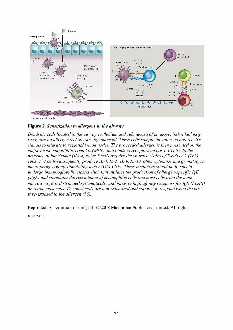
Figure 2. Sensitization to allergens in the airways
Dendritic cells located in the airway epithelium and submucosa of an atopic individual may recognize an allergen as body-foreign material. These cells sample the allergen and receive signals to migrate to regional lymph nodes. The proceeded allergen is then presented on the major histocompatibility complex (MHC) and binds to receptors on naive T cells. In the presence of interleukin (IL)-4, naive T cells acquire the characteristics of T-helper 2 (Th2) cells. Th2 cells subsequently produce IL-4, IL-5, IL-9, IL-13, other cytokines and granulocyte-macrophage colony-stimulating factor (GM-CSF). These mediators stimulate B cells to undergo immunoglobulin class-switch that initiates the production of allergen-specific IgE (sIgE) and stimulates the recruitment of eosinophilic cells and mast cells from the bone marrow. sIgE is distributed systematically and binds to high affinity receptors for IgE (FcεRI) on tissue mast cells. The mast cells are now sensitized and capable to respond when the host is re-exposed to the allergen (16).
Figure 3. Early phase of allergen induced airway inflammation
When a sensitized individual is re-exposed to the allergen, sIgE bound to FcεRI on mast cells are cross-linked by the allergen. This activates mast cells to release preformed mediators and increase the synthesis of cytokines, chemokines and growth factors. These mediators induce vasodilation, increased vascular permeability and oedema in affected organs. In asthmatic airways, bronchoconstriction and mucus hypersecretion occur. Some of the mediators released may promote local recruitment and activation of eosinophilic and other inflammatory leukocytes, initiating development of the late-phase reaction (16).
indirect tests mimic the natural pathophysiology of asthma, whereas direct stimuli are more
closely related to structural changes in the airways (66). Furthermore, markers of airway
inflammation have been shown to correlate with the extent of BHR, while anti-inflammatory
treatment may reduce BHR (67, 68).
The exercise challenge test
During heavy exercise, tidal volume and respiratory frequency are increased due to increased
demand of oxygen. Increased ventilation is accompanied by heat and water loss from the
airways that lead to cooling and dehydration of the airway mucosa (69). Intracellular
hyperosmolarity induction of mediator release has been proposed as the main mechanism of
exercise-induced bronchoconstriction (EIB) (70, 71). In addition, airway cooling, mediator
release and increased osmolarity may stimulate bronchoconstriction via parasympathic reflex
pathways (69). On cessation of hyperventilation, reactive hyperaemia and oedema of the
airways may occur that reduces the size of the airway lumen (72, 73). EIB has been shown to
reflect ongoing airway inflammation (74). In asthmatics, FENO has been proposed to be a
predictive marker of EIB (75).
In most children with current asthma EIB is triggered by exercise, although children without
asthma symptoms may demonstrate it (10, 76). Therefore, the criterion for a positive EIB test
is controversial (77). A reduction in FEV1 ≥10% after a standardized exercise test is generally
accepted as a positive test (71, 77). However, a fall in FEV1 of 15% appears to be more
diagnostic of EIB (77).
25
The latest American Thoracic Society (ATS)/European Respiratory Society (ERS) guidelines
recommend an exercise load of 80-90% of predicted maximal heart rate (calculated as 220
minus age in years) (77). It has been demonstrated that a higher exercise load is more
sensitive to reveal EIB and is better related to inflammatory activity (71).
1.3.3 Fractional exhaled nitric oxide
Exhaled breath biomarkers
Exhaled biomarkers have been explored to understand pathological mechanisms and to guide
diagnosis and treatment decisions. The most studied exhaled biomarkers are NO, carbon
monoxide, volatile organic compounds (VOC) and various biomarkers in exhaled breath
condensate (EBC). FENO is the only exhaled biomarker that has been standardized and
validated for clinical paediatric application (78, 79). FENO is a non-invasive surrogate
measurement of eosinophilic airway inflammation that is easy to perform, provides
immediate results and is well suited for children (51, 80).
Nitric oxide
NO is a free radical gas with one unpaired electron that avidly reacts with other molecules. In
1987, NO was recognized as the endothelium derived relaxing factor (ERDF) (81). In 1991
Gustafsson et al. measured endogenous NO in exhaled air of humans, and thereby started a
new area in respiratory research (82). NO is known as a messenger molecule involved in
multiple biological systems, including neurotransmission, platelet inhibition, inflammation
and immunomodulation (83).
NO is generated via oxidation of L-arginine, a process catalysed by the enzyme system NO
synthases (NOS) (84). Three isoforms of NOS have been described: inducible NOS (iNOS),
endothelial NOS (eNOS) and neuronal NOS (nNOS). The latter two are calcium and
calmodium-dependent enzymes, which are released within seconds upon receptor stimulation.
By contrast, iNOS is slowly regulated at the transcriptional level and releases large quantities
of pro-inflammatory NO (83). The signal transducer and activator of the transcription (STAT)
pathway is the main regulatory mechanism of iNOS gene transcription (85). iNOS is activated
by endogenous mediators, namely chemokines and cytokines as well as exogenous factors
such as viruses, allergens and pollutants (83). Current knowledge indicates that the induction
of iNOS in asthmatics is primarily dependent on the activity of IL-4 and IL-13 in the
26
bronchial wall (86, 87). Besides NOS-catalysed formation, NO may be formed in high
concentrations from peroxynitrite and tyrosine nitration (83, 88).
All of the three NOS isoforms are expressed in the respiratory system (83). In children, a
highly significant correlation between epithelial iNOS mRNA expression and orally exhaled
NO levels has been found (89). Nasally exhaled air contains higher NO concentrations than
orally exhaled air (90). This has been attributed primarily to higher expression of iNOS in the
paranasal sinuses than in the lower respiratory tract (91).
In the respiratory system, low NO concentrations have protective effects that promote
bronchial dilatation, mediate ciliary beat frequency and stimulate mucus secretion (83, 92,
93). On the other hand, high NO concentrations have deleterious effects and promote
inflammation via Th2-mediated mechanism and oxidizing agents. Pro-inflammatory effects of
NO include vasodilatation, plasma extravasation, mucus hypersecretion, impaired ciliary
motility and cytotoxicity (83).
FENO sampling technique
The chemiluminescence method was the first established technique to measure NO in exhaled
breath of humans, and it became the gold standard (82). This sensitive technique uses ozone
to react with NO and produces NO2 in an excited state. The reaction emits light that correlates
with the amount of NO present (94).
FENO is influenced by many factors of which the most crucial is exhaled flow. FENO is flow
dependent and increases with reduced exhalation (95). According to the 2005 ATS/ERS
guidelines, FENO should be measured at an exhalation flow of 50 mL/s (±10%) (78). The
subject should inhale NO free air to avoid contamination of ambient NO (78). Exhalation is
recommended to start immediately after inhalation to total lung capacity (TLC) to avoid
accumulation of NO in the oro-pharynx (78, 96). Nasal NO (nNO) is present in higher
concentrations relative to the lower respiratory tract (97). Therefore, it is recommended to
exhale with an oral pressure of 5-20 cmH2O to ensure closure of the soft palate (78).
Factors affecting FENO measurements
Height, age and gender have been shown to influence FENO measurements. FENO increases
with age (80). Height has been found to correlate with FENO (98). The increased FENO in taller
27
individuals probably reflects the greater airway mucosal area available for NO exchange (99).
Studies report conflicting data as to whether FENO is influenced by gender in children (80,
100-102).
Treatment with inhaled corticosteroids (ICS) reduces FENO (26, 51) as may exposure to
tobacco smoke (103), whereas exposure to air pollution (50) and intake of nitrate-rich food
may increase it (104). Kharnitov et al. found no diurnal variation in FENO in healthy and
asthmatic children (105). Population-based studies have reported either no association (100,
101) or weak association between FEV1 and FENO (106). Rhinovirus infections may induce
iNOS leading to increased FENO levels (107, 108), while FENO is slightly decreased in the
symptomatic phase of respiratory syncytial virus (RSV) and influenza virus infections (109,
110).
FENO and the relation to allergic sensitization, asthma and AR
It is well documented that FENO is increased in children with asthma compared to healthy
controls (100, 111). FENO is found to correlate with measurements of eosinophilic activity in
the airway mucosa (51). Therefore, FENO is often referred to as a surrogate marker of
eosinophilic inflammation. FENO has also been shown to correlate with the degree of IgE
sensitization, both in terms of number of SPTs (111, 112) and the sIgE levels to some
allergens (113).
The FENO level is increased in children with AR, and the highest values have been found in
children with allergic asthma (100, 101). In some studies, atopic individuals without asthma
and/or AR have equal FENO concentrations relative to non-atopics (114, 115). In other studies,
increased FENO levels have been observed in atopic individuals regardless of the respiratory
tract symptoms (51, 100, 111) . It has been suggested that this might reflect subclinical airway
inflammation (51, 111). The heterogeneity in the exhaled NO levels reported might be
explained by unlike allergen exposure, different definitions of allergic sensitization, and
whether subgroups are labelled by allergic sensitization alone or by allergic sensitization and
allergy symptoms.
The effects of common laboratory procedures on FENO measurements
Bronchodilator administration, spirometric manoeuvres and EIB tests have been proposed to
affect FENO measurements (116-118). The ATS/ERS guidelines recommend refraining from
28
exercise 1 hour before performing the FENO test because forced breathing have been shown in
most studies to reduce FENO in healthy and asthmatic adults (78). It has been argued that
increased NO elimination and reduced airway surface area during EIB are the main
mechanisms of FENO decline post exercise (117-120). In children, few reports concerning the
effects of exercise on FENO have been published and with conflicting results (117, 120, 121).
Different conclusions may partly be explained by different NO sampling techniques and EIB
tests performed (i.e., different activities and thresholds; 117, 120, 121). FENO levels have been
found to correlate with the degree of eosinophilic airway inflammation (51). Although, the
impact of allergic airway inflammation on FENO in relation to exercise has not been fully
elucidated in asthmatic and non-asthmatic children.
1.4 Diagnosing inhalant allergy
The diagnosis of allergic diseases involves both the presence of allergy symptoms and
confirmation of relevant allergic sensitization (33). Allergic sensitization is commonly
determined either by in vivo skin prick testing or by in vitro measurement of sIgE in serum
(122). Serum sIgE can be analysed for single allergens, allergenic molecules (components) of
single allergens, a mix of allergens, and by multi-allergen tests for screening purposes. These
tests identifies allergic sensitization and do not necessarily demonstrate clinical relevant
allergies (123, 124). Serum sIgE cut-off points for clinically relevant allergies may be
determined by plotting the sensitivity against 1-specificity using receiver operating
characteristic (ROC) curves.
1.4.1 Skin prick test
The core diagnostic test for Type-1 hypersensitivity is the SPT test (125, 126). The SPT test
utilizes the presence and degree of cutaneous reactivity to an allergen as a surrogate marker of
sensitization. When an allergen is introduced into the skin, sIgE bound to surface receptors on
mast cells may cross-link and induce mast cell degranulation thereby releasing histamine and
other mediators (126). This may produce a wheal that can be quantified. A positive SPT is
considered in the presence of a wheal diameter ≥3 mm larger than the negative saline control
(125). A false negative result can be seen if the individual has ongoing antihistamine therapy,
current eczema, or if topical steroids have been applied to the skin. Dermographism may lead
to a false positive result (125). SPT results have been found to correlate with those of nasal
allergen challenge (127), and very good correlations have been found between SPT results
and clinical allergy symptoms (125, 128).
29
1.4.2 Serum IgE and in vitro immunoassays
Serum sIgE antibodies can be determined by a variety of in vitro immunoassays (122). There
exist no absolute serum sIgE antibody reference standards against which to judge accuracy.
However, ImmunoCAP® (Phadia) was the first established assay and has been accepted and
validated as a quasi-standard (129-131). Allergen reagents produced by different
manufactures vary in its protein composition and have been shown to detect dissimilar sIgE
populations (130, 132). Thus, sIgE cut-off levels reported for one in vitro assay as defining
positive allergic reactivity cannot be used with sIgE results generated employing test kits
from a different manufacturer. In addition, allergens may have different cut-off values when
employing the same immunoassay (41). The analyses of serum sIgE are feasible when
patients are taking anti-histamines. However, therapeutic levels of omalizumab in sera will
interfere in several of the clinically used immunoassays (132).
The Siemens IMMULITE® 2000 system (IMMULITE®) is a four-step chemiluminescent
assay using biotinylated allergens in a liquid phase coupled to ligand-coated beads (41). Cut-
off levels for IMMULITE® to some common inhalant allergens have been reported for adults
(131), but not for children. Although IMMULITE® assays and SPT are used in some clinics,
little comparative data are available for results in children; neither have paediatric cut-off
values for sIgE using IMMULITE® to diagnose AR been established.
30
2 AIMS OF THE STUDY
The aims of the study were to investigate the prevalence and time trends of atopic diseases in
a subarctic childhood population and to quantify FENO levels in relation to asthma, AR and
exercise testing. Likewise, another object was to establish paediatric serum sIgE cut-off
values for the diagnosis of AR and to explore the relationships between serum sIgE, total IgE
and FENO.
The specific aims were:
Paper I: To explore whether or not the prevalence of asthma, AR and eczema continues to
increase in Nordland County, Norway.
Paper II: To investigate FENO levels in non-asthmatic children, and to explore whether
exercise testing affect FENO levels in non-asthmatic children with and without AR symptoms.
Paper III: To determine the effects of AR on FENO in response to a standardized treadmill
exercise test in asthmatic and non-asthmatic children.
Paper IV: To establish paediatric cut-off values for serum sIgE using the Siemens
IMMULITE® 2000 to diagnose AR, and to explore the relationships between serum sIgE,
total IgE and FENO.
31
3 METHODS
3.1 Study design and subjects
This thesis is based on data from Phase I and Phase II of the study `Asthma and allergy
among schoolchildren in Nordland´ (Figure 4).
Figure 4. Subject flow chart in Phase I and Phase II of the study. aSubjects misclassified as non-asthmatics (n=14); subject who became asthmatic from Phase
I to Phase II (n=8), subjects categorized as asthmatic in the structured interview (n=6). bSubjects categorized as non-asthmatic in the structured interview (n=64).
Phase I of the study was a cross-sectional questionnaire based survey. Schoolchildren aged 7-
14 years from 65 randomly selected schools of a total of 244 schools in Nordland County
Asthma'n=373'
Asthma'n=309'
Non0asthma1c'controls'n=373'
Non0asthma1c'controls'n=145'
Ques1onnaire'n=6505'
Responders'n=4150'(63.8%)'
Asthma'n=373'
Non0asthma'n=428'
Asthma'ever'n=164'
Asthma'current'n=145'
PHASE&I&
PHASE&II&
Matched'pairs'
n=14a'
Matched'pairs'
Paper'I'
Paper'II'
Paper'III'
n=64b'
Asthma'n=729'
Non0asthma'n=3421'
32
were invited to participate. Parents received a questionnaire (Appendix 1) regarding asthma,
AR and eczema between February and May 2008. All participants received one reminder. The
study closed four weeks after the reminder was distributed. Based on the questionnaire
responses, pupils were categorized as asthmatic or non-asthmatic (Paper I).
In Phase II of the study, pupils who reported having asthma in Phase I and lived nearby the
study locations along with two age and gender matched non-asthmatic controls were invited
to participate. Of the 1144 pupils invited, 801 children (373 of them reporting asthma in
Phase I) accepted to participate. The parents completed a questionnaire and a structured
interview. A clinical examination, spirometry, exercise treadmill testing, SPT and
measurements of FENO, serum sIgE and total IgE were obtained. Based on information given
in the structured interview and the clinical examination, the pupils were finally categorized as
asthmatic or non-asthmatic (Figure 4). The participants were examined at least two weeks
after any suspected respiratory tract infection during the school season from March 2009 to
June 2010. The examinations took place at Nordland Hospital, Bodø, and at three other
locations in Nordland County (Fauske, Mo i Rana and Sortland). PhD student Tonje E.
Hansen and the author conducted all the interviews and procedures, and the same medical
instruments were used throughout to secure standardized measurement conditions.
The study population of Paper II included 373 non-asthmatic pupils (non-asthmatic controls
to the original asthma group). These children were similar with respect to demographic data
to the non-asthmatic children who were not included in Paper II. In Paper III, the assessments
of 145 pupils with current asthma and 145 non-asthmatic age- and gender-matched controls
were compared. Of the 801 children enrolled in Phase II, 303 had measurements of serum
sIgE, total IgE, SPT and FENO and constituted the study subjects of Paper IV.
Both Phase I and Phase II studies were approved by the Regional Committee for Medical and
Health Research Ethics, and were conducted in accordance with the ethical standards of the
2000 Helsinki Declaration. In Phase I, the parents/guardians signed a written consent for their
children’s participation. In Phase II, written informed consent was obtained from all children
and their parents.
33
3.2 Definitions
Phase I, Paper I
'Asthma ever' was considered if the parent answered 'yes' to the question `Has the pupil ever
had asthma?`, and/or to the question `Does the pupil experience wheeze, periods of coughing
or acute shortness of breath (asthma) due to external factors?`
ʹ′AR everʹ′ was estimated on the basis of a positive answer to the question `Has the pupil ever
had hay fever (runny or blocked nose, sneezing, itching of the nose and/or eyes, or swollen or
red eyes)?`
ʹ′Eczema everʹ′ was recorded if the pupils reported an itchy rash lasting at least four weeks,
combined with lesions on the face, elbows or knee flexures, or a high degree of itching and
lesions elsewhere.
ʹ′Current diseaseʹ′ was considered among those answering yes to the main questions about
asthma, AR or eczema and reporting symptoms the last 12 months.
Phase II, Paper II-IV
Asthma
Asthma (Paper II-IV): at least two of the following three criteria fulfilled at any time in life:
1) recurrent dyspnoea, chest tightness and/or wheeze; 2) a doctor´s diagnosis of asthma; and
3) use of asthma medication including β-2 agonist, sodium chromoglycate, corticosteroids,
leukotriene antagonists and/or aminophylline.
Current asthma (Paper III): asthma as defined above plus symptoms and/or medication
within the last year.
Current asthma (Paper IV): asthma as defined above plus symptoms and/or medication
within the last year, and/or a positive exercise test.
Asthma in remission (Paper IV): asthma not defined as current asthma.
Allergic rhinoconjunctivitis (AR)
AR symptoms (Paper II-IV): a history of watery rhinorrhea, blocked nose, sneezing, nasal
itching accompanied by itchy watery eyes in absence of airway infection.
AR (Paper III): AR symptoms in combination with allergic sensitization.
Allergic sensitization (Paper III): a positive serum sIgE and/or a positive SPT to at least one
of the ten inhalant allergens.
Non-AR (Paper III): no AR symptoms or sensitization to inhalant allergens.
34
AR (Paper IV): a positive SPT and a history of related AR symptoms as evaluated by a
doctor.
Food allergy
Food allergy (Paper IV): a positive SPT and a history of related food allergy symptoms as
evaluated by a doctor.
3.3 Questionnaires, structured interview and clinical examination
Questionnaire Phase I (Appendix I): A questionnaire that focused on diagnosis and symptoms
of asthma, AR and eczema was created in 1985 to assess disease among schoolchildren in
northern Norway. The questions covered gender, age, family history of atopy, socio-economic
conditions, passive smoke exposure and household animals. In 2008, we used the identical
questions indicated but added some about physical activity, medical diagnosis of asthma and
asthma medication. The additional questions did not change the definition of the diseases.
Questionnaire and structured interview Phase II: The parents completed a detailed
questionnaire and a structured interview relating to asthma, AR, food allergy, urticaria,
anaphylaxis and eczema symptoms and diagnosis, the use of medications, exposure to
allergens and exposure to tobacco smoke. Additional questions regarding diet, infections,
physical activity and demographic factors were answered and recorded.
Clinical examination, Phase II: A clinical examination was performed including height and
weight measurements and assessment of the skin, the upper airways, lungs and the heart.
Inhaled corticosteroids (ICS) and short acting β-2 agonists were withheld for 12 hours (h)
prior to testing; inhaled long acting β-2 agonists for the last 48 h; leukotriene modifiers for the
last 24 h; and antihistamines in the last 5 days. No children were using oral steroids.
3.4 Allergic sensitization
Serum total IgE and sIgE: Blood samples were obtained using standard venepuncture using
Vacutainer® tubes (Becton Dickinson, Plymouth, UK). Serum was collected and stored at
-80°C until assayed. Total IgE and sIgE levels were analysed employing the IMMULITE®
2000 (Siemens Healthcare Diagnostics Inc., Deerfield, IL, USA) using 3gAllergy® kits. The
35
detection range for sIgE was ≥0.10-100 kU/L. The following were tested: sIgE to timothy,
birch and mugwort pollens; dog dander, cat and rabbit epithelial dander; house dust mite
Dermatophagoides pteronyssinus; moulds Alternaria tenius and Cladosporium herbarium
and German cockroach. Seroatopy was defined by a sIgE test ≥0.35 kU/L (132) to at least one
of the listed allergens (Paper III). Blood samples were requested for all children.
Skin prick test: SPT was performed for the above listed inhalant allergens and egg white,
milk, peanut and codfish with Soluprick® allergens (ALK Abello, Denmark). Histamine was
used as positive control and saline as negative control. SPT was considered positive in the
presence of a wheal diameter ≥3 mm larger than the negative control (125). During the initial
study period, SPT was requested for all children. Thereafter, SPT was requested for children
with asthma and/or allergy symptoms.
Allergic sensitization was not evaluated in 12 individuals without AR symptoms (Paper III).
Of a total of 2673 serum analyses, 23 measurements of sIgE were missing due to low sample
volume (Paper IV).
3.5 Fractional exhaled nitric oxide FENO: was measured online by the single breath method with a chemiluminescence analyser,
4.1 Prevalence of asthma, AR and eczema 1985-2008 (Paper I)
Of 6505 pupils invited to participate, 4150 (63.8%) answered the questionnaire and were
enrolled in the study (49.1% boys). The main findings were: an increasing prevalence of
asthma ever (7.3% in 1985 to 17.6% in 2008, p for trend <0.001), and AR ever (15.9% in
1985 to 24.5% in 2008, p for trend <0.001); and the prevalence of eczema ever, after an
increase between 1985 and 1995, remained unchanged in the last time period. The prevalence
of current disease doubled and trebled between 1995 and 2008 for all three diseases (Table 1).
The proportion of children reporting at least one disease (asthma, AR or eczema) increased
from 26.2% in 1985 to 43.3% in 2008 (p for trend <0.001).
Adapted from Hansen et al. Acta Paediatr 2012;102:47-52.
!
Table 1. The prevalence of current asthma, allergic rhinoconjunctivitis and eczema in children aged 7-14 years from the 1995 and 2008 questionnaire-based surveys in Nordland. Prevalence (%) Surveys 2008/1995 1995 2008 OR 95 % CI All Current asthma 4.8 9.9 2.21 1.86-2.62 Current rhinoconjunctivitis 6.7 21.5 3.83 3.33-4.40 Current eczema 6.4 13.5 2.27 1.96-2.64 Boys Current asthma 5.6 12.0 2.29 1.83-2.87 Current rhinoconjunctivitis 7.5 24.4 3.80 2.15-4.58 Current eczema 6.2 12.3 2.11 1.70-2.62 Girls Current asthma 3.9 8.0 2.13 1.63-2.78 Current rhinoconjunctivitis 5.8 18.7 3.70 3.01-4.56 Current eczema 6.6 14.6 2.43 1.97-2.99 The difference in prevalence between 2008/1995 is quantified with odds ratio (OR). Corresponding 95 % confidence intervals (95% CI) are presented.
39
4.2 The impact of exercise on FENO in non-asthmatic children (Paper II)
Of the 373 non-asthmatic children enrolled in this part of the study, 22 children were unable
to comply with the study protocol and 21 children had a positive EIB test and were excluded.
Three hundred and thirty children were included in the statistical calculations. Children
reporting AR symptoms (n=71) were similar to children without AR symptoms (n=259) with
respect to gender, age, height, weight and spirometric indices (all p >0.05).
Geometric mean FENO values at baseline, at 1 min and at 30 min after the treadmill exercise
test are given in Table 2. Baseline FENO was significantly increased in children reporting AR
symptoms versus no AR symptoms: 15.1 (12.6-18.1) ppb versus 9.6 (9.0-10.3) ppb (p
<0.001). Subjects with AR symptoms had a significantly higher decline in geometric mean
FENO value at 1 min post-exercise compared to children without AR symptoms: 4.2 ppb
versus 2.6 ppb (p <0.001). FENO did not return to baseline level in either of the groups at 30
min post-exercise (Table 2). Subjects with baseline FENO ≥20 ppb demonstrated a higher
decline in FENO value than subjects with baseline FENO <20 ppb at 1 min post-exercise: 9.9
(8.7-11.4) ppb versus 2.4 (2.3-2.5) ppb (p <0.001).
40
Tab
le 2
. Lev
els o
f FE N
O a
t bas
elin
e co
mpa
red
to le
vels
of F
E NO
at 1
min
and
at 3
0 m
inut
es a
fter a
stan
dard
ized
exe
rcis
e in
duce
d
bron
choc
onst
rictio
n (E
IB) t
est o
n a
tread
mill
in n
on-a
sthm
atic
chi
ldre
n w
ith a
nd w
ithou
t alle
rgic
rhin
ocon
junc
tiviti
s sym
ptom
s. __
____
____
____
____
____
____
____
____
____
____
____
____
____
____
____
____
____
____
____
____
____
____
____
____
____
____
____
B
asel
ine
FEN
O*
† F
E NO 1
min
ute
P
val
ue v
s.
FE N
O 3
0 m
inut
es
P
valu
e vs
.
pos
t exe
rcis
e*
bas
elin
e
pos
t exe
rcis
e*
ba
selin
e _
____
____
____
____
____
____
____
____
____
____
____
____
____
____
____
____
____
____
____
____
____
____
____
____
____
____
____
__
A
ll ch
ildre
n
(n
=330
)
10
.6 (9
.9-1
1.3)
7.7
(7.2
-8.2
)
<0.0
01
8.9
(8.3
-9.5
)
<0.0
01
No
AR
‡ sy
mpt
oms
(n=
259)
9
.6 (
9.0-
10.3
)
7.0
(6.5
-7.5
)
<0.0
01
8.0
(7.5
-8.6
)
<0.0
01
AR
sym
ptom
s (n
=71)
15
.1 (1
2.6-
18.1
)
1
0.9
(9.2
-12.
9)
<0.0
01
1
3.0
(10.
9-15
.5)
<0
.001
__
____
____
____
____
____
____
____
____
____
____
____
____
____
____
____
____
____
____
____
____
____
____
____
____
____
____
____
*R
esul
ts a
re g
iven
as g
eom
etric
mea
ns (9
5% c
onfid
ence
inte
rval
s).
†Fra
ctio
nal n
itric
oxi
de (F
E NO) i
s exp
ress
ed a
s par
ts p
er b
illio
n (p
pb).
‡Sel
f-re
porte
d al
lerg
ic rh
inoc
onju
nctiv
itis s
ympt
oms.
Ada
pted
from
Evj
enth
et a
l. C
lin R
espi
r J 2
013;
7:12
1-12
7.
!
41
4.3 The effects of AR on FENO in response to a standardized exercise treadmill
test in asthmatic and non-asthmatic children (Paper III) In this part of the study, matched pairs of 145 pupils with current asthma (cases) and 145 non-
asthmatic pupils (controls) were enrolled. Twenty pairs included pupils (n=23) who were
unable to comply with the study protocol, and one pair included a control with a positive EIB
test. These 21 pairs were excluded. Children who did not comply were younger than the
included children (p =0.006). The included children with current asthma (n=124) had more
frequent AR, and they had significantly lower FEV1 and FEF50 than the non-asthmatic
controls (n=124), (all p <0.05).
Baseline FENO was significantly higher in asthmatics compared to non-asthmatics, 21.0 (17.6-
24.9) ppb versus 11.1 (9.9-12.4) ppb (p <0.001) and significantly elevated in asthmatics and
non-asthmatics with AR compared to individuals without AR (Figure 5). Baseline FENO was
not significantly influenced by ICS use in asthmatics or in the subgroup of allergic asthmatics
(data not presented). Comparison of FENO levels (ppb) at each time point demonstrated
parallel time trends between asthmatics and non-asthmatics (p =0.866). Adjustment for
baseline FENO yielded no significant difference in time trends between the groups (p=0.848).
However, the time trends depicted in Figure 5 were significantly different in children with AR
compared to children without AR (p =0.039), irrespective of asthma (p =0.876). In children
with AR, FENO declined by a mean of 6.1 ppb (5.1-7.5) at 1 min post exercise. At 30 min,
FENO was reduced by a mean of 2.8 ppb (2.5-3.3). In children without AR, FENO declined by
a mean of 2.7 ppb (2.1-3.5) at 1 min post exercise, while at 30 min FENO was reduced by a
mean of 1.6 ppb (1.3-2.0) compared to baseline FENO.
The effect of exercise on FENO was evaluated by comparing the % change in LnFENO from
baseline to 1 min and 30 min post exercise (Figure 6). The time trend was dependent on AR
(p <0.001), irrespective of asthma status (p =0.795). The effect of exercise was more
pronounced in children without AR than in children with AR. In asthmatics the effect of
exercise on FENO was independent of ICS treatment (p =0.583) and a positive EIB test
(p=0.230).
Based on LnFENO, the % reduction at 1 and 30 min post exercise was less pronounced with
increasing number of positive SPT/or sIgE tests. Significant differences were observed
42
between children without AR (non-sensitized, n=83) and those with AR and 1-3 (p=0.002,
n=45) and 4-9 (p <0.001, n=78) positive tests. However, the differences between the latter
two groups were not statistically significant (p=0.633).
Baseline FENO correlated positively with maximal post exercise FEV1 decline in asthmatics
(rho=0.331, p<0.001). In asthmatics with AR a positive correlation was found (rho=0.360,
p<0.001) but not in asthmatics without AR.
Figure 5. Geometric mean FENO levels in asthmatics with allergic rhinoconjunctivitis (AR) (n=89), non-asthmatics with AR (n=34), asthmatics without AR (n=22) and non-asthmatics without AR (n=61). FENO was measured at baseline (pre) and at 1 min and 30 min after a standardized exercise induced-bronchoconstriction test on a treadmill. FENO is expressed in parts per billion (ppb). Error bars represents 95% confidence intervals.
Pre 1 300
5
10
15
20
25
30
35
Time (min)
FEN
O (p
pb)
Non-asthma and non-AR
Asthma and non-AR
Non-asthma and AR
Asthma and AR
Exercise
43
Figure 6. Changes in LnFENO (%) after a standardized exercise-induced bronchoconstriction test on a treadmill in asthmatic and non-asthmatic children. Data are presented for four subgroups: asthmatics with allergic rhinoconjunctivitis (AR) (n=89), non-asthmatics with AR (n=34), asthmatics without AR (n=22) and non-asthmatics without AR (n=61). FENO was measured at baseline (pre) and at 1 min and 30 min after the exercise test. Error bars represents 95% confidence intervals.
Pre 1 30-30
-25
-20
-15
-10
-5
0
Time (min)
% c
hang
e in
LnF
ENO
Non-asthma and non-AR
Asthma and non-AR
Non-asthma and AR
Asthma and AR
Exercise
44
4.4 Paediatric cut-off values for serum sIgE to diagnose AR and its relation to
FENO (Paper IV) Of the 303 children enrolled, 223 had AR symptoms and 80 did not. In the group with AR
symptoms, children with a reaction to the negative control (n=5), food allergy (n=23) and
individuals who did not fulfil the AR definition (n=31) were excluded. In the group without
AR symptoms, one child had food allergy and was also excluded. Children with AR (n=164)
were similar to children without AR (n=79) with respect to age, height, weight, current
eczema and urticaria (data not presented). Children with AR had more often current asthma
than children without AR (p =0.044).
Diagnostic value of serum sIgE
Cut-off values for serum sIgE for a general optimal test with the best combined sensitivity
and specificity were above the detection limit of the assay for seven of the ten allergens (0.23-
1.1 kU/L). ROC curve analysis showed that the overall accuracy of the IMMULITE® in
detecting AR was moderate to excellent, with areas under the curves (AUCs) at 0.852-0.954
(Table 3). However, the sIgEs for Alternaria tenius, Cladosporium herbarium and German
cockroach were not significant predictors of AR (data not presented). Serum sIgE cut-off
values differed according to the purpose of the test. Cut-off values for a diagnostic test at 90%
specificity and for a screening test at 90% sensitivity are presented in Table 4.
FENO levels and the correlation with serum sIgE
FENO was elevated in children with AR, irrespective of asthma (Figure 7). In children with
AR, FENO correlated moderately with total IgE (Spearmans´s rank correlation coefficient
(rho)= 0.28, p <0.001), sIgE to cat (rho= 0.38, p =0.002) and dog (rho=0.59, p <0.001). FENO
did not correlate positively with sIgE to other tested allergens (data not presented).
Pairwise comparisons of ROC curves
Serum sIgE was superior to total IgE and FENO in predicting AR to timothy, birch, mugwort,
cat, dog and house dust mite. Total IgE predicted AR to timothy, birch and rabbit, while FENO
did not. FENO and total IgE had equal power to predict AR in children sensitized to dog and
Dermatophagoides pteronyssinus (Figure 8).
45
Tab
le 3
. RO
C c
urve
stat
istic
s for
seru
m sp
ecifi
c Ig
E to
inha
lant
alle
rgen
s in
child
ren
with
alle
rgic
rhin
ocon
junc
tiviti
s __
____
____
____
____
____
____
____
____
____
____
____
____
____
____
____
____
____
____
____
____
____
____
____
____
____
____
____
____
____
___
Alle
rgen
N*/
AU
C
95%
CI
p-
valu
e C
ut-o
ff
Sens
itivi
ty
Spec
ifici
ty
LR+
LR
-
Posi
tive†
va
lue‡
____
____
____
____
____
____
____
____
____
____
____
____
____
____
____
____
____
____
____
____
____
____
____
____
____
____
____
____
____
____
_ Ti
mot
hy
24
1/96
0.
954
0.92
0-0.
977
<0
.001
1.
1
94.8
84.1
6.0
0.06
B
irch
241/
73
0.90
5
0.
861-
0.93
9
<0.0
01
0.93
91.8
85.1
6.2
0.09
M
ugw
ort
24
0/17
0.
937
0.89
9-0.
964
<0
.001
0.
59
82
.4
94
.2
14
.1
0.
19
Cat
dan
der
24
0/89
0.
924
0.88
2-0.
954
<0
.001
0.
91
95
.5
83
.4
5
.8
0.
05
Dog
dan
der
24
2/77
0.
852
0.80
1-0.
894
<0
.001
0.
27
83
.1
78
.2
3
.8
0.
22
Rab
bit d
ande
r 24
2/23
0.
856
0.80
5-0.
897
<0
.001
0.
23
78
.3
93
.6
12
.2
0.
23
D.p
tero
nyss
inus
24
2/31
0.
917
0.87
5-0.
949
<0
.001
1.
00
87
.1
97
.2
30
.6
0.
13
__
____
____
____
____
____
____
____
____
____
____
____
____
____
____
____
____
____
____
____
____
____
____
____
____
____
____
____
____
____
___
AU
C, a
rea
unde
r the
cur
ve; C
I, co
nfid
ence
inte
rval
; LR
+, li
kelih
ood
ratio
pos
itive
; LR
-, lik
elih
ood
ratio
neg
ativ
e; D
.pte
rony
ssin
us, D
erm
atop
hago
ides
pt
eron
yssi
nus.
*C
ompl
ete
resu
lt se
ts o
f ser
um sp
ecifi
c Ig
E, sk
in p
rick
test
(SPT
) and
alle
rgic
rhin
ocon
junc
tiviti
s (A
R) s
ympt
oms.
† Posi
tive
SPT
and
rela
ted
AR
sym
ptom
s as e
valu
ated
by
a do
ctor
. ‡ Se
rum
spec
ific
IgE
cut-o
ff v
alue
s (kU
/L) w
ith th
e be
st c
ombi
ned
sens
itivi
ty a
nd sp
ecifi
city
. A
dapt
ed fr
om E
vjen
th e
t al.
Act
a Pa
edia
tr 20
14;1
03:7
59-6
5.
46
Tab
le 4
. Cut
-off
val
ues
and
diag
nost
ic u
tility
of a
llerg
en-s
peci
fic Ig
E fo
r ide
ntify
ing
child
ren
with
alle
rgic
rhin
ocon
junc
tiviti
s A
llerg
en
Purp
ose
Cut
-off
va
lue*
Se
nsiti
vity
(9
5% C
I)
Spec
ifici
ty
(95%
CI)
LR
+ LR
-
Tim
othy
D
iagn
ostic
† 4.
1 78
.1 (
68.5
-85.
9)
90.3
(84
.3-9
4.6)
8.
1 0.
24
Sc
reen
ing‡
1.7
90.6
(82
.9-9
5.6)
87
.6 (
81.1
-92.
5)
7.3
0.11
B
irch
Dia
gnos
tic
2.8
80.2
(69
.1-8
8.6)
90
.2 (
84.6
-94.
3)
8.2
0.22
Scre
enin
g 1.
0 90
.4 (
81.2
-96.
1)
85.1
(78
.8-9
0.1)
6.
1 0.
11
Mug
wor
t D
iagn
ostic
0.
35
82.4
(56
.6-9
6.2)
91
.0 (
86.5
-94.
4)
9.2
0.19
Scre
enin
g 0.
16
94.1
(71
.3-9
9.9)
82
.1 (
76.4
-86.
9)
5.3
0.07
C
at d
ande
r D
iagn
ostic
7.
4 69
.7 (
59.0
-79.
0)
90.1
(84
.1-9
4.3)
7.
0 0.
34
Sc
reen
ing
1.3
91.0
(83
.1-9
6.0)
85
.4 (
78.8
-90.
6)
6.3
0.11
D
og d
ande
r D
iagn
ostic
1.
7 52
.0 (
40.3
-63.
5)
90.3
(84
.7-9
4.4)
5.
4 0.
53
Sc
reen
ing
0.1
88.3
(79
.0-9
4.5)
67
.3 (
59.5
-74.
4)
2.7
0.17
R
abbi
t dan
der
Dia
gnos
tic
0.11
78
.3 (
56.3
-93.
5)
90.4
(85
.7-9
4.0)
8.
2 0.
24
Sc
reen
ing
0.1
78.3
(56
.3-9
3.5)
88
.6 (
83.6
-92.
5)
6.9
0.25
D
.pte
rony
ssin
us
Dia
gnos
tic
0.36
87
.1 (
70.2
-96.
4)
90.5
(85
.7-9
4.1)
9.
2 0.
14
Sc
reen
ing
0.1
87.1
(70
.2-9
6.4)
82
.9 (
77.2
-87.
8)
5.1
0.16
C
I, co
nfid
ence
inte
rval
; LR
+, li
kelih
ood
ratio
pos
itive
; LR
-, lik
elih
ood
ratio
neg
ativ
e; D
.pte
rony
ssin
us, D
erm
atop
hago
ides
pt
eron
yssi
nus.
*S
erum
spe
cific
IgE
cut-o
ff v
alue
s (k
U/L
) † D
iagn
ostic
test
; spe
cific
ity a
t 90%
or t
he c
lose
st s
peci
ficity
iden
tifie
d w
ith th
e be
st c
ombi
ned
sens
itivi
ty
‡ Scre
enin
g te
st; s
ensi
tivity
at 9
0% o
r the
clo
sest
sen
sitiv
ity id
entif
ied
with
the
best
com
bine
d sp
ecifi
city
A
dapt
ed fr
om E
vjen
th e
t al.
Act
a Pa
edia
tr 20
14;1
03:7
59-6
5.
47
Figure 7. Comparison of fractional exhaled nitric oxide (FENO) levels in children without asthma (non-asthma, n=110), asthma in remission (n=60) and current asthma (n=73) with (shaded bars) or without (white bars) allergic rhinoconjunctivitis (AR). AR was defined by a positive skin prick test and related AR symptoms. FENO was measured using the single breath technique and was expressed as parts per billion (ppb). Group comparisons were analysed by independent t-test with natural logarithm transformed data. Data are given as geometric means with 95% confidence intervals. Adapted from Evjenth et al. Acta Paediatr 2014;103:759-65.
Non-asthma
Asthma in remission
Current asthma
0
10
20
30
40FE
NO
(ppb
)Without ARWith AR
p<0.001p<0.001
p<0.001
Fig 1.
48
(a)
(b)
(c)
Tim
othy
(n=2
40)
Birc
h (n
=239
) M
ugw
ort (
n=23
9)
Cat
(n=2
39)
(d)
Dog
(n=2
40)
(e)
D. P
tero
nyss
inus
(n=2
40)
(f)
____
Spe
cific
IgE
____
Tot
al Ig
E __
__ F
E NO
Sensitivity
Sensitivity
Sensitivity
Sensitivity
Sensitivity
Sensitivity
100-
Spec
ifici
ty
100-
Spec
ifici
ty
100-
Spec
ifici
ty
100-
Spec
ifici
ty
100-
Spec
ifici
ty
100-
Spec
ifici
ty
020
4060
80100
020406080100
100-Specificity
Sensitivity__
__ S
peci
fic Ig
E __
__ T
otal
IgE
____
FE N
O
020
4060
80100
020406080100
100-Specificity
Sensitivity
020
4060
80100
020406080100
100-Specificity
Sensitivity
020
4060
80100
020406080100
100-Specificity
Sensitivity
020
4060
80100
020406080100
100-Specificity
Sensitivity
020
4060
80100
020406080100
100-Specificity
Sensitivity
____
Spe
cific
IgE
____
Tot
al Ig
E __
__ F
E NO
____
Spe
cific
IgE
____
Tot
al Ig
E __
__ F
E NO
____
Spe
cific
IgE
____
Tot
al Ig
E __
__ F
E NO
____
Spe
cific
IgE
____
Tot
al Ig
E __
__ F
E NO
____
Spe
cific
IgE
____
Tot
al Ig
E __
__ F
E NO
Figu
re 8
. Re
ceiv
er o
pera
ting
char
acte
rist
ic (
ROC
) cu
rves
for
spec
ific
IgE,
tota
l IgE
and
frac
tiona
l exh
aled
nitr
ic o
xide
(FE
NO)
to
pred
ict a
llerg
ic r
hino
conj
unct
iviti
s (A
R). A
R w
as d
efin
ed b
y a
posi
tive
skin
pri
ck te
st a
nd r
elat
ed A
R sy
mpt
oms.
Spec
ific
IgE
for
(a)
timot
hy-,
(b)
birc
h-, a
nd (
c) m
ugw
ort p
olle
ns, (
d) c
at, (
e) d
og a
nd (
f) D
. pte
rony
ssin
us w
ere
anal
ysed
in s
erum
usi
ng I
MM
ULI
TE®
20
00, a
nd th
e re
sults
wer
e ex
pres
sed
as k
U/L
. Ada
pted
from
Evj
enth
et a
l. Acta%Paediatr%2
014;10
3:75
9565
.%
D. P
tero
nyss
inus
(n=2
40)
49
5 DISCUSSION
5.1 Main findings
5.1.1 Prevalence of asthma, AR and eczema
The main findings were an increasing prevalence of asthma ever and AR ever between 1985
and 2008, while the prevalence of eczema ever reached a plateau. The prevalence rates found
in 2008 were similar to those reported from the Environmental and Childhood Asthma study
conducted in Oslo (10), but somewhat higher compared to results in the OLIN (Obstructive
Lung Disease in northern Sweden) study (136). In contrast to prevalence studies in
comparable populations (10, 137, 138), we found a substantial increase in the proportion of
children reporting current diseases in the last time period.
Asthma, AR and eczema are closely related diseases (10, 139). Still the comorbidity of
asthma and AR levelled off, while the comorbidity of asthma and eczema increased. These
trends are in line with reports from the ISAAC study (6). We found a male predominance in
asthma and AR. Male gender has been proposed to be a risk factor of asthma and AR (11,
140).
The increased prevalence of current asthma and AR in this subarctic childhood population
may be related to changing environmental conditions (137). Global warming might increase
the length and severity of the pollen season (141). It is widely recognized that air pollutants
such as nitrogen dioxide (NO2) and sulphur dioxide (SO2) can damage the respiratory
epithelium and also modify the allergenic potential of pollens (142). Information on the effect
of environmental factors on respiratory allergic diseases in subarctic children is lacking, and
future studies are needed on this issue.
5.1.2 FENO levels and the relation to asthma and AR
The aims of Paper II and III were to investigate the effects of exercise on FENO, whereas the
primary aim of Paper IV was to establish serum sIgE cut-off values to diagnose AR and to
examine the relationship between serum sIgE and FENO. Nevertheless, it is appropriate to
compare FENO levels in the subgroups with other studies.
50
FENO levels in non-asthmatic children
We found that FENO was 10.6 (9.9-11.3) ppb in non-asthmatic children. FENO was
significantly increased in non-asthmatic children reporting AR. FENO levels were not affected
by gender or exposure to tobacco smoke (Paper II). In a multicentre study by Buchvald et al.,
non-asthmatic children with and without AR symptoms aged 4-17 years had geometric mean
concentrations of FENO of 9.7 ppb (80). FENO increased significantly with self-reported
rhinitis/conjunctivitis and age. FENO was not affected by passive smoke exposure or gender.
Our FENO measurements in non-asthmatic children are in line with this multi-center study
(80). However, in other investigations diverging FENO reference limits are reported, although
the correlation with AR is consistent (98, 100, 111, 143, 144). Conflicting results are reported
as to whether FENO is associated with gender (98, 100, 101, 145). In a Norwegian birth-cohort
study, FENO was similar in boys and girls (101), while a Swedish childhood population study
reported higher FENO levels in males than in females (100). They argued that larger lung
volumes and higher degree of self-reported atopy could explain elevated FENO values in
males.
FENO levels in relation to asthma and AR
In the case-control study (Paper III), we found that FENO was significantly increased in
children with current asthma compared to non-asthmatic controls. The highest FENO levels
(28.1 (23.0-34.3) ppb) were found in children with current allergic asthma. These findings are
in line with other studies (76, 101, 111, 146). However, Nordvall et al. found that FENO was
independently related in a multiple linear regression model to AR symptoms, although they
reported considerably lower FENO values compared to our results (100). This may be
explained by the use of an exhaled flow rate of 0.1 L/s rather than the recommended 0.05 L/s
(78). We found that asthmatic and non-asthmatic children without AR had similar FENO
concentrations (Paper III and IV), and that FENO levels were similar in asthmatics and non-
asthmatics in remission (Paper IV). These findings are in line with the Environmental and
Childhood Asthma study in Oslo (101), but contrasts to those of Norvall et. al who found
increased FENO levels in children with ´ever asthma´, but allergic sensitization was not taken
into account (100).
Interpreting FENO levels, the AR definition should be taken into account. In non-asthmatic
with AR, we reported higher FENO values in Paper III and IV than in Paper II. This could be
rationalized by different definitions of AR; AR was merely defined by self-reported
51
symptoms in Paper II, while it was defined by allergic symptoms and allergic sensitization in
Paper III and IV.
It is well known that ICS treatment reduces FENO values (26, 51). In our study, FENO was not
influenced by the use of ICS in asthmatics (Paper III). However, ICS treatment has been
reported to be a marker of more severe disease (146).
In conclusion, our findings are in accordance with current literature namely that FENO is a
marker of AR and allergic asthma (51, 111, 145). In clinical practice, AR and allergic asthma
should be suspected when elevated FENO levels are measured. However, FENO has been
reported to have a low positive predictive value (PPV) and a high negative predictive value
(NPV) to diagnose current asthma (101). Our findings supports that FENO cannot be used to
rule out asthma, as children with non-allergic asthma express FENO values similar to non-
asthmatics. The lower FENO production found in non-atopic asthmatics than in atopic
asthmatics supports the theory of different pathophysiological mechanism of airway
inflammation in these groups (17, 30).
FENO and the correlation with serum total IgE and sIgE
In children with AR, total IgE correlated significantly with FENO. High total IgE is a well-
known predictive marker of FENO increase in children (147, 148). However, different
correlations with FENO have been demonstrated in different phenotypes of AR and allergic
asthma (149-151). In children with AR, serum sIgE to cat and dog correlated significantly
with FENO. This may partly be explained by allergen size. Sensitization to small molecules is
associated with BHR, whereas sensitization to larger molecules such as pollen allergens is
associated with allergic inflammation in the upper airways (45, 152). Allergens inhaled to the
lower respiratory tract may induce FENO production by increased expression of iNOS (83).
Serum sIgE to pollen allergens did not positively correlate with FENO, in line with other
studies (146, 149) . On the other hand, FENO has been shown to increase substantially in the
pollen season in children with seasonal AR and asthma (144, 153). A limitation of our study
was that it was performed mainly out of the pollen season and pollen exposure is time-limited
in cold climates. In contrast to other studies, we did not find a positive correlation between
sensitization to Dermatophagoides pteronyssinus and FENO (151, 154). This may partly be
explained by the few children with AR to Dermatophagoides pteronyssinus.
52
In conclusion, the correlation between FENO and serum sIgE is dependent on the allergic
phenotype.
5.1.3 The impact of exercise on FENO
The main results were that FENO decreased in non-asthmatic and asthmatic children
immediately after a submaximal exercise challenge and did not return to baseline value within
30 min. Children with AR expressed higher baseline FENO levels and demonstrated a
significantly greater reduction in FENO value (ppb) than children without AR, irrespective of
asthma. However, the effect of heavy exercise (% change in LnFENO) was more pronounced
in subjects without AR.
The increased baseline FENO level found in children with AR and allergic asthma suggest that
AR might be linked to both the upper and the lower respiratory tract, in concordance with the
united airways disease concept (45, 155). This is supported by histochemical studies that have
demonstrated eosinophilic inflammation from the nasal mucous membrane to the bronchial
lining in subjects with AR (45). Therefore, the increased FENO at baseline in non-asthmatics
with AR is likely to reflect subclinical lower eosinophilic airway inflammation.
Eosinophilic cells are known to provoke airway-epithelium injury via oxidative damage of
proteins (156) and thereby promote the release of cytokines and other pro-inflammatory
mediators. The expressions of iNOS and cNOS are enhanced by pro-inflammatory mediators,
and the NO production is aggravated by oxidative stress (83, 88). During exercise, airway
inflammation is triggered by cooling and dehydration of the airway mucosa, and
inflammatory mediators are released in response to a hyperosmolar stimulus (69, 70). In
addition, in children with AR and allergic asthma the nose can be blocked and mouth
breathing is favoured. The reduced air-conditioning may enhance the inflammatory process
(71). Based on our results we hypothesise that airway inflammation and oxidative stress
during heavy exercise aggravate NO production in asthmatics and non-asthmatics with AR
leading to a less % reduction in LnFENO post exercise compared to children without AR.
The differences in the effect of exercise on FENO were more significant with increasing
number of positive SPT/or sIgE tests in children with AR compared to children without AR.
In children with AR, the effect was not significantly different with increasing numbers of
positive allergy tests. The few children in each of these subgroups may partly explain the non-
53
significant difference. However, AR is an index of greater atopy. It is likely that the allergic
inflammation drives both AR and FENO production in the lower airways.
Few reports regarding the effects of exercise on FENO in children have been published, and
the results are conflicting (117, 120, 121, 157). In these studies differences in response were
not reported according to AR or allergic sensitization in asthmatics and controls, and few
subjects were included compared to the present study. Likewise, unlike NO sampling
techniques may affect the results (158, 159).
The main mechanism of FENO decline post exercise has been explained by a washout of tissue
NO store due to hyperventilation (160, 161). The EIB test has been reported with different
activity and intensity (117, 120, 121). We used a high intensity load, which can explain a
marked decline in FENO post exercise. High exercise intensity entails increased pulmonary
blood flow. However, Borland et al. found that NO diffusion towards the pulmonary
circulation did not increase during exercise (162).
The overall decreased FENO after exercise may partially be explained by a lower contribution
of nNO during exercise. During exercise nNO falls rapidly, and oral breathing may contribute
to lower contamination by nNO of the lower respiratory tract (163). Studies are conflicting as
to whether nNO is altered in AR (97).
The positive relationship between baseline FENO and the severity of BHR has also been
demonstrated in other studies (75, 76, 164). Bronchoconstriction could conceivably decrease
the airway surface area, and thus decrease the diffusing capacity of NO. We found that the
time trend was not associated with EIB in asthmatics. The reduced FENO after exercise has
been found to be independent of changes in airway caliber in other studies (120, 165).
What are the clinical implications of these findings? In clinical practice, FENO is used to guide
diagnosis and treatment decisions. If children are physically active before FENO
measurements, FENO values could be underestimated. This is especially pronounced in
children with AR who have the greatest reduction in FENO value post exercise. Therefore,
FENO measurements should be performed before EIB tests, and children should be
recommended to rest at least 30 min before FENO measurements.
54
5.1.4 IMMULITE® 2000 cut-off values for serum sIgE to diagnose AR
Previous studies using different immunoassays have shown wide disparity among serum sIgE
levels (130, 166). In a proficiency survey by Hamilton et. al excellent agreement was
demonstrated for total IgE measurements between the most commonly used assays including
IMMULITE® (130). They reported a trend towards higher estimates of sIgE levels to
common inhalant allergens for the IMMULITE® compared to those of ImmunoCAP® at sIgE
levels above 1 kU/L (130). Likewise, IMMULITE® has been found to overestimate sIgE
levels to cat, dog and Dermatophagoides farina (166). Thus sIgE cut-off values reported for
one in vitro assay defining clinical allergy cannot be used with sIgE results from a different
assay. To our knowledge, no previous studies have determined paediatric cut-off values for
serum sIgE to diagnose AR using IMMULITE® 2000.
We found that serum sIgE was a powerful predictor of AR to the tested pollen, animal and
mite allergens. Cut-off values with the best combined sensitivity and specificity were above
the detection limit of IMMULITE® for seven of the ten allergens (0.23-1.1 kU/L) tested. At
these levels the sIgEs were good predictors of AR to pollens, cat and rabbit; and sIgE was a
very good predictor of AR to house dust mite. However, sIgE to dog had a low LR+ and was
therefore poor in predicting AR to dog. Cut-off values for a diagnostic test were determined at
90% specificity. At these cut-off points most individuals with AR to pollens, rabbit and house
dust mite were diagnosed. However, AR to dog and cat were under-diagnosed. Using sIgE as
a screening test with sensitivity at approximately 90% resulted in lower sIgE cut-off points.
The sIgE cut-off level for dog could not be used to rule out AR to dog. The sIgEs for
Alternaria tenius, Cladosporium herbarium and German cockroach were not significant
predictors of AR to these allergens. This may partly be explained by the few children with AR
to these allergens.
It could be argued that over-sampling of children with AR affected the results. However,
ROC curves are theoretically independent of disease prevalence (167). On the other hand, cut-
off values may be affected by the severity of AR in the schoolchildren (167).
In conclusion, labelling serum sIgE as a dichotomous variable (positive or negative) based on
the detection limit of IMMULITE® would result in over-diagnosing AR. The cut-off values
were dependent on the allergic phenotype and the purpose of the test. Consequently, the cut-
55
off values used in the clinic should be chosen according to the allergen and the purpose of the
test.
5.2 Methodological considerations
5.2.1 Phase I (Paper I)
A large representative fraction of schoolchildren in Nordland County were enrolled in Phase I
of the study. The Nordland population is mainly of Caucasian ethnicity. Therefore, the
external validity is restricted by ethnicity. The response rate of 63.8% might entail a selection
bias. However, in a Swedish study the prevalence of airway symptoms and diseases did not
differ between responders and non-responders (168). Thus, we find the study population to be
representative for the Nordland childhood population. The questionnaire has been used in
other Norwegian studies (169, 170), and its validity has been evaluated thereby proving to be
a method with high sensitivity and specificity (169). However, self-reporting is affected by
recall bias. Nevertheless, questions concerning current symptoms and diseases are expected to
be the most reliable.
5.2.2 Phase II (Paper II-IV)
Study design
No power calculations were performed beforehand, since our intention was to include as
many as possible within the scope of study. For each of the children who reported asthma in
Phase I and was invited to Phase II, two non-asthmatic controls were invited as less
attendance was expected in the control group. A relatively high attendance rate of 70% in
Phase II was an important factor to control for selection bias. However, people attending
surveys may tend to be more health-interested (171). This may be a cause of information bias
(171). As a consequence of the population-based design most asthmatics had mild or
moderate disease.
Detailed inclusion criteria are prerequisite in a case-control study (Paper III). Based on the
structured interview and the clinical examination the participants were categorized as
asthmatic or non-asthmatic. The pupils were matched according to age and gender as these
features influence FENO and spirometric values and represent potential confounding factors
(Paper III) (80, 135). One advantage of the case-control study design is the possibility to
56
study exposures associated with the diseases, whereas conclusions on causality cannot be
drawn.
Definitions
One limitation in interpreting the results is the lack of a ´gold standard´ in defining asthma.
However, asthma was defined more strictly than doctor´s diagnosis alone, or by questionnaire
reported wheeze. Thus, a detailed structured interview and the requirement of at least two of
three commonly used criteria reduced the risk of both over- and under-diagnosing, thereby
increasing the validity of the data. AR symptoms were defined according to the ARIA
guidelines (38). The reliability of the AR diagnosis may be affected by the definition of
allergic sensitization (Paper III and IV) (132). In Paper III, seroatopy was defined by a sIgE
≥0.35 kU/L to reduce over-diagnosing AR.
Procedures
A major advantage of this study was the comprehensive clinical characterization of the
children, including clinical examination, measurements of FENO, lung function, BHR and
allergic sensitization as well as the detailed questionnaire and interview data. Although an
obvious limitation was that the Phase II study was not blinded to the investigators.
Two paediatric doctors conducted all the interviews, clinical examinations and procedures and
the same medical instruments were used to secure standardized measurement conditions.
Further, the procedures were performed in accordance with validated published guidelines
(77, 78, 125, 133). Thus, the clinical assessments can be regarded as consistently reported and
reliable and thereby strengthen the statistical power and the internal validity of the results.
FENO measurements were performed with a chemiluminescence analyser that has
demonstrated to exhibit good reproducibility and accuracy (78, 172). The participating
children were examined at least two weeks after any respiratory tract infection. Hence, it was
not likely that current viral infections influenced FENO values. Physical activity and food
intake were restricted one hour prior to FENO measurements. None of the pupils smoked.
However, FENO measurements may be confounded by the asthma status, asthma severity and
use of ICS. Ideally, after the exercise FENO measurements should have been repeated until
normalization.
57
Blood samples were requested for all children. During the initial study period SPT were
requested for all children. Thereafter, SPT were requested for children with asthma and/or
allergy symptoms. This approach resulted in an oversampling of children with AR (Paper IV).
The participants went through a comprehensive program. Due to time limitations, we
prioritized SPT testing in children with asthma and/or allergy symptoms. Ideally, SPT should
have been requested for all children.
Statistical considerations
It should be noted that, in the published version of Paper II, the % change in FENO was
calculated from geometric mean FENO values, and these percentages were not included in the
statistical calculations.
5.3 Future perspectives
Asthma and AR are complex diseases and future research is needed in the areas of
epidemiology, genetics and inflammatory markers.
The present study revealed an increasing prevalence of asthma and AR in Nordland County
over the last three decades. This points to the need of repeated regional studies. Life-style and
environmental factors may contribute to the development and the severity of asthma and
KH. Time spent in vigorous physical activity is associated with increased exhaled nitric
oxide in non-asthmatic adolescents. Clin Respir J 2013;7: 64-73.
144. Henriksen AH, Sue-Chu M, Holmen TL, Langhammer A, Bjermer L. Exhaled and nasal
NO levels in allergic rhinitis: relation to sensitization, pollen season and bronchial
hyperresponsiveness. Eur Respir J 1999;13: 301-6.
145. Barreto M, Villa MP, Monti F, Bohmerova Z, Martella S, Montesano M, et al. Additive
effect of eosinophilia and atopy on exhaled nitric oxide levels in children with or
without a history of respiratory symptoms. Pediatr Allergy Immunol 2005;16: 52-8.
146. Kovesi T, Dales R. Exhaled nitric oxide and respiratory symptoms in a community
sample of school aged children. Pediatric Pulmonology 2008;43: 1198-205.
147. Cardinale F, de Benedictis FM, Muggeo V, Giordano P, Loffredo MS, Iacoviello G, et
al. Exhaled nitric oxide, total serum IgE and allergic sensitization in childhood asthma
and allergic rhinitis. Pediatr Allergy Immunol 2005;16: 236-42.
148. Banovcin P, Jesenak M, Michnova Z, Babusikova E, Nosal S, Mikler J, et al. Factors
attributable to the level of exhaled nitric oxide in asthmatic children. Eur J Med Res
2009;14 (Suppl. 4): 9-13.
149. Leuppi D. Downs SH, Dowie SR, Marks GB, Salome CM. Exhaled nitric oxide levels
in atopic children: Relation to specific allergic sensitisation, AHR, and respiratory
symptoms. Thorax 2002;57: 518-23.
72
150. Sacco O, Sale R, Silvestri M, Serpero L, Sabatini F, Raynal ME, et al. Total and
allergen-specific IgE levels in serum reflect blood eosinophilia and fractional exhaled
nitric oxide concentrations but not pulmonary functions in allergic asthmatic children
sensitized to house dust mites. Pediatr Allergy Immunol 2003;14: 475-81.
151. Barreto M, Villa MP, Martella S, Ronchetti F, Darder MT, Falasca C, et al. Exhaled
nitric oxide in asthmatic and non-asthmatic children: Influence of type of allergen
sensitization and exposure to tobacco smoke. Pediatr Allergy Immunol 2001;12: 247-56.
152. Boulet LP, Turcotte H, Laprise C, Lavertu C, Bédard PM, Lavoie A, et al. Comparative
degree and type of sensitization to common indoor and outdoor allergens in subjects
with allergic rhinitis and/or asthma. Clin Exp Allergy 1997;27: 52-9.
153. Baraldi E, Carrá S, Dario C, Azzolin N, Ongaro R, Marcer G, et al. Effect of natural
grass pollen exposure on exhaled nitric oxide in asthmatic children. Am J Respir Crit
Care Med 1999;159: 262-6.
154. Choi BS, Kim KW, Lee YJ, Baek J, Park HB, Kim YH. Exhaled nitric oxide is
associated with allergic inflammation in children. J Korean Med Sci 2011;26: 1265-9.
155. Chawes BL. Upper and lower airway pathology in young children with allergic- and
non-allergic rhinitis. Dan Med Bull 2011;58: 1-23.
156. Wu W, Samoszuk MK, Comhair SA, Thomassen MJ, Farver CF, Dweik RA, et al.
Eosinophils generate brominating oxidants in allergen-induced asthma. J Clin Invest
2000;105: 1455-63.
157. Barreto M, Zambardi R, Villa MP. Exhaled nitric oxide and other exhaled biomarkers in
bronchial challenge with exercise in asthmatic children: current knowledge. Paediatr
Respir Rev 2013. epub ahead of print.
158. Borrill Z, Clough D, Truman N, Morris J, Langley S, Singh D. A comparison of exhaled
nitric oxide measurements performed using three different analysers. Respir Med
2006;100: 1392-6.
159. Kissoon N, Duckworth LJ, Blake KV, Murphy SP, Taylor CL, DeNicola LR, et al.
Exhaled nitric oxide concentrations: Online versus offline values in healthy children.
Pediatr Pulmonol 2002;33: 283-92.
160. Shin HW, Rose-Gottron CM, Cooper DM, Hill M, George SC. Impact of high-intensity
exercise on nitric oxide exchange in healthy adults. Med Sci Sports Exerc 2003;35: 995-
1003.
161. Persson MG, Wiklund NP, Gustafsson LE. Endogenous nitric oxide in single
exhalations and the change during exercise. Am Rev Respir Dis 1993;148: 1210-4.
73
162. Borland C, Mist B, Zammit M, Vuylsteke A. Steady-state measurement of NO and CO
lung diffusing capacity on moderate exercise in men. J Appl Physiol 2001;90: 538-44.
163. Lundberg JO, Rinder J, Weitzberg F, Alving K, Lundberg JM. Heavy physical exercise
decreases nitric oxide levels in the nasal airways in humans. Acta Physiol Scand
1997;159: 51-7.
164. Lex C, Dymek S, Heying R, Kovacevic A, Kramm CM, Schuster A. Value of surrogate
tests to predict exercise-induced bronchoconstriction in atopic childhood asthma.
Pediatr Pulmonol 2007;42: 225-30.
165. Shin HW, Schwindt CD, Aledia AS, Rose-Gottron CM, Larson JK, Newcomb RL, et al.
Exercise-induced bronchoconstriction alters airway nitric oxide exchange in a pattern
distinct from spirometry. Am J Physiol Regul Integr Comp Physiol 2006;291: 1741-8.
166. Wang J, Godbold JH, Sampson HA. Correlation of serum allergy (IgE) tests performed
by different assay systems. J Allergy Clin Immunol 2008;121: 1219-24.
167. Zweig MH, Campbell G. Receiver-operating characteristic (ROC) plots: a fundamental
evaluation tool in clinical medicine. Clin Chem 1993;39: 561-77.
168. Rönmark EP, Ekerljung L, Lötvall J, Torén K, Rönmark E, Lundbäck B. Large scale
questionnaire survey on respiratory health in Sweden: Effects of late- and non-response.
Respir Med 2009;103: 1807-15.
169. Steen-Johnsen J, Bolle R, Holt J, Benan K, Magnus P. Impact of pollution and place of
residence on atopic diseases among schoolchildren in Telemark county, Norway.
Pediatr Allergy Immunol 1995;6: 192-9.
170. Dotterud LK, Odland JØ, Falk ES. Atopic dermatitis and respiratory symptoms in
Russian and northern Norwegian school children: a comparison study in two arctic areas
and the impact of environmental factors. J Eur Acad Dermatol Venereol 2004;18: 131-
6.
171. Bopal RS. Concepts of epidemiology: Integrating the ideas, theories, principles, and
methods of epidemiology. New York: Oxford University Press; 2008.
172. Boot JD, de Ridder L, de Kam ML, Calderon C, Mascelli MA, Diamant Z. Comparison
of exhaled nitric oxide measurements between NIOX MINO® electrochemical and
Ecomedics chemiluminescence analyzer. Respir Med 2008;102: 1667-71.
173. Polonikov AV, Ivanov VP, Solodilova MA. Genetic variation of genes for xenobiotic-
metabolizing enzymes and risk of bronchial asthma: the importance of gene-gene and
gene-environment interactions for disease susceptibility. J Hum Genet 2009;54: 440-9.
74
174. von Hertzen L, Haahtela T. Disconnection of man and the soil: reason for the asthma
and atopy epidemic? J Allergy Clin Immunol 2006;117: 334-44.
175. Van Bever HP, Lee BW, Shek L. Viewpoint: the future of research in pediatric allergy:
What should the focus be? Pediatr Allergy Immunol 2012;23: 5-10.
176. Agache I, Akdis C, Jutel M, Virchow JC. Untangling asthma phenotypes and endotypes.
Allergy 2012;67: 835-46.
177. Fitzpatrick AM, Higgins M, Holguin F, Brown LA, Teague WG. The molecular
phenotype of severe asthma in children. J Allergy Clin Immunol 2010;125: 851-857.
178. Sanak M, Gielicz A, Bochenek G, Kaszuba M, Niżankowska-Mogilnicka E, Szczeklik
A. Targeted eicosanoid lipidomics of exhaled breath condensate provide a distinct
pattern in the aspirin-intolerant asthma phenotype. J Allergy Clin Immunol 2011;127:
1141-7.
75
ERRATA
Published paper II: The correct name of the chemiluminescence device is ECO MEDICS
Exhalyzer®.
Paper I
Paper II
Paper III
Paper IV
Appendix I
Forskningsprosjekt om astma og allergiske sykdommer hos skolebarn i Nordland 2008.
1
Skjema nr.
INNLEDNING Dette er spørreskjemaet som vi ber dere fylle ut hvis dere vil delta i forskningsprosjektet. Spørreskjemaet inneholder 49 spørsmål. Undersøkelsen baserer seg på frivillig deltakelse, men for det beste resultatet, er det viktig at så mange som mulig deltar. Vi ønsker å delta i forskningsprosjektet: Ja Sted/dato Underskrift foreldre/foresatte
PERSONOPPLYSNINGER Gutt Jente Alder i år Fødselsdato Skole:………………………………………………………………………………………. Klasse Hvor bodde eleven det første leveåret(poststed)?.................................................................................... Hvor lenge har eleven bodd i nåværende område (antall år)? Spørreskjemaet er fylt ut av: Eleven selv Mor Far Andre
FAMILIE 1. Har noen i familien til eleven (foreldre, søsken) hatt astma, ”høysnue”, eksem, elveblest eller andre sykdommer som dere tror kan skyldes allergi? Ja Nei 2. Hvis JA: kryss av: Mor Far Søstere Brødre Astma Høysnue Elveblest Eksem Andre allergiske sykdommer 3. Hvor mange søsken har eleven?
Forskningsprosjekt om astma og allergiske sykdommer hos skolebarn i Nordland 2008.
2
LUNGESYKDOMMER 4. Har eleven hatt astma? Ja Nei 5. Hvis JA: har eleven hatt slike plager siste 12 måneder? Ja Nei 6. Har eleven brukt astmamedisiner? Ja Nei 7. Hvis JA: har eleven brukt slike medisiner siste 12 måneder? Ja Nei 8. Har lege diagnostisert astma hos eleven? Ja Nei 9. Har eleven hatt perioder med tetthet og piping i brystet, og/eller anfall med tung pust uten at dette har vært oppfattet som astma? Ja Nei 10. Har eleven hatt perioder med hoste uten å være forkjølet? Ja Nei 11. Har eleven hatt anfall med tung pust? Ja Nei 12. Får eleven piping i brysteteller blir han/hun mer tungpustet enn jevnaldrende ved anstrengelser eller i rå, kald luft? Ja Nei 13. Får eleven piping i brystet, perioder med hoste eller anfall med tung pust (astma) på grunn av ytre faktorer? Ja Nei 14. Hvis JA: kryss av: Dyr Gress Matvarer Værforandringer Infeksjoner Andre 15. Har eleven noen gang vært behandlet av lege eller innlagt i i sykehus for annen sykdom enn ovenfor nevnt i bronkier eller lunger, f. eks bronkitt eller lungebetennelse? Ja Nei
HØYSNUE 16. Har eleven hatt ”høysnue”( Perioder med plager fra nese og/eller øynene som f. eks renning fra nesen, nesetetthet, nysing, kløe i nese/øyne, hovne øyne, ”røde øyne”)? Ja Nei 17. Hvis JA: har eleven hatt slike plager siste 12 måneder? Ja Nei Hvis NEI: fortsett til spørsmål nr. 27. 18. Hvis JA: kryss av: Nesetetthet Renning fra nesen Kløe i nesen Kløe i øynene Hovne øyne Nysing Hevelse rundt øynene Rødhet i øynene Andre
Forskningsprosjekt om astma og allergiske sykdommer hos skolebarn i Nordland 2008.
3
19. Vet dere om forhold som utløser høysnueplagene? Ja Nei 20. Hvis JA: kryss av: Dyrekontakt Gress Trær Matvarer Andre 21. Er det noen årstid hvor høysnueplagene er verst? Ja Nei 22. Hvis JA: kryss av: Sommer Høst Vinter Vår 23. Elevens alder (år) da høysnueplagene begynte? 24. Dersom eleven tidligere har hatt høysnue, men nå er kvitt disse plagene: Hvor gammel var eleven da plagene forsvant? 25. Bruker eleven medisiner for sine høysnue plager? Ja Nei 26. Hvis JA: hvilke medisner bruker han/hun? …………………………………………………………………………………………………………………………………………………………………..
HUDSYKDOMMER 27. Har eleven hatt utslett som har vart i mer enn 4 uker? Ja Nei Hvis NEI: fortsett til spørsmål nr. 32. 28. Hvis JA: har eleven hatt slikt utslett siste 12 måneder? Ja Nei 29. Hvis JA: med: Mye kløe Lite kløe Ingen kløe 30. Hvis JA: hvor var utslettet lokalisert? Ansikt Mage Albuebøyer Rygg Knehaser Andre steder 31. Hvis JA: hvor gammel var eleven da utslettet begynte 32. Dersom eleven tidligere har hatt utslett som ovenfor nevnt, men nå er kvitt plagene: Hvor gammel var han/hun da utslettet forsvant? 33. Har eleven hatt elveblest (kløe og hevelse i huden – utslettet flytter seg fra sted til sted ila minutter/timer og forsvinner etter timer eller dager)? Ja Nei Hvis NEI: fortsett til spørsmål nr. 36. 34. Hvis JA: hvor mange slike perioder har eleven hatt? Mindre enn 5 Flere enn 5
Forskningsprosjekt om astma og allergiske sykdommer hos skolebarn i Nordland 2008.
4
35. Hvis JA: hvor gammel var han/hun da plagene begynte? 36. Har eleven reagert på matvarer? Ja Nei Hvis NEI: fortsett til spørsmål nr. 40. 37. Hvis JA: Bare en gang Flere ganger 38. Hvis JA: hvordan reagerte han/hun? Kløe i halsen Tungpust Utslett/elveblest Allergisjokk 39. Hvis JA: hva reagerte han/hun på? …………………………………………………………………………………………………………………………………………………………………….
BOLIG 40. Hvor mange i familien bor nå sammen? 41. I hvilket år ble boligen bygget? 42. Hvor stor er boligen (ca boligareal i kvadratmeter)?
43. Ligger boligen i et tettbebygget område med gater? Ja Nei 44. Ligger skolen så langt unna hjemstedet at eleven må ha skyss til skolen? Ja Nei 45. Røyker noen i familien daglig? Ja Nei 46. Røyker noen i familien innendørs? Ja Nei 47. Har familien selv dyr? Ja Nei 48. Hvis JA: hvilke: Hund Katt Hest Ku Geit Reinsdyr Sau Kanin Fugl (er) Marsvin Hamster Andre 49. Hvis NEI: har eleven omtrent daglig kontakt med dyr? Ja Nei Nå er spørreskjemaet ferdig. Vi ber dere om å se over at alle spørsmål som dere ønsker å besvare, er besvart. Spesielt viktig er det at spørsmålene uthevet med gult er besvar. I fase to av denne undersøkelsen ønsker vi å gjøre klinisk undersøkelse og testing av de barna som vi ut fra spørreskjemaet tenker har astma, samt kontrollbarn til disse. Dette vil bli et tilbud til disse elvene og det er frivillig om man vil delta. Vi kontakter de aktuelle elvene når spørreskjemaundersøkelsen er gjennomført. Fase to planlegges gjennomført ila av høsten -08. Vi vil da reise rundt å undersøke barna, alternativt ta dem inn til undersøkelse her hos oss ved barneavdelingen.