Gut, 1962, 3, 301 Faecal stasis in proctocolitis J. E. LENNARD-JONES, M. J. S. LANGMAN, AND F. AVERY JONES From St. Mark's Hospital, London, and the Department of Gastroenterology, Central Middlesex Hospital, London EDITORIAL SYNOPSIS Six cases of faecal stasis in active proctocolitis are reported. In each case the left colon was severely diseased but the right colon was unaffected. X-ray studies showed retention of barium in the right colon for as long as five weeks. In two patients subacute intestinal obstruction developed and in three patients constipation was a major symptom during the active phase of colitis. Usually a patient with colitis passes liquid stools, but in certain patients with colitis the liquid passed consists almost wholly of blood and pus and the faeces passed are solid. We report here six patients who passed liquid stools but in whom there was considerable faecal stasis in the proximal colon. This was sufficient to produce symptoms in four patients, and paradoxically these patients with active colitis needed treatment with laxatives. In cases 1 and 2 faecal impaction in the right colon produced acute abdominal tenderness and subacute intestinal obstruction. In cases 2, 3, and 4 a sense of constipation and the passage of hard stools were major symptoms. In cases 5 and 6 marked faecal stasis was present in the right colon but its clinical significance was doubtful. CASE REPORTS CASE 1 Four months before admission, after an episode of constipation, this woman, aged 32, noticed rectal bleeding. Diarrhoea began and increased until she was passing six or seven loose, blood-stained stools daily. She felt ill, lost I stone in weight and, two weeks before admission, began to vomit. At the time of admission she was febrile, lesions of erythema nodosum were present on botb legs, and sigmoidoscopy showed typical changes of active colitis. A barium enema showed gross changes of proctocolitis with apparent deep ulceration affecting the colon distal to the spienic flexure. She was treated with oral prednisone, sulphasalazine, and retention enemas of hydrocortisone. One week after admission a mass was noted in the right iliac fossa and was palpable for over three weeks. During this time she passed on average five loose stools daily, the number slowly decreasing as time passed. Four weeks after admission, and five weeks after the barium enema, severe griping abdominal pain developed associated with vomiting and acute tenderness and rigidity in the right iliac fossa. The mass was still palpable despite the rigidity and the diagnoses considered were faecal impaction with subacute intestinal obstruction, stercoral ulceration with pericolic inflammation, localized perforation of the colon and pericolic abscess, or acute appendicitis. A radiograph of the abdomen showed a dense mass of inspissated barium about 2 in. in diameter (Fig. 1) in the region of the hepatic flexure with accumu- FIG. 1. Case 1: Barium mass remaining in the ascending colon afier a barium enema five weeks before: the high density of the mass suggests absorption of water from the barium suspension. Faecal material is layered around the barium concretion and retained behind it. The air outline of the left colon shows that it is narrow and irregular. The barium enema had shown severe ulceration of this part of the colon. 301 on 30 September 2018 by guest. Protected by copyright. http://gut.bmj.com/ Gut: first published as 10.1136/gut.3.4.301 on 1 December 1962. Downloaded from
Transcript
Gut, 1962, 3, 301
Faecal stasis in proctocolitisJ. E. LENNARD-JONES, M. J. S. LANGMAN, AND F. AVERY JONES
From St. Mark's Hospital, London, and the Department of Gastroenterology,Central Middlesex Hospital, London
EDITORIAL SYNOPSIS Six cases of faecal stasis in active proctocolitis are reported. In each case theleft colon was severely diseased but the right colon was unaffected. X-ray studies showed retentionof barium in the right colon for as long as five weeks. In two patients subacute intestinal obstructiondeveloped and in three patients constipation was a major symptom during the active phase of colitis.
Usually a patient with colitis passes liquid stools,but in certain patients with colitis the liquid passedconsists almost wholly of blood and pus and thefaeces passed are solid. We report here six patientswho passed liquid stools but in whom there wasconsiderable faecal stasis in the proximal colon.This was sufficient to produce symptoms in fourpatients, and paradoxically these patients withactive colitis needed treatment with laxatives. Incases 1 and 2 faecal impaction in the right colonproduced acute abdominal tenderness and subacuteintestinal obstruction. In cases 2, 3, and 4 a sense ofconstipation and the passage of hard stools weremajor symptoms. In cases 5 and 6 marked faecalstasis was present in the right colon but its clinicalsignificance was doubtful.
CASE REPORTS
CASE 1 Four months before admission, after an episodeof constipation, this woman, aged 32, noticed rectalbleeding. Diarrhoea began and increased until she waspassing six or seven loose, blood-stained stools daily. Shefelt ill, lost I stone in weight and, two weeks beforeadmission, began to vomit. At the time of admission shewas febrile, lesions of erythema nodosum were presenton botb legs, and sigmoidoscopy showed typical changesof active colitis. A barium enema showed gross changesof proctocolitis with apparent deep ulceration affectingthe colon distal to the spienic flexure. She was treated withoral prednisone, sulphasalazine, and retention enemas ofhydrocortisone.One week after admission a mass was noted in the
right iliac fossa and was palpable for over three weeks.During this time she passed on average five loose stoolsdaily, the number slowly decreasing as time passed.Four weeks after admission, and five weeks after thebarium enema, severe griping abdominal pain developedassociated with vomiting and acute tenderness andrigidity in the right iliac fossa. The mass was still palpabledespite the rigidity and the diagnoses considered werefaecal impaction with subacute intestinal obstruction,
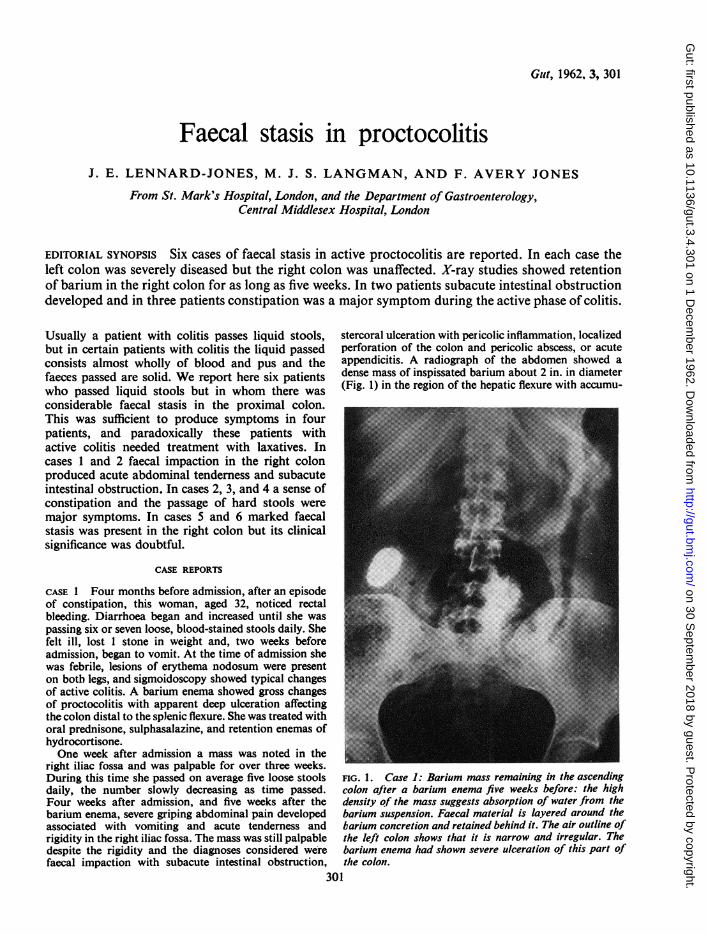
stercoral ulceration with pericolic inflammation, localizedperforation of the colon and pericolic abscess, or acuteappendicitis. A radiograph of the abdomen showed adense mass of inspissated barium about 2 in. in diameter(Fig. 1) in the region of the hepatic flexure with accumu-
FIG. 1. Case 1: Barium mass remaining in the ascendingcolon afier a barium enema five weeks before: the highdensity of the mass suggests absorption of water from thebarium suspension. Faecal material is layered around thebarium concretion and retained behind it. The air outline ofthe left colon shows that it is narrow and irregular. Thebarium enema had shown severe ulceration of this part ofthe colon.
301
on 30 Septem
ber 2018 by guest. Protected by copyright.
http://gut.bmj.com
/G
ut: first published as 10.1136/gut.3.4.301 on 1 Decem
J. E. Lennard-Jones, M. J. S. Langman, and F. Avery Jones
lated faecal material apparently layered around it andretained behind it. The ascending colon and caecum weredistended with faeces and the lower ileum was distendedwith gas. The distal colon, outlined by gas in the regionof the transverse colon and splenic flexure, showedirregularity and constriction of its lumen. Subacuteintestinal obstruction due to impacted barium and faecesin the ascending colon was diagnosed. Magnesiumsulphate was given by mouth, a large faecal mass waspassed and then the pain and tenderness subsided. A fur-ther radiograph showed that the mass of barium was nolonger present though some faecal material remained inthe ascending colon. She was discharged from hospitalthree weeks later free of symptoms. When last seen, onemonth later, she felt well, was passing one formed stooldaily and was about to return to work.
CASE 2 At the age of 47 this patient experienced a shortepisode of diarrhoea with rectal bleeding. Eight yearslater she was referred to hospital because she had becomeconstipated and had passed blood per rectum. Sigmoi-doscopy revealed active mucosal inflammation extendingfor 15 cm. above the anus; beyond this distance thecolonic mucosa appeared normal. She was admitted tohospital two months later because she became severelyill, with fever, constant nausea, weight loss, and thepassage of several dark red, liquid stools each day. Onexamination she was pale (Hb 36%), the abdomen wasdistended, and a large mass could be felt in the rightiliac fossa. Sigmoidoscopy to 13 cm. revealed an inflamedrectal mucosa, the lumen containing blood and mucus.Perforation of the colon and a pericolic abscess weresuspected. However, radiographs of the abdomen showedno abnormality other than distension of the caecum byfaeces. A few days later the patient had a rigor with afever of 103°F. and at this time constipated stools werepassed. A barium enema showed a somewhat dilated butotherwise normal colon containing many faecal masses.Constipation remained a problem until she was dis-charged from hospital free of symptoms after six weeks'treatment with corticosteroids, frequent olive oil enemas,and oral laxatives.
After two weeks she was re-admitted to hospitalbecause of severe abdominal pain. She again looked ill,the descending colon was palpable and heavily loaded;the ascending colon felt distended and was tender. Onsigmoidoscopy, the rectum appeared very inflamed andmany pseudo-polypi were seen. That evening she passeda large stool, her condition improved, and the colonbecame impalpable. During the next five days no bowelaction occurred though blood was frequently passed. Shesuddenly deteriorated with nausea, a rigor, and abdominaldistension two weeks later, despite treatment with pred-nisone and regular doses of magnesium hydroxide withliquid paraffin emulsion. A radiograph of the abdomenshowed that the whole colon was distended with faecesand some fluid levels were present in the small intestine.Daily saline washouts were instituted. Some days latershe had a severe rectal haemorrhage, necessitating im-mediate transfusion, and haemorrhage continued inter-mittently for some days. Surgical treattnent was decidedupon and subtotal colectomy with ileostomy performed.
-4
-3
-2
-o
FIG. 2. Case 2: Junction of normal and severelydiseased mucosa in the distal sigmoid colon. The colon was
normal proximal to this level.
The operation specimen (Fig. 2) showed a normal butdilated colon proximal to an area of severe disease withulceration, crypt abscesses, and numerous pseudo-polypi, involving the distal sigmoid colon and rectum.Recovery was delayed by a lung abscess but the patientis now well.
CASE 3 A woman of 35 was admitted to hospital with afour-month history of passing blood and pus per rectum.The symptoms had not improved during out-patienttreatment with prednisone and sulphasalazine. In hospitalshe complained bitterly of a sense of constipation; shepassed small quantities of blood several times a day and asmall constipated stool every few days. On palpation ofthe abdomen the ascending colon felt distended and on
sigmoidoscopy the rectal mucosa was actively inflamedbut the rectum was empty. A barium enema (Fig. 3)showed severe changes of colitis in the distal colon, withnarrowing and pseudo-polypi, and distension of theproximal colon, which appeared free of disease butcontained lumps of solid faeces. The colitis was treatedwith A.C.T.H., hydrocortisone retention enemas, andlarge doses of magnesium hydroxide emulsion withliquid paraffin by mouth. Her conditioni slowly improved
inches
ra
-7
-6
-5
302
on 30 Septem
ber 2018 by guest. Protected by copyright.
http://gut.bmj.com
/G
ut: first published as 10.1136/gut.3.4.301 on 1 Decem
FIG. 3. Case 3: The lower part of the descending colonis severely narrowed with pseudo-polypoid changes. Manysolid faecal masses are present in the dilated colon abovethe diseased area.
and the colonic distension subsided. When last seen, sixmonths after leaving hospital. she was well and passingone normal motion daily with a little blood and mucus.
CASE 4 This woman of 70 was admitted to hospitalwith a six-month history of increasing rectal bleeding,diarrhoea, weight loss, and latterly of high fever. Forseveral days after admission she ran a remittent fever of102 to 103°F. On sigmoidoscopy, the colonic mucosawas seen to be actively inflamed. A barium enema showedapparent deep ulceration involving the descending colon;the colon above this level could not be filled. She wasgiven A.C.T.H. and her condition slowly improved, theinitial diarrhoea being replaced by severe constipation;indeed, a second barium enema examination performedtwo months after admission was technically unsatis-factory because of impacted faecal masses in the ascendingcolon. The radiologist commented that these massesmust have taken weeks to accumulate. Treatment of theconstipation with oral laxatives and olive oil enemasbecame a major problem in the management of the illness.After the faecal masses were passed, constipation ceasedto be a problem and the patient was discharged homefree of symptoms six weeks after admission to hospital.A relapse of the disease occurred just over a year later.
Before treatment on this occasion, she was passing nineto 12 bowel motions daily, a soft motion in the morningand thereafter loose stools containing much mucus. One
FIG. 4a. FIG. 4b.
FIG. 4. Case 4: (a) The right colon is outlined 24 hours after giving barium suspension by mouth. (b) Nine days laterbarium is still present in the left colon. Faecal masses containing a little barium are present in the righlt colon.
303
on 30 Septem
ber 2018 by guest. Protected by copyright.
http://gut.bmj.com
/G
ut: first published as 10.1136/gut.3.4.301 on 1 Decem
J. E. Lennard-Jones, M. J. S. Langman, and F. A very Jones
2 1
20
19
17
1 6
14
1 3
1 2
I0
9
5
3
2
FIG. 5a. FIG. 5b.0
FIG. 5. Case 5: (a) Barium administered four weeks previously as an enema is retained withinfaecal masses in the rightand descending colon. (b) Operation specimen showing that the colon was almost normal proximal to the mid-transversecolon, but distally there was severe disease with extensive stripping of the mucosa leaving isolated mucosal tags.
week after starting treatment with prednisone the numberfell to three formed stools daily. Abdominal distensionnow developed and the stools became hard and infrequent,though blood-stained mucus was passed frequently. Aradiograph of the abdomen showed distension of theproximal colon with gas and faeces. Abdominal disten-sion became the main symptom and this was apparentlyassociated with faecal stasis, the hard, loaded ascendingcolon being easily palpable. A small quantity of bariumsuspension was given by mouth and its progress followedby daily radiographs. These films showed that within24 hours the barium had reached the right colon (Fig. 4a),but thereafter progress was so slow that nine days laterthe descending colon was still outlined (Fig. 4b). She wastreated with prednisone, sulphasalazine, magnesiumhydroxide, liquid paraffin and methyl cellulose by mouth,and by olive oil enemas. The distension and faecalstasis gradually resolved and she was discharged wellfive weeks after admission.
CASE 5 This patient, aged 51, was admitted to hospitalelsewhere with a short history of severe diarrhoea andsystemic illness. There was no response to treatment withcortisone and sulphasalazine and, as her condition gaverise to anxiety, she was transferred after two months toSt. Mark's Hospital for surgical treatment. When first
seen, she was pale, lethargic, vomiting frequently, andpassing about eight liquid stools each day. There wasmoderate fever; the abdomen was slightly distended butno masses were palpable. A radiograph of the abdomenshowed an unexpected degree of faecal stasis becausebarium, introduced exactly one month previously, wasstill present mixed with faecal material in the caecum(Fig. 5a). Faecal masses persisted in the caecum foralmost two weeks despite colonic wash-outs. For twoweeks the patient's condition gradually improved withsupportive treatment, but then deteriorated suddenlywith increased fever and abdominal tenderness.
Subtotal colectomy with ileostomy was performed, andthe post-operative recovery was uneventful. The operationspecimen (Fig. 5b) showed that the caecum and proximalhalf of the transverse colon were almost normal butdistally there was extensive stripping of mucosa, leavingmucosal tags. Histologically, the inflammatory processwas seen to penetrate all layers of the bowel wall in thesigmoid colon.
CASE 6 This man, aged 54, was admitted to hospitalthree months after developing diarrhoea. During the weekbefore admission he lost all desire for food, his abdomenbecame distended, and he passed about 10 motions dailyconsisting mainly of blood. He looked ill, was running a
304
on 30 Septem
ber 2018 by guest. Protected by copyright.
http://gut.bmj.com
/G
ut: first published as 10.1136/gut.3.4.301 on 1 Decem
fever up to 102°F., the abdomen was distended and tender,and on sigmoidoscopy the rectal mucosa appearedactively inflamed. Two days later he was worse and had20 bowel actions during one night. On this day radio-graphy of the abdomen showed the presence of muchfaecal material in the ascending colon. Treatment withA.C.T.H. resulted in immediate improvement but somefever persisted and several liquid, brown, blood-stainedstools were passed each day. A further radiograph of theabdomen three weeks after admission still showed faecalmasses in the right colon. Four days later a mouthful ofbarium suspension was given. Daily radiographs showedthat the barium remained in the caecum for six daysthough liquid stools were still being passed. An oraldose of Cr61 as sodium chromate, an unabsorbed tracer,was administered, and in 48 hours, despite the passage ofseven liquid stools, only 12% of the dose was excreted ascompared to a mean normal excretion of 54 3 % (Hanskyand Connell, 1962). Oral sodium sulphasuccinate wasadded to the treatment regime. Over the next few daysthe fever subsided, his appetite returned, and one weeklater a formed stool was passed for the first time sincethe illness began. A barium enema at this stage showed anapparently normal ascending and proximal transversecolon. Distal to the mid-transverse colon, the bowel wasshortened and narrowed; numerous mucosal fillingdefects were apparent in the descending colon. The patientwas discharged, feeling well and passing two semi-formedstools daily, seven weeks after admission.
DISCUSSION
Certain features were common to these six patients.All had severe ulcerative colitis of part or all of theleft colon and in all of them the right colon wasapparently normal. All passed frequent liquid stoolsat a time when the right colon was filled with faecalmaterial. In four of the six patients there was radio-logical evidence of prolonged retention of bariumsuspension in the right colon, in one case for fourand in another for five weeks. It seems possible thatthe severe colitis in the left colon acted as anobstruction to the passage of solid faeces while atthe same time the water-absorptive capacity of thenormal right colon remained unimpaired so thatinspissation of the contents of the right colonfollowed. The high density of the barium con-centration in case 1 (Fig. 1) suggests that water wasabsorbed in the ascending colon of this patient. Theslow passage of the unabsorbed tracer in case 5
suggests that the frequent liquid stools passed bythese patients consist mainly of exudate from thediseased colon.
In five of these patients faecal stasis was an un-expected finding on a plain radiograph of theabdomen. This is only part of the informationgained from such a film which can be helpful inassessing the state of the colon in patients withcolitis. A plain radiograph has revealed unexpectedfaecal stasis in other patients with colitis but wehave not assessed its frequency and do not knowhow often it is of clinical significance.
If faecal stasis is thought to be aggravating thecondition of a patient with colitis specific treatmentis indicated. Opiates and anticholinergic drugsshould be stopped and then, if necessary, orallaxatives should be given. Saline purges, liquidparaffin and wetting agents, such as dioctyl sodiumsulphasuccinate, may be helpful. Saline wash-outsare probably ineffective when the faecal accumu-lation is in the right colon but retention enemas ofolive oil seem beneficial when hard faeces in therectum are causing discomfort.There is little comment in the literature about
faecal stasis in colitis, though constipation occurs inproctitis (Lennard-Jones, Cooper, Newell, Wilson,and Avery Jones, 1962). Henderson (1954) suggestedthat constipation may be followed by colitis, andintroduced the concept of faecal stasis at St. Mark'sHospital by describing cases similar to those reportedhere as suffering from 'proximal constipation anddistal diarrhoea'. The term 'faecal stasis' seemspreferable as it is free of other connotation.
We thank Dr. Richard Asher (case 1), Mr. 0. V. LloydDavies (cases 2 and 4), and Mr. C. Naunton Morgan(case 5) for permission to report their cases. We thankDr. J. Hansky for the radio-isotope study in case 6 andthe X-ray and Photographic Departments at the CentralMiddlesex and St. Mark's Hospitals for the illustrations.
REFERENCES
Hansky, J., and Connell, A. M. (1962). Measurement of gastro-intestinal transit using radioactive chromium. Gut, 3, 187-188.
Henderson, N. P. (1954). What is ulcerative colitis? Lancet, 1, 159-160.Lennard-Jones, J. E., Cooper, G. W., Newell, A. C., Wilson, C. W.
E., and Jones, F. Avery (1962). Observations on idiopathicproctitis. Gut, 3, 201-206.
on 30 Septem
ber 2018 by guest. Protected by copyright.
http://gut.bmj.com
/G
ut: first published as 10.1136/gut.3.4.301 on 1 Decem