11
Tala Saleh M. Al-Mohtaseb Faisal Nimri 5
4
Tala Saleh
M. Al-Mohtaseb
Faisal Nimri
5
1 | P a g e
Inguinal Hernia
- An abdominal hernia is the protrusion of part of the abdominal content beyond the normal
confines of the abdominal wall through weak points in the abdominal wall. This happens due
to a combination of:
1- Weakness
2- Strain; increase in intra-abdominal pressure.
- Weakness can be in the:
1- Anterior abdominal wall muscles; they become weak in elderlies.
2- Deep inguinal ring in the transversalis fascia; considered a weak point because the
spermatic cord passes through it.
3- Umbilicus.
- The strain may result due to:
1- Chronic constipation in elderlies.
2- Chronic coughing in smokers.
➔ Increasing the abdominal pressure with strain results in the opening of weak points
allowing hernia to occur. Small intestine loops herniate most often, but also portions of
great omentum may also protrude.
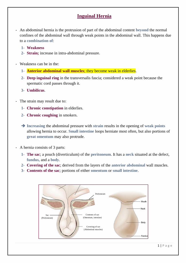
- A hernia consists of 3 parts:
1- The sac; a pouch (diverticulum) of the peritoneum. It has a neck situated at the defect,
fundus, and a body.
2- Covering of the sac; derived from the layers of the anterior abdominal wall muscles.
3- Contents of the sac; portions of either omentum or small intestine.
Contents of sac
(Omentum, intestine)
Covering of sac
(Abdominal muscles)
Sac
(Peritoneum)
Peritoneum
2 | P a g e
- There are many types of hernias, such as inguinal hernia, umbilical hernia, incisional hernia,
etc. The inguinal hernia is the commonest and it can be either indirect or direct.
1- Indirect (most common)
- Route: In males, it follows the course of
the spermatic cord, entering the inguinal
canal through the deep inguinal ring,
lateral to the inferior epigastric vessels
extending through the inguinal canal as far
as the superficial inguinal ring or even the
scrotum; the longer it persists the farther it
goes. While in females, it extends through
the superficial inguinal ring reaching the
labia majora.
- The hernia descends downwards, forward and medially. To reduce it (push it back to the
abdomen), a pressure is applied upwards, backward and laterally.
- It is mainly due to congenital defects where the processus vaginalis persists.
- Explanation: During the development of the embryo, the testes descend into the scrotum
carrying along with it a double layered peritoneal sac called ‘Processus Vaginalis’. Later
on, the proximal portion of the processus vaginalis obliterates undergoing fibrosis while the
distal portion persists as ‘Tunica Vaginalis’.
- If the processus vaginalis did not undergo obliteration it remains in open communication
between the peritoneum cavity and the scrotum, allowing herniation through it, with the
neck of the hernia positioned at the deep inguinal ring (recall the route).
- If the indirect hernia is due to a congenital origin (defect in the processus vaginalis), it happens
bilaterally, i.e. on both sides of the lower abdomen.
If it’s acquired, it happens unilaterally with it being more common on the right side;
because the right testis descends later than the left testis.
- The indirect hernia is more common in young males than in females (20:1).
Inferior Epigastric vessels Direction of hernia
The processus
vaginalis undergoes
fibrosis, closing the
communication
between the scrotum
and peritoneum.
If it does not
obliterate during
development, the
communication
remains open
allowing herniation.
3 | P a g e
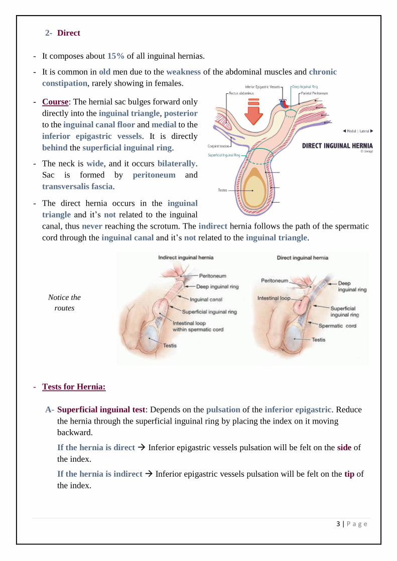
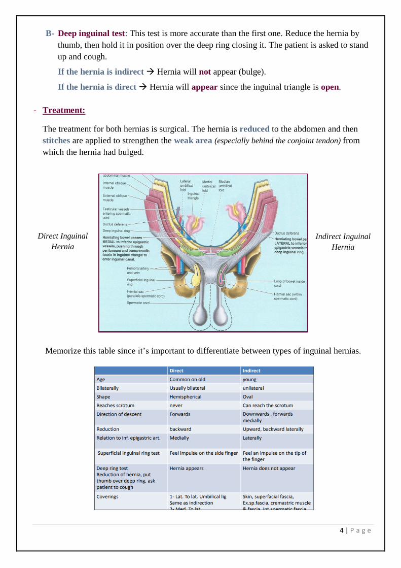
2- Direct
- It composes about 15% of all inguinal hernias.
- It is common in old men due to the weakness of the abdominal muscles and chronic
constipation, rarely showing in females.
- Course: The hernial sac bulges forward only
directly into the inguinal triangle, posterior
to the inguinal canal floor and medial to the
inferior epigastric vessels. It is directly
behind the superficial inguinal ring.
- The neck is wide, and it occurs bilaterally.
Sac is formed by peritoneum and
transversalis fascia.
- The direct hernia occurs in the inguinal
triangle and it’s not related to the inguinal
canal, thus never reaching the scrotum. The indirect hernia follows the path of the spermatic
cord through the inguinal canal and it’s not related to the inguinal triangle.
- Tests for Hernia:
A- Superficial inguinal test: Depends on the pulsation of the inferior epigastric. Reduce
the hernia through the superficial inguinal ring by placing the index on it moving
backward.
If the hernia is direct → Inferior epigastric vessels pulsation will be felt on the side of
the index.
If the hernia is indirect → Inferior epigastric vessels pulsation will be felt on the tip of
the index.
Notice the
routes
4 | P a g e
B- Deep inguinal test: This test is more accurate than the first one. Reduce the hernia by
thumb, then hold it in position over the deep ring closing it. The patient is asked to stand
up and cough.
If the hernia is indirect → Hernia will not appear (bulge).
If the hernia is direct → Hernia will appear since the inguinal triangle is open.
- Treatment:
The treatment for both hernias is surgical. The hernia is reduced to the abdomen and then
stitches are applied to strengthen the weak area (especially behind the conjoint tendon) from
which the hernia had bulged.
Memorize this table since it’s important to differentiate between types of inguinal hernias.
Direct Inguinal
Hernia
Indirect Inguinal
Hernia
5 | P a g e
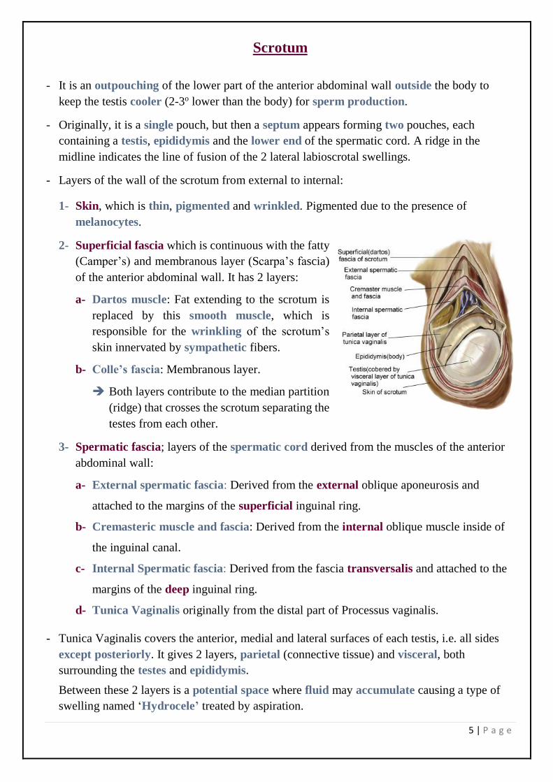
Scrotum
- It is an outpouching of the lower part of the anterior abdominal wall outside the body to
keep the testis cooler (2-3o lower than the body) for sperm production.
- Originally, it is a single pouch, but then a septum appears forming two pouches, each
containing a testis, epididymis and the lower end of the spermatic cord. A ridge in the
midline indicates the line of fusion of the 2 lateral labioscrotal swellings.
- Layers of the wall of the scrotum from external to internal:
1- Skin, which is thin, pigmented and wrinkled. Pigmented due to the presence of
melanocytes.
2- Superficial fascia which is continuous with the fatty
(Camper’s) and membranous layer (Scarpa’s fascia)
of the anterior abdominal wall. It has 2 layers:
a- Dartos muscle: Fat extending to the scrotum is
replaced by this smooth muscle, which is
responsible for the wrinkling of the scrotum’s
skin innervated by sympathetic fibers.
b- Colle’s fascia: Membranous layer.
➔ Both layers contribute to the median partition
(ridge) that crosses the scrotum separating the
testes from each other.
3- Spermatic fascia; layers of the spermatic cord derived from the muscles of the anterior
abdominal wall:
a- External spermatic fascia: Derived from the external oblique aponeurosis and
attached to the margins of the superficial inguinal ring.
b- Cremasteric muscle and fascia: Derived from the internal oblique muscle inside of
the inguinal canal.
c- Internal Spermatic fascia: Derived from the fascia transversalis and attached to the
margins of the deep inguinal ring.
d- Tunica Vaginalis originally from the distal part of Processus vaginalis.
- Tunica Vaginalis covers the anterior, medial and lateral surfaces of each testis, i.e. all sides
except posteriorly. It gives 2 layers, parietal (connective tissue) and visceral, both
surrounding the testes and epididymis.
Between these 2 layers is a potential space where fluid may accumulate causing a type of
swelling named ‘Hydrocele’ treated by aspiration.
6 | P a g e
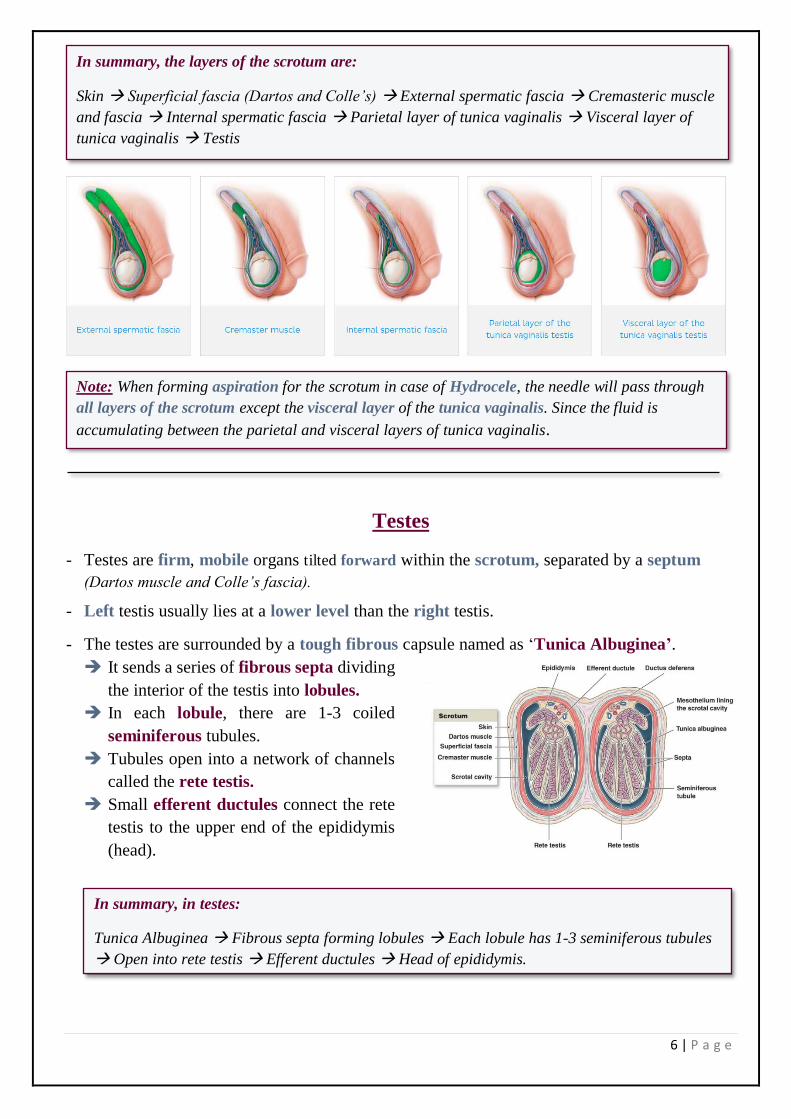
Testes
- Testes are firm, mobile organs tilted forward within the scrotum, separated by a septum
(Dartos muscle and Colle’s fascia).
- Left testis usually lies at a lower level than the right testis.
- The testes are surrounded by a tough fibrous capsule named as ‘Tunica Albuginea’.
➔ It sends a series of fibrous septa dividing
the interior of the testis into lobules.
➔ In each lobule, there are 1-3 coiled
seminiferous tubules.
➔ Tubules open into a network of channels
called the rete testis.
➔ Small efferent ductules connect the rete
testis to the upper end of the epididymis
(head).
In summary, the layers of the scrotum are:
Skin → Superficial fascia (Dartos and Colle’s) → External spermatic fascia → Cremasteric muscle
and fascia → Internal spermatic fascia → Parietal layer of tunica vaginalis → Visceral layer of
tunica vaginalis → Testis
Note: When forming aspiration for the scrotum in case of Hydrocele, the needle will pass through
all layers of the scrotum except the visceral layer of the tunica vaginalis. Since the fluid is
accumulating between the parietal and visceral layers of tunica vaginalis.
In summary, in testes:
Tunica Albuginea → Fibrous septa forming lobules → Each lobule has 1-3 seminiferous tubules
→ Open into rete testis → Efferent ductules → Head of epididymis.
7 | P a g e
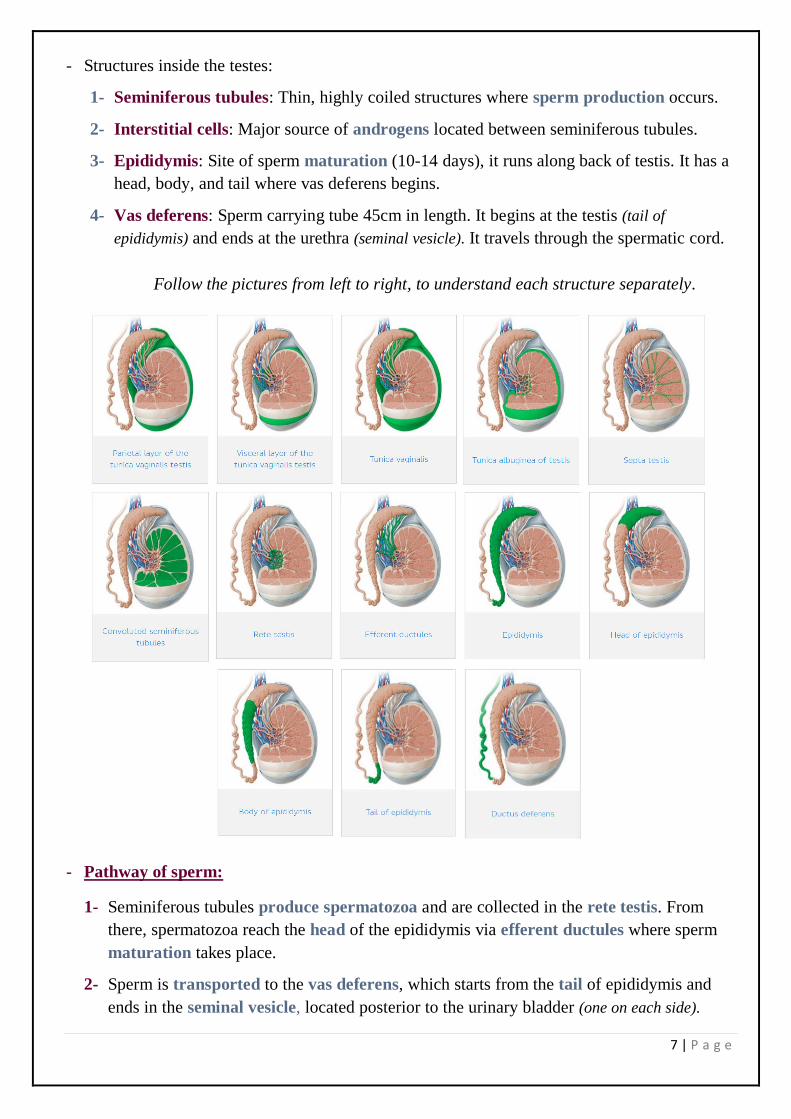
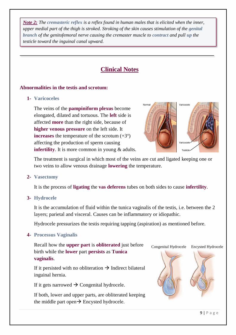
- Structures inside the testes:
1- Seminiferous tubules: Thin, highly coiled structures where sperm production occurs.
2- Interstitial cells: Major source of androgens located between seminiferous tubules.
3- Epididymis: Site of sperm maturation (10-14 days), it runs along back of testis. It has a
head, body, and tail where vas deferens begins.
4- Vas deferens: Sperm carrying tube 45cm in length. It begins at the testis (tail of
epididymis) and ends at the urethra (seminal vesicle). It travels through the spermatic cord.
Follow the pictures from left to right, to understand each structure separately.
- Pathway of sperm:
1- Seminiferous tubules produce spermatozoa and are collected in the rete testis. From
there, spermatozoa reach the head of the epididymis via efferent ductules where sperm
maturation takes place.
2- Sperm is transported to the vas deferens, which starts from the tail of epididymis and
ends in the seminal vesicle, located posterior to the urinary bladder (one on each side).
8 | P a g e
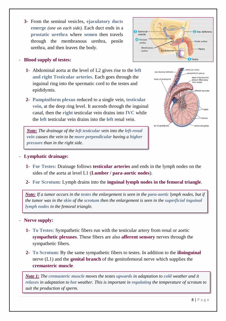
3- From the seminal vesicles, ejaculatory ducts
emerge (one on each side). Each duct ends in a
prostatic urethra where semen then travels
through the membranous urethra, penile
urethra, and then leaves the body.
- Blood supply of testes:
1- Abdominal aorta at the level of L2 gives rise to the left
and right Testicular arteries. Each goes through the
inguinal ring into the spermatic cord to the testes and
epididymis.
2- Pampiniform plexus reduced to a single vein, testicular
vein, at the deep ring level. It ascends through the inguinal
canal, then the right testicular vein drains into IVC while
the left testicular vein drains into the left renal vein.
- Lymphatic drainage:
1- For Testes: Drainage follows testicular arteries and ends in the lymph nodes on the
sides of the aorta at level L1 (Lumber / para-aortic nodes).
2- For Scrotum: Lymph drains into the inguinal lymph nodes in the femoral triangle.
- Nerve supply:
1- To Testes: Sympathetic fibers run with the testicular artery from renal or aortic
sympathetic plexuses. These fibers are also afferent sensory nerves through the
sympathetic fibers.
2- To Scrotum: By the same sympathetic fibers to testes. In addition to the ilioinguinal
nerve (L1) and the genital branch of the genitofemoral nerve which supplies the
cremasteric muscle.
Prostatic
urethra
Membranous
urethra
Penile urethra
Note: The drainage of the left testicular vein into the left-renal
vein causes the vein to be more perpendicular having a higher
pressure than in the right side.
Note: If a tumor occurs in the testes the enlargement is seen in the para-aortic lymph nodes, but if
the tumor was in the skin of the scrotum then the enlargement is seen in the superficial inguinal
lymph nodes in the femoral triangle.
Note 1: The cremasteric muscle moves the testes upwards in adaptation to cold weather and it
relaxes in adaptation to hot weather. This is important in regulating the temperature of scrotum to
suit the production of sperm.
9 | P a g e
Clinical Notes
Abnormalities in the testis and scrotum:
1- Varicoceles
The veins of the pampiniform plexus become
elongated, dilated and tortuous. The left side is
affected more than the right side, because of
higher venous pressure on the left side. It
increases the temperature of the scrotum (+3o)
affecting the production of sperm causing
infertility. It is more common in young & adults.
The treatment is surgical in which most of the veins are cut and ligated keeping one or
two veins to allow venous drainage lowering the temperature.
2- Vasectomy
It is the process of ligating the vas deferens tubes on both sides to cause infertility.
3- Hydrocele
It is the accumulation of fluid within the tunica vaginalis of the testis, i.e. between the 2
layers; parietal and visceral. Causes can be inflammatory or idiopathic.
Hydrocele pressurizes the testis requiring tapping (aspiration) as mentioned before.
4- Processus Vaginalis
Recall how the upper part is obliterated just before
birth while the lower part persists as Tunica
vaginalis.
If it persisted with no obliteration → Indirect bilateral
inguinal hernia.
If it gets narrowed → Congenital hydrocele.
If both, lower and upper parts, are obliterated keeping
the middle part open→ Encysted hydrocele.
Congenital Hydrocele Encysted Hydrocele
Note 2: The cremasteric reflex is a reflex found in human males that is elicited when the inner,
upper medial part of the thigh is stroked. Stroking of the skin causes stimulation of the genital
branch of the genitofemoral nerve causing the cremaster muscle to contract and pull up the
testicle toward the inguinal canal upward.
10 | P a g e
Congenital abnormalities of the testes:
1- Cryptorchidism
Incomplete descent of the testis although
traveling down the normal pathway, it may be
found in the abdominal cavity, inguinal canal,
superficial inguinal ring or at the upper part of the
scrotum.
2- Maldescent
Testes travel down an abnormal pathway, it may be found in the superficial fascia, the
root of the penis, perineum or in the thigh.
It is more difficult than Cryptorchidism requiring an immediate operation. If the testis
remains in an abnormal position beyond 6 years, this will impair the production of
testosterone.
Good Luck


