Farm animal reproduction: Conserving local genetic resources Proceedings from a minisymposium at Lithuanian Veterinary Academy, Kaunas, Lithuania September 13-15, 2003 Renée Båge and Aloyzas Januskauskas (editors) Uppsala 2003 CRU Report 17
Transcript
Farm animal reproduction:Conserving local genetic resources
Proceedings from a minisymposium atLithuanian Veterinary Academy, Kaunas, LithuaniaSeptember 13-15, 2003
Renée Båge and Aloyzas Januskauskas (editors)
Uppsala 2003
CRU Report 17
Farm animal reproduction: Conserving local genetic resources
Proceedings from a mini-symposium at
Lithuanian Veterinary Academy, Kaunas, Lithuania September 13-15, 2003
Development of Genetic Evaluation Systems - Hossein Jorjani 6Gene banking of endangered sheep and goat breeds in Sweden Lennart Söderquist 7Ex-situ and in-situ conservation of Lithuanian domestic animal genetic resources – lessons from past and future perspectives - Ilona Miceikiene 8Present and future actions for conservation of genetic resources in the Nordic countries - Birgitta Danell 12Evaluation of semen for long-term preservation with special reference to stallion - Stig Einarsson 13
Milk protein polymorphism in four Lithuanian cattle breeds - Jolanta Malevičiūtė 14
Collection and storing of embryos and oocytes - Renée Båge 22Age and breed-dependent variation of ovarian and follicular parameters in cows - Marius Masiulis 23
Papers: Anti-mastitis medication ‘Synolux’, Mamexine, Mastimix and Lincomycin-F efficacy in treating bovine sub-clinical mastitis - Jūratė Klimaitė 28
Porcine chalmydiosis diagnostics - Žaneta Laureckienė 32Assessment of sperm quality post-thaw and the effect of progesterone on sperm function of dairy AI bull semen - Kristina Lukoseviciute 35The effect of fetal bovine serum and bovine serum albumin on bovine embryos development in vitro - Rasa Nainienė 40Principles for conservation activities of critical farm animal breeds in Lithuania - Violeta Razmaite 41Use Cloprostenolum combine with vitamins therapy: Follicular growth and progesterone level - Arunas Rutkauskas 42
The effects of sperm morphology on boar fertility results - Neringa Sutkeviciene 45Influence of various hormones and hormonic preparations on the reproductive performance of sows - Henrikas Žilinskas 48
CRU Publication Series 55
4
Foreword The Symposium “Farm animal reproduction: Conserving local genetic resources” at the Veterinary Academy in Kaunas, Lithuania on September 13-15 is a part of the cooperative programme “Farm animal reproduction: Reducing infectious diseases and Conserving local genetic resources” between Estonian Agricultural University (EAU), Tartu, The Lithuanian Veterinary Academy of Lithuania, Kaunas, Latvia University of Agriculture, Jelgava, and the Centre for Reproductive Biology in Uppsala, Swedish University of Agricultural Sciences. The cooperation is financially supported by “Nya Visbyprogrammet” at the Swedish Institute, Stockholm. This symposium focuses on conserving local genetic resources since there is a major concern in animal husbandry today about the diminishing biodiversity among the species involved. In the Baltic States there is still a lot of local breeds considerably different from those in the Nordic countries and Western Europe. There is strong demand for preserving this unique genetic resource, and techniques or methodology from the area of animal reproduction may play a major role in meeting this demand. Here we have gathered speakers from all four countries giving several presentations in the field of conserving genes and reproductive biotechnologies. We are sure that this will provide the basis for a fruitful collegial exchange and scientific progress. On behalf of the national programme-coordinators, Drs. Toomas Tiirats in Estonia, Henrikas Zilinskas and Vita Riskeviciene in Lithuania, Vita Antane in Latvia and Ulf Magnusson in Sweden, we wish you a pleasant reading! Uppsala and Kaunas in September 2003 Renée Båge and Aloyzas Januskauskas (editors)
5
List of participants at the mini-symposium “Farm animal reproduction: Conserving local genetic resources”, Kaunas, Lithuania, September 13-15, 2003. ESTONIA Käde Kalamees Dept. of Animal Breeding, Institute of Animal Science, Estonian
Agricultural University Piret Kalmus Department of Therapy, Faculty of Veterinary Medicine,
Estonian Agricultural University Toomas Tiirats Department of Animal Health, Faculty of Veterinary
Medicine, Estonian Agricultural University LATVIA Ziedonis Grīslis Department of Animal Sciences Faculty of Agriculture Daina Jomkus Department of Animal Sciences Faculty of Agriculture Didzis Strautmanis Research Centre "Sigra" of Latvia University of Agriculture Santa Skuja Clinical Institute Faculty of Veterinary Medicine Vita Antane Clinical Institute Faculty of Veterinary Medicine LITHUANIA Henrikas Zilinskas Dept. of Obstetrics and Gynecology, LVA Aloyzas Januskauskas Dept. of Obstetrics and Gynecology, LVA Eugenijus Aniulis Dept. of Obstetrics and Gynecology, LVA Arunas Rutkauskas Dept. of Obstetrics and Gynecology, LVA Neringa Sutkeviciene Dept. of Obstetrics and Gynecology, LVA Jurate Klimaite Dept. of Obstetrics and Gynecology, LVA Jolanta Maleviciute Dept. of Animal Breeding and Genetics, LVA Ilona Miceikiene Dept. of Animal Breeding and Genetics, LVA Vita Riskeviciene Dept. of Physiology and Pathology, LVA Marius Masiulis Dept. of Physiology and Pathology, LVA Kristina Lukoseviciute Dept. of Physiology and Pathology, LVA Rasa Nainiene Institute of Animal Science Violeta Razmaite Institute of Animal Science Ana Zilinskiene Lithuanian University of agriculture Zaneta Laureckiene Lithuaniana veterinary academy, Dept. of OG SWEDEN Renée Båge Dept. Of Obstetrics and Gynaecology, SLU Birgitta Danell Dept. Of Animal Breeding and Genetics, SLU Stig Einarsson Dept. Of Obstetrics and Gynaecology, SLU Hossein Jorjani Interbull, Dept. Of Animal Breeding and Genetics, SLU Ulf Magnusson Dept. Of Obstetrics and Gynaecology, SLU Lennart Söderquist Dept. Of Obstetrics and Gynaecology, SLU
Development of Genetic Evaluation Systems
Hossein JorjaniInterbull Centre, Department of Animal Breeding and Genetics, Swedish University of Agricultural
A Genetic Evaluation System (GES), defined as each and every statistical treatment of data that has an evolutionary-genetic-breeding motivation or justification, comprises tens (if not hundreds) of components, from collection of data topublication of results. Development of a GES is complicated by the fact that for each of these components there are anumber of different alternative methods available. Making decisions as to which alternative method should be thepreferred one for each of the GES components may easily be turned into an insurmountable task. This is probably truefor any GES, whether it is in plants or animals, domestic or wild, rural or industrialized. Fortunately, the example ofdairy cattle breeding shows that the task of developing and sustaining a national GES may be easier than anticipated atthe outset. Interbull Centre1 continuously collects detailed information on its member countries’ national GES for various traits ofeconomic interest in dairy cattle. Currently available information pertains to 31 countries, 6 breeds of dairy cattle, and atleast 10 different trait groups (www.interbull.org, look for “Genetic Evaluations” and then “Description of GES asapplied in member countries”). Based on the results of a recent survey (Interbull2, 2000) it could be concluded that the“raw data” obtained from individual animals were subjected to treatments in many stages and that there were manydifferences with regard to the number, nature, model and method of such treatments in different countries.Consequently Interbull has issued a set of Guidelines (Interbull Guidelines3,4, 2001) that can be of help in developmentand / or sustaining national GES for dairy cattle in general, but maybe also for other breeds and species. The motivationfor working on a set of guidelines stems from the devotion of Interbull to a) Active utilization of domestic animalgenetic resources, and b) Effective use of genetic resources globally to obtain the largest sustainable genetic progress.To achieve these goals Interbull encourages I) Development of national GES according to the world’s best practices fora broad range of economically important traits to fit variable objectives in member countries; and II) Bi- and multi-lateral cooperation between national genetic evaluation centers. Interbull Guidelines are the result of a pragmaticcompromise between dictates of several perspectives, among others, state of art in theoretical dairy cattle breeding, andneeds and capacity of individual farmers to incorporate the recommended changes into their operations. Interbull Guidelines, divides all necessary components of a GES into three (rough) groups as Pre-Evaluation steps,Evaluation Step and Post-Evaluation Steps, each of which is divided into many smaller steps. For those people who areabout to start the development of a new GES it is interesting to know that the methods employed in each step can be assimple as, or as complicated / sophisticated as possible. However, there is no minimum requirement imposed by anynational / international or scientific / professional authority on any of these. One can choose to use a very simplemethod or even ignore a step or one can adopt such a complicated method that the majority of the experts from othercountries may consider it a waste of resources. The key recommendation is to set up and start running a GES, no matterhow simple it may be. It is not in the scope of this short abstract to come up with some numerical examples, because it may lead totrivialization of the key recommendation put forward here. Historically, all national GES have been courageouslysimple, but, in time, all of them strived towards higher efficiency which could have not been possible if the experiencesfrom simpler national GES were not available. As a commercial advertisement says: Just Do It.
1 “Interbull Centre”, housed at the Department of Animal Breeding & Genetics, Swedish University of AgriculturalSciences, Uppsala, Sweden, is the (only) operational unit for the “International Bull Evaluation Service (Interbull)”.Interbull is a permanent Sub-Committee of the International Committee of Animal Recording (ICAR). In 1996 InterbullCentre was appointed as the official reference laboratory for cattle breeding for European Union (EU). All Interbullpublications and a huge amount of other useful information are freely available through www.interbull.org 2 Jorjani, H. (2000) national Genetic Evaluation Programmes for Dairy Production Traits Practiced in Interbull MemberCountries 1999-2000. Interbull Bulletin 24, Interbull, Uppsala.3 Jorjani, H., Philipsson, J. & Mocquot, J.-C. (2001) Interbull Guidelines for National & International GeneticEvaluation Systems in Dairy Cattle with Focus on Production Traits. Interbull Bulletin 28, Interbull, Uppsala.4 Interbull Guidelines have been incorporated in ICAR’s Guidelines (available through www.icar.org ) which addressesmost of the domestic species.
Gene banking of endangered sheep and goat breeds in Sweden
Lennart Söderquist Department of Obstetrics and Gynaecology, Faculty of Veterinary Medicine, Centre for
Reproductive Biology in Uppsala (CRU), Swedish University of Agricultural Sciences, Uppsala, Sweden, e-mail: [email protected]
Since 1998 the Department of Obstetrics and Gynaecology (OG), SLU, in co-operation with the Swedish Board of Agriculture, the Swedish Sheep Breeders Association and Uppsala Fårtjänst, have collected semen samples from rams and bucks from endangered sheep and goat breeds in Sweden, in order to create a national gene bank. Up to date 20 rams and 15 bucks have contributed to the gene bank and altogether approximately 2700 AI doses are now stored in liquid nitrogen (LN2) at Svensk Avel, Skara. The males used are remains of different ancient Swedish native breeds threatened by extinction. At present, some breeds only consist of less than 200 individuals. The breeds so far participating in the project are Gutefår, Dala pälsfår, Roslagsfår, Skogsfår (Värmlandsfår, Svärdsjöfår), Göingeget och Jämtget During the normal breeding season in Sweden (October- December) the selected males are transported from different small farms situated all over the country to a specially prepared and equipped AI station for small ruminants at Kungsängens gård, SLU, Uppsala. At the station there is room for altogether 8 rams/bucks and 2 ewes/goats. The males are not allowed to mate naturally within 30 days prior to semen collection. At arrival they are examined to be clinically free from any symptoms of disease and their testes are palpated and scrotal circumferences are measured in order to exclude abnormal individuals. Furthermore, all selected males originate from flocks tested and found free from Maedi/Visna disease. Blood samples are taken at the station for analyses of Border disease (ram) and CAEV (buck). Last year also samples from rams for analyses of Scrapie were taken. During a period of one to two weeks the males are trained and allowed to get used to the life and procedures at the station for semen collection. Although the situation at the station is very artificial, very few males refuse to get stimulated by the two teaser ewes/goats (prepared artificially by hormonal treatment to show oestrus behaviour) and the majority accepts to ejaculate in the artificial vagina used for semen collection. After first having collected 3-5 ejaculates per male, another semen sample is collected and motility is assessed under a microscope and samples are prepared for assessment of sperm concentration and morphology at the sperm laboratory at our department. If the sperm motility, concentration and morphology are within what is considered normal limits, two consecutive semen samples are collected and diluted up to 7.5 ml with a milk-based extender. The cooling of the diluted samples starts at the AI station in a Styrofoam box in which the samples, as soon as possible, are further transported to the freezing laboratory at the department of OG. There, a glycerol-containing extender is added step wise at 5°C up to 15 ml. After circa 2 hours equilibration at 5°C in a water bath the samples are centrifuged (700g, 10 min) and the supernatant is removed to leave a volume resulting in a final concentration of 800 million spermatozoa per ml (ram) and 300 million spermatozoa per ml (buck). For buck semen it is essential to get rid of the egg yolk coagulating enzyme by centrifugation (1000g, 7 min) before further dilution, cooling and freezing. So far the semen from bucks has been prepared as if the AI doses are to be used for intrauterine deposition (i.e. approx. 75 million spermatozoa/dose). (Results from studies in progress, in co-operation with a group in Norway, where different deposition sites are compared, might lead to changes in the future sperm concentration needed per dose.) The cooled semen is then automatically filled into labelled mini straws (0.25 ml) and frozen in a programmable freezer and stored in LN2 at –196°C. After thawing, sperm motility routinely is said to have to be equal or higher than 50% to be approved, but since this is, from a genetic point of view,a unique material, a somewhat lower motility sometimes must have to be accepted. The frozen mini straws are transported to Svensk Avel, Skara for long time storage until future use. So far none of the frozen semen doses in the gene bank have been used for artificial insemination. Gene banking facilitates preservation of genes from endangered species for future inseminations, thereby contributing to the important bio-diversity so well needed in the world. Hopefully gene banking will help to secure that also future generations are able to enjoy the unique characters and traits of these ancient and very well adapted sheep and goat breeds.
8
Ex-situ and in-situ conservation of Lithuanian domestic animal genetic resources – lessons from past and future perspectives
Ilona Miceikiene, Natalija Krasnapiorova, Rasa Petraškienė
Summary In Lithuania the system for characterization, management, registration and conservation of plant and animal genetic resources (AnGR) is under development. Information of genetic structure of breed is very important in making decisions for conservation, origin, and relation with other breeds. The Lithuanian White back and Light Grey cattle, Žemaitukai and Large type Žemaitukai horse, Lithuanian Blackhead and coarse wool sheep, native wattle pigs and Vištinės geese are indigenous and unique breeds included into 3d World Watch List for Cattle Biodiversity. Preservation of indigenous cattle breeds was performed by forming mini populations with pure breeding, cryopreserving semen and DNA. Investigations of biological traits – maturation rapidity, meat quality, productivity, estimation within breed polymorphism and genetic distances between breeds with different markers- blood groups, plasma protein, milk protein and microsatellites and mitochondrial DNA markers was performed. Keywords: animal genetic resources, diversity, conservation.
Introduction The total global biodiversity most likely includes tens of millions of species. But the biological diversity of the planet is rapidly being depleted as a direct or indirect consequence of human actions. An unknown but large number of species are already extinct, while many others have reduced population size that put them to risk (Frankham, 1995). Mankind uses some 40 species of animals as domestic livestock to meet our needs for food, clothing, power, etc. Within these species, there are in total some 4500 breeds that are referred to as the global animal genetic resources (Barker, 1999). In recent years, changes in economic climate have promoted the use of breeds suited to intensive production systems, which has led to a few breeds becoming widespread while the breeds that they have replaced have decline in population size. In some cases native populations have been crossbred with imported stock in upgrading programmes (Blott et al, 1998). However, the dramatic decline in livestock inventories and the economic conditions clearly indicate that there is pressure to increase profitability of livestock farming by replacing less productive breeds with more productive ones. Especially high-input/high-output breeds like the Holstein have already and still are gaining importance in Poland and the Baltics, as well. At the same time, it becomes fashionable to take-up beef farming by importing diverse beef breeds from Western Europe and North America (Grigaliunaite et al, 2001). To make the current and future progressive improvement of domestic animals populations successful in intensive and extensive circumstances, the genetic variation within domesticated species must be maintained (Oldenbroek, 1999).
Material and methods Based on different sources of information and own investigations the analysis of the development, present situation and problems of conservation of Lithuanian animal genetic resources is given in this study.
Results and discussion Our farm animal breeds are disappearing at an alarming rate. Of the 6400 recognized breeds, about 1000 have become extinct in the last 100 years- 300 during the last 155 years. Now it is estimated that two livestock and poultry breeds die out every week. These are breeds that people and environment has shaped over the last 10 000 years. Local breeds may carry genetic material of immense value. When the breed becomes extinct the whole world loses ability to react to changing environmental conditions, resist unforeseen diseases and respond to changes in human dietary. In Lithuania the system for characterization, management, registration and conservation of plant and animal genetic resources (AnGR) is under development. The dynamics of the number of livestock and poultry in 1990 – 2000 shows that high rate of their decrease has been observed in the first years of the tested period.
9
Figure.1.Changes in the livestock population in Lithuania (in thousands)
441,8
751,7748,3897,8
922,81016,3
438,4500541582,8
1010,8855,6936,1
1200,1 1159
751,7
742,3674,9 637,3
557,7
24 15,8 13,8 11,5 12,30
200
400
600
800
1000
1200
1400
1998 1999 2000 2001 2002
Cattle Cow Pig Poultry, 10 thous. Sheep
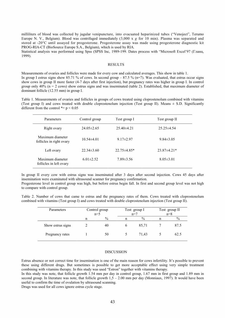
The number of livestock at the end of 2000 accounted for 31 percent of the number of the cattle bred in 1990, that of pigs 31 percent and poultry 32 percent accordingly. The main reasons for the decrease in the number of livestock are as follows restructuring of the agricultural sector and essential changes in domestic and foreign markets, the reduced consumption of meat. In 2002 – 2003 there is a tendency to slight increase of animal and poultry number. Table 1. Lithuanian livestock populations in 2003 year
Species Population (in thousands) Cattle of which cows Sheep Goats Horses Pigs of which sows Chicken of which hens Turkey Ducks Geese Rabbits Beehives
Species Total number of breeds Lithuanian Native breeds Cattle Pigs Sheep Goat Horses Poultry hens ducks geese Rabbits
20 10 5 3
16
Different crosses 1 2 8
4 2 2 1 3
1 2
In Lithuanian we have more than 20 cattle breeds of which 4 breeds are local – Lithuanian black and white and Lithuanian Red (modern breeds) and Lithuanian Light grey and Lithuanian White Backed (endangered breeds). Recently introduced breeds comprises less than 3 % from total amount of cattle – Lithuanian Black and White, German
10
Black and White, British Friesian, Dutch Black and White, Holstein, Danish Black and White, Swedish Black and White, Lithuanian Reds, Angler, Danish Reds, Ayrshire, Brown Swiss, German Red and White, Swedish Red and White. Also we have few meet cattle breeds – Charolais, Hereford, Limousine, Aubrak and Simental – that have been started to be raised few years ago. The main work in the sphere of cattle breeding outlined in the Animal Breeding Programmer for 2002 were control of cattle performance, analysis of milk content and quality, improvement of cattle breeding information system, analysis of received data, assessment of bulls according to performance values and exterior of progeny, assessment of breeding institutions and breeding herds, their data analysis, preparation of breeding documentation aiming for improvement of organization of breeding activities, their approximation with EU legal acts on breeding. There are 73 cattle breeding institutions and 233 breeding herds in Lithuania today. In 2002, one milk-recording cow averagely produced 5015 kg of milk 4,24 % fat content and 3,33 % protein content. In Lithuania mainly are bred Lithuanian white pig breed. It comprises more than 58 % o total breed pigs.The other native pig breed in Lithuania is Lithuanian native pig (Pig with wattles) which is very much diminished in numbers and is under conservation.The introduced breeds from which sows are used – Swedish Yorkshire, English Large White, Norwegian Landrace breeds.The introduced breeds from which boards are used – Large White, Pjetren, Norwegian Landrace. In Lithuania there are 3 native horse breeds – Žemaitukai, Large type Žemaitukai and Lithuanian Heavy Draught. All these breeds have status of endangered breeds. Breeding institutions and herds also keep horses of Trakėnai, Hanover, Holstein, Russian and American Trotters, Thoroughbred Mounts, Arab, Budioni and Pony. Certified horse breeding institutions are commissioned to perform primary accounting of horse breeding, issue pedigree certificates and manage herd-books. In Lithuania there are 5 sheep breeds from which 2 are native – Lithuania Black head and Lithuania coarse wool. Both breeds are under conservation. The introduced breeds are Romanov, Ostfryz, Prekos. Some farmers recently started to breed Berishon Diusher and Suffolk. Goat breeding herds mainly encompass Zaanen, Czech White and local goats. Lithuania had one second rank turkey reproduction farms, and 7 second rank chicken and goose reproduction farms. Chicken and goose reproduction farms breed laying and meet chicken breeds, hole breed geese and Big-5, But-8 and But-9 turkey crosses. Breeding herds include rabbits of French Ram, Belgian Giant, Rex, New Zealand and Viennese Giant breeds. Lithuania committed itself to conservation of its genetic resources by signing the Conservation on Biological Diversity in Rio de Janeiro on June 11, 1992. Lithuania Breeding Law indicates that one of the major tasks in animal genetics is conservation and improvement of Lithuanian animal breeds and their gene pools. In Lithuania, the improvement of indigenous animal breeds by absorptive crossbreeding was begun on a large scale as far back as end of World War I. In 1921, the Committee of the Ministry of Agriculture made a proposal to establish purebred herds of native animals with the aim to preserve the gene pools of establishing the Stud for Žemaitukai horses that was subsequently lost during World War II. ( Programme, 1997). Some of the indigenous animal and poultry breeds of Lithuania have become extinct, others, such as Žemaitukai horses, wattle pigs, ash-grey and white-backed cattle, native sheep, Vištinės geese, are on the verge of extinction. Thus, the Lithuanian Institute of Animal Science and Lithuanian Veterinary Academy took the initiative in organizing the conservation of Lithuanian native animal genetic resources. The search for native breed animals, gathering them and formation of mini populations with genealogical structure started in Lithuania after it’s restoration of independence. Lithuanian native animals- the Lithuanian White back and Light Grey cattle, Žemaitukai and Large type Žemaitukai horse, Lithuanian Black head and coarse wool sheep, native wattle pigs and Vištinės geese are indigenous and us unique breeds included into 3d World List for Cattle Biodiversity. There is a need also to preserve some modern Lithuanian animal breeds that have been created during long process of selection. Those breeds have good production and reproduction traits but because of crossbreeding under intensive farm breeding pressure and in the situation when few top breeds very productive prevail in the species in the world as Holstein in cattle are at risk of extinction. Those breeds are Danish type of Lithuanian Red, Dutch type of Lithuanian Black and White, Lithuanian White pig. Most of Lithuanian native animal breeds have FAO status of endangerment. Ex situ and in situ conservation methods are used to keep these breeds alive. Conservation herds have been formed in several keeping places. Ex situ method is mostly used for cattle Cryoconservation is used for semen, embryos and DNA. For conservation purposes certain amount of semen from each used bull is kept in artificial insemination stations. Lithuanian Farm Animal Gene Bank was created in Lithuanian Veterinary Academy in which DNA samples are gathered and kept from all native Lithuanian farm animal breeds. Earlier Lithuanian native animal breeds were investigated very little. When conservation started and they have been prevented from extinction investigations of these breeds started- phenotypical traits, production and reproduction properties, milk quality and composition, craniological testing, cytogenetical testing, within and between-breeds polymorphism and breed distancing by biochemical- blood groups, blood plasma proteins, milk proteins and molecular-
11
microsatelites, mitochondrial DNA, Y chromosome polymorphism. While evaluating it’s AnGR Lithuania together with Etonia, Latvia, Poland Scandinavion countries Ireland and some post Soviet countries participate in different international projects- “Analysis and Comparison of Genetic Diversity in Cattle Breeds of Northern European Area _ N-EURO-CAD (www.neurocad.lva.lt), “Origin and Genetic Diversity of North European sheep breeds _ NORD-SHED (www.rala.is/beta), “Guidelines for Cryopreservation AnGR in Europe”, etc.( Maleviciute et.al, 2002; Grigaliunaite et.al, 2003. According to the results obtained from different investigations short and long-term Lithuanian AnGr conservation and sustainable use programmes are prepared. In Lithuania for sustainable use, management, and conservation of AnGR are responsible Animal Breeders Associations, AnGR conservation Committee at Lithuanian Ministry of Agriculture, research institutions. The law and long term Lithuanian AnGR sustainable use and conservation programme are prepared but yet not implemented.
Conclusions If we want to prevent Lithuanian AnGR from extinction the country should have to develop effective system of monitoring, sustainable use, management and conservation and set the priority list to achieve mentioned results:
• Promotion national program “Sustainable use and conservation of Lithuanian AnGR” as national priority
through separation of the budget from animal breeding • Establishment of effective monitoring system of AnGR • Establishment of Lithuanian AnGR Cryobank • Creation and promotion of animal products from Lithuanian native cattle breeds • Creating legal framework for sustainable use and conservation of Lithuanian AnGR • Continuing education of farmers and dissemination of information about AnGR • Development and use biotechnological methods for evaluation, better use and conservation of AnGR
References
Barker, J.S.F.1999.Conservation of livestock breed diversity. Agri. 25 ;33-43 Blott, S.C., Williams, J.L. and Haley, C.S.1998.Genetic variation within the Hereford breed of cattle.
Anim.genet.29:202-211. Frankham,R.1995.Conservation genetics. Ann. Rev. Genetics 29:305-27 Grigaliūnaitė I., Malevičiūtė J., Miceikienė I., Viinalass H., Grislis Z., Slota E., Kantanen J., Eythorsdottir E., Olsaker
I., Holm L.E., Danell B., and Fimland E. 2002.Biodiversity studies Baltic-Nordic Domestic Animal Genetic Resources (ANGR).Proceedings of the 8th Baltic Animal breeding and Genetics Conference.Lithuania, Kaunas.
Grigaliūnaitė Ilma, Tapio Miika, Viinalass Haldja, Grislis Ziedonis, Kantanen Juha, Miceikienė Ilona. 2003. Microsatellite variation in the Baltic sheep breeds. Veterinarija ir zootechnika.T. 21 (43).
Malevičiūtė Jolanta, Baltrėnaitė Lina, Miceikienė Ilona. 2002. Domestic cattle breed Diversity in Lithuania. Veterinarija ir zootechnika. T. 20 (42).
Oldenbroek, J.K.1999 In: Genebanks and conservation of farm animal genetic resources, ad. By J.K.Oldenbroek, The Netherlands.
12
Present and future actions for conservation of genetic resources in the Nordic countries
Birgitta Danell
Dept of Animal Breeding and Genetics, Swedish University of Agricultural Sciences, Box 7023, 750 07 Uppsala, Sweden, e-mail: [email protected]
Concerns about the development and access of animal genetic resources have a long tradition in mankind, but these concerns have risen out of different reasons and they have over time lead to different actions. They however reflect the great importance and economic value of having access to high quality livestock. Today the convention on biological diversity have brought domestic animal and plant genetic resources under the same umbrella as all other biological diversity. Countries that have signed the diversity have agreed to take actions for conservation of their genetic resources; in particular those considered unique meaning they do not exist elsewhere. To initiate and support the national work FAO has, among other things, worked out a proposal for a global plan of action. However, it could also be said that genetic resources are equally important for all countries, regardless their signing of the convention or not. In practise, the state control and support of animal breeding had just been reduced in most countries, in particular the financial part, at the time when international conventions calls for more, at least more efficient, incentives and support for the long term management of AnGR. National plans of action are asked for. So, what could or should be in such a plan? In sorting out what should be done, several steps can be identified.
1. The identification and characterisation of existing breeds in the country 2. The identification of present areas for use of animals and the likely development of existing and new ways of
using animals 3. Management of AnGR to be used in commercially viable livestock production
a. Access and development, breeding programmes, dissemination tools, AI, recording, imports, exports, etc 4. Conservation of AnGR of low immediate economic interest
a. Stimulating the development of different, old and new, areas for active use of these AnGR b. Live gene banks
5. Ex-situ conservation – cryo preservation of semen, embryos etc 6. Information and education
All Nordic countries have worked out a country report for the global review of AnGR and they have or are in the process of developing national plans for conservation and sustainable development. The country reports can be addressed from NGH’s internet pages ( http://www.nordgen.org/english/links/links.htm). All countries have already had genetic resources committees since about 15 years back, but the economic resources have been limited and the work has been focused on supporting small local breeds. The most important, but difficult, issue is to agree on priorities. Animals are in most countries owned, used and taken care of by private people, and conservation programmes very much deals with how to make a group of people to agree on a breeding or management plan and to coordinate their efforts to achieve the set objectives. The co-operative breeding of cows, sheep, pigs etc, for commercial use has been the main alternative in all the Nordic countries. Today it is still so but they all, except Iceland, are facing more of international co-operation as well as competition. The earlier direct state economic support has gone, but a state overview remains (also called upon by EU). The challenge is to be able to maintain a route (breeding goal) of their own while also making use of the possibilities for international cooperation with some use of imported genes. Hossein Jorjani will speak about this. Iceland remains an exception by having, still, committed them not to import genetic material of dairy, sheep and the Icelandic horse from outside. Breeding of these species is therefore based on the local Icelandic breeds. The conservation of small local breeds is in all Nordic countries to a very large extent carried by the private breeders and by their breeding organisation. The public support given is e.g. by supplying information and administrative service, by subsidising collection and use of semen, by assisting in the recruitment of male breeding animals. Direct economic support per animal according to the possibilities given by EU, or by national laws, is also used. Participation of supported herds in ordinary recording schemes is required e.g. in Sweden. Special public gene bank herds have been established in Finland, Denmark and Norway. In Sweden a private gene banking system is practised in the breeding of Swedish local breeds of chicken, sheep and pigs. For information and research the Nordic Genebank for Animals (NGH) is a major contributor since 1998 when the financial possibilities was greatly improved. Genetic characterisation projects on cattle and sheep are one example. The success of the proposed plans for the small breeds seems to rely on continued public (or sector) support, on improved information and education and on the diversification of livestock production, such that economically interesting niches can be found for the local breeds. Management tools to be used in selection and mating will be available and will support the development and sustainability in small as well as commercial breeds.
13
Evaluation of semen for long term preservation with special reference to stallion
Stig Einarsson1 and Ants Kavak1,2 1Department of Obstetrics and Gynaecology, Swedish University of Agricultural Sciences, CRU,
P.O. Box 7039, SE-750 07 Uppsala, Sweden, e-mail: [email protected]. 2Department of Obstetrics and Gynaecology, Estonian Agricultural University, Tartu, Estonia
In several countries there are local horse breeds with small populations and rather small numbers of pure-bred stallions. In such cases there might be a need to maintain the existing breeds by gene banking of semen. Freezing of stallion semen is complicated. There is an obvious variation in freezability of semen between breeds, and sometimes also between individual stallions within breed. A programme for freezing stallion semen from a local horse breed must therefore be established. In many small breeds only natural mating has been praticed. When starting a programme for freezing semen for gene banking, selection of stallions is the first very important.step. The stallions must be clinically healthy, of proven fertility, and not older than 15 years. Fresh semen parameters and testicular size are some of the criteria included in the evaluation of breeding soundness. Testicular size and volume are direct measures of the amount of testicular parenchyma present, and are related to the body size of the stallion breed. Total scrotal circumference is significantly correlated with daily sperm output (DSO), which yields valuable information of the reproductive capacity. Before semen collection for fresh semen evaluation, the stallion must be trained for semen collection, mounting a teased mare in oestrus or a phantom. Evaluation of the collected ejaculate includes measurements of volume, sperm concentration, total sperm.number, sperm motility and sperm morphology. The standard procedure for evaluation of sperm morphology is performed with phase and/or light microscopy. Available computer-assisted methods can only evaluate the sperm head, not count morphological abnormalities of mid-piece, tail and acrosome. The ejaculate should contain at least 50 % morphologically normal spermatozoa, and the total number of morphologically normal spermatozoa from a stallion of medium size. should not be below 2x10⁹ spermatozoa. For freezing the gel-free semen is diluted and centrifuged. After centrifugation, the supernatant is removed and the sperm pellet resuspended in freezing medium to a final sperm concentration. Thereafter, semen is cooled to 4 0C, packaged into straws (0.25 ml, 0.5 ml, or 2.5 ml) and frozen. The straws are then stored in liquid nitrogen. .Processing of semen such as freezing and thawing is detrimental to sperm functionality and usually results in the death of large numbers of spermatozoa. Since there is a need of a certain population of viable, motile, non-capacitated spermatozoa with intact acrosome in the frozen-thawed AI dose to obtain fertility, a scrutiny of these parameters is necessary. Motility is the most widely used criteria for selection and evaluation of fresh as well as of frozen-thawed spermatozoa. Computer assisted spem analysis (CASA) is adequate to determine individual motility patterns. It is not recommended to freeze ejaculates with less than 60 % spermatozoa with progressive motility. However, sole evaluation of motility has proven inadequate to predict the fertilizing capacity of frozen-thawed semen. Plasma membrane integrity is essential for the function of the spermatozoa. Several methods to investigate the plasma membrane integrity and acrosome status have been tested. However, many of the methods, including those using fluorescent markers are slow and poorly repeatable and assess only 100-200 spermatozoa per sample, when fluorescent microscopy is performed. Further advantages of using these fluorescent dyes to assess sperm organelle function are reached if flow cytometry is used by assessing large populations of cells. Different probes have been used to measure sperm viability in stallions using flow cytometry. Thus flow cytometry has ben applied to assess viabilty and acrosome status using PI, PI and SYBR-14, PI and FITC-PsA double stating. Combinations of these fluorophores (such as Carboxy SNARF-1, PI and FITC-PSA triple staining) and flow cytometry was recently applied for evaluation of frozen-thawed stallion spermatozoa. A method for evaluation of the early capacitation changes in the membrane of spermatozoa using Merocyanine 540/Yo-Pro-I is also established for the stallion. The capacitation status is evaluated in control medium, e.g. Tyrode´s medium without bicarbonate – at 0 min and in capacitating medium – Tyrode´s medium + bicarbonate – at 30 min. Using this method it is possible to see if the non-capacitated spermatozoa are viable and functional, capable of undergoing fertilisation steps.
14
Milk protein polymorphism in four Lithuanian cattle breeds
Jolanta Malevičiūtė1, Nijolė Pečiulaitienė1, Ilma Grigaliūnaitė1, Jūratė Kučinskienė1, Lina Baltrėnaitė1, Georg Erhardt2 ,Ilona Miceikienė1
1K.Janušauskas Laboratory of Animal Genetics, Department of Animal Breeding and Genetics, Lithuanian Veterinary Academy, Tilžės 18, 3022 Kaunas, Lithuania, e-mail: [email protected] and
2Institute of Animal Breeding and Genetics, University of Giessen, Ludwigstrasse 21b, 35390 Giessen, Germany, e-mail: [email protected]
Summary Genetic polymorphism of milk proteins was studied in two native and two modern Lithuanian cattle breeds in order to characterise the variants that are characteristic for these populations. In this work different variants of the six main milk proteins were analysed by isoelectric focusing method. No high genetic diversities were found across studied breeds. In general, studied breeds presented milk protein variants most common to milk yielding European cattle breeds. Only in β-LG system the highest frequency was presented regarding in cheese making B variant. The results showed that similarity among three studied breeds is based on the selection work, restricted geographical location and gene drift. LR cattle population showed the genetic variant common to the red cattle breeds. Native Lithuanian cattle breeds do not present high genetic differences from LBW breed possible because of likely influence of this breed. Keywords: casein, lactoglobulin, lactoalbumin, milk. Abbreviations: CN-casein, LA-lactoalbumin, LG-lactoglobulin, IEF-isoelectric focusing, LWB-Lithuanian White Backed cattle breed, LBW-Lithuanian Black&White cattle breed, LR-Lithuanian Red cattle breed, LLG-Lithuanian Light Grey cattle breed. Introduction The milk proteins are most important components of milk in human nutrition. Today the dairy industry has the technological possibilities to produce much different kind of milk products. It is widely accepted that manufacturing properties of milk are related to the composition of proteins in the milk (Lunden et al. 1997). Relationships between milk protein polymorphism, production traits, composition of milk and milk manufacturing properties have been studied and described in several studies (Grosclaude 1988, Ng-Kwai-Hang&Grosclaude 1992, Lien et. al. 1992). In general milk proteins are divided into two main groups. To the first group belong four main native caseins: αs1-CN, αs2-CN, κ-CN and β-CN. Several different whey proteins form the second group with β-LG and α-LA as the most important of them. Different variants and genetic variability of milk proteins have significant effect on the physical and chemical properties of milk (Schaar et al. 1985). It has been reported that specific genetic variants of milk proteins, especially of caseins, have significant importance in cheese making while cheese yield is related to the casein content and casein amount in milk (Grosclaude 1988). For example, κ-CN is only one fraction of casein, which contains S-amino acid and is not precipitated by calcium ions. The importance of B allele of κ-CN for milk manufacturing properties is reported in several studies (Schaar et al. 1985; Marziali&Ng-Kwai-Hang 1986; Aaltonen&Antila 1987; Van den Berg et al. 1992). In dairy cattle the B variant of κ-CN is associated with milk renneting properties, quality of curd and yield of cheese. It has been suggested that identification of κ-CN genotypes could be an economically important selection criteria for dairy herds designated for industrial milk production and milk protein polymorphism can be used as selection criteria in cattle selection programs. Intensification of cattle husbandry in Lithuania leads to the predominance of few highly productive breeds, while the native breeds at the same time are pushed out. Although Lithuanian native and modern cattle breeds have been intensively improved by crossing them with imported cattle of various breeds, nevertheless there are still cattle left with qualities and traits common to the local populations. According to the Food and Agriculture Organization (FAO), two of four Lithuanian cattle breeds are classified to the endangered breeds (FAO 2000). The importance of maintenance of genetic variation of domestic cattle in different levels has been emphasized in several reports (Hall and Bradley 1995; Kantanen et al. 1999; Oldenbroek 1999). The aim of this study was to investigate milk protein polymorphism and distribution of different milk protein variants in two native (Lithuanian White Backed and Lithuanian Light Grey) and two modern (Lithuanian Red and Lithuanian Black & White) cattle breeds by isoelectric focusing (IEF) method with purpose to characterise genetic profiles of Lithuanian cattle breeds.
15
Material and methods Sampled breeds In the study 346 animals from four Lithuanian cattle breeds were included. Milk samples were collected from 63 Lithuanian Light Grey (LLG), 141 Lithuanian Red (LR), 44 Lithuanian White Backed (LWB) and 98 Lithuanian Black & White (LBW) unrelated animals. Lithuanian Light Grey and Lithuanian White Backed cattle breeds are located in the south-east, south-west and partly central regions of Lithuania. These local populations were able to survive during many years, have high value for adaptability, fine feeding and housing probabilities, good health and longevity. Lithuanian native cattle belong to the dairy type, but nowadays can be found animals with qualities common to dairy-beef cattle. Lithuanian Light Grey cattle have typically light grey or ash-grey coat colour. Some cattle differ in the colour of head which may be very light grey or even white and others in the colour of hind legs that may be white. Also animals with untypical brown coat undertone can be found. White Backed cattle were bred in north-east regions of Poland, Scandinavia, some regions of Russia and Lithuania. Lithuanian White Backed cattle can be divided into two main types: first type has black spots on the white background, while the second type has completely black head, neck and sides, but white back. Animals with untypical brown colour also can be found. For a long time Lithuanian native cattle were bred without systematic breeding. 1995 most typical animals of Lithuanian White Backed and Lithuanian Light Grey cattle from private holders were bought and the indigenous cattle herds were formed. The modern Lithuanian cattle breeds present large populations and have been intensively selected for milk production during last 50 years. Present-day Lithuanian Black&White breed was created improving local black&white cattle population with Dutch, Swedish, Danish, German, British Black & White and mostly with Holstein-Friesian cattle. Since 1951 Lithuanian Black & White breed is accepted as an independent breed. Today this breed belongs to the dairy cattle type and form 66% of the total Lithuanian cattle population. For improving local red cattle Danish Red, Angeln, Brown Swiss, Latvian Brown and Simmental breeds were used. As independent breed Lithuanian Red is know since 1951 and nowadays form 33% of the total Lithuanian cattle population. Isoelectric focusing (IEF) method Milk samples were supplied by company “Pieno tyrimai”. Phenotyping of skim milk was carried out by isoelectric focusing (IEF) in 0.3 mm thin polyacrylamide gel using carrier ampholytes according to the method developed by Erhardt (1989). The gel solution was made of 8.4 ml of gel stock solution (5.78% (wt/vol) acrylamide, 0.15% (wt/vol) NN´methylene-bisacrylamide, 51% (wt/vol) urea) and 0.644 ml of the following mixture of carrier ampholytes: 15.5% (vol/vol) Servalyte pH 2.5-5; 37.9% (vol/vol) Pharmalyte pH 4.2-4.9 and 46.6% Ampholyte pH 5-7. As catalysts 1 ml ammoniumpersulfate (0.7% wt/vol) and 15 µl TEMED were added. After prefocusing at 3000 V limit, 20 mA constant current for 150 Vh, 10 µl of each sample preparation containing 10% (vol/vol) whole milk, 2.7% (wt/vol) 2-β-mercaptoethanol in H2O were applied 3 mm in front of the anode. Final focusing was for 3000 Vh constant and 40 mA limit. Identification of the genetic variants was carried out after staining the gels with Coomassie Brilliant Blue R-250 according to Erhardt (1989). Statistical analysis The frequencies of different milk protein variants and genotypes were calculated using GENEPOP computer program (Raymond and Rousset 1995). Results Three hundred forty six animals were investigated for twenty three variants in six milk protein systems. Fourteen different milk protein variants were available for the studied breeds. The estimated frequencies of different variants of αs1-CSN, αs2-CSN, β-CSN, κ-CSN, α-LA and β-LG for each breed are shown in Table 1. The rare A, D and F variants of αs1-CN were not detected in any of studied cattle populations. The B variant of αs1-CN was found as predominant in all four studied breeds and varied from 0.888 (LBW) to 0.989 (LR). The C variant of αs1-CN was found as most common in LBW (0.112) breed, while in other populations it appeared at very low frequency.
16
The most common A variant of αs2-CN was found as a predominant in all studied cattle breeds and varied from 0.890 (LR) to 1.000 (LBW). Only three breeds were found as polymorphic for this protein, while D variant of αs2-CN in LBW breed was not detected at all. Table 1. A frequency of different milk protein variants for αs1-CN, αs2-CN, β-CN, κ-CN, α-LA and β-LG in four Lithuanian cattle breeds.
Breed Lithuanian
White Backed (n = 44)
Lithuanian Light Grey (n = 63)
Lithuanian Red (n = 141)
Lithuanian Black & White (n = 98)
αs1-CN A - - - - B 0.966 0.984 0.989 0.888 C 0.034 0.016 0.011 0.112 D - - - - F - - - - αs2-CN A 0.966 0.960 0.890 1.000 D 0.034 0.040 0.110 0.000 β-CN A1 0.489 0.603 0.663 0.510 A2 0.443 0.381 0.316 0.398 A3 0.011 - 0.011 0.066 B 0.057 0.016 0.010 0.026 C - - - - κ-CN A 0.682 0.722 0.752 0.755 B 0.295 0.254 0.231 0.158 C - - - - E 0.023 0.024 0.017 0.087 G - - - - α-LA A - - - - B 1.000 1.000 1.000 1.000 β-LG A 0.375 0.460 0.071 0.398 B 0.625 0.540 0.922 0.602 C - - 0.007 - D - - - -
The A1 and A2 variants of β-CN were detected as a predominant in all four Lithuanian cattle populations. The rare variant A3 of β-CN was observed at very low frequency in three of studied breeds, but was not available for LLG breed. B variant of β-CN at very low frequency was presented in all of studied breeds. Rare C variant was absent in all breeds. In many cattle breeds the most common A variant of κ-CN was also found as high frequent in all four investigated cattle breeds. At the same time regarding C and rare G variants of κ-CN were not found in any of Lithuanian cattle breeds. The favourable B variant of β-CN showed approximately the same moderate frequency in all studied cattle breeds. The E genetic variant of κ-CN with very low frequency was identified in all four breeds. All four cattle breeds were found as monomorphic according α-LA B varian, while from the two known genetic variants A and B of α-LA only B variant with frequency of 1.000 was detected in all four breeds. The most regarding B variant of β-LG was found in all populations and varied from 0.540 (LLG) to 0.922 (LR). C variant at very low frequency (0.007) was found only in Lithuanian Red, while D variant was not detected in any of studied breeds. The number and percent of the observed different genotypes for αs1-CSN, αs2-CSN, β-CSN, κ-CSN, α-LA and β-LG are shown in the Table 2. From two genotypes of αs1-CN the BB was observed at high frequency and BC at very low frequency in all four populations. The homozygous CC variant was not presented. The highest frequency of αs2-CN represented homozygous AA genotype. AD genotype was not detected in LBW population. DD genotype was detected only in LR breed. LBW breed was found monomorphic at the αs2-CN protein system.
17
The homozygous A1A1 genotype of β-CN was found as most common in LR cattle population. The favourable genotype A2A2 showed the highest frequency in LWB and LBW cattle. In LLG heterozygous A1A2 genotype was found as most common. Genotype A1A3 was not available for LWB and LLG and genotype A2 A3 was not found in LLG. Genotypes A1B and A2B at low frequency were detected in all four cattle breeds. AA and AB genotypes of κ-CN were detected in all four breeds at relatively high frequency. In all studied breeds only BB genotype of α-LA was detected. The BB genotype of β-LG was found as very frequent (85.10%) in LR population. The BC genotype was found only in LR. AA genotype of β-LG was detected in all of studied breeds. Table 2. A frequency of the milk protein different genotypes for αs1-CN, αs2-CN, β-CN, κ-CN, α-LA and β-LG in four Lithuanian cattle breeds.
Discussion and conclusions It is known, that all of casein proteins are the major constituencies (80%) of total proteins in cattle milk. αs1-CN A variant is known as rare and, was absent in Lithuanian cattle population, but can be found at low frequency in Holstein Friesian (Grosclaude et al. 1970), Red Danish (Larsen and Thymann 1966), Kostroma (Petrushko 1970), more recently in German Friesian (Erhardt 1976) and some other Friesian strains (Arave 1967; Hoogendoorn et al. 1969; Corradini 1970; Bianchini et al. 1973). The B allele of αs1-CN is associated with higher milk yield (Ng-Kwai-Hang et al. 1984). Over 99% of the αs1-CN variants in dairy cattle is B (Ng-Kwai-Hang et al. 1990). The αs1-CN B variant can be found with frequency of 90-95% (sometimes of 100%), only in some breeds like Jersey, Guernsey, Normande, Italian Brown, Reggiana and Modenese the frequency is a little lower - 75-85% (Grosclaude et al. 1976). Similar to the published results, the B allele of αs1-CN was found as predominant in all four Lithuanian cattle breeds. αs1-CN BB genotype has a positive influence on milk yield, is most common to the dairy cattle (Aleandri et al. 1990) and was observed at the highest frequency in all four studied Lithuanian cattle populations.
18
The C variant of αs1-CN is associated with higher proteins level in milk. It is discovered, that C variant of αs1-CN is as predominant in zebu and yak, conversely, with respect to B, with about 90% of frequency in zebu and about 63% in yak populations (Grosclaude et al. 1976). Surprisingly high frequency C variant has in Swedish Holstein (Lunden et al. 1997), while in other domesticated cattle breeds this variant is not common. Similar to the most European cattle breeds the C variant of αs1-CN was found in all four populations at typical to the dairy cattle low frequency. αs1-CN D variant is known as rare (Grosclaude et al. 1966), but was observed in Red Danish, Red Polish (Michalak 1969), Jersey (Corradini 1969), Italian Brown (Russo and Mariani; 1971; Mariani 1987), Reggiana (Mariani and Russo 1971), while in Lithuanian cattle this variant was not available. Assuming the results, Lithuanian cattle breeds have the variants of αs1-CN system most common to the dairy milk-yielding European cattle. A comparison of the results did not show significant differences between native and modern Lithuanian cattle breeds. The D variant of αs2-CN was not available only for LBW cattle breed. In respect, that LBW cattle for a long time were improved with Holstein-Friesian cattle from different countries, the absence of D variant is probably typical to breeds, belonging to the Holstein Friesian type. LR breed showed difference from other breeds having the highest frequency of the D variant and existence of the DD genotype. D variant and DD genotype were found in two French red bovine breeds (Grosclaude 1976; Grosclaude et al. 1978), some German red cattle breeds (Erhardt 1993) and Finnish Airshyre (Ikonen et al. 1996), but not detected in German Holstein-Friesian cattle (Erhardt 1989). Theoretically, that difference from other Lithuanian breeds can be explained as LR dependence to the red European cattle population. High frequency of β-CN A1 variant detected in all four Lithuanian cattle breeds is typical, because A1 and A2 variants are known as most prevail in many cattle breeds especially in Nordic cattle (Grosclaude 1988). A3 variant of β-CN is not common, but at very low frequency was found in several breeds, like Italian Friesian (Di Stasio, 1983), German Friesian, Jersey, some German breeds (Erhardt, 1993), Simmental (Seibert et al. 1987) and Grey Alpine (Di Stasio and Merlin 1979). B variant also is known as diffused, but generally at a lower frequency with respect to A1 and A2. Normande and Jersey breeds have the highest β-CN B frequency values (30-45%), Italian Brown, Reggiana, Modenese and Italian Red Pied (10-25%) but in most cattle breeds the frequency of B variant is near to 10% (Aschaffenburg 1963). The low frequency of the B and absence of regarding in cheese making C variant at β-CN system may indicate high similarity of the whole Lithuanian cattle population. Small geographical region, long-term selection mostly for the milk yield, improvement using only few high-productive breeds restricted the random gene flow and affected differentiation between Lithuanian native and modern cattle breeds. The comparison of β-CN variants did not show significant difference between commercial and native Lithuanian cattle breeds. κ-CN plays an important role in protecting other caseins from precipitation and specific genetic variants of κ-CN affect the properties of cheese and curd formation. The most diffused κ-CN variants A and B are presented in all breeds at variable frequency: prevails in Friesian, Ayrshire, Red Danish and in Irish Kerry its frequency is near to 93% (Murphy and Downey, 1969). The κ-CN A variant is associated with the milk yield and has a negative influence on cheese-making (Ikonen et al. 1999). It has been reported that in a large group of Holstein cattle a higher daily milk production is related to κ-CN AA genetic variant (Ng-Kwai-Hang et al. 1984). Modern and local Lithuanian cattle breeds presented the κ-CN A variant as most frequent as it was observed in most European cattle breeds oriented to the milk yielding capacity (Jakob 1991; Van Eenennaam&Medrano 1991). B variant of κ-CN is associated with milk renneting time and has a favorable effect on the concentration of milk components, as well as physico-chemical and technological properties of milk. (Lunden et al. 1997). Cheese produced from milk of κ-CN BB cows have been exposed to contain more protein, give higher yield of the product and be of a better quality than those produced from AA or AB cows’ milk (Ng-Kwai-Hang et al. 1984). For example, in Polish Black and White cattle, cows of κ-CN AA genotype were characterized by higher overall milk production, while those of AB and BB genotypes yielded milk with higher protein, fat and total solids content (Walawski et al. 1994). B variant is prevalent in Jersey, Normande and African zebu. Beef cattle breeds have a marked prevalence of B variant (Russo and Mariani, 1978), while in Lithuanian dairy cattle important B variant was found at low frequency. κ-casein E, nevertheless is considered as not very common variant, in Finnish Ayrshire was found at high frequency (30%) (Ikonen et al. 1996). Recently this variant has been detected in Italian Brown and Italian Friesian breeds (Leone et al. 1998; Caroli et al. 2000 ). Surprising, rare E variant, originates to Lowland cattle breeds (Erhardt 1993), at very low frequency was identified in all four Lithuanian cattle breeds. The detection of E allele in all studied Lithuanian breeds shows that those breeds might belong to Lowland breeds or might be improved by using Lowland cattle breeds. EE genotypes, as well as the genotypes with C and G variants, are known as very rare in black & white cattle and were not detected in Lithuanian cattle. From two known prevail genetic variants A and B of α-LA only B variant which is known as common in many western cattle breeds (Blumberg and Tombs 1958) was detected in all four Lithuanian cattle breeds. The most important for milk manufacturing properties BB genotype of α-LA was detected in three breeds with very low frequency except LBW population.
19
β-LG is a major whey protein. Of its known seven genetic variants only A and B types are diffused in many dairy cattle breeds. B variant is most common to European cattle breeds, like Ayrshire, Shorthorn and Red Danish (Grosclaude et al. 1982) and is known as predominant in the Holstein breed (Eigel et al. 1984). Previous studies have shown that A and B variants of β-LG affect milk composition and manufacturing properties. Milk produced by β-LG AA-genotype cows contains more β-LG, less caseins and less fat than obtained from BB cows (Hill 1993; McLean et al. 1984). Conversely, milk produced by BB genotype cows yields significantly more cheese than that by AA cows (Van der Berg et al. 1992). A study in New Zealand showed that milk from cows of the β-LG AA genotype contains 28% more whey protein, 7% less casein, 11% more fat and 6% less total solids than milk from β-LG BB cows (Hill 1993). It has to be remembered, however, that higher whey protein content in milk is useless for cheesemaking industry. B variant of β-LG is associated with high casein content in milk (Lunden et al. 1997). The BB genotype of β-LG has a favourable effect on fat and casein percentage in milk. All four Lithuanian cattle breeds were found having high frequency of B variant and BB genotype. Especially high frequent favourable BB genotype was observed in Lithuanian Red cattle population. The detection of C variant and BC genotype only in LR breed shows that this breed might belong to the same group as German Red. The first time milk protein polymorphism was investigated in Lithuanian cattle breeds. The obtained results and predominance of the variant and genotypes which have positive effect on milk yield and are typical for most European dairy cattle breeds show, that all studied Lithuanian cattle populations are mainly oriented for milk yield production. Theoretically, it can be explained as intensive selection of Lithuanian cattle breeds for milk production that has occurred in the high productivity breeds since more than 50 years, and should affect frequencies of milk protein variants positively correlated to an increase in milk production. As exceptional was found only Lithuanian Red cattle breed having higher amount of milk proteins regarding in cheese making. In general, all studied breeds might be grouped into two groups: Holstein Friesian group, including two native (LWB and LLG) and one modern (LBW) cattle breeds, and LR group. The grouping of breeds is based more on the historical background. Lithuanian native cattle have had high reduction in population size and populations were almost extinct. The reduction of animal number was compensated by crossing them mostly with black & white cattle at the same time increasing the possibility of the gene flow from these breeds. Thus, the similarity of three Lithuanian breeds could be based on the possible mixing between breeds. We can assume, that three Lithuanian cattle breeds are closely related because of unadvised human practise. Present day genetic variation of the milk proteins in Lithuanian cattle breeds suggest selection of these breeds more for milk yield purpose in milk manufacturing industry. The favorable effect of the specific �-CN, �-CN and �-LG variants on milk manufacturing properties could be improved by increasing frequency of them in dairy cattle breeds. Before the milk protein polymorphism can be used in cattle selection, is needed to estimate the distribution of different haplotypes of milk proteins in Lithuanian cattle breeds. The probability that favorable variants and genotypes of the milk proteins might be in linkage disequilibrium due to the close location of the casein loci on chromosomes, polymorphism of the other casein variant has to be taken into account as well. Milk protein polymorphism is determinated of allelic variation at many loci and might be used as informative molecular markers for cattle selection. At present day the genetic variation of milk proteins is based on polymerase chain reaction (PCR) enabling the detection of milk proteins loci in still not lactating cows, both sexes, very young animals or even embryos. The results provide a view on the present Lithuanian cattle breeds and gives understanding how it is important to maintain still available part of history for future, to explore present-day cattle recourse in right way as well as improvement of cattle productivity and adaptability under changing environmental and management circumstances without damage.
Acknowledgements The authors wish to thank the Lithuanian State Science and Studies Foundation for the partial funding, the milk recording centre “Pieno tyrimai” (Kaunas, Lithuania) for the help with milk samples and data collection and co-workers from the Institute for Animal Breeding and Genetics at Justus-Liebig University (Giessen, Germany) for the help with the laboratory analysis.
References
1. Aaltonen M.L. and Antila V. Milk renneting properties and the genetic variants of proteins. Milchwissenshaft.
1987. N. 42. P. 490-492. 2. Aleandri R., Butazzoni G., Scneider J.C., Caroli A. and Davoli R. The effect of milk protein polymorphisms on
milk components and cheese-producing ability. 1990. Dairy Science. N. 73. P. 241-255. 3. Arave C.W. Evidence for caseins linkage in Holsteins. 1967. Journal of Animal Science. N. 26. P. 883. 4. Aschaffenburg R. Inherited casein variants in cow’s milk. II. Breed differences in the occurrence of β-casein
variants. 1963. Journal of Dairy Research. N. 30. P. 251-258.
20
5. Van Den Berg G., Escher J.T.M., De Konig P.J. and Bovenhuis H.. Genetic polymorphism of κ-casein and β-lactoglobulin in relation to milk composition and processing properties. 1992. Neth. Milk Dairy. N. 46. P. 145.
6. Bianchini F., Crimella C., Rognoni G., Carenzi C. Distribuzione delle varianti genetiche di caseina del latte nella popolazione bovina Frisona delle provincie di Milano, Cremona e Mantova. 1973. Proceedings Società Italiana delle Scienze Veterinarie. N. 27. P. 526-535.
7. Blumberg B.S. and Tombs M.P. Possible polymorphism of bovine α-lactalbumin. 1958. Nature. N. 181. P. 683-684.
8. Bovenhuis H. and Van Harendonk J.A.M. Estimation of milk protein gene frequencies in crossbred cattle by maximum likelihood. 1991. Journal of Dairy Science. N. 74. P. 2728-2736.
9. Caroli A., Bolla P., Budelli E., Barbieri G., Leone P. Effect of k-casein E allele on clotting aptitude of Italian Friesian milk. 2000. Zootechnica de Nutrizione Animale.
10. Corradini C. Distribution of the genetic variants of �s1-, �- and k-casein in milk from Jersey cows in the Netherlands. 1969. Netherlands Milk Dairy Journal. N. 23. P. 79-82.
11. Corradini C. Distribuzione delle varianti genetiche delle caseine �s1, �, k nel latte di vacche di razza Frisona. 1970. Scienza e Tecnica Lattiero-Casearia. N. 21. P. 166-170.
12. Van Eenennaam A.L. and Medrano J.F. Differences in allelic protein expression in the milk of heterozygous κ-casein cows. 1991. Dairy Science. N. 74. P. 1491.
13. Eigel W.N., Butler J.E., Ernstorm C.A., Farrel H.M. jr., Harwalkar V.R., Jennesse R., Whitney R. Nomenclature of proteins of cow’s milk: fifth revision. 1984. Journal ofDairy Science. N. 67. P. 1599-1631.
14. Erhardt G., Godovac-Zimmermann J., Juszczak J., Prinzenberg E-M., Krick-Saleck H., Panicke L. Milk protein polymorphism in Polish and German Red Cattle and the characterization of a new genetic β-lactoglobulin variant. 1997. Proceeding of the 48th EAAP Meeting, 25 th - 28 th August. Vienna, Austria. P. 1-7.
15. Erhardt G. k-Kaseine in Rindermilch–Nachweis eines weiteren Allels (k-CnE) in verschiedenen Rassen. 1989. Journal of Animal Breeding and Genetics. N. 106. P. 225-231.
16. Erhardt G. Allele frequencies of milk proteins in German cattle breedsand demonstration of αS2– casein variants by isoelectric focusing. 1993. Archiv fur Tierzucht. Dummersdorf. N. 36. P. 145-152.
17. FAO. World Watch List for Domestic Animal Diversity, third edition. Food and Agriculture Organization, Rome. 2000.
18. Grosclaude F. Le polymorphisme genetique des principales lactoproteines bovines. 1988. INRA Production Animales. N. 1. P. 5-17.
19. Grosclaude F., Mahé M.F., Mercier J.C., Ribadeau-Dumas B. Localisation dans la partie NH2-terminale de la caséine αS1 bovine, d'une délétion de 13 acides aminés différenciant le variant A des variants B et C. 1970. FEBS letters. N. 11. P. 109-112.
20. Grosclaude F., Pujolle J., Garnier J., Ribadeau-Dumas B. Mise en évidence de deux variants supplémentaires des protéines du lait de vache: αS1-Cn D et LgD. 1966. Annales de Biologie Animale, Biochimie et Biophysique. N. 6. P. 215-222.
21. Grosclaude F., Mahé M.F., Mercier J.C., Accolas J.P. Note sur le polymorphisme genetique des lactoproteines de bovines et de yaks Mongols. 1982. Annales de Genetique et de Selection Animale. N. 14. P. 545-550.
22. Grosclaude F., Mahé M.F., Mercier J.C., Bonnemaire J., Teissier J.H. Polymorphisme des lactoprotéines de Bovinés Népalais. I. Mise en évidence, chez le yak, et caractérisation biochimique de deux nouveaux variants: β-Lactoglobuline D yak et caséine αS1 E. 1976. Annales de Génétique et de Sélection Animale. N. 8. P. 461-479.
23. Grosclaude F., Joudrier P., Mahé M.F. Polymorphisme de la caséine αS2 bovine: étroite liason du locus αS2-Cn avec les loci αS1-Cn, β-Cn et κ-Cn; mise en evidence d'une délétion dans le variant αS2-Cn D. 1978. Annales de Génétique et de Sélection Animale. N. 10. P. 313-327.
24. Hill J.P. The relationship between β-lactoglobulin phenotype and milk composition in New Zealand dairy cattle. 1993. Journal of Dairy Science. N. 76. P. 282-286.
25. Hall S.J.G. and Bradley D.G. Conserving livestock breed biodiversity. 1995. Ecology and Evolution. N. 10. P. 267-270.
26. Hoogendoorn M.P., Moxley J.E., Hawes R.O., MacRae H.F. Separation and gene frequencies of blood serum transferrin, casein and beta-lactoglobulin loci of dairy cattle and their effects on certain production traits. 1969. Canadian Journal of Animal Science. N. 49. P. 331-341.
27. Ikonen T., Ruottinen O., Erhardt G., Ojala M. Allele frequencies of the major milk proteins in the Finnish Ayrshire and detection of a new k-casein variant. 1996. Animal Genetics. N. 27. P. 179-181.
28. Ikonen E. and K. Najim. Learning control and modelling of complex industrial processes. 1999. Overview report for the ESF/COSY.
29. Jakob E. Frequencies of casein phenotypes and genotypes in different breeds in Switzerland and the effect of κ-casein C and E on renneting properties of milk. 1991. Proceedings to the Specialists Meeting on Genetic Polymorphism of Milk Proteins. Zurich.
21
30. Kantanen J., Olsaker I., Adalsteinsson K., Sandberg K. and Eythorsdottir E. Temporal changes in genetic variation of North European cattle breeds. 1999. Animal Genetics. N. 30. P. 16-27.
31. Krause I., Buchberger J., Weiß G., Klostermeyer H. Screening methods for genetic variants of milk proteins. Milk Proteins: nutritional, clinical, functional and technological aspects. 1988. Eds. C.A. Barth and E. Schlimme, Steinkopff Verlag Darmstadt, Germany. P. 171-173.
32. Larsen B., Thymann M. Studies on milk protein polymorphism in Danish cattle and the interaction of the controlling genes. 1966. Acta Veterinaria Scandinavica. N. 7. P. 189-205.
33. Leone P., Scaltriti V., Sangalli S., Caroli A., Pagnacco G. Polimorfismo della k-caseina nei bovini: identificazione dell'allele E in torelli di razza Bruna e Frisona Italiana. 1998. Proceedings IVth National Congress Biodiversity, Alghero, Italy, 8-11 September.
34. Lien S., Alestrom P., Klungland H. and Rogne S. Detection of multiple β-casein (CASB) alleles by Amplification Created Restriction Sites (ACRS). 1992. Animal Genetics. N. 23. P. 333-338.
35. Lunden A., Nilsson M., Janson L.. Marked Effect of β-Lactoglobulin Polymorphism on the Ratio of Casein to Total Protein in Milk. 1997. Dairy Science. N. 80. P. 2996-3005.
36. Lunden A., Nilsson M., Janson L. Marked effect of β-lactoglobulin polymorphism on the ratio of casein to total protein in milk. 1997. Journal of Dairy Science. N. 80. P. 2996-3005.
37. Mariani P., Russo V. Distribuzione delle varianti genetiche delle caseine e della beta-lattoglobulina nelle vacche di razza Reggiana. 1971. Rivista di Zootecnia. N. 44. P. 310-321.
38. Mariani P. Il polimorfismo genetico delle caseine in vacche di razza Bruna: frequenza della variante C al locus k-Cn. 1987. Annali Facoltà di Medicina Veterinaria, Università di Parma. N. 7. P. 317-332.
39. Marziali A.S. and Ng-Kwai-Hang K.F. Effects of milk composition and genetic polymorphism on coagulation properties of milk. 1986. Dairy Science. N. 69. P. 1793-1798.
40. McLean D.M., Graham E.R.B., Ponzoni R.W. Effects of milk protein genetic variants on milk yield and composition. 1984. Journal of Dairy Research. N. 51. P. 531-546.
41. Michalak W. Hereditary polymorphism of milk proteins in some breeds of cattle raised in Poland. II. 1969. Biuletyn Zaktadu Hodowli Doswiadczalnej Zwierzat. Polska Akademja Nauk. Warsaw. N. 15. P. 89-111.
42. Murphy R.F., Downey W.K. Milk protein polymorphism in the Kerry breed of cattle. 1969. Journal of Dairy Science. N. 52. P. 1113-1115.
43. Ng-Kwai-Hang K.F., Hayes J.F., Moxley J.E. and Monardes H.G. Associations of genetic variants of casein and milk serum proteins with milk, fat and protein production by dairy cattle. 1984. Dairy Science. N. 67. P. 835-840.
44. Ng-Kwai-Hang K.F. and Grosclaude F. Genetic polymorphism of milk proteins. 1992. Advanced Dairy Chemistry. N. 1. P. 405-455.
45. Ng-Kwai-Hang K.F., G. Monardes and J.F. Hayes. Association between genetic polymorphism of milk proteins and traits during three lactations. 1990. Dairy Science. N. 73. P. 3414-3420.
46. Oldenbroek J.K. Introduction in Oldenbroek J.K. 1999. Genebanks and the management of farm animal genetic resources. P. 1-10.
47. Petrushko S.A. Polymorphism of bovine αS1-caseins and some aspects of its use in selection. 1970. "Voprosy Genetiki i Selektsii. P. 147-154.
48. Raymond M. & Rousset F. GENEPOP Version 1.2. population genetics software for exact tests. 1995. Journal of Heredity. N. 86. P. 248-9.
Russo V., Mariani P. Polimorfismo delle proteine del latte e relazioni tra varianti genetiche e caratteristiche di interesse zootecnico, tecnologico e caseario. 1978. Rivista di Zootecnia e Veterinaria. N. 5, 6. P. 289-304, P. 365-379.
49. Russo V., Mariani P. Polimorfismo genetico delle proteine del latte nelle vacche di razza Bruna Alpina. 1971. Scienza e Tecnica Lattiero-Casearia. N. 22. P. 167-183.
50. Shaar J., Hansson B. and Pettersson H.-E. Effects of genetic variants of κ-casein and β-lactoglobulin on cheesemaking. 1985. Dairy Researches. N. 52. P. 429.
51. Seibert B., Erhardt G., Senft B. Detection of a new κ-casein variant in cow's milk. 1987. Animal Genetics. N. 18. P. 269-272.
52. Di Stasio L. Indagine genetica sulle razze bovine Modicana e Cinisara mediante l'analisi dei sistemi proteici del latte. 1983. Rivista di Zootecnia e Veterinaria. N. 11(1). P. 70-74.
53. Di Stasio L., Merlin P. Polimorfismi biochimici del latte nella razza bovina Grigio Alpina. 1979. Rivista di Zootecnia e Veterinaria. N. 7(2). P. 64-67.
54. Walawski K., Sowicki G., Czarnik U., Zabolewicz T. β-lactoglobulin and κ-casein polymorphism in relation to production traits and technological properties of milk in the herd of Polish Black and White cows. 1994. Genetica Polonica. N. 1-2. P. 93-108.
22
Collection and preservation of female gametes and embryos
Renée Båge Dept of Obstetrics and Gynaecology, P.O. Box 7039, SLU, SE-75007 Uppsala, Sweden, Centre for
Reproductive Biology in Uppsala, CRU, e-mail: [email protected] In the transition from traditional, small-scale farming to intensive production there is usually a rapid change in the domestic animal population. Local breeds will become mixed with or displaced by commercial, high-performance breeds. For many reasons, it is necessary to preserve the native breeds. It is important to maintain genetic diversity and to recognize the qualities of the native breeds concerning their adaptation to the local conditions as well as their potential use in agricultural niche production. Economic conditions and market requirements can change in the future and the genetic merits of the old breeds may again be in demand. According to the Rio de Janeiro convention from 1992, all countries should make up plans and programs for conservation of animal genetic resources and promote their sustainable utilisation. Genetic resources can be conserved both for direct use and, by long term preservation, for future use. Rare breeds may be preserved in situ, in living herds, for the immediate use to improve livestock populations. In short- and long-term perspectives, assisted reproduction techniques can be used for the collection of gametes and production of offspring. Cryopreservation can be applied to preserve the produced gametes and embryos in genetic resource banks. Some of the techniques available are artificial insemination (AI), multiple ovulation and embryo transfer (MOET), collection of immature oocytes from ovaries of animals after slaughter or from live animals by transvaginal oocyte recovery (so-called ovum pick-up, OPU), in vitro maturation and fertilization of oocytes and in vitro culture of embryos, micromanipulation for the production of twins embryos or clones by nuclear transfer, intracytoplasmic sperm injection (ICSI), sperm sexing, and cryopreservation of gonad tissue for in vitro development of gametes. Some of these techniques are since long in commercial use (mainly in the bovine species), but many remain to be further developed before they can be used on a large scale. There may also be considerable species differences with respect to reproduction and sensitivity to the various techniques. The most commonly used reproductive techniques in the bovine species are presented below: • AI: Despite the progressive advances in the science of quantitative and molecular genetics, AI still remains one of
the most important (and cost effective) assisted reproductive technologies. It is an important tool for dissemination of genes achieved in selective breeding programmes, in combination with more advanced reproductive techniques.
• MOET: Where normally only one oocyte develops to ovulation per oestrous cycle, hormonal stimulation renders numerous oocytes from one female that can be fertilised and develop into embryos. During the 20 years that the technique has been in use, gradual improvements in the procedures result in increased numbers of viable embryos produced by session. Still, the numbers and quality of embryos varies greatly (the average is 5 per session), and there are physiological limitations to how frequent a selected donor female can be used. Unwanted side effects from the hormonal treatment are common, and about 20% of the donors do not respond to the given treatment at all.
• OPU: The most flexible and repeatable technique to produce embryos from a donor cow is OPU. Immature oocytes are collected and submitted to in vitro maturation, fertilisation and embryo culture, resulting in an in vitro-produced (IVP) embryo. With a twice-weekly scheme, there is an average of 1.2-1.3 transferable embryos produced. Alternatively, collection of oocytes every 2 weeks after hormonal stimulation averages 1.5-3 transferable embryos per session. The calving rate of IVP embryos is however lower than that of MOET embryos and the incidences of dystocia and malformations are higher.
• Cryopreservation: By cryopreservation, organic tissue can be preserved for an infinite time, and cryopreservation of spermatozoa and embryos is a well established method, with successful freezing of bovine embryos since the early 1980s. There are two main procedures in cryopreservation, slow-rate freezing and vitrification. Slow-rate freezing of embryos in either glycerol or ethylene glycol has been the routine method for several years, but the interest for vitrification is advancing increasingly. It has turned out to work exceptionally well for extra sensitive embryos, i.e. early stage embryos and IVP- and micromanipulated embryos. Cryopreserved IVP embryos generally have 10-20% lower pregnancy rate than fresh embryos and perform more poorly than MOET embryos. Cryosurvival is also reduced in micromanipulated embryos. Porcine embryos are more chill sensitive due to their high lipid content, but improved protocols have recently resulted in live offspring born. Equine embryos with their unique embryology still present a challenge for cryopreservation.
• Oocytes are extremely chill sensitive, and successful cryopreservation is a highly topical research subject. The most plentiful source of oocytes is ovarian tissue, as it contains many thousands of primordial follicles. Cryopreservation of primordial follicles and ovarian tissue for further culture in vitro in the future thus represents another opportunity to preserve female gametes, but it is still experimental and far from being clinically applicable.
23
Age and breed-dependent variation of ovarian and follicular parameters in cows
Marius Masiulis, Henrikas Žilinskas, Vita Riškevičienė Lithuanian Veterinary Academy, Tilžės 18, LT - 3022, Kaunas, Lithuania, e-mail: [email protected]
ABSTRACT
The aim of the present investigation was to determine variation in ovarian parameters and follicular growth dynamic of different age groups of Lithuanian Black and White (LJ) and German Black and White (VJ) breed cows. In the Lithuanian Veterinary Academy’s “Practical training and research centre” 110 LJ and VJ breed cows were examined. The measurement of ovarian and follicular parameters where made by linear ultrasound scanner. The results indicate that in comparison with the other age groups, the right ovary was the biggest in six year age cows. The ovaries of LJ breed cows were bigger than these of VJ breed cows. Key words: ovaries, follicles, cows.
INTRODUCTION
There are many different native and imported breeds of cows in Lithuanian farms. They adapt differently to the environment, vary in sexual maturation, production and reproduction quality. However, the largest numbers of cows kept are of two breeds: Lithuanian Black and White (LJ) and German Black and White (VJ) breed cows. In the latest years no measurements of cows ovaries were made, neither follicular growth dynamic studied in these cow breeds. Follicular growth in cows occurs in waves. These waves are very characteristic (Bao et al., 1998). Follicular growth occurs in two or three waves during the bovine estrous cycle with the maturation of dominant follicle. Waves of follicular development occur in seven-to-eight day intervals throughout the estrous cycle. Each wave produces dominant follicle that is capable to ovulate (Evans, 1997; Henschel, 2001). In the first wave it is possible to determine dominant follicle on the second–third day of oestrous cycle (Sirous, 1988; Adams et al., 1993; Rollosson et al., 1995). Each follicular wave comprises of periods of follicular recruitment, selection, dominance and turnover or atresia (Pierson, 1988; Binelli et al., 1999, Beg et al., 2001). Follicular wave begins from the recruitment of growing follicles, usually 4.0 mm in diameter, where one of the follicles begins to grow and becomes dominant and the rest of the follicles drop down in growth and regress (Adams et al., 1993; O‘Connor, 1993). It was determined with ultrasound scanner that follicular daily growth rate is between 1.5 to 2.0 mm (Monniaux et al., 1997), or 1.5 – 2.5 mm (Kähn, 1991). On the average, dominant follicle grows from about 4.0 mm in size to about 8.0 mm, and largest subordinate follicle (second follicle on the size) – from 4.0 mm to about 7.5 mm (Gibbons et al., 1999). The dominant follicle from the first wave reaches its maximum size on day 7 or 8 of the estrous cycle (day of estrus designated as day 0). Dominant follicle maintains its morphological and functional dominance until around day 11, and then begins to regress. The second wave of follicular growth begins between days 11 and 14 (Yang, 2000). The second wave of follicular growth begins on about day 10 after the ovulation and the third follicular wave of the three-wave oestrous cycle, begins on day 16 after the ovulation. During two or three waves estrous cycle capable of ovulating dominant follicle mature in the last follicular wave (Ginther et al., 1996). The number of follicular waves during the estrous cycle is dependent on the length of the luteal phase. During its growth, follicle protrudes to the surface of the ovary in tight, filled with fluid vesicle, that is between 1.0 to 2.0 cm in diameter (up to 2.5 cm) (Peters, 1994; Grunert, 1995, VOSt-ET, 1997; Monniaux et al., 1997). Objectives: Determine variation in ovarian parameter and dynamics of follicular growth in various age groups of Lithuanian Black and White (LJ) and German Black and White (VJ) breeds cows.
MATERIALS AND METHODS
The research was made during the 2000 – 2002 year period in the Lithuanian Veterinary Academy’s ”Practical training and research centre”. Research animals. For the research we used 110 three to thirteen year age LJ and VJ breeds’ cows. All research animals were held in the same living conditions and were fed the same diets. Researched cows were held stanchioned indoors, and released daily for 1-hour ling promenade. Research methods. All cows where monitored during day 7 to day 60 interval after the parturition. All cows were examined by means of rectal palpation and ultrasonografic examination with ultrasound scanner (Scanner 100 LC Vet, Netherlands). Topography of genital organs and of cervix, uterine body, uterine horns and ovaries were examined by means of rectal palpation. Primary assessment of variation in ovarian parameters and dynamic in follicular growth was performed during ultrasonografic examination, which was made by hand-held ultrasound scanner Scanner 100 LC Vet (Maastricht, The Netherlands). This scanner consists of monitor and linear (6 MHZ and 8 MHZ) transducer that emits and collects ultrasound waves. The ultrasonografic examination of cow’s uterus and ovaries was made by trans-rectal ultrasound
24
scanning at 6 MHZ power. All faeces from the rectum were evacuated. The transducer face was lubricated with a suitable coupling medium. The transducer was taken in the hand between first and fourth finger and safely inserted to the rectum. Following the ultrasonografic examination of uterus, the ovaries were examined. In the ovaries, length and width were measured and documented. The presence of functional formation in the ovary: number of follicles and the length and width of every follicle present. Follicular growth dynamic was followed daily starting at day 7 to day 60 after parturition.
RESULTS
Cow’s age had a significant influence on the size of the ovaries. We observed that compared to the other age groups, the biggest right ovary had six years old cows, being on the average 23.95±3.34 mm (means ± standard deviation), and the biggest left ovary had cows between seven and nine years of age, being 22.80±2.94 mm and 22.88±2.50 mm respectively. This difference between the age groups was statistically significant (p<0.05). Ten, eleven, twelve and thirteen year age cows were excluded from the analyses because of the small number of animals (n≤2). Variations in several parameters of ovaries of cows aging from three to nine years of age cows are shown in the Figure 1.
19
20
20
21
21
22
22
23
23
24
24
25
3 4 5 6 7 8 9
Cows age, years
Ave
rage
siz
e of
the
ovar
y, m
m
DKM
KKM
Figure 1. Variation in ovarian size of three to nine-year old cows. DKM – Average size of right ovary, KKM – Average size of left ovary.
We found, that with the age, right ovary tends to grow bigger than the left ovary (p<0.001), and that three and four year old cows’ left ovary is bigger than her right ovary. Also, the right ovary enlarges till cow becomes six years and then declines in size (p<0.05). After analyzing age influence to the follicular development we determined, that the biggest follicles in the right ovary was of six years old cows (9.57±3.36 mm) and in the left ovary – of three and five years old cows (8.81±3.47 mm and 8.27±3.32 mm respectively) (p<0.05). Ten, eleven, twelve and thirteen year age cows were excluded from the analyses because of the small number of animals (n≤2). One thirteen year’s age cow in her right ovary had a cyst. The average size of cyst and follicles was 19.28±7.65 mm (smallest follicle was 5.90 mm and the maximal average cyst size was 32.25 mm). The variations in follicles parameters of three to nine years of age cows are presented in Figure 2.
25
8,618,08
8,829,22
8,367,97,83
8,18
9,57
8,81
7,788,27 7,52
7,27
0
2
4
6
8
10
12
3 4 5 6 7 8 9
Cows age, years
Aver
age
size
of t
he fo
llicle
s, m
m
DKFM
KKFM
Figure 2. Variation in follicular size of three to nine-year old cows. DKM – Average size of right ovary, KKM – Average size of left ovary.
We observed that in the right ovary size of the follicles enlarges in three to six year old cows and after six years of age the become smaller in size. The size of the follicles in the left ovary varies they size depending on cows age (p<0.05). After analyzing breed and age influence to the ovarian size we found, that compared to the other age groups, the biggest right ovary was of seven years old LJ breed cows (25.53±3.29 mm) (p=0.0001), and six years old VJ breed cows - (24.33±3.65 mm) (p=0.0001). On the average, the biggest left ovary was of four years old LJ breed cows (26.10±1.82 mm) and in seven years old VJ breed cows (22.6±3.1 mm) (p=0.0001) (Figure 3).
0
5
10
15
20
25
30
3 4 5 6 7 8 9
Cows age. years
Ave
rage
siz
e of
the
ovar
ies,
mm
LJ DKMVJ DKMLJ KKMVJ KKM
Figure 3. Influence of age on the size of ovaries from Lithuanian Black and white (LJ ) and German Black and white VJ breed cows. . DKM – Average size of right ovary, KKM – Average size of left ovary.
After analyzing breed and age influence to the follicular size we found, that biggest follicles were of five years old LJ breed cows (9.77±4.79 mm) and in six years old VJ breed cows (9.55±3.29 mm) (p=0.0001). The biggest (8.65±3.25 mm) follicles were found on the left ovary of three years old LJ breed cows and of three years old VJ breed cows (8.98±3.69 mm) (p=0.0001) (4 Figure).
26
0,00
2,00
4,00
6,00
8,00
10,00
12,00
3 4 5 6 7 8 9
Cows age, years
Ave
rage
siz
e of
the
follic
les,
mm
LJ DKFMVJ DKFMLJ KKFMVJ KKFM
4 Figure. Influence of age on the size of folicles from Lithuanian Black and white (LJ ) and German Black and white VJ
breed cows. . DKM – Average size of right ovary, KKM – Average size of left ovary.
We found, that the biggest follicle on estrus day was 17.4 mm ± 9.34 mm (p<0.05). On the average, the follicles’ growth rate was 1.62 mm per day (p<0.05), and varied from 0.2 mm to 2.6 mm per day.
DISCUSSION AND CONCLUSIONS
The data shows that ovarian and follicular parameters differ at different ages and breeds of cows. The age has highest influence to the size of right ovary and the breed influence to the size of booth ovaries. The changes in ovarian activity are very dependent from cow’s age. This may be regarded as physiological process, during which the ovaries grow until they reach their maximal size and then atresia begins (Morrow, 1993). We found, that follicular growing rate was 1.62 mm per day, which agrees with the findings of others, being 1.5 – 2.0 mm (Monniaux et al., 1997) or 1.5 to 2.5 mm (Kähn, 1992)). Hereby we can make the following conclusions:
1. The right ovary of six years old cows was the biggest in comparison to right ovary of cow from other age groups. This result is statistically significant (p<0.05).
2. The ovaries of LJ breed cows are bigger than VJ breed cows. This result is statistically significant (p<0.05).
3. The biggest follicle on estrus day was found 17.54 mm ± 9.34 mm (p<0.05). 4. The follicular growth rate of LJ and VJ breed cows was 1.62 mm per day (average) (p<0.05), and
varied between 0.2 mm to 2.6 mm per day.
REFERENCES
1. Adams G. P., Kot K., Smith C. A., Ginther O. J., 1993: Selection of a dominant follicle and suppression of follicular growth in heifers. Journal of Animal Reproduction Science, No. 30. 259-271
2. Bao B., Calder M. D., Xie S., Smith M. F., Salfen B. E., Youngquist R. S., 1998: Expression of Steroidogenic Acute Regulatory Protein Messenger Ribonucleic Acid is Limited to Theca of Healthy Bovine Follicles Collected during recruitment, Selection, and Dominance of Follicles of the First Follicular Wave. Biology of Reproduction, No. 59. 953-959
3. Beg M. A., Bergfelt D. R., Kot K., Wiltbank M. C., Ginther O. J., 2001: Follicular – Fluid factors and Granulosa – Cell Gene Expression Associated with Follicle Deviation in Cattle. Biology of Reproduction, No. 64. 432-441
4. Binelli M., Hampton J., Buhi W. C., Thatcher W. W., 1999: Persistent Dominant Follicle Alters Pattern of Oviductal Secretory Proteins from Cows at Estrus. Biology of Reproduction, No. 61. 127-134
5. Evans A. C. O., Fortune J. E., 1997: Selection of the Dominant Follicle in Cattle Occurs in the Absence of Differences in the Expression of Messenger Ribonucleic Acid for Gonadotropin Receptors. Endocrinology, Vol. 138, No. 7. 2963-2971
6. Gibbons J. R., Wiltbank M. C., Ginther O. J., 1999: Relationship between Follicular Development and the Decline in the Follicle-Stimulating Hormone Surge in Heifers. Biology of Reproduction, No. 60. 72-77
27
7. Ginther O. J., Wiltbank M. C., Fricke P. M., Gibbons J. R., Kot K., 1996: Selection of the dominant follicle in cattle. Biology of Reproduction, No. 55. 1187-1194
8. Grunert E., Berchtold M., 1995: Fertilitätsstörungen beim weiblichen Rind, 2., unveränderte Auflage. Berlin, Wien: Blackwell Wissenschafts-Verlag. 29, 258-277
9. Henschel O., 2001: Welchen Einfluß hat der Entwicklungsstand des Dominanten Follikels am Rinderovar auf die Entwicklungskompetenz von Eizellen aus dazugehörigen untergeordneten Follikeln? Berlin: Inaugural – Dissertation. 10-20
10. Yang M. Y., Rajamahendran R., 2000: Involvement of Apoptosis in the Atresia of Nonovulatory Dominant Follicle During the Bovine Estrous Cycle. Biology of Reproduction, No. 63. 1313-1321
11. Kähn W., 1992: Ultrasonography as a diagnostic tool in female animal reproduction. Animal Reproduction Science, Vol. 28. 1-10
12. Monniaux D., Huet C., Besnard N., Clement F., Bosc M., Pisselet C., Monget P., Mariana J. C., 1997: Follicular growth and ovarian dynamics in mammals. Journal of Reproduction and Fertility, Vol. 51. 3-23
13. Morrow D. A., 1980: Current Therapy in Theriogenology. Philadelphia, London, Toronto: W. B. Saunders Company. 193, 199-209
14. O’Connor M., 1993: New concepts in follicular development in cattle. http://www.inform.umd.edu/EdRes/Topic?AgrEnv/ndd/reproduc/NEW_CONCEPTS_. 1-4.
15. Peters A. R., Ball P. J. H., 1994: Reproduction in cattle. Australia: Blackwell Science. 14-19, 25-52. 16. Pierson R. A., Ginther O. J., 1988: Ultrasonic imaging of ovaries and uterus in cattle. Theriogenology, No. 29.
21-37 17. Rollosson M. M., Crim J. W., Kiser T. E., 1995: Density of 125l-hCG binding to the dominant follicle of the first
wave of the estrous cycle during discrete phases of follicular development in the cow. Annual Report. 92-101. http://www.ads.uga.edu/annrpt/1995/95_092.htm
18. Sirois J., Fortune J. E., 1988: Ovarian follicular dynamics during the estrous cycle in heifers monitored by real – time ultrasonography. Biology of Reproduction, No. 39. 308-317
19. VOSt-ET, 1997: Unterlagen zum Lehrgang über Embryotransfer. Fachtechnischer Bereich. VOSt-ET. 2-8.
28
Anti-mastitis medication ‘Synolux’, Mamexine, Mastimix and Lincomycin-F efficacy in treating bovine sub-clinical mastitis
Jūratė Klimaitė
Department of Obstetrics and Gynecology, Lithuanian Veterinary Academy, Tilžės 18, LT-3022 Kaunas, Lithuania, e-mail:[email protected]
ABSTRACT
Sub-clinical mastitis is caused by single and mixed microorganisms. In testing milk from cows having sub-clinical mastitis, our investigation methods show that 31.37% cases were caused by single microorganisms and mixed – 68.63%. Candida genus fungi are associative with microbes in 21.56% of cases. This investigation analyzes changes in milk components (fat, lactose, proteins, somatic cells, bacterial infection) in treating cows with the following medications: ‘Mastimix’, ‘Mamexine’, ‘Synulox LC’ and ‘Lincomycin–F’. Streptococcus spp. and conditionally pathogenic Staphylococcus spp. were sensitive to these medications. 14 days post treatment, S.aureus and Candida genus fungi repeatedly caused inflammation of the udder quarter. Treatment efficacy using medications was as follows: ‘Lincomycin-F’ - 50.0%, ‘Mastimix’ - 69.2%, ‘Mamexine’ - 43.7 % and ‘Synulox LC’ - 44.4 %. In our examined farms, according to increases in milk fat, proteins and lactose during treatment, and decreases in SCC and bacterial infection, the most effective medications were ‘Mastimix’ and Lincomycin–F’. Key words: subclinical mastitis, bacteria, antibacterial compounds, cows
INTRODUCTION
Mastitis in dairy cows is a significant economic and animal welfare issue in the dairy industry. The biggest losses are caused by various forms of sub-clinical mastitis. (White et al., 2001) During inflammation, activity of glandular milk synthesis decreases and component amount changes occur (decrease of protein, lactose, fat) while Somatic Cell Count (SCC) increases. The SSC is a main indicator used in determining milk grade and condition of udders (Saloniemi, 1995). According to State Laboratory for Milk Control “Pieno tyrimai” data (June, 2003), in control testing of 141,909 milk samples, fat content was 4.19%, proteins - 3.42% and lactose - 4.78%. The average SCC was 580× 103 scc/ cm3. 89.69% of milk samples were of the highest grade, second grade – 4.71% and non-graded – 3.85%. Sub-clinical mastitis can be caused by S.aureus, S.agalactiae, S.dysgalctiae, S.uberis, E.coli, mycoplasms, pseudomonas, Candida and others. However most cases are caused by mixed microflora (Siugzdaite, 1995). Staphylococci constituted 57% of the isolates, of which the predominant cause of bovine mastitis was Staphylococcus aureus (40.5%). Other mastitis pathogens isolated include Streptococci (16.5%), coliforms (9%) and Corynebacterium spp. (5%) (Cattell et al., 2001). Antibiotic therapy is an important tool in the scheme of mastitis control. The treatments are more effective when correct drug selection can be enhanced using an appropriate antimicrobial susceptibility test.(Gianneechini et al., 2002). Purpose: To determine causes of sublinical mastitis and efficacy of medications ‘Lincomycin–F’, ‘Synulox LC’, ‘Mamexine’ derived by milk component changes during treatment.
MATERIALS AND METHODS
The experiments were carried out in the Lithuania Veterinary Academy´s Centre of Experiments and Practical training, Agricultural Enterprise “Bernatonys” and the Laboratory of Animal Reproduction during period of 2000. Milk composition and quality estimation was calculated in State Laboratory for Milk Control “Pieno tyrimai”. Somatic cells count was estimated by “Fossomatic” instrument (Denmark) and “Somascop MK 2” (Holland). Fat, protein and lactose content in milk was estimated by “Lactoscope 550” (Holland). Total bacteria count in milk was analysed by “Cobra 2024 –Asterija” (France). Infected cow milk samples were inoculated on McConkey (for Gram-negative bacteria), sheep blood (staphylococci) agars and Edwards medium (streptococci) (“Oxoid”, England), Sabouraud dextrose agar (yeast) ("Oxoid", England). For S.aureus bacteria estimation, latex kit “Staphytest Plus Test DR 850” (“Oxoid”, England).was used. The affected quarters were determined with express-diagnostic kit Mastitest-N. According to the methodology, the change in color and consistency after mixture of equal parts of milk and reagents gives positive reaction to sub-clinical mastitis. Cows were selected for the experiment that had a somatic cell count above 500 × 103 scc/cm3 and bacterial infection above 700 × 103 scc/cm3 of the milk. 50 cows (76 quarters) with sub-clinical mastitis were treated by different anti-mastitis preparations. Milk samples were taken from every quarter at the end of milking 4 times: before treatment, following each treatment, and 7 and 14 days
29
after the last injection of the substance. The quantitative changes of milk constituents and in bacterial. Results of treatment were decided by changes in somatic cells count, lactose, proteins. fat, total bacterial count. Treatment was performed using the following preparations: ‘Lincomycin –F’ (lincomycin chlorhydratum 200 mg, neomycin sulphate 200 mg, dexamethasone –21 phosphate 1g; “Lek”, Slovenia) — 12 cows (23 quarters), ‘Mastimix’ (oxytetracycline hydrochloratum 400 mg., neomycin sulphate 200 mg, (Spain) — 13 cows (18 quarters), ‘Mamexine’ (cephalexin 200 mg, kanamycin sulphate 100.000 IU, (Austra) — 16 cows (23 quarters), ‘Synulox LC’ (clavulanic acid 50 mg, amoxycillin 200 mg., prednisolene 10 mg; “Pfizer animal health”, Belgium) — 9 cows (12 quarters). Antibacterial drugs were selected according to a antibiogram. Date was analyzed by various statistical methods. “Bioban’’ computer program and “Microsoft Excel 97” were used. Anti – mastitis preparations were injected in to teat canal of the affected quarters 3 days in a row subsequentl to the last evening milking.
RESULTS
Treating with “Lincomycin - F” bacteriological investigation revealed that sub-clinical mastitis in 31.37% cases was caused by single bacterial infections and in 68.63% of the cases by mixed bacterial infections. In 17.65% of the cases mixed bacterial infections were caused by streptococcus spp. and staphylococcus spp. infections, in 17.65% by streptococcus spp., staphylococcus spp. and enterobacter infections, in 11.76% by staphylococcus spp. and enterobacter infections, in 9.8% by streptococcus spp., staphylococcus spp. and Candida type fungi infections, in 7.84% by staphylococcus spp. and Candida type fungi infections and in 3.92% of the cases by staphylococcus spp., enterobacter and Candida type fungi infections. E.coli were isolated in 29.4% of the cases and Candida type fungi in 1.97% of the cases. In several cases microorganisms Stapylococcus spp. were isolated (25%) amongst them S. agalactiae –26.5% and S. dysgalactiae in 24.5%. They were further subdivided to coagulase resistant (S. aureus) and coagulase-negative staphylococcus. Before treatment, milk fat was 4.267 ± 0.24%, protein — 3.641 ± 0.101%, lactose — 4.86 ± 0.04%, SCC — 1948.08 ± 361.43 x 10 3 scc/cm3 , total bacterial count — 738.0 ± 77.024 x 10 3 scc/cm3 Seven days following the last application of the substance to the affected quarters, milk fat content of the samples was slightly reduced, but was by 14.06% higher (P<0.025) compared to the onset of the treatment. Milk protein content decreased by 6.5%, compared to the assessment after the 3rd application, but was by 1.2% higher (P>0.5) compared to the onset of the treatment. Lactose content steadily decreased and was by 1.0% (P>0.5) higher compared to the onset of the treatment. SCC steadily increased, but was by 10.5% (P>0.5) lower compared to the onset of the treatment. The increment in SCC was followed by the increase in bacterial contamination of the milk samples (11.8%) compared to the 3rd application of the substance, but was by 31.8% lower compared to the onset of the treatment (0.05<P<0.1). Bacteriological evaluation of the samples revealed S. aureus, on the 16.0% of the cases, S. aureus and Candida on the 8.33% of the cases, Candida on 8.33% of the cases. Mixed bacterial infection was isolated on the 50% of the cases evaluated. Fourteen days following treatment, milk fat content progressively decreased (7.5%), compared to the time period of 7 days following the treatment, but was by 5.6% higher (P>0.2) compared to the onset of the treatment. Milk protein content decreased (2.7%, P<0.5), but increased in lactose content (1.2%, P>0.2). SCC averaged 1472.25 ± 30.52 × 10³ scc/cm3, but was by 24.5% lower (P>0.2) compared to the onset of the treatment. Total bacterial count increased by 190,4% compared to the bacterial content after the 3rd application, but was 9.1% lower compared to the onset of the treatment. Bacteriological evaluation of the samples revealed coagulase-negative staphylococcus sp. (33.33%), S. aureus, (16.67%) and mixed bacterial infection (50%). During the treatment with Lincomycin-F, there was no statistically significant relationship (P>0.05) of SCC with bacterial contamination of milk samples. Bacterial contamination of the milk was independent on the type of bacteria and was 788.2 ± 28 and 712.8 ± 39.4 × 10³ scc/cm3 for S. aureus and coagulase- negative microorganisms respectively. Summarizing the results of the treatment, it is noteworthy to mention that out of 12 cows treated, the affected quarters of 6 cows did not respond to ‘Mastitest-N’ following the 3rd application of the substance. Based on the SCC, treatment had positive effects (cured) in 50% of the treated cows, and based on bacterial contamination – 75% of the treated cows were cured from sub-clinical mastitis. Treating with ‘Mamexine’ bacteriological investigation revealed that subclinical mastitis single bacterial infections in 18,75% were caused by S.aureus et streptococcus spp. infections, in 31,25% — S.aureus, streptococcus spp., E.coli, in 12,5% by S.aureus, streptococcus spp. et Candida type fungi infections, in 18,75% to coagulase negative staphylococcus spp., streptococcus spp., and enterobacter infections, in 6,25% — streptococcus spp. infections. Before treatment, milk fat was 5.073 ± 0.385%, protein — 3.361 ± 0.089%, lactose 4.51 ± 0.078%, SCC 1468.8 ± 234.02 x 10 3 scc/cm3 , total bacterial count 735.68 ± 147.75 x 10 3 scc/cm3 Seven days following the last application of the substance to the affected quarters, milk fat content of the samples were slightly reduced, but 5,5% (P>0,5)less in comparison to the onset of the treatment. Milk protein content was stabile. Lactose content steadily increased by 4,44% (P>0.2). SCC steadily decreased, but was by 1344,4±334x10³ scc/cm3. The total bacterial content decreased and was by 285,5±42,3x10³/ml (61,3% P<0,01)
30
Bacteriological evaluation of the samples revealed S. aureus, in 18,75% of cases, S. aureus and sreptococcus - 6,25%, S.aureus, streptococcus and Candida - 12,5%, S.aureus and E.coli — 12,5%, streptococcal — 18,75%, E.coli — 12,5% and mixed bacterial infection was isolated on the 50% of the cases evaluated. Fourteen days following the treatment milk fat content progressively decreased (17.6% P<0,05). Milk protein and lactose content increased (1,6%, P>0.5). SCC averaged 1575,5 ± 345,2 × 10³ scc/cm3, but was by 7,28% (P>0.5) major compared to the onset of the treatment. Total bacterial count was 19.2% (P>0,4) lower compared to the onset of the treatment. Bacteriological evaluation of the samples revealed coagulase negative staphylococcus spp. et streptococcus spp. — 25%, S. aureus, streptococcus spp., E.coli — 18,75% and mixed bacterial infection —56,25%. During the treatment with ‘Mamexine’, there was no statistically significant relationship (P>0.05) of SCC with bacterial contamination of milk samples. Bacterial contamination of the milk was independent on the type of bacteria. Summarizing the results of the treatment, it is noteworthy to mention that out of 16 cows treated, the affected quarters of 7 cows (43,7%) did not respond to ‘Mastitest-N’ following the 3rd application of the substance. Based on the SCC, treatment had positive effect (cured) in 43,7% of the treated cows, and based on bacterial contamination – 100% of the treated cows were cured from sub-clinical mastitis. Treating with ‘Mastimix’ bacteriological investigation revealed that sub-clinical mastitis single bacterial infections, in coagulaze negative staphylococcus spp. in 15,38 %,in 15,33 % streptococcus spp. infections, in 7,69 % by S. aureus and in 61,55 % — by mixed bacterial infection. Before treatment, milk fat was 3.889 ± 0.279%, protein — 3.045 ± 0.108%, lactose 4.378 ± 0.05%, SCC 1586.4 ± 338.1 x 10 3 scc/cm3 , total bacterial count 744 ± 457.6 x 10 3 scc/cm3 Seven days following the last application of the substance to the affected quarters, milk fat content of the samples was slightly reduced by 30,1 % (P<0.001), protein content by 13,49 % (P< 0,025)and lactose content by 3,74 % (P<0.05).SCC steadly decreased by 60,01 % ( P < 0,01).The total bacterial content decreased by 42,4 % ( P>0,4). After seven days following the last application, bacteriological evaluation of the samples revealed reductions of S. aureus, coagulase negative staphylococcus sp. In 15,38 % of cases, coagulase-negative staphylococcus spp. and streptococcus spp. by 15,38 %, mixed bacterial infection - by 53,86%. Fourteen days following the treatment milk fat content progressively decreased 1,1%. Milk protein and lactose increased (P<0,025 P < 0,05).SCC averaged 1291,23 ± 292,76× 10³ scc/cm3. Bacteriological evaluation of the samples revealed coagulase negative staphylococcus spp. in 30,77% of cases, S. aureus, and streptococcus spp. and 38,48 % mixed bacterial infection. During the treatment with ‘Mastimix’, there was no statistically significant relationship of SCC with bacterial contamination of milk samples. Bacterial contamination of the milk was independent on the type of bacteria . Summarizing the results of the treatment, it is noteworthy to mention that out of 13 cows treated, the affected quarters of 9 (69,23%) did not respond to ‘Mastitest-N’ after 3 applications of the substance. Based on the SCC, treatment had a positive effect (cured) — 69,23 % of the treated cows, and based on bacterial contamination – 69,23 % . Treating with “Synulox LC” bacteriological investigation revealed that subclinical mastitis single bacterial infections, in coagulase negative staphylococcus in 22.22 %, in 22.22 % by S. aureus and in 55.56 %- by mixed bacterial infection. Before treatment milk fat was 5,457 ± 0.232%, protein — 3.643 ± 0.15%, lactose 4.57 ± 0.06%, SCC 1533 ± 70.88 x 10
3 scc/cm3, total bacterial count 840 ± 96.2 x 10 3 scc/cm3 Seven days following the last application of the substance to the affected quarters, milk fat content of the samples was slightly reduced by 23.2 % (P<0.05) lactose content by 5.7 % (P<0.01). SCC increased 32.0 % ( P < 0,05).The total bacterial content increased by 32.69 % (p< 0.01). After seven days following the last application, bacteriological evaluation of the samples revealed S. aureus, coagulase negative staphylococcus. Mixed bacterial infection - by 11.11%. Fourteen days following the treatment milk fat content progressively decreased 1,1%. Milk protein and lactose increased (P<0,025 P < 0,025). SCC average decreased 27.5% (p<0.01). Bacteriological evaluation of the samples revealed coagulase negative staphylococcus (44.44%), 55.56 % mixed bacterial infection . During the treatment with ‘Synulox LC’, there was no statistically significant relationship of SCC with bacterial contamination of milk samples. Bacterial contamination of the milk was independent on the type of bacteria . Summarizing the results of the treatment, it is noteworthy to mention that out of 9 cows treated, the affected quarters were 6 (44.4 %). In comparison, non-effective treatment can be explained by S.aureus resistance to ‘Synulox -LC’ components.
DISCUSSION Our investigations show that sub-clinical mastitis cases are usually caused by mixed microflora – 68.63% and 31.37% by pure cultures. This data is in agreement with J.Siugzdaite (1997), W. Wawron, M. Szczubial (2002). Sub-clinical mastitis is caused by mixed pathogenic micro-flora and pure-cultured microbes. In treating sub-clinical mastitis it is important to select antibiotics that are effective against identified and common milk micro-flora. In using such preparations, the basal ingredient must not irritate udder tissue.
31
The changes in milk components during treatment depends on the causative species, it’s virulency and pathogenic effect on the mammary gland. Our results are in accordance with other literature findings (Jonson, 1993) The variations of medication efficacy depended on antibiotic content and whether or not the causative agent was sensitive to it. Prolonged use of antibiotic substances in treatment of mastitis makes the pathogens adapt to them in udder tissues, more than 85% of isolated pathogens show some resistance against antibiotics (Jemeljanows et al., 2003). To cease the spread of S.aureus on the farm, cows need to be culled (White et al., 2001). Our investigation showed that following a 14-day treatment, S. aureus caused re-inflammation of affected quarter. This can be explained by S.aureus being resistant to the active ingredient of the medication.
CONCLUSIONS
1. Medications ‘Lincomycin–F’, ‘Synulox LC’, ‘Mamexine’ and ‘Mastimix’ show little efficacy against S.aureus and
Candida genus fungi. 2. The medications showing highest efficacy were ‘Mastimix’ (69.2%), ‘Lincomycin–F’ (50.0%), ‘Synulox LC’
treatment: in vitro sensitivity and bacteriologic cure. J.Daity Sci., 84 (9), 2036 – 2043. 2. Gianneechini R.E., Concha C., Franklin A., 2002:Antimicrobial susceptibility of udder pathogens isolated
from dairy herds in the West Littoral region of Uruguay. Acta vet. scand. 43, 31-41. 3. Jemeljanows A., Bluzmanis J., Lusis I, 2003: Mastitis bacteriological diagnosis and its specific prophylaxis
in dairy cows. CRU report 16, 14 –16. 4. Jonson A.P.,1993: Mastitis control in dairy herds. Med. Vet. Pract. 3 243-246 ] 5. Saloniemi H.,1995: Use of somatic cell count in udder health work. The bovine udder and mastitis.
Helsinki.105 – 110. 6. Šiugždaitė J., 1995. Karvių mastittas, mikoplazmų reikšmė jo etiologijoje. Daktaro disertacija. Kaunas. P.
112 7. Watson Dl., McColl Mi., Davies HL, 1996: Field trial of a Staphylococcal mastitis vaccine in dairy herds:
clinical, subclinical and microbiological assessments. Am J. Vet. Res. 74, 447 - 450. 8. Wawron W., Szczubial M.,2002: porownanie wražliwošci na chloramfenikol, Tiamfenicol I rifaksimine
drobnoustrojow wyizolovanych z przypadnow mastitis u krow. Med. Wet. 58 (8), 614 – 615. 9. White LJ., Schukken YH., Lam TJ., Medley GF., Chapell MJ, 2001: A multspecies model for the
transimssion and control of mastitis in dairy cows. Epidemiol Infect 127, 567-76.
32
Porcine chlamydiosis diagnostics
Žaneta Laureckienė Department of Obstetrics and Gynecology, Lithuanian Veterinary Academy,Tilžės 18, LT-3022
Kaunas, Lithuania
ABSTRACT
Chlamydia can be contracted via the alimentary tract, aerogenically or by direct contact. At the Limited Joint Stock Company ‘Sistem’ hog complex, Chlamydiosis spread amongst sows and boars was investigated using three test methods: TIF , CFT and ICFT. 62 first-time birthing sows and 67 older sow blood serums were used to compare the three test methods. 129 sow vaginal mucosa prints and 37 boar sperm samples were tested using the TIF method. In testing the 129 sows via the CFT, ICFT and TIF methods, we determined that using the ICFT method, 21 sow blood serums tested positive (16.3%), via CFT – 15 (11.6%) and via TIF – 29 (62.25%). A battery of tests was employed to examine the hogs in regards to Chlamydiae. We observed that the most sensitive method was TIF, which was on average 1.9 times more sensitive than the Complement Fixation Test (CFT), and 1.3 times more sensitive than the Indirect Complement Fixation Test (ICFT).
Keywords: Chlamydiae, sows
INTRODUCTION
Porcine Chlamydiosis is anthropomorphic, caused by the Chlamydophila abortus genus. Chlamydia are widespread in nature. Man, livestock, wildlife and 139 species of birds have been infected. Chlamydia is characterized as various organism and organ system tissue trophic. They cause various types of illness, which can be acute or latent forms. Often times Chlamydiosis can be asymptomatic. Hogs of all ages are sensitive to Chlamydia. Initial outbreaks usually always begin with pregnant sows and newborn piglet illnesses; later other age group hogs contract the disease. (Done S.at all 1998). The incubation period can be from a few weeks to a year or more. Clinical onset of Chlamydiosis can be a change in the animal’s physiological state, activated by various stressors, under poor hygiene, nutrition, etc. Most all investigators indicate that a significant factor of the onset of this disease is caused by introduction of an infected and under treatment animal to the farm or healthy animals introduced to unfavorable conditions. The epizootic situation is related to sanitary-hygienic conditions, wholesome nutrition and exercise. Irrelevant of these conditions, the disease affects a large number of juveniles, is known for difficulty in treatment, a large percentage of abortions and birthing of expired piglets (up to 90%). Seasons, meteorological and climatic conditions can influence enzootic outbreaks. (Bagdonas J., 1996). Pregnant sows are less affected during summer and autumn when maintained outdoors. The farm’s origin and reservoir of Chlamydia are infected and previously-infected animals which discharge Chlamydia via excretions and secretions. Previously ill though clinically healthy animals have been observed to carry and discharge Chlamydia for long periods. Such animals can be new sources of enzootic and epizootic outbreaks. In analyzing the source of genital hog Chlamydia, one must pay attention to constant conditions. Especially dangerous situations are caused by diseased sows, which discharge Chlamydia with embryonic fluids and newborn piglets. Chlamydia are also discharged along with boar sperm. In the disadvantaged farm, a closed circle forms: causative agent – sensitive organism. The degree of infection within the farm depends on many actions. It is more distinct on farms that have a high animal concentration. There are multiple passages of Chlamydia and it’s virulency becomes more active (Bortničiuk V.A., 1991). Chlamydiosis can be contracted via the alimentary tract, aerogenically and by contact. One route of infection is embryonic, when uterine Chlamydia pass the placental barrier and infect the embryo. As a result the embryo dies. It is proven that the is a copulatory route also. Porcine Chlamydiosis is diagnosed as a composite - consideration of epizoological conditions, clinical aspect, patho-anatomic finding analysis and laboratory test results (Ščerban G.P., 1982, Bagdonas J., 1998). Laboratory Chlamydiosis diagnostics are obtained from pathological tissue smears, which are stained with modified Romanovsky-Gimzo, Stempo methods and analyzed using a light microscope with immersion. Characteristic elementary bodies and interim forms are detected within and beyond infected cells (Ragovskij P.J., 1986) To isolate this causative agent, it is recommended to use 6-day old chick embryos, white mice and guinea pigs. In performing serological diagnostics, an evaluation of different farm epizootic situations should be employed. One of the most currently accepted methods is the complement-joining reaction (CJR) and the indirect-complement-joining reaction (ICJR). These reactions are used to identify the hidden forms of Chlamydiosis. According to V.A. Bortničiuk
33
(1979) findings, the anti-chlamydial complement antibodies were detected in approximately 60% of hog blood serum samples. As many author investigations support the CJR diagnostic method, one can find literature stating basic shortfalls in CJR testing. The diagnosis of Genital Porcine Chlamydiosis is difficult as the disease is not intensely researched, the diagnostic methods are not fully prepared, the disease forms are varied and the non-detection of classic Chlamydial Therefore Genital Porcine Chlamydiosis is often not diagnosed (Гpaнтов В. М., 2002) Final diagnosis is reached using laboratory tests: finding EB and interim bodies in organ tissue prints, specific antibodies in hog blood serum, also infecting chick embryos, laboratory animals, later determining morphological and developing cell characteristics. Investigations of various ages and physiological states of hog blood serum tests have shown that the titers of complement-joining antibodies depend on disease stage, the causative agent’s virulency and immunogenicity. Titers of aborted sows are 1:10 – 1:40 and higher. Low antibody titers are found in animals infected with a latent form of this disease. This is determined via testing conjugated serums. In repeatedly testing, an antibody increase is found only after an acute infection. Anti-chlamydial complement-joining antibody findings in low titers show Chlamydia infection (Ragovskij P.J.,1986, Andersen A.A.,1996) To explain to Limited Stock Company BLRĮ ,, Sistem,, hog complex spread of Chlamydiosis amongst sows and boars. To compare the efficacy of TIF, KSR ir NKSR tests in diagnosing Chlamydiosis.
MATERIALS AND METHODS
The occurrence of Chlamydia was analyzed at the BLRĮ UAB ,,Sistem,, hog complex during 1997- 2001. Serological tests were performed at the Lithuanian Veterinary Academy’s Dept. of Contagious Disease’s Virusology Laboratory and the Veterinary Institute’s Virusology Dept. For comparison, three methods were used in investigating 62 first-time birthing sows and 67 older sow’s blood serum. For testing, commercially available “Biovet“ (Czech) was used and according to M. Volkert ir P. Christensen (1955) methods, modified V.J. Černovsky and O.M.Popov (1958) antigens. Using the TIF method, 129 sow vaginal musoca prints were used and 37 boar sperm samples. For KSR blood serum tests we prepared “KSR Chlamydia Serodiatest” diagnostics. We evaluated reactions by using 50 percent erythrocyte hemolysis. Serums were diluted from 1:4 – 1:128. Indirect KSR was performed using both macro and micro methods. Doubled serum dilutions in physiological solution were poured into test tubes for the macro method and/or into ‘U’-shaped discs for the micro method. In each serum dilution 2 units of antigen and complement were added. Thoroughly pipeted, they were placed in a 37 0C thermostat for 20 min. Then in each test tube we added 1 unit of previously titrated and known immune serum (each unit was the highest dilution, which allowed for 4+ hemolysis reaction in the direct KSR with homologous antigen). It was re-incubated for 40 min. in the thermostat, sensibilised erythrocytes were added, and again incubated for 30 min. Investigating with the TIF method, we used specific antibody conjugating fluorescein. Monoclonic antibodies in reacting with Chlamydia proteins showed EK and RK glow. For the reaction we used fluoresceinizotiocianate (FITC) marked specific immunoglobulin, which joins with test sample having Chlamydia antigens. For these tests we used ‘KIT Imagen Chlamydia’ (France) and ‘Chlamitest’ diagnostics. Sterile tampons and slides soaked in Nikiforov solution were used for the smear prints. Boar Chlamydia tests were performed using sperm samples. Upon testing suspected Chlamydia infected boars, KSR and those reacting to Chlamydia antigen samples were used. Sperm samples were diluted 1:2 and 1:10. Suspension drops were smeared on slides and dried. They were fixated for 10 min. using cooled to 4 °C 96 proc. ethanol or methanol.
RESULTS
In testing 129 sows using KSR, NKSR and TIF methods, we found the following positive reactions: NKSR 21 sow blood serums (16,3%), KSR –15 sows (11,6%), o TIF- 29 sows (62,25%). The test data show that the results of the different methods are not identical. TIF method is 1.9 times sensitive than KSR, and 1.3 times than NKSR. In investigating whether the age of sows has an influence regarding infection of Chlamydia, using NKSR, KSR and TIF, we tested 62 first-time birthing sows and 67 second-time and older sows. Results show that of the 62 first-time sows, 7 positive NKSR reactions were obtained (11,3%), and of the latter 67 sows - 14 (20,8%). In using the KSR reaction, respectively: 5 (8,1%) and 10 (14,9%). Using the TIF method, a non-marked increase was found: first-time birthing sows - 17,7% tested positive, second-time & older sows – 20,9%. In summarising we can state that of the NKSR, KSR and TIF methods used, the latter is most reliable. In comparison of the mentioned methods accuracy, we tested boar sperm smears via the TIF method. Of 37 boar sperm samples, 9 showed antigen-glow which comprised 24,3%.
34
DISCUSSION
Eb.F (1998) and Done S. (1998) investigations show that sow urogenital diseases can be caused by Chlamydia especially as the sows are obtained from different farms. In testing 129 sows for Chlamydia, 11.6% - 22.5% tested positive (7) depending on the method used. Less affected are first-time sows (8.1-17.7%). As the number of birthings increase, Chlamydia infection counts increase too. Second-time and older sows had a higher infection rate:14.9 – 20.9%. Test results indicate that 24.3% of boars are infected with Chlamydia.
CONCLUSIONS
1. In Testing with KSR, NKSR and TIF methods, the latter is most accurate. 2. Chlamydia infected boars are one of the carriers of this disease.
REFERENCES
1. Andersen A., Rogers D., 1996: Are chlamydiae swine pathogens?. 4, 286-288. 2. Bagdonas J., Tamašauskienė B., Liutkevičienė V., 1996: Kiaulių chlamidiozė ir jos diagnostikos ypatybės.
Veretinarija ir zootechnika 1, 20-25, 3. Bagdonas J., 1998: Kiaulių chlamidiozė ir jos diferencinė diagnostika.
Assessment of sperm quality post-thaw and the effect of progesterone on sperm function of dairy AI bull semen
K. Lukoseviciute, A. Januskauskas a
Department of Obstetrics and Gynecology, Lithuanian Veterinary Academy, Tilžės 18, LT-3022 Kaunas, Lithuania
ABSTRACT
We studied the effect of effects of free progesterone on sperm function in bovine spermatozoa. Changes in the numbers of acrosome-reacted spermatozoa and in sperm viability following incubation under capacitating conditions were used as measures to evaluate the response of spermatozoa to progesterone. The (mean ± SD) percentage of motile spermatozoa post-thaw was 61.5 ± 8.2 %, and the mean percentage of viable spermatozoa was 58.7 ± 5.9 %. At the beginning of incubation, 58.0 ± 10.9 % of spermatozoa were viable, and then progressively lowered to 44.1 ± 6.7% at the end of 180 min culture. Analysis of variance showed that there were significant bull and time (p<0.001) effects on the incidences of viable spermatozoa, as well as sperm capacitation and acrosome reaction. Treatment with progesterone significantly (p<0.001) affected the incidence of spontaneous acrosome reaction, but not sperm viability or capacitation (p>0.05). The only significant interaction effect on all parameters tested was bull × time. From the results obtained we can draw a conclusion that at the concentrations used, progesterone induces capacitation in bovine spermatozoa and has no negative effect on sperm viability.
INTRODUCTION
Acrosome reaction (AR) is calcium-dependent exocytotic event that is required for mammalian fertilization. Ejaculated spermatozoa require preparatory changes collectively termed capacitation to be able to undergo the AR and to participate in the fertilization process (Yanagimachi, 1994). Capacitation occurs in vivo during the passage of spermatozoa through the female genital tract, or in vitro, during the incubation of washed spermatozoa under certain conditions. In addition to zona pellucida, natural initiator of the AR, various other biological substances, might contribute to the AR induction required for a successful fertilization. In the bovine, follicular fluid was able to both capacitate and to induce the AR in spermatozoa (McNutt and Killian, 1991). Similarly, follicular fluid and cumulus cells have been shown to induce the acrosome reaction in human sperm (Tesarik, 1985). One of the acrosome reaction inducers in the human follicular fluid has been identified as protein-bound progesterone (Osman et al, 1989). It has been also shown that free progesterone is capable of inducing the AR in human (Meizel and Turner, 1991), stallion (Cheng et al, 1996), mouse (Roldan et al, 1994) and pig (Melendrez et al, 1994) spermatozoa. No studies, however, reported the effect of free progesterone on the acrosomal status of bovine spermatozoa. The aim of the present study was to investigate the effects of progesterone on sperm function in bovine spermatozoa.
MATERIALS AND METHODS
Treatment and pre-incubation of spermatozoa
Frozen semen samples from 4 commercially used Lithuanian black-and-white AI bulls used in the present study. The collected semen had ≥70% initial sperm motility and a total concentration of at least 4×10⁶ spermatozoa. Semen was diluted with a commercial TRIS-based extender (TCF [Tris-Citrate-Fructose]), Foote, 1970, processed and frozen in 0.25-ml plastic straws, each containing about 15×106 motile spermatozoa. Only frozen semen doses with ≥40% post-thaw motility were accepted for use in field artificial insemination. For the analysis, two mini-straws from each of the bull were thawed for 10 sec. at 38°C in a water bath. Semen was then pooled in a pre-warmed tube and re-suspended with 2 ml of pre-warmed (37°C) culture medium, SP-TALP (Parrish et al., 1988) and centrifuged twice (5 min at 600 g) at room temperature. After decanting the supernatant, sperm pellet was re-suspended in SP-TALP to a final concentration of 5×10⁶ spermatozoa/ml and incubated at 39° C in a humidified atmosphere saturated with 5% CO2. Sperm motility was subjectively assessed by visual estimation of motile spermatozoa under a phase-contrast microscope (×200) at 38°C immediately after thawing.
Correspondence to: Aloyzas Januskauskas, Lithuanian veterinary academy, Tilzes 18, LT-3022 Kaunas, Lithuania (e-mail: [email protected])
36
Experimental design Each sample was subjected to the following tests: 1) subjective motility analysis, 2) viability by supra-vital fluorescence staining, and 3) co-incubation with progesterone. Progesterone stock solution was prepared on daily basis by dissolving 10 mg progesterone (Sigma) in 1ml dimethyl sulfoxide (DMSO, Sigma). The contents of the stock solution were thoroughly mixed and diluted with SP-TALP to a final concentration of 100µg/ml progesterone. In the experiment, four treatments were used: A) SP-TALP containing 0.1% DMSO, served as a vehicle control, B) SP-TALP containing 0.1µg/ml progesterone, C) SP-TALP containing 1.0µg/ml progesterone, and D) SP-TALP containing 10µg/ml progesterone. The samples were further incubated for 180 min at 38°C in a humidified atmosphere saturated with 5% CO2. Aliquots of sperm suspension were examined at 1-hour intervals following the start of the incubation.
Assessment of sperm viability
The LIVE/DEAD® kit, calcein-AM in combination with ethidium homodimer (EthD-1) (Viability/Cytotoxicity, Molecular Probes Inc. Eugene, OR, USA) was used to assess sperm viability as described by Januskauskas et al, (1999).
Assessment sperm capacitation
Capacitation was assessed with Merocyanine assay, as described by Harrison et al, 1996.
Assessment of acrosomal status
For the assessment of acrosomal status, a modified procedure of Tao et al, (1993) was used. Briefly, 45 µl of sperm suspension was mixed with 5 µl of 23.33 µM EthD-1 in SP-TALP and incubated for 5 min at 37oC. Then, 150µl of solution containing 0,025mg/ml in PBS was added to bind the excess EthD-1. Immediately after, 200 µl of 1% paraformaldehyde in PBS was added and mixed to fix the sample. The sperm suspension was then centrifuged at 600 x g for 5 min. After removal of supernatant, 25 µl of DNR was added to bind the residual EthD-1. Subsequently, 25 µl of 2% Igepal was added and mixed to permeabilize sperm membranes. After 5 min, 200 µl of PBS was added and the sperm suspension was centrifuged at 600 x g for 5 min to remove remaining Igepal. After removal of supernatant, 5 µl of 0.1 mg/ml PNA-FITC in PBS was added for 10 min to stain the sperm acrosome. Samples were then kept in the dark and cold place at 4oC. Sperm pellet was transferred to a glass slide. To estimate the proportion of AR in spermatozoa, 200 spermatozoa were assessed in randomly selected fields according to Cheng et al, (1996) under an epifluorescence microscope CETI Spectrum (CETI n.v. Antwerp, Belgium) equipped with standard set of filters.
Statistical Analysis
All experiments were undertaken in triplicate and the results represent the mean value of three evaluations made for each incubation condition. The results (expressed as mean ± SEM) were analyzed by two-way ANOVA, using the multivariate general linear models of SPSS, considering fixed effects of bull, treatment, and incubation time. When ANOVA revealed significant effect, values were compared by Tukey test.
RESULTS
The (mean ± SD) percentage of motile spermatozoa post-thaw was 61.5 ± 8.2 %, and the mean percentage of viable spermatozoa was 58.7 ± 5.9 % as assessed by supra-vital fluorescent staining. At the beginning of incubation, 58.0 ± 10.9 % of spermatozoa were alive, as indicated by EthD-1 exclusion. This value was then progressively reduced to 44.1 ± 6.7% at the end of 180 min culture, under described capacitating conditions (Figure 1).
37
40
45
50
55
60
0 60 120 180
incubation time, min
% v
iabl
e ce
lls control0,1µg/ml1µg/ml10µg/ml
Figure 1. Effect of time and concentration of progesterone on sperm cell viability. Spermatozoa were incubated in SP-TALP containing final concentrations of 0µg/ml, 0.1µg/ml, 1.0µg/ml and 10.0µg/ml progesterone. N=4 bulls.
Analysis of variances showed that there were significant bull and time (p<0.001) effects on the incidences of viable spermatozoa, as well as sperm capacitation and acrosome reaction. Treatment with progesterone significantly (p<0.001) affected the incidence of spontaneous acrosome reaction, but not sperm viability or capacitation (p>0.05). The only significant interaction effect on all parameters tested was bull × time. Compared to the initial time, there was a significant increase in the number of capacitated spermatozoa at 60 min incubation time, and then values remained more or less constant (Figure 2), whereas numbers of acrosome-reacted spermatozoa constantly rose during the incubation (Figures 3 and 4).
5101520253035
0' 60' 120' 180'
Incubation time, min
% V
iabl
e, h
igh
mer
ocya
nine
cel
ls
Contr0,1µg/ml1 µg/ml10 µg/ml
Figure 2. Effect of time and concentration of progesterone on sperm capacitation assessed with Merocyanine. Spermatozoa were incubated in SP-TALP containing final concentrations of 0µg/ml, 0.1µg/ml, 1.0µg/ml and 10.0µg/ml progesterone. N=4 bulls.
Compared to control samples, samples, containing progesterone, had slightly higher percent values of viable spermatozoa (Figure 3). As it is seen from Figure 3, progesterone induced AR in spermatozoa irrespectively of the concentration used. Still, the highest incidence of reacted spermatozoa was observed with 10µg/ml progesterone at 180 min incubation time.
38
0
5
10
15
20
0 60 120 180
incubation time, min
% li
ve, a
cros
ome
reac
ted
sper
mat
ozoa
control0,1µg/ml1µg/ml10µg/ml
Figure 3. Effect of time and concentration of progesterone on the incidence of acrosome-reacted live spermatozoa. Spermatozoa were incubated in SP-TALP containing final concentrations of 0µg/ml, 0.1µg/ml, 1.0µg/ml and 10.0µg/ml progesterone. Data is presented as means ± SD, N=4.
Dead acrosome-reacted spermatozoa constituted to nearly half of the population of spermatozoa expressing the acrosome-reacted pattern, (Figures 3 and 4), averaging to about 21% at 180 min of incubation.
05
1015202530
0 60 120 180Incubation time, min.
% a
cros
ome-
reac
ted
sper
mat
ozoa control
0,1µg/ml1µg/ml10µg/ml
Figure 4. Function of time and concentration of progesterone on the incidence of acrosome-reacted spermatozoa. Spermatozoa were incubated in SP-TALP containing final concentrations of 0µg/ml, 0.1µg/ml, 1.0µg/ml and 10.0µg/ml progesterone. Data is presented as means, n=4.
DISCUSSION
Capacitation has been described as the functional changes that affect the ejaculated spermatozoa in the female genital tract which render them capable of undergoing the acrosome reaction (Yanagimachi, 1994). The results of the present study indicate that progesterone is capable to induce capacitation in frozen-thawed spermatozoa. Similarly it seems to induce the acrosome reaction, but the incidence of AR was only mildly affected by progesterone, unlike the results reported for human sperm (Osman et al., 1989). We here observed, the presence of progesterone during incubation did not markedly accelerate capacitation. Numbers of capacitated cells rose during the first hour of incubation, and remained steady afterwards. The effect of progesterone was very mild nevertheless, still statistically significant at 60 min of incubation, but not clearly dose-dependent. Only highest concentration of progesterone (10µg/ml) slightly increased the incidence of AR at 180 min of incubation, still, statistically not significant increase if compared to the other concentrations used. Spermatozoa, co-incubated with progesterone did not exhibit any change in plasma membrane integrity when compared to those untreated sperm suspensions. Similar effect of progesterone on sperm cell viability was observed on boar spermatozoa (Barboni et al, 1995). Whether this observed in vitro influence of progesterone on spermatozoa has a real physiological role remains to be established. Our experiment demonstrates that in bovine this steroid accelerates the capacitation without affecting the acrosome reaction. Further research is needed to ascertain the precise contribution of progesterone to successful fertilization.
39
From the results above we can draw several conclusions that progesterone induces capacitation in bovine spermatozoa and has no negative effect on sperm viability.
ACKNOWLEDGMENTS
We acknowledge Joined Stock Company “Litgenas” for generously supplying frozen bull semen samples for the analyses. This work was supported by grant from Lithuanian Science and study foundation.
REFERENCES
1. Barboni B., Mattioli M. Seren E. Influence of progesterone on boar sperm capacitation. Journal of Endocrinl. 1995 144, 13-18.
2. Cheng FP, Fazeli A, Voorhout WF, Marks A, Bevers MM, Colenbrander B. Use of peanut agglutinin to assess the acrosomal status and the zona pellucida-induced acrosome reaction in stallion spermatozoa. J Androl 1996; 17: 674-682.
3. Harrison RA, Ashworth PJ, Miller NG. Bicarbonate/CO2, an effector of capacitation, induces a rapid and reversible change in the lipid architecture of boar sperm plasma membranes. Mol Reprod Dev. 1996; 45: 378-91.
4. Januskauskas, A., Gil, J., Söderquist, L., Håård, M.G., Håård, M.Ch., and H. Rodriguez-Martinez: Effect of cooling rates on post-thaw motility, membrane integrity, capacitation status and fertility of dairy bull semen used for artificial insemination in Sweden. Theriogenology 1999; 52: 641-58.
5. Foote RH. Fertility of bull semen at high extension rates in TRIS-buffered extenders. J. Dairy Sci. 1970; 53: 1475-1477.
6. McNutt TL, Killian GJ. Influence of bovine follicular and oviduct fluids on sperm capacitation in vitro. J Androl. 1991;12:244-52.
7. Johnson GD, Gloria M. A simple method of reducing the fading of immunofluorescence during microscopy. J Immunol Meth 1981; 43: 349-350.
8. Meizel S, Turner KO. Progesterone acts at the plasma membrane of human sperm. Mol Cell Endocrinol. 1991; 77: R1-5.
9. Melendrez CS, Meizel S, Berger T. Comparison of the ability of progesterone and heat solubilized porcine zona pellucida to initiate the porcine sperm acrosome reaction in vitro. Mol Reprod Dev. 1994; 39: 433-8.
10. Parrish JJ, Susko-Parrish J, Winer MA, First NL. Capacitation of bovine sperm by heparin. Biol Reprod 1988, 38: 1171-1180.
11. Roldan ER, Murase T, Shi QX. Exocytosis in spermatozoa in response to progesterone and zona pellucida. Science. 1994; 266: 1578-81.
12. Osman RA, Andria ML, Jones AD, Meizel S. Steroid induced exocytosis: the human sperm acrosome reaction. Biochem Biophys Res Commun. 1989; 160: 828-33.
13. Tao, J., Crister ES., Crister JK. Evaluation of mouse sperm acrosomal status and viability by flow cytometry. Mol Reprod Dev 1993; 36:183-194.
14. Tesarik J. Comparison of acrosome reaction-inducing activities of human cumulus oophorus, follicular fluid and ionophore A23187 in human sperm populations of proven fertilizing ability in vitro. J Reprod Fertil. 1985; 74: 383-8.
15. Yanagimachi R. Mammalian fertilization. In: Knobil E, Neill JD, (eds), The Physiology of Reproduction. New York: Raven Press LTD, 1994; 189-315.
40
The effect of fetal bovine serum and bovine serum albumin on bovine embryos development in vitro
Rasa Nainienė*, Jonas Kutra, Algirdas Urbšys, Artūras Šiukščius, Vidmantas Pileckas
Lithuanian Institute of Animal Science, R. Zebenkos 12, LT-5125 Baisogala, Radviliskis distr., Lithuania, e-mail: [email protected]
A study was designed to investigate the effect of protein ingredient in the medium TCM-199 (with L-glutaminum, 25 mM Hepes and 26.2 mM Na-bicarbonate) – fetal bovine serum (FBS) and bovine serum albumin (BSA) – on bovine oocyte maturation, development and embryo culture in vitro. The comparison of 10 and 25% FBS supplemented mediums for oocyte maturation and embryo culture in vitro indicated that lower (10%) concentration of FBS resulted in better maturation and development of oocytes, i.e. 66.6% of oocytes matured, 67.2% developed further and 13.3% of embryos reached the stage of morula and blastula. Addition of 25% FBS resulted in maturation of 44.6% of oocytes, further development of 52.4% of embryos and formation of 1.6% of morulas and blastulas. The results from the study indicated that there was no difference in the effects of FBS produced by two different companies – “Sigma” and The Institute of Biochemistry. 68.1% and 63.7% of oocytes developed in the medium with 10% FBS produced, respectively, by “Sigma” and The Institute of Biochemistry. The studies of the effects of FBS and BSA on the development of the embryos in vitro indicated that BSA had a positive influence on the first divisions of the embryos and the quality of embryos and, therefore, it can be used in the culture medium in place of FBS. 74% of oocytes developed in the BSA medium after fertilization and 14.8% of embryos reached the morula stage. 59.7% of oocytes successfully developed in the FBS medium, and 18.9% of embryos reached the morula stage. FBS has a higher positive effect on the embryos in their subsequent stages of development. Key words: bovine embryos, fetal bovine serum, bovine serum albumin.
41
Principles for conservation activities of critical farm animal breeds in Lithuania
V. Razmaite* and V. Macijauskiene Lithuanian Institute of Animal Science, R. Zebenkos str. 12, LT-5125 Baisogala, Radviliskis distr.,
Lithuania, e-mail: [email protected] Conservation of Lithuanian farm animal genetic resources at the Lithuanian Institute of Animal Science was initiated in 1993 by professor J.S. Sveistys and based upon the primary goal to save the old breeds. Some of the indigenous farm animal breeds become extinct, others are at risk of extinction. In situ conservation was preferred, because it proved to be effective in attaining such objectives as opportunities to meet future demands, insurance against future changes in production circumstances, cultural and historical value, ecological value and opportunities for research. There is vide consensus on conservation by maintaining populations when the development of the breed can continue, which means selection as far as it possible within small populations. Therefore, Lithuanian Institute of Animal Science is of the opinion that the sequence of first conservation phase conservation in situ and research of critical breeds should be:
- formation of the closed herds as minimal populations and maintenance of their genealogical structure; - complete investigation of biological and farming qualities; - preparation of evaluation systems and search for possibilities of wider use and their introduction into the
general breeding system of corresponding animal species. Due to the low present socio-economic value of old breeds, high needs in guidelines for conservation activities, signaling schemes and continuous monitoring, herds of conserved critical Lithuanian animal breeds (Zemaitukai horses, wattle and Lithuanian white pigs, ash-grey and white-backed cattle, native sheep, Vistines geese) were established at the Lithuanian Institute of Animal Science. For cattle and horses it is possible to cryo-conserve semen. Conservation of the ash-grey and white-backed cattle semen was introduced in breeding enterprises. Conservation of the semen of Zemaitukai horses have been started in the gene bank of the Lithuanian Institute of Animal Science. This conservation ex-situ activity will be decrease the risk of in situ conservation schemes.
42
Use Cloprostenolum combine with vitamins therapy: Follicular growth and progesterone level
This study investigated the ovarian function, efficiency of Cloprostenolum using together with vitamins therapy and using double injection in cows with corpus luteum persistens. Using ultrasound scanner was estimated diameter of ovaries and follicles and calculated averages. Follicles of cows in control group were growth 1.54 mm per day, and this rate was higher in first and second groups. Was established, that using single injection of cloprostenolum together with vitamins therapy pregnancy rates was higher but follicles growth smaller. Progesterone level in first and second groups was similar, but compare with control group not so high. Keywords: cow, cloprostenolum, follicle, pregnancy rate, progesterone.
INTRODUCTION
Fertility is an economically important trait in dairy production. It causes lowered production, additional inseminations and veterinary costs, increased culling rate and replacement costs. Impaired fertility is in general one of the two major causes of involuntary culling of dairy cows. To maintain an optimum 365 days calving interval, cows must reinitiate estrus cycles and conceive by 85 days postpartum (Morrow, 1980). The long postpartum anestrum or days open (> 100 days) results in prolonged calving interval. Often problems occur by corpus luteum persistens (CL). The corpus luteum of the ovary is one of the biological clocks of the reproductive cycles that determine the time of ovulation, the expression of estrus, the maintenance of pregnancy and the process of parturition in the cow. Its action is primarily through the production and increase in circulating concentrations of the steroid hormone progesterone. The luteal clock is set at the time of the preovulatory surge of luteinizing hormone (LH) that occurs at the beginning of behavioral estrus. Following the surge in LH the cells of the follicle undergo luteinization that leads to their differentiation to luteal cells and a corpus luteum. As was note, sometimes corpus luteum became to persistens and stop all estrus cycle. To prevent economical costs is important to determinate corpus luteum persistens by time. If CL persistens is occurring, it’s possible, that follicle is not growing and follicles development is stopped. Follicular dynamics is one of the most important subjects on ovarian physiology, and was largely studied in many cows’ breeds. It’s described by follicular waves. Each wave is preceded by a small increase on serum FSH concentrations, and the following recruitment of a pool of follicles within 2 to 5 mm, which progress development (Günter et al., 1997). After the initial growth period, a functional dominant follicle is selected and subordinate follicles begin atresia. Bovine usually shows two (Taylor & Rajamahendran, 1991) or three (Sirois & Fortune, 1988) follicular waves during estrous cycle, but cycles with one (Savio et al., 1988) or four (Sirois & Fortune, 1988) follicular waves are also found. The main characteristics of follicular growth and atresia are affected by wave order and can also change among animals due to many factors like reproductive stage (Roche & Boland, 1991), season (Zeitoun et al., 1996), heat stress (Wilson et al., 1998), energy balance (Rhodes et al., 1995). Long time in Lithuania was used PGF2α analog – “Estron”. It’s used alone or combines with vitamins therapy. The objective of this study was to evaluate efficiency of prostaglandin’s, combine with vitamins therapy, also follicular growth and regression characteristics during the estrous cycle in Lithuanian Black and White cows.
MATERIALS AND METHODS
Experiment was conducted at the South Lithuania part, in Padovinys agricultural company at the Marijampole region. Cows were 3 – 5 years old, Lithuanian Black and White breed. Cows were kept in a lot with minimal pasture and fed hay and corn silage. Ovaries and reproductive tracts were palpated per rectum before initiation of the treatment to confirm presence of a CL and eliminate any cow presenting gross morphological anomalies. In the first group (n = 7) cows with persistens CL was treated with 2 ml of “Estron” (Cloprostenolum natrium – 0,25 mg, Chlorocresolum – 1,0 ml. Bioveta Plc, Czech Republic) and 15 ml of “Trivitum” (Solutio oleosa vitaminorum ADE injectionibus. Kiew, Russia) single injection. Second group (n=8) was group of cows with second injection of drugs after 11 day post I injection. And control group (n=5) don’t received any treatment. After first day of experiment cows twice weekly was examinated by rectal palpation and ultrasonography (Scanner 100 LC Vet, Holland) respectively. At the same time ten
43
milliliters of blood was collected by jugular venipuncture, into evacuated heparinized tubes (“Venoject”, Terumo Europe N. V., Belgium). Blood was centrifuged immediately (3,000 x g for 10 min). Plasma was separated and stored at -20°C until assayed for progesterone. Progesterone assay was made using progesterone diagnostic kit PROG-RIA-CT (BioSource Europe S.A., Belgium), which is used by RIA. Statistical analysis was performed using Spss (SPSS Inc, 1989-199. Dates process with “Microsoft Excel’97 (Гланц, 1999).
RESULTS Measurements of ovaries and follicles were made for every cow and calculated averages. This show in table 1. In group I estrus signs show 85.71 % of cows. In second group – 87.5 % (n=7). Was evaluated, that estrus occur signs show cows in group II more faster (4-7 days after first injection), but pregnancy rates was higher in group I. In control group only 40% (n = 2 cows) show estrus signs and was inseminated (table 2). Established, that maximum diameter of dominant follicle (12.55 mm) in group I. Table 1. Measurements of ovaries and follicles in groups of cows treated using cloprostenolum combined with vitamins (Test group I) and cows treated with double cloprostenolum injection (Test group II). Means ± S.D. Significantly different from the control *= p < 0.05
Parameters Control group Test group I Test group II
Right ovary 24.05±2.65 25.40±4.21 25.25±4.54
Maximum diameter follicles in right ovary 10.54±4.01 9.17±2.97 9.84±3.05
Left ovary 22.34±3.60 22.75±4.85* 23.87±4.21*
Maximum diameter follicles in left ovary
6.01±2.52 7.89±3.56 8.05±3.01
In group II every cow with estrus signs was inseminated after 3 days after second injection. Cows 45 days after insemination were examinated with ultrasound scanner for pregnancy confirmation. Progesterone level in control group was high, but before estrus begin fall. In first and second group level was not high to compare with control group. Table 2. Number of cows that came to estrus and the pregnancy rates of them. Cows treated with cloprostenolum combined with vitamins (Test group I) and cows treated with double cloprostenolum injection (Test group II).
Control group n=5
Test group I n=7
Test group II n=8
Parameters
n % n % n %
Show estrus signs 2 40 6 85,71 7 87.5
Pregnancy rates 1 50 5 71,43 5 62.5
DISCUSSION
Estrus absence or not correct time for insemination is one of the main reason for cows infertility. It’s possible to prevent these using different drugs. But sometimes is possible to get more acceptable effect using very simple treatment combining with vitamins therapy. In this study was used “Estron” together with vitamins therapy. In this study was note, that follicle growth 1.54 mm per day in control group, 1.67 mm in first group and 1.89 mm in second group. In literature was note, that follicle growth 1,5 – 2.00 mm per day (Monniaux, 1997). It would have been useful to confirm the time of ovulation by ultrasound scanning. Drugs was used for all cows ignore estrus cycle stage.
44
CONCLUSION
Follicles cows in control group were growth 1.54 mm per day, and this rate was higher in first and second groups. Was established, that using single injection of “Estron” together with vitamins pregnancy rates was higher but follicles growth smaller. Progesterone level in first and second groups was similar, but compare with control group not so high.
REFERENCES 1. Ginther O. J., Wiltbank M. C., Fricke P. M., Gibbons J. R., Kot K. Selection of the dominant follicle in cattle.
Biology of Reproduction, No. 55, 1996. P. 1187-1194 2. Kähn W. Ultrasonography as a diagnostic tool in female animal reproduction. Animal Reproduction Science, Vol.
28, 1992. P. 1-10 3. Monniaux D., Huet C., Besnard N., Clement F., Bosc M., Pisselet C., Monget P., Mariana J. C. Follicular growth
and ovarian dynamics in mammals. Journal of Reproduction and Fertility, Vol. 51, 1997. P. 3-23 4. Morrow D. A. Current Therapy in Theriogenology. Philadelphia, London, Toronto: W. B. Saunders Company,
1980. P. 193, 199-209 5. Roche J. F., Crowe M. A. Boland M. P. Postpartum anoestrus in dairy and beef cows. Animal Reproduction
Science, Vol. 28, 1992. P. 371-378 6. Гланц C. Медико – биологическая статистика. Москва: “Практика”, 1999. 30.
45
The effects of sperm morphology on boar fertility results
Neringa Sutkeviciene and Henrikas Zilinskas Department of Obstetrics an Gynaecology, Lithanian Veterinary Academy, Tilzes 18, LT-
This study investigated semen quality and determinated relationship between semen quality and fertility in porcine species. In total 12 randomly selected boars from Joined stock company „Lekeciai“ were included into the analysis. Quality of 47 ejaculates was assessed and reproductive data from 209 sows were included into the analysis. None of initial semen quality parameters showed significant correlation with non-return rate % or litter size. Only some of semen quality traits (morphological defects) of the spermatozoa - loose abnormal heads, short broad, big head and acrosomal defects correlated significantly wiht non-return rate % and the litter size. Semen quality parameters differed significantly between the breeds. Danish Landrace boars, compared to the boars of Duroc breed, had lower incidence of pathological spermatozoa in their semen (P < 0.001), but greater percentage of sows conceived. Key words: boar, semen, non-return rate, litter size.
INTRODUCTION
In animal production, an ejaculate is divided into multiple doses for AI (artificial insemination); therefore, it would be economically beneficial to know the functional quality (fertility: non- return rate %, litter size) of the semen before insemination (Tardif, 1999). An accurate prediction of fertility would be of great economic and practical value, since low quality ejaculates or doses could be readily discarded. The aim of this study was to determine the relationship between various semen quality parameters and fertility rezults (non-return rate % and litter size). In our study, the correlation between different semen quality parameters and non-return rate % within 60 days of first inseminations (NR %) and litter size primiparous (LS. PRIM) and multiparous (LS. MULT) sows were studied.
MATERIALS AND METHODS
Semen from 12 randomly chosen AI boars (8 Danish Landrace and 4 Duroc) at Joined stock kompany “Lekeciai” was examined over 6 months period. The average age of animals was 18.79 ± 5.59 months. All boar were used for artificial insemination (AI). Ejaculates were collected 3 times during the two week period. Volume of ejaculate (ml) was recorded and freshly ejaculated semen was extended in the BTS extender. Subjective motility was estimated at 37o C by use a microscope Olympus BH2 with a prewarmed 37oC table (Olympus Optical Co., Ltd., Japan) using a 400 × magnification. Sperm motility was analyzed by placing a 5-µl aliquot fresh semen on a prewarmed 37oC microscope slide, covered with a coverslip and examinated by microscope. Subjective motile value was recorded. Morfology was studied by Williams and Formol-saline (Hanckok) solution methods. To determine sperm tail defects was used Formol-saline solution method. Proximal and distal droplets, loose heads, acrosome defects, pouch formations, abnormal midpieces and the incidences of tail abnormalitie were determined. Sperm head defects (pear shape, narrow at base, abnormal countour, undeveloped, loose abnormal head, norrow, big, little normal, short – broad and abaxial) were determined in dry preparations, stained according to Williams. Sperm koncentration (density) was determined in blood cel counting (Goriajev) chamber. Fertility data were obtained from Joined stock company „Lekeciai”. Fertility of the boars was determined by the non-retur rate within 60 days of first inseminations (NR%) and by litter size (at primiparous and multiparous farrowing). In total, 209 first inseminations were recorded. The average number of litter size in primiparous sows was 11.3 piglets (89 litters) and 10.9 piglets in multiparous sows (120 litters). Statistical analyses were carried out using the SPSS software (version 7.0 for windows, SPSS Inc., Chicago, IL, USA). Spearman rank correlations were used to calculate the relationships between sperm parameters and fertility. Values are presented as mean ± standart deviations (SD), and were concidered statistically significant when P<0.05.
RESULTS
A summary of semen and fertility parameters is shown in Table 1. Not all semen quality parameters (defects) correlated significantly with non-return rate (NR %). Correlations beween morphological defects of spermatozoa and fertility parameters are depicted in Table 2.
46
None of initial semen quality parameters showed significant correlation with NR % or litter size. Animal age was correlated with ejaculate volume (r=0.588, P<0.001) and sperm motility (r=0.287; P<0.05). Many of the semen quality parameters were strongly intercorrelated. Boar breed and age has no significant effect on non-return rate % and the litter size. There was also a marked difference in semen quality (P <0.001) and non-return rate (P >0.05) between the two breeds. The NR% in Danish Landrace was 79.44 ± 21.98 and 89.85 ± 12.35 in Duroc breed respectively. In total, sperm defects ammounted to 8.84 ± 11.5% in Danish Landrace and in Duroc breed to 25.52 ± 19.6%. Table 1. Summary of semen and fertility parameters
AGE = animal age (months) VOLUM = volume of ejaculate (ml) DENS = sperm density (Goriajev counting chamber, mlrd/ml) MOTSUBJ = subjective motility (%) PROX = proximal droplets (%) DIST = distal droplets (%) ACROS = acrosomal defects (%) LOAH = loose abnormal heads (%) BIG = big head (%) PEAR = pear shape (%) SHORT = short broad (%) NR% = non-return rate within 60 days of first insemination LS.PRIM = litter size of primiparous sows LS.MULT = litter size of multiparous sows
Table 2. Correlation of semen parameters and fertility parameters PROX DIST LOAH PEAR SHORT BIG ACROS NR% 0.204 0.117 0.125 0.219 0.424** 0.323* -0.625*** LS.PRIM 0.137 -0.041 0.405* 0.079 -0.176 0.042 0.419* LS.MULT 0.057 0.191 0.150 -0.070 -0.087 0.077 -0.173
PROX = proximal droplets (%) DIST = distal droplets (%) LOAH = loose abnormal heads (%) PEAR = pear shape (%) SHORT = short broad (%) BIG = big head (%) ACROS = acrosomal defects (%) NR% = non-return rate within 60 days of first insemination LS. PRIM = litter size of primiparous sows LS. MULT = litter size of multiparous sows
P<0.05 -*; P<0.01-**; P<0.001-***
47
DISCUSSION
Good correlation between many semen evaluation parameters have been recorded in many studies (Januskauskas and Rodriguez - Martinez, 1995; Juonala et al., 1998). In our study only loose abnormal heads, and acrosomal defects correlated significantly with litter size of primiparous sows and short, broad, big and abnormal spermatozoa correlated significantly wiht non-return rate. We found that litter size shuld not be used for semen evaluation studies, because there are many more important factors that influence the litter size, such as: age, breed, helth and nutritional status of gilts, the number of gilts estrus, genetics factors, etc. Results of the present study demonstrated that the percentage of total sperm defects of Danish Duroc is higter as compared to Danish Landrace. That difference have been also documented in some previos studies (Huang et al., 2000). We observed that in order to maintain conception at a steady niveau, ejaculates wit poor motility must be compensated including higher numbers of spermatozoa to the insemination dose. Simultaneously, some of the characteristics of the spermatozoa, like acrosome defects cannot be compensated for by increasing the number of spermatozoa, but affect the pregnancy rate and litter size in all insemination doses. Other charactersitics (most classical parameters) of the spermatozoa affect their ability to reach and fertilize the oocites, and increasing the number of spermatozoa in the insemination dose can compensate for bad performance with respect to these parameters.
CONCLUSION
Our results stressess out the importance of quality control on semen production. We suggest routine morfological examinations on boars intended for AI use at least before taking the boar into the regular collection scheme, and preferably regularly, 4 times a year on a regular basis.
REFERENCES
1. Huang S. Y., Kuo Y. H., Lee Y. P., Tsou H. L., Lin E. C., Ju C. C. ,Lee W. C., 2000: Association of
heat shock protein 70 with semen quality in boars. Animal Reproduction Science 63, 231–240. 2. Januskauskas A, Rodrigues - Martinez H, 1995: Assessment of sperm viability by measurement of ATP,
membrane integrity and motility in frozen/thawed bull semen. Acta Vet Scand 36, 571-574. 3. Juonala T, Lintukangas S., Nurttila T., Andersson M., 1998:Relationship between semen quality and
fertility in 106 AI-boars. Reprod Dom Anim 33, 155-158. 4. Tardif S., Laforest J.-P., Corrnier N., Bailey J.L.,1999: The importance of porcine sperm parameters on
fertility in vivo. Thereogenology 52, 447-459.
48
Influence of various hormones and hormonic preparations on the reproductive performance of sows
Henrikas Žilinskas, Ana Žilinskienė¹
Lithuanian Veterinary Academy, Tilžės 18, 3022 Kaunas, Lithuania ¹Lithuanian University of Agriculture, Studentų 11, 4324 Akademija, Kauno r. Lithuania
ABSTRACT
This article surveys research on regulation of reproductive cycles in sows in Lithuania, carried out during the period of 1980-1985, the time of intensive industrialized swine production. This experimentation was the first approach to study the effects of estrus and ovulation stimulation with substances, such as suisynchron, Evertas-P. The possibilities to use progesterone and megestrole acetate of pirogliutaminic acid, PMGS, chorionic gonadotropin to improve reproductive characteristics of sows are also discussed.
INTRODUCTION
Organizing industrialized production of pork and introducing new technologies, much attention now is being paid to regulation of sexual cycle. Group farrowing under various conditions of pig housing have been practiced for many years. It is easier to achieve the goal with primiparous sows, because of synchronized estrus in about 3- 4 days after weaning. It is more complicated to provoke synchronization oestrus in gilts and quite impossible to achieve this by changing conditions of housing. Intensifying swine-breeding and introducing inseminations efficiency and profitable production have been obtained by simple but effective means of synchronization. This investigation of estrus synchronization is aiming at two practical problems:
1. To regulate time of ovulation so that sampling and insemination could be done on foreseen terms. 2. To achieve high conception rates in swine considerable results in regulating estrus and ovulation by
pharmacological substances have been achieved during the last two decades. According to the present investigation four methods to synchronize estrus can by pointed out:
2.1. The induction of ovulation. It is the best means to provoke ovulation in group of pigs on exact time. However of the method depends on the phase of a sexual cycle, where preparation ought to be used. E.g. an injection of PMGS given only once at the follicular phase of sexual cycle stimulates estrus in 3 – 4 days. During the stimulation ovulation takes place spontaneously and many ova are fertile. Whereas using injections with preparations at the luteal phase of sexual cycle, spontaneous ovulation is impossible. Although the PMSH stimulates the development of follicle, the conception rates of inseminated pigs are rather low. Thus, the present method is preferred only when the exact phase of the sexual cycle is known.
2.2. Sexual induction and the supporting of rhythm in a following sexual cycle meaning that formation of corpora lutea is stimulated, irrespective the phase of the sexual cycle. Therefore, the following estrus depends on the duration of functioning of the corpora lutea. In this way, synchronization estrus can be attained, but it is less practiced because of enormous expenditure of labor.
2.3. The destruction of corpora lutea. 2.4. The luteal phase of the sexual cycle of pigs is the longest of al phases. Therefore, the practical means that
stimulate the regression of corpora lutea could suit for synchronization estrus at this phase. However, the method is not effective as long as there is no substance that could block the function of corpora lutea in the ovary.
2.5. Inhibition of estrus and ovulation. The essence of this method is that estrus and ovulation are inhibited using pharmacological means through hypothalamus during a certain period of time. Following withdrawal of the preparations, synchronization oestrus is attained in 5 – 6 days.
MATERIALS AND METHODS
The aims of our investigation were the following:
1. Improvement of the organization of swine reproduction at large pig farms. 2. Definition of the efficiency of the preparations suisynchron, Evertas - P, chorionic gonadotropin and
PMGS to synchronize estrus and ovulation of gilts. 3. Determination of the influence of the preparation progesterone and megestrole acetate with the aim of
diminishing embryonic mortality. 4. Possibilities to increase fertility in swine following application of pirogliutaminic acid.
49
Table 1. Principal scheme of experiments on the influence of various hormones and hormone-based preparations on the reproductive performance of sows.
Initial material - primiparous gilts
Comparative investigation of the influence of preparations on the reproductive performance
of sows
Establishment of the optimal doses of preparations and PMSG for the efficiency of the synchronization
of estrus suisynchron evertas-P suisynchron evertas-P
The decrease of the embryonic mortality after the synchronization of estrus by using
The influence of preparations on the physiological status of animals and
hematological changes
progesterone
megestrol acetate
Main sows The influence of chorionic gonadotrophin and PMSG on
the post-weaning synchronization of ovulation The influence of piroglutamin acid on the
reproductive performance of sows
Investigation was carried out on Lithuanian White bred pigs of 8 to 9 months age, weighing 110-120 kg and on primiparous sows after the farrowing. Every group of numbered 15 to 20 gilts were selected according to the principle of analogues. The preparation suisynchron (Germany) was dozed at 2.5, 5.0, 7.5 g and Evertas-P (Czech Republic) was dosed at 25, 50, 75 g accordingly in order to synchronize the oestrus in gilts. The preparations were used every day non stop for 20 days period. Every 24 hours after the last preparation, PMSG was injected at doses of 1250, 1500, 2000, 2500, and 3000 IU. The ratio of FSH: LH in the PMSG was 3:1. The above mentioned preparations were admixed to the conventional forage. Following the 10 days of feeding the animals with the preparation, estrus was checked with the help of the teaser boar. Pigs were inseminated 12 to 24 hours after the standing heat was determined. Influence of the preparation on the endocrine glands, i.e. thyroid gland, adrenal, ovary has been also investigated. For this reason, we performed a control slaughter on the 18th day after the onset of feeding pigs with the preparations. We have also investigated several biochemical markers in the blood, such as: hemoglobin, erythrocytes, calcium, phosphorus, glucose, billirubin, cholesterol, thyreotropin, thyroxin. We used progesterone and megestrodiol acetate after the induced estrus by suisynchron and Evertas – P, in order to induce embryonic mortality. At different periods of time, namely 24, 48, 72 and 96 hours after the onset of estrus, we injected the solution of oil progesterone dozed at 100mg. Megestrole acetate melted in alcohol was dozed at 5, 10, 15, 20mg and used in the fodder. Treatment started 48hours after insemination, and lasted during the farrowing. In order to improve reproductive characteristics of primiparous sows, we used PMSG at 2000IU. Following the insemination, at various periods - 40, 60, 80, 120, 160 min we used pirogliutaminic acid at 300, 500 or 700µg). For synchronization ovulation of primiparous sows, we used PMSG in chorionic gonadotropin. During our first experiment, we inseminated pigs without paying any attention to the clinical signs of estrus 24 hours after the injection once again after 12hours. The details of the experiment are given in Table 2.
Table 2. A scheme of experiment on ovulation synchronization of the main sows
PMSG injection after weaning the piglets chorionic gonadotrophin given after the injection of
PMSG
Groups Dose, in I.U. Hours after DOSE, d Hours after
0 CONTROL 2000 24 - -
1 2000 24 500 48
2 2000 24 500 56
3 2000 24 500 64
4 Expe
rimen
tal
2000 24 500 72
RESULTS
The preparation suisynchron dozed at 2.5, 5 and 7.5g enabled to stimulate the synchronization oestrus for 66.6 to 80% of pigs. One should notice that in many cases pigs were in synchronization oestrus on the 6th or 7th day when feeding on
50
the preparation has been already discontinued. Suisynchron at 5 and 7 g produced almost identical results on the synchronization of estrus. However, in groups where the preparation was dozed at 7.5g the induced conception rates are evident: 13.3% (Table 3). The efficiency of Suisynchron dozed at 2.5 g on estrus synchronization is clearly insufficient. Thus, the most efficient doze of suisynchron is 5g given for 20 days non stop. Injection of PMSG after discontinuation of feeding with suisynchron, increases the efficiency of synchronization (Table3). The increase of the PMSG dose from 1250 IU up to 2000 IU enabled to increase fertility up to 19% and the number of synchronizes pigs up to 20%. It has been proved that the optimal dose of the universal preparation Evert - P is 25g when it is used for 20 days and it’s withdrawal on day 20 is followed by the PMSG injection with 2000IU (Table 4). As far as 3rd group is concerned, the occurrence of estrus for 80% of pigs has been noticed 10 days after feeding on the preparation was discontinued, i.e. by 26.7% more compared to that of the control group. The percentage of not fertile pigs in all the examined groups coincided with the control group (P>0.05). There has not been noticed any considerable differences in the weight of newly born pigs. There were no marked changes in the size and mass of the thyroid gland after the 18 day-long feeding of different doses of both suisynchron and Evert – P. No degenerations have been observed as well. We should admit that due to the preparations of suisynchrone and Evert - P., the swine had worsened appetite, as well as loss in the body mass, become apathetic and want to lie down. It should be taken into consideration that the preparations have influenced most of blood indices as well. Under the influence of suisynchron the following indices have diminished: glucose, erythrocytes, common proteins, proteins and globulins, the amount of thyroxines and thyreothropines.
Table 4. The influence of various doses of Evertas – P and PMSG on the reproductive performance of sows
Occurrence of estrus after the completion of
feedings The weight of a piglet,
kg
Groups A dose of evertas-p
A dose of
pmsg in 10 days, %
The farrowing rate, %
of all the sows Fertility Birth
weight Weaning weight
1 CONTROL - - 53.3 46.6 9.9 1.2 18.0
2 25 2000 80.0 73.3 9.5 1.3 18.5
3 50 2000 73.3 60.0 9.1 1.3 17.9
4 Expe
rime
ntal
75 2000 66.7 46.7 8.9 1.3 18.1 The amount of globuline, alaninaminotransaminase and lactodehydrogenase has been increased. Dynamics of these changes is based on a dose of the preparations used. The preparations Evert - P and suisynchron have the same influence on blood indices. The blood changes came to the physiological norm again when feeding on the preparations was discontinued.
The use of progesterone and megestrole acetate following the induction of synchronized estrus by suisynchrone.
Injections of 10mg of progesterone haven’t had statistically significant influence on increase in fertility (Table5). However in group 3, fertility increased by 5.7% (<0.05 compared with the control group) and farrowing 10% when progesterone was injected after insemination. Compared to the control group, 5mg of megestrole acetate increased farrowing rates by 6.7% (P<0.05). The higher the dose of MGA was used, the fewer percentage of pregnant sows was achieved, the values being 3.3, 13 and 23.3% in groups in groups 3, 4 and 5 respectively. In groups 2 and 3 where 5mg and 10mg of MGA were given, fertility increased to 16.5, 13.3% (P<0.05). Fertility was diminished 14.4% (P<0.05) in group 5 due to 20mg of MGA. If we compare the group that was given 5mg of MGA to groups 3 and 4, we see the increase in fertility accordingly by 19 and 25% (P<0.05). Sucking pigs significantly P<0.05) increased in their weight (7.0; 7.0; and 7.8 %) in the groups 3, 4 and 5, where MGA was used. This was due to the prolonged period (about 1.62days) of farrowing in all experimental groups. Thus, the injection of oil progesterone made only once in 24, 48, 72, 96 hours after the insemination did not influence the increase in fertility Megestrol acetate given at 5mg with fodder for 48 hours after insemination has had positive effect on fertility. It has also prolonged the farrowing period.
The use of progesterone and megestrole acetate following the induction of synchronized estrus by Evert - P Progesterone was given only once, 48 hours following the insemination, and increased the number of swine that farrowed by 15% in group 3 (Table 6). Fertility was almost the same in all the groups. Weight of newly born sucking pigs was similar (P<0.05)
51
MGA at a dose of 5mg produced the best results (Table 6). The increase in MGA concentration did not influence fecundation. The period of farrowing was extended in about 19.4 days (P<0.05) in all the groups due to increase in MGA. The highest fertility rates were been achieved in group 2 (10.9% sucking pigs) i.e. it’s 14.4% more than in the control group (P<0.05). Whereas 20mg of MGA diminished fertility to 10.9% (P<0.05). Thus, Evert - P did not improve reproductive characteristics. In comparison to this, 5mg of MGA given during all the period of farrowing increased fertility by 14.4%.
Possibilities of increase in fertility under the influence of pirogliutaminic acid
Experiments have been carried out on 180 primiparous sows. They were given 2000IU injections of PMSG (FSH;LH – 4.6:1) after weaning of suckling pigs. Injections of pirogliutaminic acid were given at different periods of time after insemination (Table 7). Occurrence of estrus has been noticed in 90-100% of swine in all the groups after weaning. The highest index of conception rates was achieved in group 5 (makes up 85%). Fertility in the control group makes up to 9.6% of pigs and in group 2 it is by 16.4% more (P<0.05). Increased fertility has been noticed in the parallel group 5 as well. Fertility hasn’t been increased by injections of greater amount of pirogliutaminic acid at further stages (P<0.05). Thus, we can draw the conclusion than the pirogliutaminic acid injected at 300mkg 40 minutes after insemination can effectively improve reproductive characteristics of pigs.
Table 7. The influence of various doses of injections of pirogliutamin acid on the reproductive performance of sows
The dose of piroglutamin acid and the interval between the insemination and the
PMSG and chorionic gonadotropin have been used for synchronization of ovulation (Table 8). An experiment on 87 pigs has been carried out. The highest positive results have been observed in group 3. Farrowing has been noticed with 88.2% of inseminated pigs in their groups. There are almost no differences in fertility in the groups 100 inseminated pigs have brought 953 sucking pigs (group3) i.e. 27.8% more than that in the control group. This synchronization method can be applied in farm, because it does not require controlling exact time of estrus detection.
52
Table 8. Influence of the moment of injection of choriogonic gonadotropin on some indicators of the reproductive performance of Sows
Control group Testing groups
Indicators 2 3 4 5 Interval between the injection HCG after injection PMSG - 48 56 64 72
A dose of PMSG, I.U 2000 2000 2000 2000 2000
Farrowing rate of the inseminated sows, % 66.6 80 88.23 75 65
Fertility 10.33 10.43 10.79 9.93 10.07
Piglets born from 100 inseminated sows 688 835 935 745 610
DISCUSSION The preparations suisynchrone and Evert - P are an effective means in regulating the sexual cycle under the conditions of industrial pig housing. There is no necessity to apply synchronization of sexual cycle at smaller farms. General suppression of organisms has been noticed during the 20 days of consumption of the preparations. Swine become empathic, had worsened apetite and loose their weight. The phenomena ceased 2-3 days after feeding on the preparations suisynchrone and Evert - P discontinued. It should be pointed out that many hematological parameters have been changed during the feeding period. No pathological cases have been noticed. PMSG (FSH:LH – 3:1) improves estrus efficiency of synchronized pigs. Progesterone at a dose of 100mg given at different times after insemination hasn’t got positive influence upon fertility of sows. Fertility of sows has been increased by 15.4% under stimulation of estrus by suisynchron and Evert -P was followed by megestrole acetate. In order to improve reproductive characteristics of sows one should use pirogliutaminic acid as well as PMSG mixed with chorionic gonadotropin.
REFERENCES
1. ColeDJA,Foxcroft GR. Conrol of Pig Reproduction.London:Butterworth;1982. 2. Drost M,Thatcher WW. Application of gonadotrophin releasing hormone as therapeutic agent in animal
reproduction. In:Dieleman SJ.ColenbranderB,BoomanP,LendeT van der, editors. Clinical trends and basic research in animal reproduction.Amsterdam:Elsevier,1992:11-9.
3. FoxcroftGR,ColeDJA,WeirBJ.Control of pig reproduction II.J of Reproduction and Fertility 1985;Suppl 33. 4. GemcikP. Reguliacia poclavenko cyklu vohovech osipanych//Veterinaria Spofa.1974.N.1.165-170. 5. Growes T.W. Methallibure in synchronization of oestrus in the gilts//Vet.rec.1977.Vol.80.N.15.470-475. 6. Zilinskas H. Investigation of use various hormones and hormonic preparation on the reproductive performance of
sows. Tartu.1981.Doctoral thesis. 1-211.
53
54
55
CRU publication series
CRU is publishing a series of reports from mini-symposia or other activities within the centre. The series starts with number 10, number 1 to 9 have been published by the former centre of reproductive biology at SLU. Single copies of the reports may be ordered free of charge from [email protected]. Report 1 Proceedings of the Inauguration of the Centre for Reproductive Biology, 1997 by Andersson H, Kindahl H, and Neil M (editors) Report 2 (In Swedish) Svinforskning vid SLU - Presenteras för Sveriges grisproducenter vid föreningens årsmöte på Ultuna 25-27 juni, 1998 by Andersson K (editor) Report 3 Advances in Canine Reproduction - Minisymposium at SLU, September 3, 1998 by Linde-Forsberg C (editor) Report 4 Sperm behaviour prior to fertilization in animals - Special symposium at SLU, November 16, 1998 by Larsson B and Rodriguez-Martinez H (editors) Report 5 Aspects of Equine Reproduction - symposium at SLU, April 15, 1999 by Rodriguez-Martinez H (editor) Report 6 Impaired Reproductive Performance in Postpartum Cows- symposium at SLU, May 26, 1999 by Kindahl H, (editor) Report 7 Dairy production in Estonia - Today and Tomorrow. Symposium at Estonian Agriculture University, Tartu, June 7, 1999 by Tiirats T and Magnusson U (editors) Report 8 Aspects of Ovine Reproduction - Special symposium at SLU, June 8, 1999 by Söderquist L, Forsberg M and Rodriguez-Martinez H (editors) Report 9 Symposium I: Reproduction in Aquatic Organisms Symposium II: Bird Reproduction Symposium: Avian Fertility - Mechanisms and Application by Madej A, Waldenstedt L and Norrgren L (editors)
56
Report 10 Proceedings of the Inauguration of CRU:Reproduction and Our Environment. Uppsala November 4, 1999 by Ulf Magnusson (editor) Report 11 Feeding, metabolism and infections in farm animals with special reference on reproduction. Proceedings from a symposium at Estonian Agriculture University, Tartu, February 24-25, 2000 by B Aasmäe, T Tiirats and U Magnusson (editors) Report 12 Sexual Biology from Fish to Humans. Proceeding from a symposium in Uppsala, May 18, 2000 by U Magnusson, M Neil and M Olovsson (editors) Report 13 Hormones in the Environment and Animal Production - a Public Health Concern? Proceeding from a symposium in Uppsala, March 13, 2001 by B Brunström and U Magnusson (editors) Report 14 Reproductive Failure in Farm Animals. Proceedings from a symposium at Estonian Agriculture University, Tartu, June 14-15, 2001 by T Tiirats (editor) http://www-cru.slu.se/CRUre14.pdf (2436 kB). Report 15 Envirovet Baltic: Workshop on Ecosystem Health. Proceedings from a workshop in Saaremaa, Estonia, August 15-19, 2002 by Börje K. Gustafsson and Ulf Magnusson (editors) http://www-cru.slu.se/CRUre15.pdf (482kB) Report 16 Farm animal reproduction: Reducing infectious diseases. Proceedings from a symposium at the Faculty of Veterinary Medicine, Jelgava, Latvia, januari 22-23, 2003 by Vita Antane and Ulf Magnusson (editors) http://www-cru.slu.se/CRUre16.pdf (743 kB)