CLINICAL RESEARCH Fascial release effects on patients with non-specific cervical or lumbar pain Paolo Tozzi, Bsc (Hons) Ost, D.O., FT a, *, Davide Bongiorno, M.D., D.O. b , Claudio Vitturini c a Centro di Ricerche Olistiche per la Medicina Osteopatica e Naturale, C.R.O.M.O.N., Via Pasquale Fiore 18, Rome, Italy 1 b Andrew Taylor Still Academy Italia, A.T.S.A.I., Bari, Naples, Milan, Italy 2 c Universita`la Sapienza - Dipartimento di Psicologia, Rome, Italy Received 14 January 2010; received in revised form 22 November 2010; accepted 24 November 2010 KEYWORDS Connective tissue; Real-time ultrasound; Fascial imaging; Soft tissue manipulation; Pain perception; Osteopathy Summary Background: Myofascial Release (MFR) and Fascial Unwinding (FU) are widely used manual fascial techniques (MFTs), generally incorporated in treatment protocols to release fascial restrictions and restore tissue mobility. However, the effects of MFT on pain perception, and the mobility of fascial layers, have not previously been investigated using dynamic ultra- sound (US) in patients with neck pain (NP) and low back pain (LBP). Objectives: a) To show that US screening can be a useful tool to assess dysfunctional alteration of organ mobility in relation to their fascial layers, in people with non-specific NP or LBP, in the absence of any organ disease; b) To assess, by dynamic US screening, the change of sliding movements between superficial and deep fascia layers in the neck, in people with non-specific NP, before and after application of MFTs c) To assess, by dynamic US screening, the variation of right reno-diaphragmatic (RD) distance and of neck bladder (NB) mobility, in patients with non- specific LBP, before and after application of MFTs d) To evaluate ‘if’ and ‘at what degree’ pain perception may vary in patients with NP or LBP, after MFTs are applied, over the short term. Methods: An Experimental group of 60 subjects, 30 with non-specific NP and 30 with non- specific LBP, were assessed in the area of complaint, by Dynamic Ultrasound Topographic Anatomy Evaluation (D.US.T.A.-E.), before and after MFTs were applied in situ, in the corre- sponding painful region, for not more than 12 min. The results were compared with those from the respective Sham-Control group of 30 subjects. For the NP sub-groups, the pre- to post- US recorded videos of each subject were compared and assessed randomly and independently by two blinded experts in echographic screening. They were asked to rate the change observed in the cervical fascia sliding motions as ‘none’, ‘discrete’ or ‘radical’. For the LBP sub-groups, * Corresponding author. Via Festo Avieno 150, 00136, Rome, Italy. Tel.: þ39 3486981064 (mobile); fax. þ39 06 97749900. E-mail address: [email protected](P. Tozzi). 1 www.cromon.it. 2 www.atsai.it. available at www.sciencedirect.com journal homepage: www.elsevier.com/jbmt Journal of Bodywork & Movement Therapies (2010) xx,1e12 + MODEL Please cite this article in press as: Tozzi, P. et al., Fascial release effects on patients with non-specific cervical or lumbar pain, Journal of Bodywork & Movement Therapies (2010), doi:10.1016/j.jbmt.2010.11.003 1360-8592/$ - see front matter ª 2010 Elsevier Ltd. All rights reserved. doi:10.1016/j.jbmt.2010.11.003
Transcript
Journal of Bodywork & Movement Therapies (2010) xx, 1e12
+ MODEL
ava i lab le at www.sc iencedi rec t .com
journa l homepage: www.e lsev ier .com/ jbmt
CLINICAL RESEARCH
Fascial release effects on patients with non-specificcervical or lumbar pain
Paolo Tozzi, Bsc (Hons) Ost, D.O., FT a,*, Davide Bongiorno, M.D., D.O. b,Claudio Vitturini c
aCentro di Ricerche Olistiche per la Medicina Osteopatica e Naturale, C.R.O.M.O.N., Via Pasquale Fiore 18, Rome, Italy1bAndrew Taylor Still Academy Italia, A.T.S.A.I., Bari, Naples, Milan, Italy2cUniversita la Sapienza - Dipartimento di Psicologia, Rome, Italy
Received 14 January 2010; received in revised form 22 November 2010; accepted 24 November 2010
* Corresponding author. Via Festo AvE-mail address: pt_osteopathy@ya
1 www.cromon.it.2 www.atsai.it.
Please cite this article in press as: ToBodywork & Movement Therapies (20
1360-8592/$ - see front matter ª 201doi:10.1016/j.jbmt.2010.11.003
Summary Background: Myofascial Release (MFR) and Fascial Unwinding (FU) are widely usedmanual fascial techniques (MFTs), generally incorporated in treatment protocols to releasefascial restrictions and restore tissue mobility. However, the effects of MFT on pain perception,and the mobility of fascial layers, have not previously been investigated using dynamic ultra-sound (US) in patients with neck pain (NP) and low back pain (LBP).Objectives: a) To show that US screening can be a useful tool to assess dysfunctional alterationof organ mobility in relation to their fascial layers, in people with non-specific NP or LBP, in theabsence of any organ disease; b) To assess, by dynamic US screening, the change of slidingmovements between superficial and deep fascia layers in the neck, in people with non-specificNP, before and after application of MFTs c) To assess, by dynamic US screening, the variation ofright reno-diaphragmatic (RD) distance and of neck bladder (NB) mobility, in patients with non-specific LBP, before and after application of MFTs d) To evaluate ‘if’ and ‘at what degree’ painperception may vary in patients with NP or LBP, after MFTs are applied, over the short term.Methods: An Experimental group of 60 subjects, 30 with non-specific NP and 30 with non-specific LBP, were assessed in the area of complaint, by Dynamic Ultrasound TopographicAnatomy Evaluation (D.US.T.A.-E.), before and after MFTs were applied in situ, in the corre-sponding painful region, for not more than 12 min. The results were compared with those fromthe respective Sham-Control group of 30 subjects. For the NP sub-groups, the pre- to post- USrecorded videos of each subject were compared and assessed randomly and independently bytwo blinded experts in echographic screening. They were asked to rate the change observed inthe cervical fascia sliding motions as ‘none’, ‘discrete’ or ‘radical’. For the LBP sub-groups,
Please cite this article in press as: ToBodywork & Movement Therapies (20
a pre- to post- variation of the right RD distances and NB mobility were calculated on USimaging and compared. For all four sub-groups, a Short-Form McGill Pain Assessment Question-naire (SF-MPQ) was administered on the day of recruitment as well as on the third dayfollowing treatment.Results: The Chi square test has shown a significant correlation (0.915) with a p-Value < 0.0001between the two examiners’ results on US videos in NP sub-groups. The ANOVA test at repeatedmeasures has shown a significant difference (p-Value < 0.0001) within Experimental andControl groups for the a) pre- to post- RD distances in LBP sub-groups, b) pre- to post- NBdistances in LBP sub-groups; as well as between groups as for c) pre- to post- SF-MPQ resultsin NP and LBP sub-groups.Conclusions: Dynamic US evaluation can be a valid and non-invasive instrument to assess andmonitor effective sliding motion of fascial layers in vivo. MFTs are effective manual techniquesto release area of impaired sliding fascial mobility, and to improve pain perception over a shortterm duration in people with non-specific NP or LBP.ª 2010 Elsevier Ltd. All rights reserved.
Introduction
Fascia and MFTs
Fascia is a connective tissue organized in a three-dimen-sional network, that surrounds, supports, suspends,protects and connects muscular, skeletal and visceralcomponents of the body. Studies suggest that fascia reor-ganizes along the lines of tension imposed or expressed inthe body at both molecular (Dunn and Silver, 1983; Mosleret al., 1985) and macroscopic level (Sasaki and Odajima,1996). Myers (2000) describes fascial meridians as tensilemyofascial bands, that comprise a single continuous struc-ture. From this perspective, the repercussion of a fascialrestriction may be body-wide, and may potentially createstress on any structures enveloped by fascia (Greenman,1989). The consequent distortion of the body’s three-dimensional alignment may lead to biomechanically ineffi-cient function (Rolf, 1977). It has been suggested thatfascial strains can slowly increase, requiring progressivebody adaptation at a local and global level (Levin, 1990).The pressure exerted with subsequent stress on thesurrounding soft tissues may have mechanical and physio-logical effects. This is evident mechanically in the collag-enous framework of the body, which is organized asa tensegritive structure (Levin, 1990), as well as at thecellular level (Ingber and Chen, 1999; Pischinger, 1991).The ground substance changes to a more ‘sol’-like consis-tency (the fluid state of living colloids, reversible intoa more solid, ‘gel’-like state), while fibrous infiltration andcross links between collagen fibers may develop at thenodal points of fascial bands, together with a progressiveloss of elastic properties (Chaitow, 1999). Fascial tech-niques aim to release such tensions, decrease pain andrestore function. The proposed mechanism for fascialtechniques is based on various studies that looked at theplastic, viscoelastic and piezoelectric properties ofconnective tissue (Fratzl, 2008). As the collagen fibers arereleased, they reorganize themselves in the underlyingsubstance, whose viscosity changes so permitting tissueremodelling (Cantu and Grodin, 1992). This change inviscosity seems to involve an increase in the production ofhyaluronic acid, together with the flow of as well as
zzi, P. et al., Fascial release effec10), doi:10.1016/j.jbmt.2010.11.
improved drainage of inflammatory mediators and meta-bolic wastes (Schultz and Feltis, 1996); together withreduced chemical irritation of the ANS endings and noci-ceptive stimuli to somatic endings (Lund et al., 2002;Mense, 1983).
To better understand the clinical implications of fascialrestrictions in cases of acute and chronic NP or LBP, thequality of sliding motion between fascial layers in vivoappears to be of great importance (Langevin 2006).
FU is a commonly used, but seldom researched, techniquein osteopathic practice (Ward, 2003), aimed to release fasciarestrictions and to restore tissue mobility and function.
MFR is defined by Manheim (2001) as the facilitation ofmechanical, neural, and psychophysiological adaptivepotential as interfaced via the myofascial system. Itrepresents a widely employed manual technique specificfor fascial tissues, to reduce adhesions, restore and/oroptimise fascia sliding mobility in both acute and chronicconditions (Barnes, 1996; Martin, 2009; Sucher, 1993;Walton, 2008). Some studies have shown the efficacy ofMFR to decrease pain, improve posture, and quality of life(Barnes, 1990; Fernandez de las Penas et al., 2005; LeBaueret al., 2008; Lukban, 2001; Radjieski et al., 1998).However, according to Remvig (2008) “There are no pub-lished reliability studies documenting that the diagnosticmethod is reproducible and valid.”
US screening
In many different studies and areas of practice, US is widelyused to screen and diagnose for various:
a) Acute (Nelson et al., 1980) and chronic conditions (DeMiguel et al., 2009; Falsetti et al., 2004): infective(Gandolfo et al., 1993; Harr et al., 1982; Simons et al.,1983), genetically transmitted (Heckmatt et al., 1982),inflammatory (Karabay et al., 2007; Kenney and Hafner,1977), degenerative (Heers and Hedtmann, 2002) andneoplastic (Nishimura et al., 1992) diseases;
b) As well as to perform real-time investigation ofdysfunctional syndromes, still not well-understood byother methods of screening (Cvitkovi�c-Kuzmi�c et al.,2002; Wong and Li, 2000).
ts on patients with non-specific cervical or lumbar pain, Journal of003
Fascial release effects on patients with non-specific cervical or lumbar pain 3
+ MODEL
US is also shown to be a reliable tool:
c) To assess the presence and the extent of surgery-relatedsequelae (Kullmer et al., 1997; Mann et al., 1989; Wieneret al., 1987), as well as the consequences of traumaticinjuries (Bokhari et al., 2004; Murphy et al., 2005);
d) To monitor the procedure of invasive techniques ofinvestigation and surgical intervention (Bassi et al.,1996; Gandolfo et al., 1993; Sinha and Chan, 2004);
e) To evaluate the follow up of patients under manualtherapies in real-time (Hutzschenreuter et al., 1989;Park et al., 2007; Quere et al., 2009; Torstensenet al., 1994), or under specific therapeutic protocols(Wang et al., 2008);
f) To treat musculo-skeletal conditions when applied ina therapeutic form (Dogru et al., 2008; Downing andWeinstein, 1986; Esposito et al., 1984).
However, few studies have relied on US screening toinvestigate alterations of the mobility of organs on theirfascial layers, and even fewer have related such impairedmobility with pain on the correspondent spinal level. Noresearch has ever assessed, by real-time US screening, anypossible change in vivo of the range of sliding movementsbetween superficial and deep fascial layers, before andafter MFTs are applied in situ, on patients with non-specificNP or LBP: as has been the scope of this study.
US screening of cervical organs mobility in patients withNP e Hypothesis 1 (H1)Up to now, most of research has assessed thyroid mobility,esophageal motility and larynx mobility, by US screening, inpeople with NP in concomitance of a disease of the organobserved: thyroid mobility and shape have been evaluatedin patient complaining of NP and suffering of subacutethyroiditis (Yamashita et al., 1993) and thyroglossal ductabscesses (Rohn and Rubio, 1980); additionally, esophagealsensory and motor function has been studied by US inves-tigation, in dysfunctional (Hirano and Pandolfino, 2007),pathological (Takebayashi et al., 1991) as well as in normalconditions (Mittal, 2005); mobility and anatomy of thehealthy larynx and perilaryngeal structure have beenobserved by US screening (Valente et al., 1996) mainly inthe paediatric field (Friedman, 1997).
For the scope of this study, instead, the generalmobility of cervical organs within the superficial and deepfascia complexes of the neck were investigated in relationto non-specific NP, in the absence of any cervical organdisease, before and after MFTs were applied in situ.Because the patient’s discomfort or pain should be taken inaccount as clinically relevant phenomena, in addition, thisstudy has questioned whether changes in fascial mobility,following manual therapy, might influence pain perceptionin symptomatic patients. Thus this study’s first hypothesis:
H1: i) US screening can be used to assess a dysfunctionalalteration of cervical organ mobility on their fascial layers,in people with non-specific NP and without cervical organdisease; ii) The application of MFTs to the symptomaticcervical region improves the quality and quantity of suchfascial layers mobility, observable by US screening; iii) Theapplication of MFTs decreases NP perception in the shortterm.
Please cite this article in press as: Tozzi, P. et al., Fascial release effecBodywork & Movement Therapies (2010), doi:10.1016/j.jbmt.2010.11.
US screening of kidney and bladder mobility in peoplewith LBP e Hypothesis 2 of this study (H2)Research has shown the relation between lumbar pain andaltered renal mobility and shape in patients with frankacute (Barbagelata Lopez et al., 2008) and chronic (Riveraet al., 2008) kidney pathology, as well as in cases ofinherited (Bajwa et al., 2004) and acquired conditions(Watkins et al., 2009), by using US methods of screening.However, no study has established the criteria for “normal”kidney mobility. There is also no established neither ifthere is a correlation between renal mobility and lumbarpain in the absence of renal pathologies (although onestudy (Morgan and Dubbins, 1992) screened for pancreasand, partially, for renal mobility, using US, on patients withunrelated symptomatology).
With regard to US assessment of bladder mobility,research studies have investigated the degree of bladderdescent in primiparae (Sartori et al., 2004), nulligravid andmultiparae (Meyer et al., 1996), as well as in women withstress urinary incontinence (Pregazzi et al., 2002), thelatter during both Valsalva manoeuvre and maximal pelvicfloor contraction. However, only a few have questioneda relationship between bladder pathology and LBP, such asin a case of bladder prolapse (Heit et al., 2002), or generalurological disease (Tilscher et al., 1977). There have beenno such studies reported in the absence of bladderpathology.
Furthermore, no studies have investigated how backpain perception and kidney/bladder mobility varies aftermanual therapy is applied, in patients with no frank organicpathologies (the literature reports a preliminary study ofchiropractic decompression (Browning, 1989) in six caseswith pelvic dysfunction, although clinical signs were used asindicators for pre and post assessment). Therefore, thisstudy has investigated the possible relationship betweennon-specific LBP and renal/bladder mobility, and theirmyofascial suspending and supporting structures, inpatients with healthy kidneys and bladder, before and afterMFTs were applied in situ. In addition, this study hasquestioned whether possible changes in fascial mobility,following manual therapy, may influence pain perception insymptomatic patients. Thus this study’s second hypothesis:
H2: i) US screening can be used to assess dysfunctionalchanges in kidney and bladder mobility and their fasciallayers, in people with non-specific LBP and without organdisease; ii) The application of MFTs to the symptomaticlumbo-pelvic region improves the quality and quantity ofsuch organs mobility, measurable by US screening; iii) Theapplication of MFTs decreases LBP perception over theshort term.
Materials and methods
Population
During the one year period during which this study wasconducted, out of the 356 subjects who came to the clinicpresenting with NP or LBP, a total of 120 were recruitedafter examination and meeting the inclusion criteria. Theinclusion criteria were an age between 18 and 60 years;a complaint of non-specific pain in the cervical or lumbar
ts on patients with non-specific cervical or lumbar pain, Journal of003
4 P. Tozzi et al.
+ MODEL
region, with or without associated neurological symptoms,with a duration of at least 3 weeks and of not more than 6months; an MRI/US documented absence of inherited oracquired pathologies of the spine, or the neck, kidneys andbladder. Exclusion criteria were pregnancies, concomitantreceipt of physical or manual therapy, the use of analgesicsand/or anti-inflammatory drugs in the previous 72 h.
Out of the 120 people included in this study, 60 weresuffering from non-specific NP, while the remainderreporting non-specific LBP. The subjects were randomlyselected and assigned to Experimental and Sham-Controlgroups. A block randomization was applied at this phase:a block size of 6 was established and a random choice of thepossible balanced combination in each block was made todetermine the assignment of the two sub-groups (NP andLBP) into their respective main groups (Experimental andControl).
The male-female ratio as well as the age range andmean for each group are shown on Table 1.
Setting
This study was conducted over a period of 13 months, fromSeptember 2008 to October 2009 at the C.R.O.M.O.N.centre in Rome, Italy.
Real-time US screening
Each subject underwent a US scanning of the area ofcomplaint, performed by a blinded, medical doctor with 15years experience of specialised US screening. ESAOTE MyLAB 25 GOLD device was used for this purpose. A DynamicUltrasound Topographic Anatomy Evaluation (D.US.T.A.-E.)was performed on each subject: This offered a method ofUS screening that included recordings of real-time USvideos, with a specific focus on anatomical margins andmorphologies of the organs assessed, together with theireffective sliding motion on surrounding connective tissuestructures in vivo.
Neck US screening
A D.US.T.A.-E. was performed on each subject of the NPExperimental and Sham-Control groups in supine position,with the head, in mild extension and right side-bending-
Table 1 A list of the number of subjects, male (M) andfemale (F), age range and age mean values for each maingroup (Experimental and Control) and the respective sub-groups (NP and LBP) is shown.
Please cite this article in press as: Tozzi, P. et al., Fascial release effecBodywork & Movement Therapies (2010), doi:10.1016/j.jbmt.2010.11.
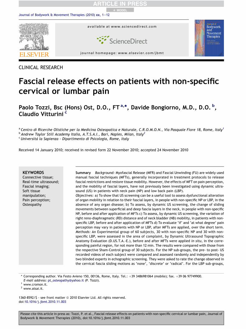
rotation, rested on the couch, before and after MFTs or thesham treatment had been applied. A linear probe was usedat 7.5e13 MHz. It was always positioned on the sagittalplane at the left antero-lateral region of the neck, betweenthe sternocleido-mastoid muscle and the ipsilateral neu-rovascular bundle, as shown in Figure 1. The aim was toobserve any quantitative and/or qualitative change inmobility between fascial layers of the neck region, such aspretracheal and retropharyngeal fascia, during quietrespiration, maximal inspiration-expiration, and swallow-ing, before and after treatment.
Two medical doctors, of 19 and 21 years experience inUS screening and diagnosis, were asked to compare theresults independently. They were blind to the groups(Experimental and Control) from which the images wereobtained. After having randomly viewed and compared thepre- and post- US videos for every NP subject, they wereasked to rate any possible change in quality and quantity ofthe cervical fascia sliding motions as ‘none’, ‘discrete’ or‘radical’. The values obtained by the first examiner werecalled Ultrasound Qualitative Scale 1 (US-QS1) results,whereas those collected from the second examiner werecalled Ultrasound Qualitative Scale 2 (US-QS2) results.
Lumbar and pelvic US screening
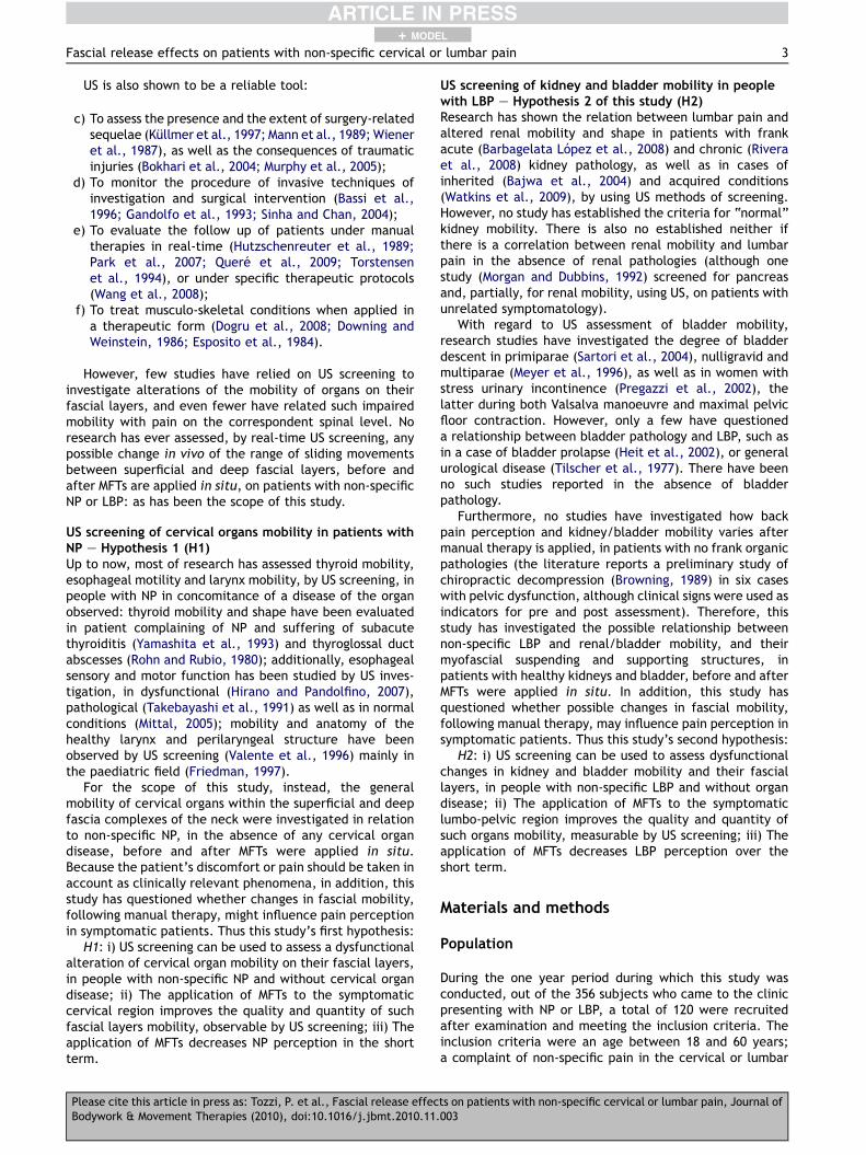
A similar procedure was applied to LBP subjects: withpatients supine, the probe was positioned in the laterallumbar region, for a sagittal scan. A convex probe was usedat 5 MHz and THI. The distance between the superior poleof the right kidney and the origin of the respective dia-phragmatic crura (RD distance) was taken during bothmaximal inspiration (RdI) and maximal expiration (RdE), asshown on Figure 2, before and after treatment (seefigure 2) was applied. The aim was to measure and compare
Figure 1 Standard procedure for the neck US screening in NPsubjects. The standard procedure for US screening of the neckregion for NP sub-groups is shown: the patient lies supine withthe head resting on the couch, in a mild extension, and rightside-bending-rotation. The probe is positioned on the leftantero-lateral region of the neck, between the sternocleido-mastoid muscle and the ipsilateral neurovascular bundle, alongthe sagittal plane. A US recorded video was taken duringswallowing, quiet and forced breathing, before and aftertreatment was applied.
ts on patients with non-specific cervical or lumbar pain, Journal of003
Figure 2 US RD distance measurement on LBP sub-groups.The distance between the superior pole of the right kidney andthe origin of the respective diaphragmatic crura was takenduring maximal inspiration and expiration in both LBP sub-groups, before and after treatment was applied.
Fascial release effects on patients with non-specific cervical or lumbar pain 5
+ MODEL
pre- to post- range of kidney’s supero-inferior slidingmotion, during forced respiration.
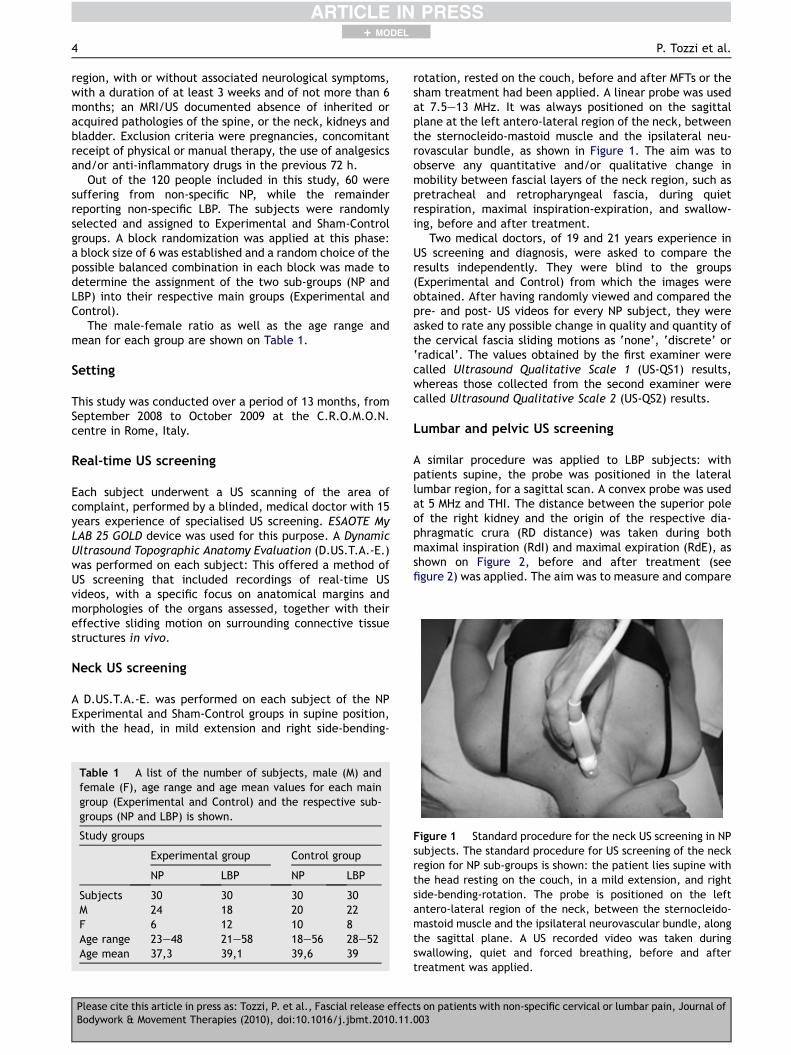
Successively, the same subjects were also assessed attheir pelvic region, in supine position, using the same typeof probe. In this case, the probe was always positionedabove the pubic symphysis, for transverse and sagittalscanning. The distance between the neck of the bladderand the anterior vesical wall on the perpendicular line (NBdistances) was taken during maximal relaxation (NbR) andcontraction (NbC) of the pelvic floor muscles, as shown inFigure 3 before and after treatment was applied. Allpatients were asked to urinate 2 h prior the session and
Figure 3 US NB distance measurement on LBP sub-groups.The distance between the neck of the bladder and the anteriorvesical wall, on the perpendicular line, was taken duringmaximal relaxation and contraction of the pelvic floor musclesin both LBP sub-groups, before and after treatment wasapplied.
Please cite this article in press as: Tozzi, P. et al., Fascial release effecBodywork & Movement Therapies (2010), doi:10.1016/j.jbmt.2010.11.
then drink 500 cc. of water an hour before the samesession. Bladder filling influences the position and mobilityof the bladder neck and the proximal urethra, which areboth more mobile when the bladder is nearly empty (Dietzand Wilson, 1999).
Pain assessment
Pain perception was measured using the Short-Form McGillPain Assessment Questionnaire (SF-MPQ), a responsivescale giving both reliable and valid data (Melzack, 1987).The SF-MPQ consists in a 15-point descriptor of averagepain, articulated in 11 points of sensory experience and 4of affective experiences. An intensity scale of 0e3 repre-senting mild, moderate or severe pain, is given for eachdescriptor. The sensory and affective pain rating scores(ranging from 0 to 33 and from 0 to 12 respectively) areadded together to give a value for total pain experience(ranging from 0 to 45). The total score has been used as theoutcome of this study. The SF-MPQ was administered toevery subject on the day of recruitment, as well as threedays later.
Osteopathic assessment
An Osteopathic assessment was performed by an Osteo-path, of 5 years experience, in the symptomatic region ofthe NP and LBP Experimental subjects, to locate thespecific area of major fascial restriction of mobility,respectively in the neck and lumbar regions.
Treatment
The Experimental group received MFTs on the painful areas,by the same Osteopath who had previously assessed them.The treatment consisted of application of MFR and FUtechniques:
MFR treatmentMFR consists in the application of a low load, long durationstretch along the lines of maximal fascial restrictions(Barnes, 1990). The latter are palpated by the practitionerand the pressure is applied directly to the skin, into thedirection of restriction just until resistance (tissue barrier)is felt. Once found, the collagenous barrier is engaged for90e120 s, without sliding over the skin or forcing the tissue(Manheim, 2001), until the fascia complex starts to yieldand a sensation of softening is achieved.
a) For the Experimental NP group: MFR was applied in twostages, for not more then 2 min each. The aim was torelease the deep and superficial cervical myofascialstructures, having an effect on their reciprocal slidingmotion, in both the anterior and the posterior neckregion. The hold used with patient supine, was with theoperator’s caudal hand on the sternum and the cranialhand on the forehead, when MFR being applied to theanterior neck structures. The cranial hand was sup-porting the head at the subocciput when MFR wasapplied to the posterior neck structures (Stanborough,2004).
ts on patients with non-specific cervical or lumbar pain, Journal of003
6 P. Tozzi et al.
+ MODEL
b) For the Experimental LBP group: MFR was applied in twostages, for not more then 2 min each. The aim was tofirstly release the right and then left psoas major andminor as well as the iliacus muscles and related lumbarorgans, by using the cross-handed hold shown in Figure 4(Stanborough, 2004). The kidneys are embedded andsuspended by the renal fascia that is anatomicallyrelated to the diaphragm and psoas fascia, that is in turna continuation of the thoraco-lumbar fascia (Bogduk,2005). Secondly, the pelvic floor muscles and relatedpelvic organs were targeted to be released by theapplication of MFR through a global pelvic A/P hold.With the patient supine, one operator’s hand on thesacrum, between patient legs, and one hand just abovethe pubic symphysis (Stanborough, 2004).
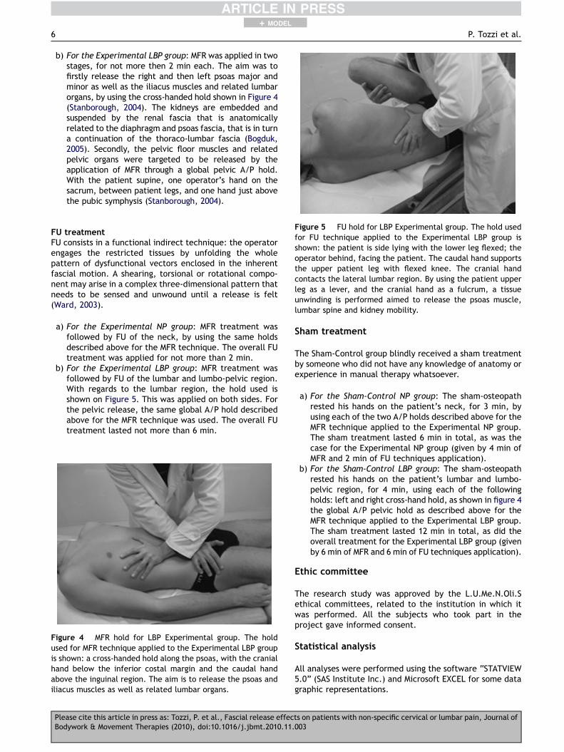
Figure 5 FU hold for LBP Experimental group. The hold usedfor FU technique applied to the Experimental LBP group isshown: the patient is side lying with the lower leg flexed; theoperator behind, facing the patient. The caudal hand supportsthe upper patient leg with flexed knee. The cranial handcontacts the lateral lumbar region. By using the patient upperleg as a lever, and the cranial hand as a fulcrum, a tissueunwinding is performed aimed to release the psoas muscle,lumbar spine and kidney mobility.
FU treatmentFU consists in a functional indirect technique: the operatorengages the restricted tissues by unfolding the wholepattern of dysfunctional vectors enclosed in the inherentfascial motion. A shearing, torsional or rotational compo-nent may arise in a complex three-dimensional pattern thatneeds to be sensed and unwound until a release is felt(Ward, 2003).
a) For the Experimental NP group: MFR treatment wasfollowed by FU of the neck, by using the same holdsdescribed above for the MFR technique. The overall FUtreatment was applied for not more than 2 min.
b) For the Experimental LBP group: MFR treatment wasfollowed by FU of the lumbar and lumbo-pelvic region.With regards to the lumbar region, the hold used isshown on Figure 5. This was applied on both sides. Forthe pelvic release, the same global A/P hold describedabove for the MFR technique was used. The overall FUtreatment lasted not more than 6 min.
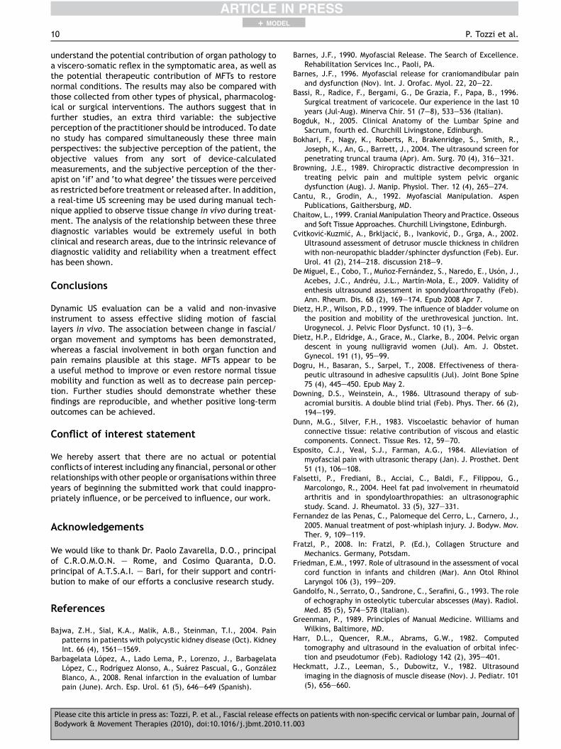
Figure 4 MFR hold for LBP Experimental group. The holdused for MFR technique applied to the Experimental LBP groupis shown: a cross-handed hold along the psoas, with the cranialhand below the inferior costal margin and the caudal handabove the inguinal region. The aim is to release the psoas andiliacus muscles as well as related lumbar organs.
Please cite this article in press as: Tozzi, P. et al., Fascial release effecBodywork & Movement Therapies (2010), doi:10.1016/j.jbmt.2010.11.
Sham treatment
The Sham-Control group blindly received a sham treatmentby someone who did not have any knowledge of anatomy orexperience in manual therapy whatsoever.
a) For the Sham-Control NP group: The sham-osteopathrested his hands on the patient’s neck, for 3 min, byusing each of the two A/P holds described above for theMFR technique applied to the Experimental NP group.The sham treatment lasted 6 min in total, as was thecase for the Experimental NP group (given by 4 min ofMFR and 2 min of FU techniques application).
b) For the Sham-Control LBP group: The sham-osteopathrested his hands on the patient’s lumbar and lumbo-pelvic region, for 4 min, using each of the followingholds: left and right cross-hand hold, as shown in figure 4the global A/P pelvic hold as described above for theMFR technique applied to the Experimental LBP group.The sham treatment lasted 12 min in total, as did theoverall treatment for the Experimental LBP group (givenby 6 min of MFR and 6 min of FU techniques application).
Ethic committee
The research study was approved by the L.U.Me.N.Oli.Sethical committees, related to the institution in which itwas performed. All the subjects who took part in theproject gave informed consent.
Statistical analysis
All analyses were performed using the software “STATVIEW5.0” (SAS Institute Inc.) and Microsoft EXCEL for some datagraphic representations.
ts on patients with non-specific cervical or lumbar pain, Journal of003
Fascial release effects on patients with non-specific cervical or lumbar pain 7
+ MODEL
a) With regards to H1 i) and ii): the results of the US-QS1and US-QS2 were compared using the Chi square test,with a p value accepted at <0.05.With regards to H1 iii) as well as to H2 i), ii), iii), : theANOVA test at repeated measures was used, with a pvalue accepted at <0.05, to calculate if betweenExperimental and Control groups there was a significantdifference for the:
b) RD-T0 and RD-T1 distances in LBP sub-groups, byconsidering RD-T0Z RdI-T0 � RdE-T0 and RD-T1Z RdI-T1 � RdE-T1;
c) NB-T0 and NB-T1 distances in LBP sub-groups, consid-ering NB-T0 Z NbR-T0 � NbC-T0 and NB-T1 Z NbR-T1 � NbC-T1;
d) pre- to post- SF-MPQ results.
Results
a) US-QS: The US-QS results (US-QS1 and US-QS2) for the NPstudy population are shown on Figure 6 with theirrespective frequency. A significant difference is shownwithap-Value< 0.0001.TheChi square test betweenUS-QS1 and US-QS2 results, after they have been normalizedin z points, has shown a significant correlation (0.915)with a p-Value < 0.0001 (confirming H1 i) and ii));
b) US kidney values: RD-T0 and RD-T1 distances in the LBPgroups are shown on Figure 7 A significant difference isshownwith an F-ValueZ 76.637 and a p-Value< 0.0001.In the Experimental group the mean value of RD-T0 was10.33, St. Dev. 4.70, against the RD-T1 mean value of21.60, St. Dev. 7.06. In the Control group themean valueof RD-TOwas 8.93, St. Dev. 2.01, against the RD-T1meanvalue of 10.10, St. Dev 4.49. The range of the all RD-T0values was �3/þ21 mm, mean 9.63, St. Dev. 3.65; the
Figure 6 Chi square p-Values for US-SQ1 and US-SQ-2 results in Nsub-groups are shown with their respective observed frequency. A
Please cite this article in press as: Tozzi, P. et al., Fascial release effecBodywork & Movement Therapies (2010), doi:10.1016/j.jbmt.2010.11.
range of all RD-T1 values was �2/þ32 mm, mean 15.85,St. Dev. 5.78 (confirming H2 i) and ii));
c) USbladder values: NB-T0 andNB-T1 distances in LBP sub-groups are shown on Figure 8. A significant difference isshown with an F-Value Z 577.349 and a p-Val-ue< 0.0001. In the Experimental group the mean valuesof NB-T0 was 12.70, St. Dev. 4.18, against the NB-T1mean value of 22.73, St. Dev. 3.73. In the Control groupthe mean value of NB-TO was 12.20, St. Dev. 3.81,against theNB-T1mean value of 12.90, St. Dev. 4.23. Therange of NB-T0 values wasþ4/þ21 mm, mean 12.45, St.Dev. 3.98; the range of NB-T1 values was þ3/þ30 mm,mean 17.82, St. Dev. 3.98 (confirming H2 i) and ii));
d) SF-MPQ: Pre- to post-differences between Experimental(NP þ LBP) and Control (NP þ LBP) groups are shown onFigure 9. A significant difference with anF-Value Z 167.742 and a p-Value < 0.0001 is shown onTable 2. Means and St. Dev. values are shown on Table 3.The mean difference between groups was 4.883; themean difference between pre- and post- was 4.483. Nosignificant difference was found either between NP andLBP sub-groups (p-Value < 0.8582), or between genders(p-Value < 0.4866) or between age classes(p-Value < 0.5031), with respect to the study pop-ulation (confirming H1 iii) and H2 iii)).
Discussion
This study shows that cervical and some lumbo-pelvicorgans mobility, with respect to the surrounding myofascialstructures, may be assessed by US screening; that suchmobility changes are related with pain in the correspondingspinal area; that such mobility may be reduced or alteredwithout frank organic pathology; that MFTs can improve
P sub-groups. The US-QS results (US-QS1 and US-QS2) for the NPsignificant difference is shown with a p-Value < 0.0001.
ts on patients with non-specific cervical or lumbar pain, Journal of003
Figure 7 US kidney results in LBP sub-groups. The significantdifference between RD-T0 and RD-T1 distances in LBP sub-groups are shown. In the Experimental group the mean valuesof RD-T0 was 10.33, St. Dev. 4.70, against the RD-T1 meanvalue of 21.60, St. Dev. 7.06. In the Control group the meanvalue of RD-TO was 8.93, St. Dev. 2.01, against the RD-T1mean value of 10.10, St. Dev 4.49.
Figure 9 SF-MPQ results in the two study group. The signif-icant difference between Experimental and Control groups forthe pre- to post- SF-MPQ results is shown.
8 P. Tozzi et al.
+ MODEL
such fascia related organs mobility as well as reduce painperception over a short term period.
H1 e neck pain, US screening of cervical fasciamobility and MFTs
Most research that has investigated the efficacy of manualtherapies on subjects with neck pain have used US for shamtreatment only, as de-tuned device (Koes et al., 1993;Schwerla et al., 2008), very few as a tool for measure-ment or monitoring (Licht et al., 1998). This study, instead,shows that US evaluation is a valid, non-invasive method tomonitor and assess organs mobility in the cervical and
Figure 8 US bladder results in LBP sub-groups. The signifi-cant difference between NB-T0 and NB-T1 distances in LBPsub-groups are shown. In the Experimental group the meanvalues of NB-T0 was 12.70, St. Dev. 4.18, against the NB-T1mean value of 22.73, St. Dev. 3.73. In the Control group themean value of NB-TO was 12.20, St. Dev. 3.81, against the NB-T1 mean value of 12.90, St. Dev 4.23.
Please cite this article in press as: Tozzi, P. et al., Fascial release effecBodywork & Movement Therapies (2010), doi:10.1016/j.jbmt.2010.11.
abdomino-pelvic region, in vivo and real-time. Further-more, intraluminal impedance and intramural or endo-scopic US and ultrasonography have been mainly recruitedin the last decades of research, because of advances intransducer technology, computerization, and graphic datapresentation. This study also shows that release obtainedby MFTs in the superficial and deep myofascial structures ofthe neck allowed a better motion of the organs related tothose structures:
a) Radical findings: The two blinded examiners of USrecorded videos have both separately indicated as‘radical’ the change between the same pre and postimages, in 7 subjects of the Experimental group,accounting for the 23.33% of the study group, whereasno ‘radical’ change was found in the Control group.
b) Discrete findings: a mean value of 15 of ‘discrete’change was found in the Experimental group, versusa mean value of 1.5 in the Control group
c) Nonefindings:With regards to the ‘none’ change, ameanvalue of 8 was found in the Experimental group,compared to a mean value of 28.5 for the Control group.
However, limitations of this part of the study were:
a) Method-related: the US property of scanning all planesreduces the chance of standardization (and often ofquantification) of distance measurements;
b) Examiner-related: because of humanmargins of error, isextremely difficult to obtain and reproduce two images,‘pre’ and ‘post’, in the same plane and angulation;
c) Patient-related: position, breathing, inter and intratissue mobility, viscoelastic changes. The need fora mathematical model capable of comparing similar USimages is paramount to analyse pre to post changes.
H2 e lumbar pain, US screening of kidney mobilityand MFTs
This study has also investigated the range of sliding motionof the right kidney in people with lumbar pain and absenceof renal pathology, before and after specific MFTs were
ts on patients with non-specific cervical or lumbar pain, Journal of003
Table 2 ANOVA table for SF-MPQ values.
DF Sum of squares Mean square F-Value P-Value Lambda Power
Fascial release effects on patients with non-specific cervical or lumbar pain 9
+ MODEL
applied on psoas muscles and lumbar region. The releasefollowed by the unwinding of the fascial restrictions mayhave restored the optimal tissue elasticity of thesurrounding myofascial structures, rebalanced the intra andinter visceral pressure, re-established an optimal renalmobility, and via fascial continuation, have improvedlumbar spine mobility. Although we could concluded thatapplication of MFTs significantly improves kidney mobilityand reduces pain perception, at this stage it is inappro-priate to state that people with non-specific lumbar painmay present with a relative reduction of right kidney’smobility, due to the fact that no study has ever assessed“normal” kidney mobility during respiration and/or estab-lished an index of kidney mobility. Therefore, no compar-ison is possible between the values obtained (RD-T0 rangevalues �3/þ21 mm, mean 9.63, St. Dev. 3.65; RD-T1 rangevalues �2/þ32 mm, mean 15.85, St. Dev. 8.25) and those in“normal” conditions.
H2 e lumbar pain, US screening of bladder mobilityand MFTs
This study has also investigated the range of neck bladdermobility in people with non-specific lumbar pain anda healthy bladder. The restriction identified may havecontributed to or maintained LBP, via viscero-somaticreflex, and/or via venous and lymphatic drainage conges-tion, or more simply via mechanical tension throughconnective tissue connections (Ward, 2003). In fact, thebladder ‘sits’ on the pelvic floor and is partially supportedand suspended by the endopelvic fascia via its extensions,such as the pubovesical ligaments, together with the pubo-sacral laminae from the levator ani muscle (Paoletti,2003). MFTs have been shown to be effective atimproving bladder mobility, and may have balanced pelvicfloor tensions on the transverse and sagittal planes,restoring optimal bladder mobility and possibly generalpelvic adaptive capacity. The latter meaning the potentialability of the pelvic girdle and its contents maintaina functional and mechanical balance against possible dis-rupting action of internal and external forces. This may
Please cite this article in press as: Tozzi, P. et al., Fascial release effecBodywork & Movement Therapies (2010), doi:10.1016/j.jbmt.2010.11.
have offered, in turn, a balanced and mobile support to thelumbar spine, possibly improving its mobility and reducinginflammation and pain. The range of neck bladder mobilityfound in this study was þ4/þ21 mm, mean 12.45, St. Dev.3.98 at T0; and þ3/þ30 mm, mean 17.82, St. Dev. 6.35 atT1. Some studies have shown “normal” bladder mobility,although in women only and exclusively with regards tobladder descent during the Valsalva manouvre (Dietz et al.,2004). The degree of mobility was found to range from 1.2to 40.2 mm (mean 17.4 mm). Other studies (Pregazzi et al.,2002) have, investigated bladder mobility during maximalpelvic floor contraction, using different electronic distancemeasurements, such as that between the bladder neck andthe pubic symphysis, the bladder neck and the symphysispubis line, the midline of the symphysis (alpha angle) andthe angle between the proximal and distal urethra (betaangle). Most of these studies have used perineal ultraso-nography that allows far more details and precision thanthe more traditional external US investigation methodchosen for this study. Therefore, comparisons of the resultsof this study with those from previous ones are inappro-priate at this stage. However, much research has relied onUS investigations, especially perineal and introital, toassess for prolapses (cystoptosis, bladder neck and urethralmobility), confirming that US remains the first line exami-nation for pelvic morphology and bladder function.
H1 and H2 e neck or lumbar pain and MFTs
In both NP and LBP sub-groups, MFTs have shown to beeffective in reducing pain perception regardless of age,gender and pain location, with an SF-MPQ mean values of24.65 at T0 and 15.51 at T1 in the Experimental groupagainst the mean values ranging from 24.88 at T0 to 25.05at T1 for the Control group. A significant difference wasfound (p-Value < 0.0001).
Suggestions for further research
In this study, pain assessment was performed over a shortperiod of 3 days following treatment, on a relatively smallstudy population (although the small p-values obtainedsupport the statistical notion that the small study populationdoesn’t minimise the validity of the study itself). Futurestudies should evaluate whether these findings are repro-ducible, in a larger population, and whether positive long-term outcomes can be achieved in both US findings and painassessment. Future research should also consider investi-gating the effect of MFTs on specific NP or LBP, to evaluatetheir efficacy when a specific organ pathology is present atthe corresponding spinal level. This may help to better
ts on patients with non-specific cervical or lumbar pain, Journal of003
10 P. Tozzi et al.
+ MODEL
understand the potential contribution of organ pathology toa viscero-somatic reflex in the symptomatic area, as well asthe potential therapeutic contribution of MFTs to restorenormal conditions. The results may also be compared withthose collected from other types of physical, pharmacolog-ical or surgical interventions. The authors suggest that infurther studies, an extra third variable: the subjectiveperception of the practitioner should be introduced. To dateno study has compared simultaneously these three mainperspectives: the subjective perception of the patient, theobjective values from any sort of device-calculatedmeasurements, and the subjective perception of the ther-apist on ‘if’ and ‘to what degree’ the tissues were perceivedas restricted before treatment or released after. In addition,a real-time US screening may be used during manual tech-nique applied to observe tissue change in vivo during treat-ment. The analysis of the relationship between these threediagnostic variables would be extremely useful in bothclinical and research areas, due to the intrinsic relevance ofdiagnostic validity and reliability when a treatment effecthas been shown.
Conclusions
Dynamic US evaluation can be a valid and non-invasiveinstrument to assess effective sliding motion of fasciallayers in vivo. The association between change in fascial/organ movement and symptoms has been demonstrated,whereas a fascial involvement in both organ function andpain remains plausible at this stage. MFTs appear to bea useful method to improve or even restore normal tissuemobility and function as well as to decrease pain percep-tion. Further studies should demonstrate whether thesefindings are reproducible, and whether positive long-termoutcomes can be achieved.
Conflict of interest statement
We hereby assert that there are no actual or potentialconflicts of interest including any financial, personal or otherrelationships with other people or organisations within threeyears of beginning the submitted work that could inappro-priately influence, or be perceived to influence, our work.
Acknowledgements
We would like to thank Dr. Paolo Zavarella, D.O., principalof C.R.O.M.O.N. e Rome, and Cosimo Quaranta, D.O.principal of A.T.S.A.I. e Bari, for their support and contri-bution to make of our efforts a conclusive research study.
References
Bajwa, Z.H., Sial, K.A., Malik, A.B., Steinman, T.I., 2004. Painpatterns in patients with polycystic kidney disease (Oct). KidneyInt. 66 (4), 1561e1569.
Barbagelata Lopez, A., Lado Lema, P., Lorenzo, J., BarbagelataLopez, C., Rodrıguez Alonso, A., Suarez Pascual, G., GonzalezBlanco, A., 2008. Renal infarction in the evaluation of lumbarpain (June). Arch. Esp. Urol. 61 (5), 646e649 (Spanish).
Please cite this article in press as: Tozzi, P. et al., Fascial release effecBodywork & Movement Therapies (2010), doi:10.1016/j.jbmt.2010.11.
Barnes, J.F., 1990. Myofascial Release. The Search of Excellence.Rehabilitation Services Inc., Paoli, PA.
Barnes, J.F., 1996. Myofascial release for craniomandibular painand dysfunction (Nov). Int. J. Orofac. Myol. 22, 20e22.
Bassi, R., Radice, F., Bergami, G., De Grazia, F., Papa, B., 1996.Surgical treatment of varicocele. Our experience in the last 10years (Jul-Aug). Minerva Chir. 51 (7e8), 533e536 (Italian).
Bogduk, N., 2005. Clinical Anatomy of the Lumbar Spine andSacrum, fourth ed. Churchill Livingstone, Edinburgh.
Bokhari, F., Nagy, K., Roberts, R., Brakenridge, S., Smith, R.,Joseph, K., An, G., Barrett, J., 2004. The ultrasound screen forpenetrating truncal trauma (Apr). Am. Surg. 70 (4), 316e321.
Browning, J.E., 1989. Chiropractic distractive decompression intreating pelvic pain and multiple system pelvic organicdysfunction (Aug). J. Manip. Physiol. Ther. 12 (4), 265e274.
Cantu, R., Grodin, A., 1992. Myofascial Manipulation. AspenPublications, Gaithersburg, MD.
Chaitow, L., 1999. Cranial Manipulation Theory and Practice. Osseousand Soft Tissue Approaches. Churchill Livingstone, Edinburgh.
Cvitkovi�c-Kuzmi�c, A., Brkljaci�c, B., Ivankovi�c, D., Grga, A., 2002.Ultrasound assessment of detrusor muscle thickness in childrenwith non-neuropathic bladder/sphincter dysfunction (Feb). Eur.Urol. 41 (2), 214e218. discussion 218e9.
De Miguel, E., Cobo, T., Munoz-Fernandez, S., Naredo, E., Uson, J.,Acebes, J.C., Andreu, J.L., Martın-Mola, E., 2009. Validity ofenthesis ultrasound assessment in spondyloarthropathy (Feb).Ann. Rheum. Dis. 68 (2), 169e174. Epub 2008 Apr 7.
Dietz, H.P., Wilson, P.D., 1999. The influence of bladder volume onthe position and mobility of the urethrovesical junction. Int.Urogynecol. J. Pelvic Floor Dysfunct. 10 (1), 3e6.
Dietz, H.P., Eldridge, A., Grace, M., Clarke, B., 2004. Pelvic organdescent in young nulligravid women (Jul). Am. J. Obstet.Gynecol. 191 (1), 95e99.
Dogru, H., Basaran, S., Sarpel, T., 2008. Effectiveness of thera-peutic ultrasound in adhesive capsulitis (Jul). Joint Bone Spine75 (4), 445e450. Epub May 2.
Downing, D.S., Weinstein, A., 1986. Ultrasound therapy of sub-acromial bursitis. A double blind trial (Feb). Phys. Ther. 66 (2),194e199.
Dunn, M.G., Silver, F.H., 1983. Viscoelastic behavior of humanconnective tissue: relative contribution of viscous and elasticcomponents. Connect. Tissue Res. 12, 59e70.
Esposito, C.J., Veal, S.J., Farman, A.G., 1984. Alleviation ofmyofascial pain with ultrasonic therapy (Jan). J. Prosthet. Dent51 (1), 106e108.
Falsetti, P., Frediani, B., Acciai, C., Baldi, F., Filippou, G.,Marcolongo, R., 2004. Heel fat pad involvement in rheumatoidarthritis and in spondyloarthropathies: an ultrasonographicstudy. Scand. J. Rheumatol. 33 (5), 327e331.
Fernandez de las Penas, C., Palomeque del Cerro, L., Carnero, J.,2005. Manual treatment of post-whiplash injury. J. Bodyw. Mov.Ther. 9, 109e119.
Friedman, E.M., 1997. Role of ultrasound in the assessment of vocalcord function in infants and children (Mar). Ann Otol RhinolLaryngol 106 (3), 199e209.
Greenman, P., 1989. Principles of Manual Medicine. Williams andWilkins, Baltimore, MD.
Harr, D.L., Quencer, R.M., Abrams, G.W., 1982. Computedtomography and ultrasound in the evaluation of orbital infec-tion and pseudotumor (Feb). Radiology 142 (2), 395e401.
Heckmatt, J.Z., Leeman, S., Dubowitz, V., 1982. Ultrasoundimaging in the diagnosis of muscle disease (Nov). J. Pediatr. 101(5), 656e660.
ts on patients with non-specific cervical or lumbar pain, Journal of003
Fascial release effects on patients with non-specific cervical or lumbar pain 11
+ MODEL
Heers, G., Hedtmann, A., 2002. Ultrasound diagnosis of the acro-mioclavicular joint (Mar). Orthopade 31 (3), 255e261 (German).
Heit, M., Culligan, P., Rosenquist, C., Shott, S., 2002. Is pelvicorgan prolapse a cause of pelvic or low back pain? (Jan). Obstet.Gynecol. 99 (1), 23e28.
Hirano, I., Pandolfino, J., 2007. New technologies for the evalua-tion of esophageal motility disorders: impedance, high-resolu-tion manometry, and intraluminal ultrasound (Sep).Gastroenterol. Clin. North Am. 36 (3), 531e551 (viii. Review).
Hutzschenreuter, P., Brummer, H., Ebberfeld, K., 1989. Experi-mental and clinical studies of the mechanism of effect ofmanual lymph drainage therapy (Jul). Z. Lymphol. 13 (1), 62e64(German).
Ingber, D.E., Chen, C.S., 1999. Tensegrity and mechanoregulation:from skeleton to cytoskeleton. J. Osteoarthr. Res. Soc. Int. 7,81e94.
Kenney, A.H., Hafner, J.N., 1977. Ultrasonic evidence of inflam-matory thickening and fluid collection within the retrobulbarfascia: the T sign (Dec). Ann. Ophthalmol. 9 (12), 1557e1563.
Koes, B.W., Bouter, L.M., van Mameren, H., Essers, A.H.,Verstegen, G.J., Hofhuizen, D.M., Houben, J.P., Knipschild, P.G.,1993. A randomized clinical trial of manual therapy and physio-therapy for persistent back and neck complaints: subgroupanalysis and relationship between outcome measures (May).J. Manip. Physiol. Ther. 16 (4), 211e219.
Kullmer, K., Olivier, L., Eysel, P., Rompe, J.D., Schmit-Neuerburg, K.P., 1997. Traumatically-induced compartmentsyndrome of the tibia. Ultrasound diagnosis for qualitativeassessment of late sequelae for musculature after dermato-fasciotomy (Jun). Unfallchirurgie 23 (3), 87e91 (German).
Langevin, H., 2006. Connective tissue: a body-wide signalingnetwork? Medical Hypotheses 66, 1074e1077.
LeBauer, A., Brtalik, R., Stowe, K., 2008. The effect of myofascialrelease (MFR) on an adult with idiopathic scoliosis. J. Bodyw.Mov. Ther. 12, 356e363.
Levin, S.M., 1990. The myofascial skeletal truss: a system scienceanalysis. In: Barnes, J.F. (Ed.), Myofascial Release. Rehabilita-tion Services Inc, Paoli, PA cM.
Licht, P.B., Christensen, H.W., Højgaard, P., Marving, J., 1998.Vertebral artery flow and spinal manipulation: a randomized,controlled and observer-blinded study (Mar-Apr). J. Manip.Physiol. Ther. 21 (3), 141e144. Erratum in: J ManipulativePhysiol Ther 1998 May; 21(4), inside back cov.
Lukban, J., 2001. The effect of manual physical therapy in patientsdiagnosed with interstitial cystitis, high-tone pelvic floordysfunction, and sacroiliac dysfunction. Urology 57 (Suppl. 6A),121.
Lund, I., Ge, Y., Yu, L.C., et al., 2002. Repeated massage-likestimulation induces long-term effects on nociception: contri-bution of oxytocinergic mechanisms. Eur. J. Neurosci. 16,330e338.
Manheim, C.J., 2001. The Myofascial Release Manual, third ed.Slack Incorporated, New Jersey, USA.
Mann, W., Riechelmann, H., Gilsbach, J., 1989. The state of thefrontal sinus after craniotomy. Acta Neurochir (Wien) 100 (3e4),101e103.
Martin, M.M., 2009. Effects of the myofascial release in diffusesystemic sclerosis (Oct). J. Bodyw. Mov. Ther. 13 (4), 320e327.Epub 2008 Jun 17.
Melzack, R., 1987. The short-form Mc-Gill pain questionnaire. Pain30, 191e197.
Mense, S., 1983. Basic neurobiologic mechanisms of pain andanalgesia. Am. J. Med. 75, 4e14.
Meyer, S., De Grandi, P., Schreyer, A., Caccia, G., 1996. Theassessment of bladder neck position and mobility in continent
Please cite this article in press as: Tozzi, P. et al., Fascial release effecBodywork & Movement Therapies (2010), doi:10.1016/j.jbmt.2010.11.
nullipara, mulitpara, forceps-delivered and incontinent womenusing perineal ultrasound: a future office procedure? Int. Uro-gynecol. J. Pelvic Floor Dysfunct. 7 (3), 138e146.
Mittal, R.K., 2005. Motor and sensory function of the esophagus:revelations through ultrasound imaging (Apr). J. Clin. Gastro-enterol. 39 (4 Suppl 2), S42eS48. Review.
Mosler, E., Folkhard, W., Knorzer, E., Nemetschek-Gansler, H.,Nemetschek, T.H., Koch, M.H., 1985. Stress-induced moleculararrangement in tendon collagen. J. Mol. Biol. 182, 589e596.
Paoletti, S., 2003. Le fasce: il ruolo dei tessuti nella meccanicaumana. Esomm, Italy.
Park, S.J., Lee, H.K., Yi, B.H., Cha, J.G., Joh, J.H., Hong, H.S.,Kim, H.C., 2007. Manual reduction of torsion of an intrascrotalappendage under ultrasonographic monitoring (Mar). J. Ultra-sound Med. 26 (3), 293e299.
Pischinger, A., 1991. Matrix and Matrix Regulation: Basis fora Holistic Theory in Medicine. Haug International, Brussels.
Pregazzi, R., Sartore, A., Bortoli, P., Grimaldi, E., Troiano, L.,Guaschino, S., 2002. Perineal ultrasound evaluation of urethralangle and bladder neck mobility in women with stress urinaryincontinence (Jul). BJOG 109 (7), 821e827.
Quere, N., Noel, E., Lieutaud, A., d’Alessio, P., 2009. Fas-ciatherapy combined with pulsology touch induces changes inblood turbulence potentially beneficial for vascular endothe-lium (Jul). J. Bodyw. Mov. Ther. 13 (3), 239e245. Epub 2008Aug 12.
Radjieski, J.M., Lumley, M.A., Cantieri, M.S., 1998. Effect ofosteopathic manipulative treatment of length of stay forpancreatitis: a randomized pilot study (May). J. Am. Osteopath.Assoc. 98 (5), 264e272. Erratum in: J Am Osteopath Assoc 1998Jul; 98(7):408.
Remvig, L., 2008. Myofascial release: an evidence-based treatmentconcept? J. Bodyw. Mov. Ther. 12, 385e396.
Rivera, M., Rioja, M.E., Burgos, F.J., Ortuno, J., 2008. Chroniclumbar pain and urinary infections in a young woman. Nefro-logia 28 (2), 222e223 (Spanish).
Rohn, R.D., Rubio, T., 1980. Neck pain due to acute suppurativethyroiditis and thyroglossal duct abscess (Dec). J. Adolesc.Health Care 1 (2), 155e158.
Rolf, I., 1977. The Integration of Human Structure. Harper andRow, London.
Sartori, J.P., Sartori, M.G., Baracat, E.C., De Lima, G.R.,Girao, M.J., 2004. Bladder neck mobility and functional evalu-ation of the pelvic floor in primiparae according to the type ofdelivery. Clin. Exp. Obstet. Gynecol. 31 (2), 120e122.
Sasaki, N., Odajima, S., 1996. Elongation mechanism of collagenfibrils and force-strain relations of tendon at each level ofstructural hierarchy. J. Biomech. 29, 1131e1136.
Schultz, R.L., Feltis, R., 1996. The Endless Web: Fascial Anatomyand Physical Reality. North Atlantic Books, Berkeley, CA.
Schwerla, F., Bischoff, A., Nurnberger, A., Genter, P.,Guillaume, J.P., Resch, K.L., 2008. Osteopathic treatment ofpatients with chronic non-specific neck pain: a randomisedcontrolled trial of efficacy (Jun). Forsch Komplementmed. 15(3), 138e145. Epub 2008 Jun 4.
ts on patients with non-specific cervical or lumbar pain, Journal of003
Sinha, A., Chan, V.W., 2004. Ultrasound imaging for popliteal sciaticnerve block (Mar-Apr). Reg Anesth. Pain Med. 29 (2), 130e134.
Stanborough, M., 2004. Direct Release Myofascial Technique. AnIllustrated Guide for Practitioners. Churchill Livingstone.
Sucher, B.M., 1993. Myofascial manipulative release of carpaltunnel syndrome: documentation with magnetic resonanceimaging (Dec). J. Am. Osteopath. Assoc. 93 (12), 1273e1278.
Takebayashi, S., Matsui, K., Ozawa, Y., Nozawa, T., Fujioka, E.,1991. Cervical esophageal motility: evaluation with US inprogressive systemic sclerosis (May). Radiology 179 (2),389e393.
Tilscher, H., Bogner, G., Landsiedl, F., 1977. Visceral diseases ascause of lumbar syndromes (May-Jun). Z. Rheumatol. 36 (5e6),161e167 (German).
Torstensen, T.A., Meen, H.D., Stiris, M., 1994. The effect ofmedical exercise therapy on a patient with chronic supra-spinatus tendinitis. Diagnostic ultrasoundetissue regeneration:a case study (Dec). J. Orthop. Sports Phys. Ther. 20 (6),319e327.
Valente, T., Farina, R., Minelli, S., Pinto, A., Rossi, G., Tecame, S.,Caranci, F., 1996. The echographic anatomy of the larynx and
Please cite this article in press as: Tozzi, P. et al., Fascial release effecBodywork & Movement Therapies (2010), doi:10.1016/j.jbmt.2010.11.
the perilaryngeal structures (Mar). Radiol. Med. 91 (3), 231e237(Italian).
Walton, A., 2008. Efficacy of myofascial release techniques in thetreatment of primary Raynaud’s phenomenon (Jul). J. Bodyw.Mov. Ther. 12 (3), 274e280. Epub 2008 Mar 5.
Wang, H.K., Ting-Fang Shih, T., Lin, K.H., Wang, T.G., 2008. Real-time morphologic changes of the iliotibial band during thera-peutic stretching; an ultrasonographic study (Aug). Man. Ther.13 (4), 334e340. Epub 2007 Aug 9.
Ward, R.C., 2003. Fondamenti di medicina osteopatica. Casa Edi-trice Ambrosiana, Pavia, Italia.
Watkins, C.T., Tao, C., Yochum, T.R., 2009. Renal cell carcinoma ina 44-year-old man: an etiology for low back pain (Sep).J. Manip. Physiol. Ther. 32 (7), 597e600 (Review).
Wiener, M.D., Bowie, J.D., Baker, M.E., Kay, H.H., 1987. Sonog-raphy of subfascial hematoma after cesarean delivery (May).Am. J. Roentgenol. 148 (5), 907e910.