Page 1
Fasciola hepatica: Isolation and
characterisation of a cathepsin L proteinase.
Thesis Presented for the Degree of
DOCTOR OF PHILOSOPHY
by Angela M. Smith, B.Sc.
under the supervision of
John P. Dalton, Ph.D.
School of Biological Sciences
Dublin City University.
Page 2
I hereby certify that this material, which I now submit for assessment on the
programme of study leading to the award of Ph.D. is entirely my own work and
has not been taken from the work of others save and to the extent that such
work has been cited and acknowledged within the text of my work.
Signed: l/v Date
Angela 1VI. Smith
Date:.
Page 3
ACKNOWLEDGEMENTS
I would like to thank Dr. John Dalton for all he has done for me during
the last four years. His help, guidance and enthusiasm were much
appreciated. I would also like to thank Dr. Paul Brindley for his help and advice
during my six month visit to his laboratory in Australia, Dr. Alan Trudgett and his
colleagues in Queen's University Belfast for their collaboration in the
immunolocalisation studies, and Dr. Carlos Carmona of the Universidad de la
República, Montevideo, Uruguay for his contribution to the antibody inhibition
studies during his visit to our laboratory in Dublin.
I would also like to express my gratitude to my parents for giving me so
many opportunities, and to all my friends for their support and encouragement,
especially during the last six months.
Finally, I wish to thank the members of the Parasitology lab. in D. C. U.
for the good times and the never boring lab excursions!
Page 5
CONTENTS
Abstract 1
Abbreviations 2
1.0 Introduction 4
2.0 Materials and Methods 36
2.1 Materials 37
2.2 Methods 41
2.2.1 Preparation of in vitro-released products from
adult Fasciola hepatica 41
2.2.2 Sodium-dodecyl-sulphate polyacrylamide gel
electrophoresis (SDS-PAGE) 41
2.2.3 Gelatin-substrate gel analysis of fluke in vitro
released products 42
2.2.4 Protein estimation 42
2.2.5 HPLC analysis of E/S products 43
2.2.6 Assay for lgG2a cleaving activity 43
2.2.7 Proteinase assays with synthetic fluorogenic
peptide substrates 43
2.2.8 Fluorogenic visualisation of proteinases in
SDS-PAGE 44
2.2.9 Inhibition studies using diethylpyrocarbonate
(DPC) and Z-F-A-CHN2 44
2.2.10 Purification of F. hepatica\gG cleaving
cysteine proteinase 45
2.2.11 N-terminal sequence determination 46
2.2.12 Production of a polyclonal antiserum 46
2.2.13 Immunoblotting 47
Page 6
2.2.14 Immunolocalisation studies 47
2.2.15 Inhibition of proteinase activity using anti-
cathepsin L-like proteinase antibody 48
2.2.16 RNA isolation from adult F. hepatica worms 49
2.2.17 mRNA isolation 50
2.2.18 cDNA preparation 51
2.2.19 Construction of oligonucleotide primers 53
2.2.20 Polymerase chain reaction (PCR) 55
2.2.21 Subcloning of PCR gene fragments 55
2.2.22 Screening of recombinant colonies 56
2.2.23 Sequencing of subcloned PCR gene fragments 57
3.0 Results 58
3.1 Characterisation of IgG cleaving enzyme in adult fluke
E/S products 59
3.1.1 Demonstration of IgG cleavage 59
3.1.2 HPLC analysis of E/S products 61
3.1.3 Direct visualisation of proteinases in HPLC
fractions 61
3.2 Inhibition studies with DPC and Z-F-A-CHN2 64
3.2.1 Inhibition of the active site histidine residue
with DPC 64
3.2.2 Inhibition with Z-F-A-CHN2 67
3.3 Purification of the cathepsin L-like cysteine proteinase 67
3.4 N-terminal sequence determination 69
3.5 Immunoblotting studies 74
3.6 Light- and electron-microscope immunolocalisation
studies 78
Page 7
3.7 Inhibition of proteinase activity with anti-cathepsin
L-like proteinase antibodies 81
3.7.1 Inhibition of GS-PAGE proteolytic activity 81
3.7.2 Inactivation of the IgG cleaving ability
of the proteinase 84
3.7.3 Antibody-mediated eosinophil attachment
to juvenile flukes 84
3.8 Cloning and sequencing of PCR amplified cysteine
proteinase gene fragments 85
3.8.1 PCR amplification of cysteine proteinase
gene fragments 86
3.8.2 Subcloning and sequence analysis 86
4.0 Discussion 93
5.0 References 122
6.0 Appendix 147
Page 8
ABSTRACT
Fasciola hepatica, a parasitic trematode, is the causative agent of liver fluke
disease. It has been shown previously, that both the migratory and adult worm
stage of the parasite secrete multiple cysteine proteinases when they are
cultured overnight (Dalton & Heffernan, 1989). In this study, one of these
proteinases has been purified by standard chromatographic techniques. The
purified enzyme was characterised as a cathepsin L-like proteinase using
synthetic substrates, inhibition studies, N-terminal sequencing and
immunolocalisation studies. This is the first cathepsin L-like proteinase to be
identified in a parasitic trematode. This cathepsin L-like proteinase is capable
of cleaving immunoglobulin molecules, and is able to protect newly excysted
juveniles from destruction by immune-effector cells when it is included in an
eosinophil adherence assay. Antibodies to the purified proteinase are able to
neutralise its proteolytic activity in vitro. A partial gene fragment encoding the
cathepsin L-like proteinase has been obtained using PCR and subcloning
techniques. The cathepsin L-like proteinase is present in all stages of F.
hepatica and, hence, is considered an ideal target molecule at which to design
a vaccine and/or drug, for use in the control of this agriculturally important
parasitic disease.
1
Page 9
ABBREVIATIONS
BCIP 5-bromo-5-chloro-3-indolyl phosphate
Bisacrylamide N, A/“-Methylene bisacrylamide
BSA Bovine serum albumin
DMSO Dimethyl sulphoxide
DPC Diethylpyrocarbonate
DTT Dithiothreitol
EDTA Ethylenediaminetetraacetic acid disodium salt
E-64 fra/7s-epoxysuccmyl-L-leucylamido(4-guanidino)
butane
FCS Foetal calf serum
FITC Fluorescein isothiocyanate
Hepes N-[2-hydroxyethyl] piperazine-N’[2-ethane
sulphonic acid]
IPTG Isopropyl-B-thiogalactopyranoside
NBT Nitro blue tétrazolium
PAGE Polyacrylamide gel electrophoresis
PBS Phosphate buffered saline
PMSF Phenylmethylsulphonyl fluoride
RPMI Roswell Park Memorial Institute
SDS Sodium dodecyl sulphate
TEMED N, N, N’, N’-tetramethylethylenediamine
Tris tris-(hydroxymethyl)-methylamine (2-amino-
hydroxylmethyl) propane-1,3-diol
Z-F-A-CHN2 /V-benzyloxcarbonyl-L-phenylalanine-L-alanine-
2
Page 10
Z-F-R-AMC
Z-R-AMC
Z-R-R-AMC
diazomethylketone
A/-benzyloxcarbonyl-L-phenylalanine-L-arginine-7-
amino-4-methylcoumarîn.HCI
/V-benzyloxcarbonyl-L-arginine-7-amino-4-
methylcoumarin.HCI
A/-benzyloxcarbonyl-L-arginine-L-arginine-7-amino-
4-methylcoumarin.HCI
3
Page 11
CHAPTER ONE INTRODUCTION
4
Page 12
1.0 INTRODUCTION
In 1947, Professor Stoll drew attention to the worldwide presence of
helminth parasites in his article “This wormy world”. Helminth parasites
infected 70 % of the then world population of approximately 2 billion (Stoll,
1947). Since that time, the prevalence of helminth infections has kept pace
with the growth of the world population. If the trend continues till the year 2100,
a predicted world population of 7-15 billion would harbour 5-10 billion
helminth infections, unless special control measures are undertaken
(Crompton, 1987).
The term “helminth” (derived from the Greek words helmins or helminthos),
literally means “worm”, zoologically speaking however, it has a more precise
connotation and is currently restricted to members of the phyla
Platyhelminthes, Nematoda and Acanthocephala (Smyth, 1976). The study of
helminths is now regarded as being confined to the study of parasitic worms.
Helminths typically parasitise vertebrates, although invertebrates act as
intermediate hosts. The helminth diseases in man and domestic animals are
caused by three groups of parasites belonging to the classes of trematoda
(flatworms), nematoda (roundworms), and cestoda (tapeworms), and are
distributed throughout the world (Singh & Sharma, 1991). There are
approximately 200 recognised helminth parasites of man. Table 1.1 lists the
parasites which are most common in humans.
For most helminth infections the relationship between between infection and
disease is complex, and disease is not necessarily an automatic outcome of
infection (Bundy etal., 1992). Only a small proportion of those individuals with
heavy infections are likely to develop overt disease. There is a low
mortality/high morbidity rate associated with helminth infections, so although
5
Page 13
Table 1.1
Parasitic helminth infections which are common to man, an example of a
causative agent of each infection, and the numbers infected. Data obtained
from Hopkins, (1992).
Parasite infection & example Millions infected
Ascariasis (Ascaris lumbricoides) 1000Hookworm (Ancylostoma duodenale) 900Trichuriasis (Trichuris trichiura) 750Schistosomiasis (Schistosoma mansoni) 250Filariasis (Wuchereria bancrofti) 90Taeniasis (Taenia saginata) 70Onchocerciasis (Onchocerca volvulus) 30Fascioliasis (Fasciola hepatica) 17Trichinosis (Trichinella spiralis) 11
millions of people may be infected with helminths, relatively few will actually
die as a result of infection, which seems to prevent well-focused investigation
into their control and treatment (Parkhouse & Harrison, 1989). In fact it is
estimated that at least one quarter of the worlds population is infected with
helminthic parasites (Bundy, 1992), and about 150,000 die each year as a
result of these infections (Bundy, 1990).
One feature in the evolution of some animals is the increasing complexity of
their alimentary, respiratory and circulatory systems. The development of such
systems was, of course, advantageous to these evolving organisms, but it was
not without some inherent disadvantages. As each new organ system evolved,
6
Page 14
especially those containing cavities or surfaces, it presented a habitat for
potential parasites. These cavity containing organs appeared especially in
vertebrates and every part of the vertebrate body capable of supporting
parasitic life has been invaded (Smyth, 1976). A majority of helminths use the
gastrointestinal tract as their favourite niche; however some parasites may also
invade musculature, the blood circulatory system, and other parts of the body
such as lungs, liver, lymphatics, and eyes, producing serious clinical
complications.
Traditionally the control of helminth infections has relied heavily on the use
of anthelmintic drugs, along with improvements in hygiene and reductions of
vector populations. However, within a few years of their introduction, cases of
resistance to anthelmintic drugs were reported (reviewed in Craig, 1993 and in
Jackson, 1993). Resistance occurs when a portion of a population is able to
tolerate doses of a compound that is effective against other populations (Craig,
1993). Resistance has been reported in many countries throughout the world,
against anthelmintic drugs which are commonly used by the livestock
industries (Jackson, 1993). More recently resistance to praziquantel treatment
has been induced in laboratory mice infected with Schistosoma mansoni
worms (Paul Brindley, personal communication).
The increasingly widespread problem of resistance to chemotherapeutic
agents, has made the search for new ways of combating these helminth
diseases even more important in terms of controlling helminthic infections. The
successful eradication of all helminth diseases would involve more effective
and economically viable drugs, with new modes of action, broad specificity and
minimal toxicity to the host, combined with an immunisation program designed
to enhance host resistance to reinfection. The final effective vaccines would be
7
Page 15
multivalent with broad specificity. However to optimise the chances of success
such vaccines would have to be closely modelled on each individual parasite
life-cycle.
To invade the body of another species of animal, and to live and multiply in
or on it, could not have been achieved without considerable morphological,
physiological, biochemical and immunological adaptations by the parasite.
Proteinases are enzymatic molecules which hydrolyse peptide bonds, and as
such can be associated with all the adaptations which a parasite may have to
undergo in order to survive in its parasitic environment. Proteinases are
essential for life. The study of parasite antigens has focused mainly on surface
molecules and secretions- both easily accessible targets. However it is unlikely
that a parasite would express essential molecules on its surface. Indeed such
an act would be suicidal, and would inevitably lead to the disappearance of the
parasite through evolution. Excretory/secretory molecules have been shown to
contain a variety of enzymatic activities. Some of these molecules may be
essential to the survival of the parasite and would be suitable candidates for
studies as targets for vaccine or rational drug design.
Table 1.2 lists the proteinases that are associated with helminths. In this
report, we review the proteinases which have been well characterised but not
extensively reviewed previously, and assess their potential as targets for
immuno- or chemo- therapy in the eradication (full or partial) of helminthic and
helminth associated diseases.
8
Page 16
Table 1.2
A list of the proteinases activities which have been identified in helminth
parasites.
Species Class of proteinase Ref
Trematoda
Schistosoma mansoniEgg cysteine Asch & Dresden, ‘79
Sung & Dresden, ‘86
Cercariae serine McKerrow & Doenhoff, ‘88 McKerrow etal., ‘91
47 kDa serine Chavez-Olortegui etal., ‘92
Schistosomula cysteine Zerda et a!., '88
Miracidia cysteine Yoshino etal., ‘93
Adult cysteine Timms & Bueding, ‘59 Dresden & Deelder, ‘79 Chappell & Dresden, ‘86 Lindquist etal., ‘86 Chappell etal., ‘87 Chappell & Dresden, '87 Ruppel etal., ‘85, ‘87 Davis etal., ‘87 el Meanway etal., ‘90 Klinkert etal., ‘87, ‘88, ‘89 Felleisen etal., ‘88 Felleisen & Klinkert, ‘90 Gotz & Klinkert ‘93 Smith etal., '94b
metalloproteinase Auriault etal., ‘81
calpain Andresen etal., ‘91
leucine aminopeptidase Xu & Dresden, '86
9
Page 17
Species Class of proteinase Ref.
S. mansoni (cont) Adult
Fasciola hepatica NEJ
Adults
dipeptidyl amino- peptidase I and II
cysteine
serinedipeptidylpeptidase
cysteine
serinedipeptidylpeptidase
Fasciola giganticacysteine
Fasciola sp.cysteine
Paragonimus westermanicysteine
Bogitsh & Dresden, ‘83
Dalton & Heffernan, ‘89 Carmona etal., ‘93 McGinty et al., '93
Carmona etal., '94
Howell, '66, ‘73 Simpkin etal., ‘80 Chapman & Mitchell, '82 Dalton & Heffernan, ‘89 Rege etal., '89a McGinty etal., ‘93 Carmona etal., ‘93 Smith etal., ‘93a, ‘93b, '94a Dowd etal., ‘94a Heussler & Dobbelaere, ‘94
Carmona etal., ‘94
Fagbemi & Hillyer, ‘91
Aoki etal., ‘83 Yamasaki etal., ‘89, ‘92 Yamasaki & Aoki, ‘93
Yamakami & Hamajima, '87,‘89 and '90Song & Dresden, '90
10
Page 18
Species Class of proteinase Ref.
Nematoda
Ancylostoma caninum
Dictyocaulus viviparus
Haemonchus contortus
metalloproteinase
cysteine
metalloproteinase cysteine & serine cysteine
metalloproteinasecysteine
Nippostrongylus brasiliensismetalloproteinase
Necator americanusinvasiveproteinase
Ostertagia ostertagicysteine
Strongyloides stercoralismetalloproteinase
Ascaris suumhemoglobinaseserine
Anisakis simplexproteinase
11
Hotez & Cerami, '83 Hotez etal., ‘85, '90 Dowd etal., ‘94b
Britton etal., ‘92
Rege etal., ‘89b
Gamble etal., ‘89 Cox etal., ‘90 Pratt et a i, ‘90, ‘92a Knox & Jones, ‘90 Knox etal., ‘93
Healer etal., ‘91
Matthews, ‘82
Pratt etal., ‘92b
McKerrow etal., ‘90
Maki et al., '85 Knox & Kennedy, ‘88
Kennedy etal., ‘88
Page 19
Species
Brugia malayi
Brugia pahangi
Dirofilaria immitis
Onchocerca volvulus
Cestoda
Spirometra mansoni
Taenia solium
proteinase
metalloproteinase
cysteine
proteinase
cysteine
metalloproteinaseasparticcysteine
Class of proteinase
Petralanda etal., ‘86
Hong etal., ‘93
Maki et al., '85 Tamashiro etal., ‘87
Petralanda etal., ‘86
Song & Chappell, ‘93
White etal., ‘92
Ref.
12
Page 20
Schistosomes, or blood flukes, are the causative agent of the parasitic
disease schistosomiasis, also known as Bilharzia, which afflicts more than 250
million people in tropical regions. There are three species of schistosome,
Schistosoma mansoni, Schistosoma japonicum and Schistosoma
haematobium. Proteinases of S. japonicum and S. haematobium are less well
characterised than those of S. mansoni. For this reason, only the proteinases
associated with S. mansoniwlW be dealt with in detail in this review.
Infection follows penetration of the skin by cercariae, the aquatic larvae.
Cercariae develop in an intermediate host, the fresh-water snail, and find their
human host by following a thermal gradient (Stirewalt, 1974). During human
infection, cercariae transform into schistosomula which migrate to the lungs,
and then to the liver, finally taking up residence in the vasculature of the
intestines or bladder. Here adult females release numerous eggs each day,
and can do so for many years. The eggs move through the intestinal wall and
are liberated into the lumen of the bowel. Eggs are also carried with the
circulation and are deposited in various body organs particularly in the liver.
The hosts inflammatory response to the eggs causes the tissue pathology
associated with schistosomiasis (Mahmoud & Wahals, 1990)
Proteinases are known to be secreted from S. mansoni parasites at several
stages during migration in the mammalian host. Serine proteinases released
by transforming cercariae and adult schistosomes are thought to be involved in
a variety of functions including skin penetration and nutrition. These
proteinases have been extensively reviewed previously (McKerrow &
Doenhoff, 1988; McKerrow, 1989; McKerrow eta i, 1991) and for this reason
will not be dealt with in this report.
13
Page 21
Schistosomes feed on red blood cells, providing the parasites with the
nutrients they require for growth and development. In the worm’s digestive
tract, ingested red blood cells are lysed and the hemoglobin released (Bogitsh,
1978). Proteolytic degradation of hemoglobin was first described by Timms
and Bueding, (1959). They established that the proteinase was an acidic
enzyme, found in highest concentration in female worms, which hydrolysed
hemoglobin, but not natural blood proteins. Dresden & Deelder (1979),
characterised the enzyme further by showing it was inactivated by inhibitors of
thiol proteinases, but not by agents which inactivate serine, metallo, or
carboxyl proteinases. Two forms of the cysteine proteinase have been purified
from S. mansoniextracts. The more active form is capable of degrading
hemoglobin, has a high specific activity on the synthetic substrate
carbobenzoxy-arginyl-arginyl-7-amino-4-trifluoromethylcoumarin and is also
highly immunogenic in infected animals (Chappell & Dresden, 1986a).
Reduced glutathione, which in addition to the major constituent, hemoglobin, is
also present in host red blood cells, has been shown to be effective in the
activation of this proteinase (Chappell etal., 1987). It is possible that the
“hemoglobinase" is activated in vivo by this mechanism. Immunofluorescence
studies using monoclonal antibodies have confirmed the gut localisation of the
proteinase in adult worms (Chappell & Dresden, 1987). In addition there is
strong evidence that this proteinase is also expressed at days 8-10 in in vitro
cultured larvae, but it has not been detected in cercariae or eggs (Zerda etal.,
1988).
Ruppel etal., (1985) demonstrated that natural infections in mice led to the
early and predominant formation of antibodies against a 31 kDa protein of
adult S. mansoni, the origin of which appeared to be the gut rather than the
14
Page 22
tegument, suggesting that this protein may be present in the
excretory/secretory products of worms. S. mansoni adult antigens were tested
for cross-reactions with sera obtained from patients infected with S. japonicum
using immunoblotting techniques. The sera consistently recognised a doublet
of bands, which had molecular weights of approximately 31 and 32 kDa.
Immunofluorescence assays performed with sera of S. japonicum patients
confirmed the localisation of the Sm 31 and Sm 32 antigens to the gut of S.
mansoni (Ruppel eta!., 1987).
Hence, the Sm 31 and Sm 32 antigens induced a strong and consistent
antibody response in prepatent as well as long-standing infections of man and
experimental animals. These antigens were considered to be potential targets
for sero-diagnosis under field conditions, and to this end were expressed as
fusion proteins with the 13-galactosidase gene of Escherichia coli. Using mouse
and human infection sera, recombinant clones specific for a 31/32 kDa doublet
were selected (Klinkert etal., 1987). However, the fusion proteins were found
to be unsuitable for use, as the 13-galactosidase protein cross reacted with anti-
R-galactosidase antibodies present in human sera. In order to be effective in
immunodiagnosis the sera would have to be preadsorbed with E. coli extracts
before use, making the employment of these recombinant antigens for routine
diagnosis impractical.
To overcome this difficulty the Sm 31 and Sm 32 antigens were expressed
as fusion proteins with the bacteriophage MS2 RNA polymerase (Klinkert et
al., 1988). However, other problems arose when trying to purify the fusion
proteins free of contaminating E. coli antigens. These E. coli antigens were
recognised by human infection sera and it was possible that if used, they
would lead to false positives and hence, incorrect diagnosis. Infection sera did
15
Page 23
not recognise the fusion proteins as well as it did native proteins, hence
sensitivity was also quite low. Tests revealed that an array of epitopes was
probably required for the reliable immunodiagnosis of schistosomiasis in the
field (Klinkert etal., 1988; Felleisen etal., 1988). It was thought that the failure
to reproduce the full reactivity of the native Sm 31 protein using fusion proteins
was possibly due to conformational modifications in the antigenic sites of the
recombinant molecules (Felleisen etal., 1988). Alternative methods of
expressing the molecules in their native conformation were sought. This would
serve to develop the use of the antigens as diagnostic proteins and to
characterise the molecules further.
Davis etal., (1987) isolated cDNA clones encoding S. mansonigenes by
immunologically screening an expression cDNA library with antisera raised
against purified hemoglobinase. The recombinant fusion protein encoded by
one cDNA clone exhibited the ability to degrade globin and was
immunologically cross reactive with hemoglobinase isolated from adult worms.
It was proposed that this molecule was responsible for hemoglobin digestion in
the adult schistosome (Davis etal., 1987). In 1989, Klinkert etal. published the
primary structures of the Sm 31 and Sm 32 diagnostic proteins of S. mansoni.
These sequences were derived from the nucleotide sequences of cDNA
clones, isolated from a cDNA library which was screened with mouse and
human infection sera. Both molecules were identified as acid proteinases.
Based on the nucleotide and deduced amino acid sequence data, Sm 31 was
found to be similar to the mammalian lysosomal enzyme, cathepsin B, and Sm
32 was found to be identical to the proposed schistosome “hemoglobinase”
described by Davis etal., (1987). This report by Klinkert etal., (1989) seemed
to provide conclusive evidence that the “hemoglobinase” and the cathepsin B-
16
Page 24
like enzyme were two separate proteinases.
However, there was no evidence to prove that the Sm 32 molecule, “the
hemoglobinase”, was responsible for hemoglobin degradation. It had been
shown that purified preparations of the “hemoglobinase” were found to be
contaminated with S. mansonicathepsin B activity (el Meanawy eta!., 1990),
and also that the sequence of the Sm 32 molecule did not exhibit homology
with published sequences of any other proteinases (Davis et al., 1987). In
contrast, since cathepsins had been implicated in the breakdown of
hemoglobin as a source of nutrition in other parasites, Felleisen & Klinkert
revised their original theory, that the cathepsin B proteinase and the
hemoglobinase were separate enzymes, and suggested that the schistosome
cathepsin B (Sm 31) was in fact responsible for the hemoglobinase activity of
adult worms (Felleisen & Klinkert, 1990).
Purification of the Sm 31 and Sm 32 proteinases from the
excretory/secretory products or extracts of adult S. mansoni worms was not
possible due to the presence of contaminating proteins with similar physical
properties (Chappell & Dresden, 1986b; Lindquist etal., 1986). Expression of
both molecules would provide the opportunity to characterise them further.
Following its successful expression in insect cells, the Sm 31 molecule was
shown to be capable of degrading hemoglobin (Gotz & Klinkert, 1993). Its
substrate specificity, as well as its sensitivity to naturally occurring and
synthetic inhibitors in vitro, proved it to have characteristic properties of the
cysteine proteinase, cathepsin B. Hence, it was concluded that the Sm 31
molecule was a cathepsin B-like proteinase, and it was proposed that this
proteinase was involved in hemoglobin degradation in the schistosome
digestive tract (Gotz & Klinkert, 1993). The identification of the Sm 32 molecule,
17
Page 25
the original “hemoglobinase", as a cysteine proteinase remains unverified, and
its true function remains unknown (Gotz & Klinkert, 1993).
Whilst the recombinant Sm 31 molecule is capable of cleaving hemoglobin
(Gotz & Klinkert, 1993), there has been no evidence to prove that the native
proteinase is also capable of degrading hemoglobin in vivo. Furthermore, the
participation of other proteinases in hemoglobin degradation has not been
overruled (Bogitsh & Dresden, 1983; Kramer & Bogitsh, 1985; Bogitsh and
Kirschner, 1987).
The work carried out, to date, on the Sm 31 molecule concentrated on
recombinant fusion proteinases because of the problems encountered when
purification of the native molecule was attempted. Using partially purified
material from adult schistosomes, an enzyme was shown to be capable of
degrading hemoglobin and synthetic peptides containing arginine (Dresden
etal., 1981). Sm 31 was believed to be the proteinase responsible for this
activity, and it was thought that this cathepsin B proteinase was the principle
enzyme responsible for proteolytic hydrolysis in adult worms.
Indeed it was considered, that helminths in general only synthesised
cathepsin B-like proteinases. However, in a series of studies on the related
parasitic trematode Fasciola hepatica, Smith etal., (1993a, 1993b) and
Carmona etal., (1993) isolated and characterised a cathepsin L-like
proteinase from medium in which adult and juvenile F. hepaticawere
maintained. This was the first trematode cathepsin L-like proteinase to be
characterised and will be dealt with in more detail later. The identification of
cathepsin L activity in F hepatica led to a study on the proteolytic activities in
S. mansoniand S. japonicum. A full length, cathepsin L cDNA clone has been
isolated from an adult S. mansoni cDNA library, using PCR gene fragments
18
Page 26
which encoded a cathepsin L-like proteinase, as a probe (Smith etal., 1994b).
Cathepsin L-like activity has been demonstrated as being dominant over
cathepsin B-like activity in extracts of both adult S. mansoni and S. japonicum
worms. The specific activity of the cathepsin L proteinase in these extracts was
shown to be sixty-fold greater than that of the cathepsin B proteinase (Smith et
at., 1994b). These authors believe that it would not be impossible for the
cathepsin L-like proteinase to play a greater role in hemoglobin digestion than
cathepsin B.
So although it seemed that the roles of the Sm 31 and Sm 32 molecules
had been resolved, in that the Sm 31 recombinant protein was proposed as a
hemoglobinase, while there was little proof that the Sm 32 molecule was even
a proteinase, now the proposed role of the Sm 31 proteinase as a major factor
in hemoglobin degradation is questioned by the discovery of the more
powerful and highly active cathepsin L-like proteinase in extracts of adult S.
mansoni worms.
Fasciola hepatica, a parasitic trematode, related to S. mansoni, is the
causative agent of liver fluke disease in mammals. The most common hosts for
F. hepatica are agriculturally important animals such as cattle and sheep.
Human fascioliasis has become an increasing problem in some tropical and
developing countries (Apt etal., 1992). Liver fluke infection occurs when the
animal ingests vegetation contaminated with metacercarial cysts. The
metacercaria excysts in the duodenum of the animal, migrates through the wall
of the hosts digestive tract, and then enters the liver where it causes extensive
damage over a 7-8 week period. The parasite then enters the immunologically
safe environment of the bile ducts.
19
Page 27
Howell, (1966) demonstrated that Immature F. hepatica release enzymes
in vitro, and postulated that in vivo these enzymes were involved in the
penetration of the liver tissue. Locatelli & Berretta, (1969) showed that flukes
can disrupt gelatin sheets in vivo, but are prevented from doing this when their
pharynx is ligated, and concluded that the proteinases responsible for this
activity reached “the outside” of the parasite as a result of regurgitation. In
1973, Howell localised the proteolytic activity involved in extracellular
digestion to the gut cells, and confirmed the theory of Locatelli & Berretta,
(1969).
Rupova & Keilova, (1979) and Simpkin eta!., (1980) described acidic
proteinases in F. hepatica. However, no attempt was made to assign these
enzymes to a particular class of proteinase. In 1982, Chapman & Mitchell
described the presence of a thiol proteinase activity in immature and mature F.
hepatica capable of cleaving immunoglobulin G into Fab and Fc fragments in a
manner similar to the action of papain. They suggested that these enzymes
may prevent antibody activating effector functions such as complement fixation
in the vicinity of the migrating fluke affording them some protection from
immune attack.
Rege eta i, (1989a) reported the purification of a cysteine class proteinase
of 14.5 kDa from extracts of adult F. hepatica worms. Their preliminary data
suggested that this enzyme was capable of digesting hemoglobin, collagen
and immunoglobulin G. Also at this time, Dalton & Heffernan, (1989) observed
that when immature and mature F. hepatica were maintained in culture for 16
hours they released proteolytic enzymes, and they speculated that these
enzymes were important in the feeding and migration of the parasite. All of
these enzymes were classified as cysteine proteinases due to their inactivation
20
Page 28
by cysteine proteinase inhibitors and their enhanced activity in the presence of
reducing agents. The proteinases were divided into two groups based on the
pH range in which they were most active (Group 1, 60-90 kDa, pH 3.0-4.5;
Group 2, 27.5-50 kDa, pH 4.5-8.0). In further studies it was shown that the
Group 1 proteinases were capable of cleaving IgG molecules in a similar
fashion to that reported in Chapman and Mitchell’s earlier study (Smith eta!.,
1993a). This immunoglobulin cleaving activity was classified as a cathepsin L-
like proteinase based on inhibitor studies, which was in contrast to the
cathepsin B-like activity described by Chapman & Mitchell, (1982). This was
the first cathepsin L-like proteinase activity to be described for a parasitic
trematode.
Smith etai, (1993b) purified this 27 kDa proteinase to homogeneity, and
demonstrated that it was one of the two major proteins released by adult F.
hepatica in vitro. N-terminal sequencing analysis confirmed the identification of
the proteinase as a cathepsin L-like enzyme and immunolocalisation studies at
the light- and electron-microscope level revealed that these cathepsin L-like
proteinases were concentrated in vesicles in the gut epithelial cells of adult F.
hepatica. It was proposed that whilst the flukes migrated through the host liver,
this proteinase was secreted to the exterior of the parasite, where it may play a
role in immunoevasion by cleaving host immunoglobulin and thus preventing
antibody-mediated immune effector cell attachment (Smith etal., 1993b).
McGinty etai., (1993) described the identification of E/S proteinases of adult
and juvenile F. hepatica. They observed that a 25-26 kDa proteinase activity
was a major released protein with a classical cysteine proteinase inhibitor
profile. It was also capable of hydrolysing synthetic substrates, which indicated
that it had a subsite specificity similar to that of the mammalian lysosomal
21
Page 29
proteinase, cathepsin B. They speculated that this cathepsin B-like proteinase
was identical to a 27 kDa cysteine proteinase which Yamasaki eta i, (1989)
purified from the Japanese Fasciola spp. This 27 kDa proteinase was capable
of degrading hemoglobin and was implicated in the feeding of the parasite
(Yamasaki etal., 1989). The performance of the proteinase when used in
ELISAs suggested that it could be used as an important immunodiagnostic and
prognostic tool (Yamasaki etal., 1989). Immunocytochemical studies have
since verified that the isolated enzyme is localised to the secretory granules of
the intestinal epithelial cells, and suggest that it is secreted as a digestive
enzyme into the intestinal lumen, where it may play an important role in the
extracellular degradation of host proteins, including hemoglobin (Yamasaki et
al, 1989).
Recently Heussler & Dobbelaere, (1994) described the cloning of a
proteinase gene family of F. hepatica by the polymerase chain reaction. Using
degenerate oligonucleotide primers derived from conserved cysteine
proteinase sequences, they amplified and isolated seven clones from cDNA
prepared from RNA of adult worms. Five of these clones showed homology to
cathepsin L type proteinases, while the remaining two clones were similar to
cathepsin B type proteinases. One of the gene fragments, which was similar to
cathepsin L-like proteinases was subcloned and expressed as a GST-fusion
protein in E. coli. This fusion protein was purified and used to raise antibodies.
Immunoblotting, with these antibodies, revealed a 30 kDa form of the
proteinase, believed to represent the mature enzyme, in whole worm extract as
well as in the excreted/secreted products of adult F. hepatica, and a 38 kDa
parent form of the proteinase in the whole worm extracts only. These
immunoblotting studies also indicated that the cathepsin L-like proteinase is
22
Page 30
expressed or processed in a stage specific manner (Heussler & Dobbelaere,
1994).
Using information derived from the N-terminal sequence, which had been
determined in an earlier study (Smith etai, 1993b), a specific PCR
oligonucleotide primer was designed and employed along with a generic
cysteine proteinase primer (Eakin eta!., 1990), to amplify cathepsin L-like
proteinase gene fragments, from cDNA isolated from adult F hepatica worms
(Smith etai, 1994b). The sequences isolated by this technique were similar to
the cysteine proteinase cDNA isolated from a cDNA library of Fasciola spp. by
Yamasaki & Aoki, (1993), indicating that the proteinase purified in their earlier
study was indeed a cathepsin L-like proteinase and not a cathepsin B-like
proteinase as had been indicated by McGinty eta i, (1993).
A second cathepsin L-like (CL2) proteinase activity has been isolated from
the E/S products of adult F. hepatica worms (Dowd etai., 1994a). This
proteinase has a molecular size of 29.5 kDa and shows a different substrate
specificity to the cathepsin L-like proteinase, now termed CL1, isolated
previously (Smith eta i, 1993a and 1993b). Using immunoblot techniques the
possibility that both these proteinases arose from a larger sized parent
molecule was ruled out. These two cathepsin L-like proteinases are the
predominant molecules secreted by F. hepatica into the culture medium and
represent greater than 80% of the secreted protein (Dowd etai., 1994a). The
ability of CL1 to cleave immunoglobulin in vitro, implicates that proteinase in
immunoevasion mechanisms. It is likely that CL2, another dominant molecule,
also has an important function, and plays an essential role in the survival of
the parasite, possibly in feeding or in tissue penetration.
23
Page 31
In a study by Carmona eta!., (1993) the role of the immunoglobulin
cleaving, CL1 proteinase released in vitro by F. hepatica (Smith etal., 1993a
and 1993b) was investigated. They demonstrated that newly excysted
juveniles, 3 week old, and 5 week old fluke E/S products also exhibited
cathepsin L-like activity, and secreted an enzyme capable of cleaving
immunoglobulin G. Using in vitro cell attachment assays they showed that the
cathepsin L-like proteinase, purified from E/S products of adult F. hepatica
worms can prevent the antibody-mediated attachment of eosinophils to newly
excysted juveniles. They concluded that the cathepsin L-like proteinase was
implicated in a key role In the immune evasion mechanism of F. hepatica, and
regarded it as a potential target for vaccine and/or drug design (Carmona et
al, 1993).
Recently, this work has been followed by a study which looked at the ability
of anti-cathepsin L antibodies to neutralise the activity of the cathepsin L1
proteinase. The ability of the enzyme to digest gelatin, in gelatin-substrate
polyacrylamide gels, and immunoglobulin was inhibited by preincubation of
the proteinase with antibodies, raised against the purified enzyme. The ability
of these antibodies to neutralise the activity of the proteinase was also tested in
an in vitro assay in which they were shown to interfere with the ability of the
cathepsin L1 proteinase to prevent eosinophil attachment to juvenile F.
hepatica (Smith etal., 1994a). By indicating that antibodies, raised in response
to immunisation with the CL1 molecule, were capable of neutralising the
activity of the proteinase, and more importantly its immunoglobulin cleaving
activity, this study confirmed the potential of the cathepsin L1 proteinase of both
immature and mature F. hepatica worms as an ideal vaccine candidate.
In another study, Carmona etal., (1994) have isolated and characterised a
24
Page 32
dipeptidylpeptidase activity secreted by all stages of F. hepatica worms. It is
classified as a serine proteinase of molecular weight greater than 200 kDa,
and although similar in some properties to previously characterised
dipeptidylpeptidases is different in its substrate preference and its susceptibility
to inactivation by inhibitors. It is believed that the proteinase may function in the
latter stages of the proteolytic digestion of host macromolecules, and could be
essential for providing the fluke with dipeptides that could be absorbed through
the intestine of the parasite (Carmona etal., 1994). Dipeptidylpeptidase
activities have been identified in the related trematodes S. mansoni and S.
japonicum (Bogitsh & Dresden, 1983), but these enzymes have not been
isolated and there Is little known about them.
The nematodes, or “round worms”, make up a large assemblage of worms
of relatively simple structure with a widespread distribution, their cylindrical
non-segmented bodies distinguishing them easily from other helminths. In
vertebrates, they may parasitise the eye, mouth, tongue, alimentary canal, liver,
lungs or body cavity, often causing destructive diseases and considerable
hardship (Smyth, 1976). Hookworm infections are common in the rural
population of the agrobased regions of the underdeveloped countries in the
tropics, and are acquired by walking barefoot in damp soil contaminated with
infective larvae (Singh & Sharma, 1991).
Ancylostoma caninum is a parasitic nematode which causes hookworm
disease in dogs and humans. Its general symptoms are hyperchromic anaemia
leading to general weakness, fatigue, and lack of physical and mental growth.
The patient may also experience abdominal pain, constipation, anorexia and
giddiness (Singh & Sharma, 1991). Adult parasites fastened to villi in the small
25
Page 33
intestine of the host ingest host blood cells. The anaemia is a direct result of
this blood loss (Roche & Layrisse, 1966).
In 1983, Hotez & Cerami described the release of a proteinase from adult
A. caninum. This proteinase catalysed the hydrolysis of a number of plasma
proteins and a peptide substrate known to be degraded by elastase. The
purification of this proteinase was described by Hotez etal., (1985), and
consisted of three chromatographic steps in a defined order. It is believed that
the adult worm probably uses this proteinase in three ways; (a) to degrade the
bolus of intestinal mucosa lodged in the worms buccal capsule; (b) to destroy
capillary walls and hence function as a hemorrhagic proteinase and (c) to
function as an anticoagulant proteinase. The human (Ancylostoma duodenale)
and zoonotic {A. caninum) hookworm larvae have been shown to exhibit
histological similarities in the manner by which they migrate through
connective tissue, and biochemical similarities in proteinase composition. Both
A. caninum and A. duodenale larvae homogenates contain 38 and 68 kDa
metalloproteinase activities (Hotez etal., 1990).
The exact function of the larvae proteinase is unclear. McKerrow etal.,
(1990) have shown that the metalloproteinase of Strongyloides stercoralis
effects skin and connective tissue invasion. The Strongyloides proteinase is
more enzymatically active than the hookworm metalloproteinase in vitro. It was
thought that rather than representing a change in specificity, the difference in
enzymatic activities of the two organisms reflected the kind of skin penetration
that takes place in the two species. The Strongyloides entering the hosts body
solely through skin penetration, whereas the Ancylostoma species are also
orally infective (Hotez etal., 1990).
The mechanics of skin penetration by the larval stages of nematode
26
Page 34
infections are not very well understood, but it would seem that penetration
does depend on parasite-derived proteinases (Matthews, 1982; McKerrow et
al., 1990). There is a controversy surrounding exsheathment and the role it
plays in skin penetration in hookworm infections. Results by Hotez et al.,
(1990) suggested that exsheathment, in Ancylostoma infections, occurs when
the larvae encounter resistance to penetration i.e. unbroken skin, but when
little resistance or broken skin was met the ensheathed larvae were able to
achieve some degree of penetration. It was hypothesised that the timing of
exsheathment may determine whether proteinases are released upon entry
into the skin or at some later point in connective tissue migration (Hotez etal.,
1990).
Dowd etal., (1994b) have shown that excretory/secretory products and
soluble adult and larval extracts of A. caninum possess cysteine proteinase
activity. This proteinase is actively secreted by adult parasites In vitro.
Substrate specificity analysis revealed this enzyme to be cathepsin L-like in its
proteinase activity. Cathepsin Ls play a role in lysosomal metabolism in
mammalian cells (Barrett & Kirschke, 1980), this along with the mildly alkaline
pH optimum of the enzyme reported in the study of Dowd etal., (1994b) led
them to suggest a role in feeding for the cathepsin L-like proteinase. The
enzymes ability to degrade synthetic substrates in a manner similar to plasmin,
also implicated the cathepsin L-like proteinase in anti-coagulant activities
(Dowd etal., 1994b).
Human eosinophilic enteritis has been shown to result from canine
hookworm infection of the human gut (Prociv & Croese, 1990). The
metalloproteinase isolated by Hotez etal., (1990) was implicated as one of the
causes of the pathogenicity of Ancylostoma infection because of its suggested
27
Page 35
roles in capillary wall degradation and anti-coagulation. Dowd et ai., (1994b)
have now described a cysteine proteinase activity, similar in properties to
cathepsin L. Cathepsin L is one of the most powerful protelnases in the
mammalian lysosome (Barrett & Kirschke, 1980). The identification of such a
proteinase in the E/S products and in extracts of A. caninum, and the
implication that this enzyme is also involved in anticoagulant activities, leads
us to question the roles, combined or otherwise, of both these proteinases in
the pathogenicity of hookworm infections.
Haemonchus contortus, another member of the nematode order
Strongylata, is a highly pathogenic parasite that resides in the digestive tract of
its host and feeds on host blood components. It is primarily a parasite of sheep
although it also infects cattle, goats and other ruminants.
Gamble et ai, (1989) purified and characterised a proteinase which
mediates the ecdysis of H. contortus. This enzyme, classified as a zinc
metalloproteinase, was shown to hydrolyse a specific circular region of the
second stage cuticle which results in the removal of the cuticular cap and
allows the transition of the infective larvae from free living to parasitic
environments.
In 1990, Cox etal. hypothesised that the blood feeding parasite, H.
contortus, might possess an anticoagulant mechanism to prevent the hosts
blood from clotting during feeding. They subsequently described a thiol
proteinase activity that was able to degrade fibrinogen, and reported the
primary structure of a 35 kDa cysteine proteinase that was believed to be
responsible for the cleaving of fibrinogen in vitro. However, there is no
evidence to prove that this is the case.
28
Page 36
Results indicated that the cysteine proteinase was expressed primarily by
blood-feeding H. contortus adult worms (Pratt etal., 1990), but low levels of
expression were also detected in all the blood-feeding stages with the
exception of the L4 stage. This pattern of expression resembles that of the
cathepsin B-like proteinase of S. mansoni, which is expressed by adult worms
and late stage schistosomula but not by eggs or cercariae (Zerda etal., 1988).
The H. contortus proteinase was shown to be most closely related to cathepsin
B-like proteinases, when compared to sequences of other known cysteine
proteinases (42 % similarity to human and S. mansoni cathepsin B proteinases
[Cox etal., 1990; Pratt etal., 1990]).
Further studies indicated that H. contortus adult worms express mRNAs for
multiple cysteine proteinases. These are all closely related, in that they are
similar to cathepsin B, but they are clearly distinct from one another (Pratt et
at., 1992a). This is different to the situation reported for both cathepsin B and
cathepsin L-like proteinases of adult S. mansoni worms, which appear to be
single copy genes (Klinkert etal., 1989; Smith etal., 1994b) as is the case for
human cathepsins B and L (Chan etal., 1986; Chauhan etal., 1993). However,
Heussler & Dobbelaere, (1994) have recently reported a gene family encoding
cathepsins L and B in adult F. hepatica, and Eakin etal., (1993) have also
reported a gene family encoding cathepsin L-like proteinases in the protozoan
parasite Trypanosoma cruzi.
Concurrent with the work described above Knox & Jones, (1990) also
provided evidence that the excretory /secretory products of H. contortus
possessed elastinolytic activity. They also postulated that this activity could act
as an anticoagulant and hence be responsible for the continued bleeding from
29
Page 37
damaged mucosal capillaries for extended lengths of time after the detachment
of adult parasites. In a further study, they carried out extensive analysis to
broaden the information available about these potentially important
proteinases (Knox etai., 1993). They confirmed the presence of several active
proteinases of differing molecular size, inhibitor sensitivity and substrate
specificity in extracts of adult parasites. The majority of these enzymes had an
acidic pH optimum. Hemoglobin degradation appeared to be primarily
catalysed by cysteine proteinases, and hence provides further evidence for the
role of the cathepsin B-like proteinase of H. contortus in feeding (Knox etai.,
1993).
Vaccination of lambs with extracts of adult parasite enriched on the basis of
fibrinogen-degrading activity confers significant protection against challenge
infections with H. contortus. It was not known whether this protection was due
to the neutralisation of the proteinase by antibodies, or to immunological
reactions directed against other proteins in the extracts used to immunise the
lambs (Cox etai., 1990). Smith etai., (1992) immunised lambs with gut antigen
extracts, which contained a proteinase component, and they noticed a
resistance to challenge infection. Sera from these lambs completely inhibited
proteolytic activity of some of the cysteine proteinase activities. There was also
an indication that parasites retrieved from Immunised lambs had a modified
proteinase expression, both in terms of total enzyme content and expression of
differing molecular forms. It was thought that the modifications in proteinase
expression may have a central role to play in parasite survival, in the face of
host immune attack (Knox etai., 1993).
30
Page 38
In order to be considered as potential targets for either vaccine or drug
design, proteinases must meet certain criteria. Proteinases are ubiquitous in all
living organisms. There are many general proteinases, all of which perform
similar functions, and hence, can be substituted for one another, making no
one enzyme indispensable. Parasite proteinases being used as candidates for
vaccines, or targets for drug inhibition must be indispensable to the parasite,
performing a function which is solely that proteinase’s responsibility. Cercariae
of the parasitic trematode S. mansoni release a serine proteinase which is
responsible for tissue invasion, inhibition of this proteinase, by synthetic
inhibitors prevented the cercaria from penetrating the skin (reviewed in
McKerrow & Doenhoff, 1988, McKerrow, 1989 and McKerrow etal., 1991). The
cathepsin L-like proteinase of F. hepaticais believed to play a role in protecting
newly excysted juveniles from antibody-mediated eosinophil attachment and
hence destruction (Carmona etal., 1993).
It is critical that the proteinase in question is released early in the life cycle of
the infectious stage of the parasite. Much of the physiological damage is
caused by the migratory or burrowing stages of helminths. In order to decrease
the effect of the parasite and to be of maximum benefit to the patient, it is
crucial that the parasite is arrested at the earliest possible stage, be it by either
vaccine and/or drug control. S. mansoni (reviewed in McKerrow & Doenhoff,
1988; McKerrow, 1989; McKerrow etal., 1991), F. hepatica (Dalton &
Heffernan, 1989; Carmona etal.,1993; McGinty etal., 1993) and A. caninum
(Hotez etal., 1990) have all been shown to release proteinases in the early
stages of their life cycles within their definitive hosts.
Specific drugs which may be administered and host antibodies which may
be raised as a result of vaccination or drug use, must be able to reach the
31
Page 39
target molecule and neutralise its activity. Proteinases of the helminth parasites
F. hepatica, Ascaris suum and Dictyocaulus viviparus, have been shown to be
inactivated by specific antibodies (Smith etai, 1994a, Knox & Kennedy, 1988
and Britton eta i, 1992 respectively), the serine proteinase of S. mansoni
cercariae is inactivated by specific synthetic inhibitors (Cohen eta i, 1991), and
recently Klinkert etai, (1994) and Wasilewski & McKerrow, (1994) have
observed the ability of cysteine proteinase inhibitors to reduce the survival of
S. mansoni in culture. Murine malaria caused by the protozoan parasite
Plasmodium vinckei, has been cured using synthetic inhibitors of the cysteine
proteinase found to be responsible for the digestion of host hemoglobin
(Rosenthal eta i, 1993).
The proposed use of these antibodies and/or drugs in clinical situations
raises the important point of specificity. Most parasite proteinases that have
been characterised to date bear a significant degree of similarity to their
mammalian homologues. This similarity is primarily around the active site
residues of the proteinases. Antibodies/drugs which bind to and neutralise the
active site of parasite proteinases may also exert the same effect on host
proteinases. One answer, is the design of drugs or antibodies directed at
dissimilar regions of the proteinases, binding of these molecules would have to
alter the structure-function relationship of the proteinase, and in that way
inactivate the proteinase. A second answer would be to control the dose level
of the drug or vaccine, this would be effective providing that there was a critical
low dosage, capable of inhibiting parasitic proteinases without exerting too
many damaging side effects on both the host proteinases and the host itself.
Drugs and vaccines would ideally be easy to administer, and would not
require careful monitoring in the case of every infected individual. The majority
32
Page 40
of these parasitic infections are prevalent in underdeveloped countries where
facilities are limited, and resources are very low, making such monitoring and
frequent clinic/hospital visits impractical. However, helminth parasites do not
replicate in their vertebrate hosts, and hence only a partial, nonsterilising,
immunity or resistance is required. Complete sterilising immunity is not
necessary. Ideally booster vaccine/drug doses would not be required as
subsequent trickle infections would provide continuous restimulation of
immunity. A cross reactive vaccine would be multiprotective to a host of
helminthic diseases, and would be a solution to the problem of multiple
infections which tends to occur with these parasites. It is unlikely that one
antigen alone will confer adequate protection, and incorporation of both
surface and secreted antigens into composite vaccines may prove to be more
effective. Vaccination with a combination of three synthetic peptides into a
polymeric synthetic hybrid peptide, SPf66, conferred protection on Aotus
monkeys against Plasmodium falciparum malaria (Patarroyo, 1987). Protection
has also been reported for this synthetic molecule in human trials (Patarroyo,
1988; Valero eta i, 1993).
One of the most fascinating aspects of parasites is their continued survival in
the face of all of the defensive mechanisms at the disposal of the vertebrate
host. Parasites do not passively submit to the onslaught of the hosts immune
response, they suppress, subvert and evade that response in a wide variety of
ways. The fact that parasites can themselves subvert host immune responses
to their own advantage makes the task of vaccine development that much
harder. Any induced immune response (be it vaccine or drug induced) in the
host, must be capable of overcoming a sufficient number of the parasites
immune evasion mechanisms in order that the parasite becomes susceptible
33
Page 41
to the immune responses of the host and is destroyed. In some cases however,
the pathogenicity of the helminth infection is a direct result of the hosts immune
response, e.g. the hosts inflammatory response to the eggs causes the tissue
pathology associated with schistosomiasis (Mahmoud & Wahals, 1990).
Vaccination or drug use should avoid the exacerbation of any immune
responses which are associated with the pathology of the diseases caused by
parasitic helminths.
There is a need for less toxic compounds whose chemical synthesis is
cheap enough to allow mass treatment in underdeveloped and developing
countries. These synthetic compounds must also have desirable
pharmacological properties including solubility, lack of systemic toxicity and
high oral absorption. Although there are examples of synthetic inhibitors which
inactivate proteinase activity in vitro (Cohen etal., 1991; Klinkert etal., 1994;
Wasilewski & McKerrow, 1994) - or even in laboratory models such as the
murine malaria model (Rosenthal etal., 1993), there are few reports of
inhibitors, natural or synthetic, being used to treat any illnesses. Once synthetic
inhibitors have been developed for pharmacological use, these compounds
will probably be more selective and less toxic than agents which are currently
employed for treatment.
In order to fulfil all the criteria mentioned briefly above, it is clear that the full
elucidation of the host-parasite relationship is required. The mechanisms
controlling pathogenicity are for the large part unknown, as are those
mechanisms responsible for immune evasion, and indeed even the basic
functions of feeding and mobility are little understood. An overwhelming body
of evidence shows that malnutrition, results in depressed immunological
34
Page 42
competence and defective ability to combat infection (Wakelin, 1989). This
affects the ability of the infected host to control infection and to respond to
vaccine or drug therapy. Few people are aware of the socioeconomic impact of
parasitic helminths in underdeveloped and developing countries.
Improvements in nutrition, hygiene, education and medical aid as well as the
understanding of the parasites interaction with its host and how we can
effectively interfere with it, will help us to control, as Ken Mott of the WHO put it,
“humanity’s most widespread but hidden scourges” (Maurice, 1994).
35
Page 43
CHAPTER TWO MATERIALS and METHODS
36
Page 44
2.1 MATERIALS
Aldrich Chemical Company
Ethidium bromide, Triton X-100.
Bachem
A/-benzyloxcarbonyl-L-phenylalanine-L-arginine-7-amino-4-
methylcoumarin.HCI (Z-F-R-AMC), /V-benzyloxcarbonyl-L-phenylalanine-L-
alanine-diazomethylketone (Z-F-A-CHN2).
Bethesda Research Laboratories (BRL)
Ultra pure caesium chloride, mRNA isolation system.
Bio-rad Laboratories
Bradford reagent.
Biotrin Research
Protein-A agarose.
British Drug House
Acetic acid, acrylamide, bisacrylamide, 2-mercaptoethanol, sodium dodecyl
sulphate (SDS).
Flow Laboratories
Foetal calf serum.
37
Page 45
Fiuka
Guanidinium thiocyanate.
Invitrogen
pCR II direct cloning vector.
Gibco
NaHC03, RPMI-1640.
Kodak
667 Polaroid film.
labscan
Ethanol, chloroform, glutaraldehyde, methanol, propan-2-ol.
Nunc
24-well tissue culture plates, 96-multiwell plates.
Oxoid
Agar (technical grade), bacto-tryptone, bacto-yeast extract.
Pharmacia
QAE-Sephadex A-50, sephacryl S-200 HR.
Promega
Agarose (molecular biological grade), anti-rabbit IgG (Fc) alkaline
38
Page 46
phosphatase conjugate, 5-bromo-4-chloro-3-indolyl-B-galactoside (X-Gal),
dNTPs, EcoRI, Hind\\\, isopropyl-B-thiogalactopyranoside (IPTG), Riboclone
cDNA synthesis kit, RNase ONE, Taq DNA polymerase, T4 DNA ligase, 0X174
digested with Hae III markers.
Reidel-de-Haen
Ammonium persulphate, ammonium acetate, bromophenol blue, citric acid,
dimethyl formamide (DMF), di-sodium EDTA, di-sodium hydrogen phosphate,
glucose, glycerol, glycine, hydrochloric acid, phenol, potassium dihydrogen
phosphate, sodium acetate, sodium chloride, sodium dihydrogen phosphate,
sucrose, tetramethylenediamine (TEMED), tris-(hydroxymethyl)-methylamine
(2-amino-hydroxymethyl) propane-1,3-diol (Tris).
Schleicher & Schull
Nitrocellulose paper.
Sigma
Bovine serum albumin (BSA), 5-bromo-5-chloro-3-indolyl phosphate (BCIP),
coomassie brilliant blue R, diethylpyrocarbonate (DPC), dithiothreitol (DTT),
Freund’s adjuvant (complete), Freund’s adjuvant (incomplete), gelatin (type B:
from bovine skin), gentamicin (10 mg mM), 4-(2-hydroxyethyl)-1 -piperazine
ethanesulphonic acid (Hepes), horseradish peroxidase, lgG2a, lead citrate,
lysozyme, methylene blue, nitro blue tetrazolium (NBT), papain,
phenylmethylsulphonyl fluoride (PMSF), pre stained molecular weight
markers, sacrosyl, trans-epoxysuccinyl-L-leucylamido-(4-guanidino)-butane
(E-64), tri-sodium citrate, Tween-20, uranyl acetate, xylene cyanol, N-
39
Page 47
benzyloxcarbonyl-L-arginine-L-arginine-7-amino-4-methylcoumarin.HCI (Z-R-
R-AMC), A/-benzyloxcarbonyl-L-arginine-7-amino-4-methylcoumarin.HCI (Z-R-
AMC).
University College Cork
New Zealand White rabbits.
Waters
TSK3000SW column
Whatman
Whatman No. 1 filter paper.
40
Page 48
2.2 METHODS
2.2.1 Preparation of in-vitro -released products from adult F.
hepatica.
Mature flukes were obtained from the infected livers of condemned animals
at a local abattoir (Anglo-Irish Meat Processor’s abattoir, Ballymun, Dublin).
The flukes were washed 6 times in phosphate-buffered saline (PBS), pH 7.3,
and then maintained (one mature fluke mM) in RPMI-1640, pH 7.3, containing
2% glucose, 30 mM Hepes and 25 mg H gentamicin overnight. The culture
medium (excretory/secretory (E/S) products) was removed, centrifuged at
12,000 x g for 30 min, aliquoted and stored at -20°C.
2.2.2 Sodium-dodecyl-sulphate polyacrylamide gel
electrophoresis (SDS-PAGE).
Samples were analysed by one dimensional SDS-PAGE according to the
method of Laemmli, (1970), on gels containing 10% or 12% (w/v) acrylamide,
0.27% (w/v) bisacrylamide, 0.373 M Tris-HCI, pH 8.8, 0.1% (w/v) SDS, 0.03%
(w/v) ammonium persulphate and 0.008% TEMED. The stacking gel contained
3% (w/v) acrylamide, 0.08% (w/v) bisacrylamide, 0.125 M Tris-HCI. pH 6.8,
0.075% (w/v) ammonium persulphate, 0.1% (w/v) SDS and 0.023% (w/v)
TEMED. Samples were prepared in non-reducing sample buffer (0.12 M Tris-
HCI, pH 6.8, 5% (w/v) SDS, 10% (w/v) glycerol and 0.01% (w/v) Bromophenol
Blue) or reducing sample buffer (same as non-reducing buffer except that 5%
2-mercaptoethanol is included, and the samples are boiled in reducing sample
buffer for 2 min). The sample and sample buffer were combined in a 1:1 ratio.
Gels were run in a vertical slab gel apparatus (Atto Corporation) in electrode
41
Page 49
buffer containing 0.024 M Tris-HCI, 0.186 M glycine and 0.1% (w/v) SDS pH
8.3 at 25 mA, at room temperature. They were removed when the dye front was
within approximately 1 cm of the bottom of the gel (approximately 3-4 h). The
proteins were visualised by soaking the gel in a solution containing 0.1% (w/v)
Coomassie Brilliant Blue R, 20% (v/v) methanol and 10% (v/v) acetic acid for 1
h at room temperature. Destaining was carried out in 20% (v/v) methanol, 10 %
(V/V) acetic acid.
2.2.3 Gelatin-substrate gel analysis of fluke in vitro released
products.
Gelatin-substrate PAGE (GS-PAGE) was carried out exactly as described by
Dalton & Heffernan, (1989). Briefly, samples were mixed with non-reducing
sample buffer (Section 2.2.2) applied to a 10% SDS-polyacrylamide gel
containing 0.1% gelatin. After electrophoresis the gels were washed for 1 h in
0.1 M sodium citrate, pH 4.5, containing 2.5 % Triton X-100 with one change.
The gels were then incubated in 0.1 M sodium citrate, pH 4.5 for 24 h at 37°C,
and stained in Coomassie Brilliant Blue R solution as described in section
2.2.2.
2.2.4 Protein estimation.
A micro-Bradford assay was employed to determine the protein
concentration of samples. Using a 96-well microtitre plate, standards (bovine
serum albumin (BSA)) and samples were assayed by mixing 200 |il of 1 X
Bradford (commercially obtained) reagent with 10 (il of test solution. The
absorbance was read at 595 nm after 10 min incubation at room temperature,
and the protein values of the samples extrapolated from the graph of the
42
Page 50
protein values of the standards.
2.2.5 HPLC analysis of E/S products.
One hundred |ig of mature fluke E/S products (Section 2.2.1) were
subjected to molecular sieve HPLC on a TSK3000SW column. The mobile
phase was 0.1 M potassium-phosphate, pH 7.0, the flow rate was 0.3 ml min-1
and the eluted proteins were monitored by absorbance at 280 nm using a
sensitivity range of 0.05. The molecular sizes of proteins were determined by
calibrating the column with the following proteins; lgG2a (150 kDa), bovine
serum albumin (67 kDa), horseradish peroxidase (45 kDa) and lysozyme (14.3
kDa).
2.2.6 Assay for lgG2a cleaving activity.
Five (xl of a purified lgG2a murine monoclonal antibody (a gift from Dr. E.
Pearce. NIAID, National Institutes of Health, MD, U.S.A.) was mixed with 40 \i\
of adult F. hepatica E/S products (Section 2.2.1) or HPLC fractions (Section
2.2.5), 40 |o,l PBS, 1 |il 6 M 2-mercaptoethanol and 1 (il 100 mM EDTA, and
incubated for 1 h at 37°C. Fragments derived from the proteolytically cleaved
lgG2a were visualised by SDS-PAGE run under reducing conditions (Section
2.2.2).
2.2.7 Proteinase assays with synthetic fluorogenic peptide
substrates.
The synthetic fluorogenic peptide benzyloxycarbonyl-L-phenylalanine-L-
arginine-7-amido-4-methylcoumarin.HCI (Z-F-R-AMC) was used as a substrate
to detect cathepsin L-like activity (Barrett & Kirschke, 1980). The assay mixture
43
Page 51
(1 ml volume) contained 5 mM dithiothreitol (DTT) and 13 (iM substrate in 0.1
M sodium citrate pH 4.5. Five to 20 |il samples were added to the mixture at
37°C for 1 h. The release of the fluorescent leaving group, 7-amino-4-
methylcoumarin (AMC), was monitored in a Perkin-Elmer Luminescence
Spectrometer model LS 50, at exciter and analyser wavelengths of 370 nm
and 440 nm, respectively. Enzyme activity was expressed as (imol AMC
released min-1 mM, where one unit of activity releases 1 |a,mol of AMC min-1 at
37°C.
2.2.8 Fluorogenic visualisation of proteinases in SDS-PAGE.
Proteinases capable of cleaving various fluorogenic substrates were
visualised directly in SDS-PAGE using the method described by Robertson et
al., (1990). Samples of E/S products (Section 2.2.1) and pooled HPLC
fractions (Section 2.2.5) were applied to GS-PAGE gels (Section 2.2.3).
Following electrophoresis the gels were washed in 2.5% Triton X-100 in 0.1
M sodium acetate, pH 5.5, for 30 min at room temperature and then transferred
to 0.1 M sodium acetate, pH 5.5, containing 1 mM DTT and 10 jiM fluorogenic
substrate. After an incubation time of between 10 and 30 min the proteinases
could be visualised on a ChromatoVue model TL-23 UV Transilluminator and
recorded immediately by photography. The fluorogenic substrates used were,
Z-F-R-AMC, Z-R-R-AMC and Z-R-AMC.
2.2.9 Inhibition studies using diethylpyrocarbonate (DPC) and Z-F-
A-CHN2.
DPC was used to determine if it affected the activity of the adult fluke
proteinase on the fluorogenic substrate Z-F-R-AMC. 2 (il of pooled enzyme
44
Page 52
fractions from the HPLC column (Section 2.2.5) were incubated in various
concentrations of DPC (0.35-50 mM) in 0.1 M sodium citrate, pH 4.5, for 5 min
before addition to the enzyme assay mixture (Section 2.2.7). To test the
inhibitory activity of DPC (25 mM) at different pHs the following buffers were
used: 0.1 M sodium citrate, pH 3.0, 3.5, 4.0, 4.5 and pH 5.0, 0.1 M sodium
phosphate, pH 6.0, and 0.1 M glycine, pH 7.0 and 8.0. The substrate Z-F-R-
AMC was also prepared in these buffers.
DPC inhibition of the lgG2a cleaving ability of the adult fluke proteinases
was also investigated. Twenty jil of pooled HPLC fractions (Section 2.2.5) were
incubated in a final concentration of 200 mM DPC in PBS for 30 min at room
temperature before addition to the IgG cleaving assay (Section 2.2.6). Adult
fluke E/S products (Section 2.2.1), incubated with or without 200 mM DPC, for
10 min were analysed by GS-PAGE (Section 2.2.3) to determine which of the
multiple proteolytic bands observed on these gels were inactivated by the
inhibitor. Adult fluke E/S was incubated with 2, 10 and 20 p.mol of the
diazomethylketone, Z-F-A-CHN2, a specific inhibitor of cathepsins B and L
(Rosenthal etal., 1989) for 10 min prior to addition of the E/S products to the
fluorogenic assay and before analysis by GS-PAGE.
2.2.10 Purification of F. hepatica IgG cleaving cysteine proteinase.
E/S products (Section 2.2.1) (500 ml) were concentrated in an Amicon 8400
ultrafiltration unit (Danvers) with a YM3 membrane (3000 Da cut-off) to 10 ml
and the sample applied to a 120 ml Sephacryl S-200 column (1.9 x 42 cm)
equilibrated in 0.1 M Tris-HCI, pH 7.0. Fractions of 5 ml were collected after the
void volume (110 ml) had been passed. The column eluate was monitored at
280 nm using a LKB Uvicord monitor. Fractions were analysed for enzymatic
45
Page 53
activity using the synthetic substrate Z-F-R-AMC (Section 2.2.7) and by GS-
PAGE (Section 2.2.3). Those fractions containing the Group 1 cysteine
proteinases (Dalton & Heffernan, 1989) and having IgG cleaving activity were
pooled and applied to a 50 ml QAE Sephadex column equilibrated in 0.1 M
Tris-HCI, pH 7.0. The run through fraction (unbound proteins) (150 ml) was
collected and concentrated in an Amicon ultrafiltration unit to a volume of 10
ml, dialysed against ultra-pure water and freeze dried. Purified enzyme was
analysed by SDS-PAGE (Section 2.2.2) and GS-PAGE (Section 2.2.3). Protein
concentration was determined by the micro-Bradford method (Section 2.2.4).
2.2.11 N-terminal sequence determination.
F. hepatica proteinase, purified as described in section 2.2.10 above, was
sent to the Protein Sequencing Facility, Department of Biochemistry, Tennis
Court Rd., Cambridge CB2 IQW, where the N-terminal sequence was
determined using an Applied Biosystems 477A Protein sequencer.
2.2.12 Production of a polyclonal antiserum.
Polyclonal antisera against purified enzyme was prepared by injecting a
New Zealand white rabbit subcutaneously 5 times, with 50 |ig of purified
enzyme (Section 2.2.10) per injection, in Freund’s complete (initial injection) or
incomplete (subsequent injections) adjuvant. Equal volumes of the antigen
and Freund’s complete or incomplete adjuvant were sonicated on ice to form
an even emulsion. This was then injected at several points along the back of
the rabbit. One week after the final injection the rabbit was sacrificed and the
blood collected. The blood was left to clot overnight at 4°C and the serum was
drawn off aliquoted and stored at -20°C. The IgG fraction of the serum was
46
Page 54
purified using a protein-A column according to the procedure outlined in the
Pharmacia manual.
2.2.13 Immunoblotting
Adult F. hepatica E/S products (Section 2.2.1) and purified enzyme (Section
2.2.10) were separated by SDS-PAGE (Section 2.2.2) and electrophoretically
transferred to nitrocellulose paper using an Atto semi-dry blotting system. 1%
foetal calf serum, and 0.5% Tween-20 in PBS was used to block non-specific
binding sites. The nitrocellulose was incubated in anti-cathepsin L-like
proteinase serum or normal rabbit serum (1 : 500 dilution), and the bound
immunoglobulin was visualised using alkaline phosphatase-conjugated anti
rabbit IgG. Nitro blue tetrazolium (NBT) and 5-bromo-5-chloro-3-indolyl
phosphate (BCIP) prepared in dimethyl formamide (DMF) were used as
substrate.
2.2.14 Immunolocalisation studies.
This was carried out in collaboration with Dr. A. Trudgett’s laboratory in the
Medical Biology Centre, Queens University Belfast, Northern Ireland,
according to the method described by Smith eta!., (1993b).
Immunolocalisation studies at light microscope level were carried out on 3-
(j.m JB-4 plastic embedded adult fluke sections using FITC-conjugated goat
anti-rabbit serum to detect bound antibody according to the procedure
described previously (Hanna, 1980). Electron immunocytochemistry was
performed using an indirect immunogold labelling method. Transverse slices 1
mm thick were cut from the mid-region of flukes (freshly obtained from the
abattoir) so as to include gut and reproductive tissues. These were lightly fixed
47
Page 55
in 2% double-distilled glutaraldehyde in 0.1 M cacodylate buffer, pH 7.2,
containing 3% sucrose, for 40 min at 4°C. The tissue slices were then washed
in several changes of cold buffer, dehydrated in graded ethanol at -20°C,
infiltrated overnight in LRGold resin, and polymerised under a quartz halogen
visible light source, for 28 h at -20°C. Thin sections (60 nm-70 nm in thickness)
were cut and mounted on bare 200 mesh nickel grids. Following incubation
with normal goat serum for 30 min at room temperature, the sections were
transferred to primary antibody, diluted 5000-fold with 20 mM Tris-HCI, pH 8.2,
containing 0.1% (w/v) BSA and Tween 20, for 18 h at room temperature. After
thorough washing in Tris/BSA the grids were transferred to the gold labelled
solution, a 25 jil droplet of 15 nm gold-conjugated goat anti-rabbit IgG, for 1 h.
Following this incubation the sections were buffer-washed, lightly fixed with 2%
double-distilled glutaraldehyde (3 min), and finally washed with buffer and
rinsed with distilled waster. The sections were double-stained with alcoholic,
uranyl acetate (15 min) and aqueous lead citrate (8 min) and viewed in a JEOL
100CX transmission electron microscope.
2.2.15 Inhibition of proteinase activity using anti-cathepsin L-like
proteinase antibody.
Purified anti-cathepsin L IgG (2-20 (ig) (Section 2.2.12) was incubated with
the purified proteinase (3 units) (Section 2.2.10) for 4 h at 4°C and then this
was analysed by GS-PAGE (Section 2.2.3). Purified enzyme (6 units) was also
incubated with anti-cathepsin L-like proteinase IgG (270 |ig) or control IgG in a
total volume of 700 \i\. After 4 h at 4°C, seven fil of 6 M 2-mercaptoethanol and
7 \i\ of 100 mM EDTA were added to the reaction mixtures to activate the
48
Page 56
enzyme. A 100 |il sample was removed from each reaction at this time (t0),
E-64 (0.5 |ig) was added immediately to stop the enzyme reaction and it was
stored at -20°C. The activated proteinase-antibody mixtures were transferred
to 37°C, and a second sample from each reaction was removed after 30 min
and treated as described above. The samples were analysed using reducing
SDS-PAGE (Section 2.2.2). After staining the gels with Coomassie Brilliant
Blue R they were analysed using a GS 300 Transmittance/Reflectance
scanning densitometer (Hoefer Scientific Instruments).
Purified cathepsin L-like proteinase (6 units) can prevent antibody-mediated
attachment of eosinophils to newly excysted juveniles (NEJ) in an in vitro
assay (Carmona eta i, 1993.).
In collaboration with Dr. C. Carmona, we incubated anti-cathepsin L IgG (55
|ig) and control IgG with cathepsin L-like proteinase for 30 min at 4°C prior to
addition to the assay. The in vitro assay involves incubating the juvenile flukes
with serum obtained from liver fluke infected rats (diluted 1:100) or control
serum and 8 x 10⁶ eosinophils mM in wells of a 24-well plate. After 2 h the
juvenile flukes are transferred to a microscopic slide and examined at X 40 and
X 100 magnifications. Individual flukes are examined and the number of bound
eosinophils are assessed by counting. Due to their three dimensional structure
and the difficulties associated with manipulating the NEJ to count the number
of eosinophils bound to the entire surface of a single fluke, juvenile flukes with
more than 20 eosinophils attached in the plain of the dissecting microscope
are considered positive (Carmona etal., 1993).
2.2.16 RNA isolation from adult F hepatica worms.
RNA was isolated from adult flukes according to the method of Chomczynski
49
Page 57
& Sacchi, (1987). All glassware, eppendorfs and pipette tips were treated as
described (Maniatis etal., 1982). Sterile, disposable plastic ware was used
whenever possible and all chemicals were of the highest grade available. All
solutions were treated with 0.1% DPC at 37°C overnight and autoclaved,
unless otherwise stated.
The denaturing solution was 5 M guanidinium thiocyanate, 25 mM sodium
citrate, pH 7.0, 0.5% sterile-filtered sacrosyl (w/v), 0.1 M 2-mercaptoethanol.
Adult F. hepatica worms were isolated from the bile ducts of infected cattle and
washed in PBS as described previously (Section 3.2.1). One g of tissue
(approximately 10 flukes) was minced on ice and homogenised at room
temperature with 10 ml of denaturing solution. 1 ml of 2 M sodium acetate pH
4.0, 10 ml of phenol (water saturated) and 2 ml of chloroform-isoamyl alcohol
mixture (49:1) were added and the solution mixed by inversion after each
addition. The final suspension was shaken vigorously for 10 s, cooled on ice
for 15 min, and centrifuged at 10,000 x g for 20 min at 4°C. The aqueous
phase was transferred to a fresh tube, mixed with 10 ml of isopropanol and
stored at -20°C for 1 h (at least) to precipitate RNA. Following centrifugation at
10,000 x g for 20 min, the resulting RNA pellet was dissolved in 3 ml of
denaturing solution, and precipitated with 1 volume of isopropanol at -20°C for
1 h. The tubes were spun in a microcentrifuge at top speed for 10 min at 4°C,
and the RNA pellet was washed in 75% ethanol, centrifuged, vacuum dried
and stored under 100% ethanol at -70°C until required for use.
2.2.17 mRNA isolation.
mRNA was prepared from total RNA isolated as described above, by oligo-
50
Page 58
dT cellulose chromatography. 3 ml of binding buffer (10 mM Tris-HCI (pH 7.5),
1 mM EDTA, 0.3 M NaCI, 0.1 (w/v) SDS) was used to dissolve the RNA pellet
(500 jig). This solution was heated in a 70°C water bath for 5 min and then
chilled on ice for 5 min. The dissolved RNA was loaded onto an oligo-dT
cellulose column (100 mg oligo-dT cellulose) under gravity flow and washed
with 4 ml of binding buffer to elute non-messenger RNA. The mRNA was eluted
with 1.5 ml of elution buffer (10 mM Tris-HCI (pH 7.5), 1 mM EDTA, 0.1% (w/v)
SDS), this was collected as one fraction and the column was regenerated
using 4 ml of binding buffer. The RNA was heated at 70°C for 5 min, chilled on
ice for 5 min and left at room temperature for 20 min. 90 (il of 5 M NaCI was
added and then the solution was immediately reloaded onto the column which
was subsequently washed with 4 ml of binding buffer. The mRNA was eluted
into one fraction with 1.5 ml of elution buffer as buffer. 90 |il of NaCI and 3 ml of
100% ice cold ethanol were added and the tube was placed at -20°C
overnight or until needed.
2.2.18 cDNA preparation.
cDNA was prepared from mRNA using the Promega Riboclone cDNA
synthesis system. mRNA stored at -70°C was centrifuged at top speed
(microcentrifuge), washed with 1 ml of 95% ethanol, respun and then vacuum
dried for 15 min. The pellet was resuspended in 55 }il of DPC treated water
and was used for cDNA synthesis.
In a sterile RNase-free microcentrifuge tube, the mRNA (5.5 |ig) was added
to the first strand primer (0.5 |ig primer/(ig mRNA) and was heated to 70°C for 5
min. This was cooled to room temperature and the tube was spun briefly to
51
Page 59
collect the solution. First strand buffer and rRNasin ribonuclease inhibitor were
added, the mixture heated to 42°C for 5 min and sodium pyrophosphate and
AMV reverse transcriptase (also pre heated to 42°C) were added to a final
volume of 100 |il. The contents of the tube were mixed gently by flicking and
incubated at 42°C for 60 min and then stored on ice. The final reaction
conditions for first strand synthesis are : 50 mM Tris-HCI, pH 8.3, 50 mM KCI,
10 mM MgCI2, 0.5 mM spermidine, 10 mM DTT, 4 mM sodium pyrophosphate,
1 mM each dNTP, 1 U rRNasin ribonuclease inhibitor / |j,l reaction, 0.3 - 0.5 p.g
primer / |ig mRNA and 15 U AMV reverse transcriptase / jig mRNA.
To the first strand reaction, second strand buffer, E. coli DNA polymerase I,
E. coli RNase H, and DPC treated water were added to a final volume of 500
|il. The solution was mixed gently and incubated at 14°C for 2 h. The reaction
was heated to 70°C for 10 min and the contents were collected at the bottom of
the tube by brief centrifugation and then placed on ice. 2 U of T4 DNA
polymerase/[ig input mRNA were added to the reaction which was incubated at
37°C for 10 min. The reaction was stopped by adding 50 (il of 200 mM EDTA
and then placed on ice. The final reaction conditions for second strand
synthesis are as follows: 50 mM Tris-HCI, pH 7.6, 100 mM KCI, 5 mM MgCI2, 50
(xg mM BSA, 5 mM DTT, 8 U ml -1 RNase H, 230 U mM E. coli DNA
polymerase I; from the first strand reaction are : 0.2 mM each dNTP, 0.1 mM
spermidine and 0.8 mM sodium pyrophosphate. The cDNA was extracted twice
with an equal volume of TE-saturated phenol/chloroform, ethanol precipitated
and stored at -70°C under 100% ethanol until required for use.
52
Page 60
2.2.19 Construction of oligonucleotide primers.
Generic oligonucleotide primers for cysteine proteinases were prepared as
described by Eakin etal., (1990). The design of the primers used to amplify the
cysteine proteinase gene fragments from Fasciola hepaticals based upon the
conserved amino acid sequence in the region flanking the active site
asparagine-175 residue of cysteine proteinases (Eakin etal., 1990) (Figure
2.1). Mixed oligonucleotides were synthesised, using inosines in positions
where all four bases were possible in the codon, in order to minimise the
degeneracy of the DNA primer, but to maximise its ability to form stable hybrids
with the target DNA. An oligonucleotide sense primer was also designed using
the N-terminal sequence of the purified cathepsin L-like cysteine proteinase
(Section 2.2.11). A codon bias towards F. hepatica was used for the sequence
of this primer (Figure 2.1). The codon bias was worked out using published
DNA sequence data and the method described by Hyde etal., (1989).
The degeneracies of the oligonucleotides were 1024 for the cysteine
(sense) primer, 384 for the asparagine (anti-sense) primer described by Eakin
etal., (1990), and 4 for the second sense primer which used the N-terminal
sequence for its design. Recognition sites for the restriction endonucleases
EcoRI and Hind\\\ were added to the 5’ ends of the forward and reverse primers
respectively to allow for rapid subcloning in a known orientation for double
stranded DNA sequencing. Three additional bases (ACA in the sense primers
and TTA in the anti-sense primer) were added to the 5’ ends to ensure
polymerisation through the restriction sites. These three primers were used in
combination with one another in polymerase chain reactions to amplify
cysteine proteinase gene fragments from cDNA of F. hepatica. The
oligonucleotides were synthesised by Oswell DNA Service, Edinburgh.
53
Page 61
Asn 3’ primer [Eakin et aL, 1990].
IGA5' TTA AAG CTT CCA RTT YTT IAC RAT CCA RTA 3 '
RCTHind III W S N K V I W Y
N-terminal F. hepatica cathepsin L primer.
5 1 ACA GAA TTC GGY TAT GTG ACT GGY GTG AAG G 3'EcoRl G Y V T G V K
Figure 2.1
The oligonucleotide primers constructed in order to amplify the cathepsin
L-like proteinase from F. hepatica, where /= Inosine, R = A o r G and Y= T or C.
Details of their construction are given in Methods, section 2.2.19.
54
Page 62
2.2.20 Polymerase chain reaction (PCR).
A master mix containing 100 |xM dNTPs, 2.5 U of Taq DNA polymerase
(where one unit is defined as the amount of enzyme required to catalyse the
incorporation of 10 nmol of dNTP into an acid-insoluble form in 30 min at
74°C), 5 mM MgCI2, 10 X reaction buffer (500 mM KCI, 100 mM Tris-HCI, pH
9.0 (at 25°C) and 1.0% Triton X-100) and autoclaved ddH20 to a final volume
of 50 |il was prepared and added to 50 ng of cDNA from adult worms (section
2.2.18), and 100 nM of both primers in 0.5 ml eppendorf tubes. These were
used in a polymerase chain reaction (PCR) which was carried out as follows :
40 cycles of denaturation at 94°C for 45 s, primer annealing at 40°C for 1 min
and extension at 72°C for 2 min, with an initial denaturing step at 94°C for 2
min and a final extension time of 10 min at 72°C. Reactions were immediately
chilled to 4°C. PCR gene fragments were visualised on 4% agarose-TAE gel
electrophoresis.
2.2.21 Subcloning of PCR gene fragments.
The PCR amplified products were extracted twice with an equal volume of
TE-saturated phenol/chloroform, precipitated with 100% ice cold ethanol,
washed with 75% ethanol, and the pellet was resuspended in 9 |il of
autoclaved ddH20. Amplified gene fragments were subcloned directly into the
Invitrogen pCR II direct cloning vector. Extracted fragments were also double
digested with 1 U jig*1 of each of the restriction enzymes Hind III and EcoRI for
1 h at 37°C, and were then subcloned into Puc19 plasmid which had been
similarly digested.
The ligation conditions were as follows 50 ng of vector was mixed with
55
Page 63
approximately 1 (ig of extracted PCR products, in the presence of 0.3 Weiss U
of T4 DNA ligase (where 1 Weiss unit is equivalent to 1 ligation unit which
catalyses greater than 95% ligation of 1|ig of lambda/H/nd III fragments at
16°C in 20 min), 30 mM Tris-HCI, pH 7.8, 10 mM MgCI2, 10 mM DTT and 5 mM
ATP. The contents of the tube were mixed by tapping the side of the tube gently
and the ligation was allowed to proceed overnight at 16°C. The ligase was
heat inactivated by heating to 70°C for 10 min, extracted twice with TE-
saturated phenol/chloroform, ethanol precipitated and resuspended in 10 (il of
autoclaved ddH20.
Electro-competent cells (Dower et al., 1988) were incubated on ice for at
least 30 min with 5 |il of the extracted ligation reaction. The cells were then
transformed by electroporation; the pulse generator was set to the 25 (0.F
capacitor, 2.5 kV, and 200 Q. in parallel with the sample chamber, one pulse at
these settings should result in a pulse of 12.5 kV cm-1 with a time constant of
4.5 to 5 ms. 500 (il of LB medium was added immediately following
transformation and the cells were shaken gently at 37°C for 1 h to allow the
cells to recover. The cells were concentrated by centrifuging the culture for 1
min in a microcentrifuge before plating out on an LB plate containing 50
|ig ml'1 ampicillin, 0.5 mM IPTG, and 40 ng ml'1 X-Gal. The plates were
incubated at 37°C overnight.
2.2.22 Screening of recombinant colonies.
Positive or recombinant colonies were picked from the LB plates and
inoculated into 5 ml of LB containing 50 |ig mM ampicillin and grown
overnight. Plasmid DNA from 0.5 ml of this mini-prep culture was isolated
56
Page 64
according to the alkali-lysis method described by Maniatis eta i, (1982). The
DNA was double digested with the restriction enzymes EcoRI and Hind\\\, and
the inserts, if any, were visualised by electrophoresis on 2% agarose-TAE gels.
Positive clones were taken to be those recombinants with inserts of the
expected size i.e. approximately 500 bp.
2.2.23 Sequencing of PCR gene fragments.
Plasmid DNA from positive recombinants with inserts of the expected size
was purified by centrifugation to equilibrium in caesium chloride-ethidium
bromide gradients as described in Maniatis etal., (1982). The DNA was sent to
The Sequencing Facility in Durham University where it was sequenced
commercially.
57
Page 65
CHAPTER THREE RESULTS
58
Page 66
3.0 RESULTS
3.1 Characterisation of IgG cleaving enzyme in adult fluke E/S
products.
The presence of an immunoglobulin cleaving enzyme was first indicated by
Chapman & Mitchell, (1982). Such an enzyme/enzymes may play a significant
role in the flukes immune evasion mechanism. Using comparisons to papain,
HPLC analysis and various substrate hydrolysis techniques we have
characterised the proteinase responsible for the IgG cleaving activity in E/S
products.
3.1.1 Demonstration of IgG cleavage.
A murine monoclonal antibody lgG2a was incubated with a sample of adult
F. hepatica E/S products, or with the cysteine proteinase papain, at 37°C for
1 h. SDS-PAGE analysis revealed that proteinases in the E/S products cleave
the mouse lgG2a heavy chain into two fragments (Figure 3.1, lanes 2, 4 and 6).
These fragments were similar in molecular size to the fragments produced by
papain. Papain is a cysteine proteinase that cleaves the heavy chain of
antibody molecules in the hinge region (Smyth & Utsumi, 1967). The hinge
region of immunoglobulins is the site which is most susceptible to proteinase
cleavage because it is not folded into domains like the rest of the molecule.
Therefore, adult flukes secrete an enzyme that is capable of cleaving lgG2a
close to the papain cleavage site, that is, within the hinge region of the
antibody heavy chain. Papain appears to have an additional cleavage site to
that of the F. hepatica E/S proteinase, note the additional bands at
approximately 22 kDa when the IgG molecule is cleaved with papain (Figure
3.1, lanes 3 and 5).
59
Page 67
MWxIOl-3 1 2 3 4 5 668-
45-
29-
20 .1-
H
Figure 3.1
Cleavage of mouse monoclonal antibody lgG2a by papain and a cysteine
proteinase in adult F. hepatica E/S products. Fourteen fig of lgG2a were
incubated with PBS (control, lane 1), adult fluke E/S products, 1.0, 2.0 and 5.0
li! (lanes 2, 4 and 6) and papain, 1.5 and 3 x 10⁻⁴ units (lanes 3 and 5
respectively). Cleavage fragments were analysed by 12% SDS-PAGE. H, IgG
heavy chain; L, IgG light chain.
60
Page 68
3.1.2 HPLC analysis of E/S products.
To characterise the enzyme responsible for the IgG cleaving activity we
subjected the adult fluke E/S products to size exclusion chromatography on
HPLC. Analysis of the proteins eluted from HPLC yielded 3 major peaks of >
150 kDa (Peak I), 45 kDa (Peak II), and a broad peak ranging from 35 to <10
kDa (Peak III) (Figure 3.2A). A sample of each fraction was incubated with the
lgG2a monoclonal antibody and the products of the reaction analysed by SDS-
PAGE. The lgG2a cleaving enzyme was associated with Peak III (Figure 3.2B).
We also tested each fraction for cathepsin proteinase activity using the
synthetic fluorogenic peptide substrate Z-F-R-AMC. Cathepsin-like activity was
also associated with Peak III (Figure 3.2A).
GS-PAGE analysis revealed that Peak III consisted of several enzymes with
apparent molecular sizes ranging between 60 and 90 kDa (Figure 3.3A, lane
2). These enzymes correlated with the Group 1 cysteine proteinases described
by Dalton & Heffernan, (1989).
3.1.3 Direct visualisation of proteinases in HPLC fractions.
In order to further characterise the proteinases in the apparent molecular
size range 60-90 kDa we employed a method described by Robertson etal.,
(1990), to directly visualise, using fluorogenic substrates different types of
proteinases within polyacrylamide gels. GS-PAGE was carried out as normal,
except that the gels were not incubated overnight, thereby avoiding digestion
of the gelatin within the gel matrix by the proteinases. Instead they were
washed immediately after electrophoresis was complete and then incubated
for 10 to 30 min in fluorogenic substrate. Figure 3.3A shows GS-PAGE analysis
of adult fluke E/S products and Peak III. Similar samples run on polyacrylamide
61
Page 69
0 5 10 15 20 25 30F ra c tio n No.
^ ' 1 kB
16 ^17 18 19 20 21 22 23 24 25
Figure 3.2
A. Size permeation HPLC analysis. Adult fluke E/S products (20 fig) were
size separated on a TSK3000SW gel filtration column. Eluted proteins were
monitored by absorbance at 280 nm. Each fraction (300 (i!) was then assayed
for cathepsin activity using the fluorogenic substrate Z-F-R-AMC.
B. An aliquot (10 ¡il) of each fraction from HPLC size separation was
mixed with monoclonal antibody lgG2a and the mixture was then analysed by
10% SDS-PAGE. L, IgG light chain, H, IgG heavy chain.
62
Page 70
■>-3
B
1 2M W x IO
. 97.4----------------1
/
6 8 -----------
2 9 -------------------
*
Figure 3.3
Analysis of cathepsin L-like proteinases of Peak III.
A. Total adult fluke E/S products (lanes 1) and samples of HPLC Peak III
(lanes 2) were analysed by GS-PAGE and proteinases visualised by staining
with Coomassie Blue.
B. Proteinases reactive with specific peptide substrates were
characterised by incubating gels, following electrophoresis, in the substrate
Z-F-R-AMC for 10-30 min. Gels were then placed on a transilluminator and
photographed.
63
Page 71
gels and overlayed with fluorogenic substrates revealed that all the enzymes in
the 60-90 kDa size range cleaved the substrate Z-F-R-AMC (Figure 3.3B, lane
2). The enzymes were incapable of cleaving the fluorogenic substrates, Z-R-R-
AMC and Z-R-AMC.
Each of the proteinases in the 60-90 kDa range showed identical substrate
specificity. Of particular importance is the demonstration that these enzymes do
not cleave the substrate Z-R-R-AMC, thus classifying these proteinases as
cathepsin L-like rather than cathepsin B-like proteinases. The preference of
this enzyme for the hydrophobic amino acid, phenylalanine, in the P2 position
of the substrate is typical of cathepsin proteinases.
3.2 Inhibition studies with DPC and Z-F-A-CHN2.
3.2.1 Inhibition of the active site histidine residue with DPC.
Previous studies on cathepsin proteinases have shown that these enzymes
contain a histidine residue in their active site (Barrett & Kirschke, 1980). To
attribute the lgG2a cleaving activity in adult fluke E/S products to the cathepsin
L-like enzyme, we carried out inhibition studies with DPC, a specific
irreversible inhibitor of enzymes having histidines involved in their catalytic
activity (Dickenson & Dickinson, 1975).
When DPC was incubated with samples from Peak III which contained the
cathepsin L-like activity, it inhibited the enzymes’ ability to cleave the substrate
Z-F-R-AMC; greater than 30% inhibition of enzyme activity was observed at
DPC concentrations of 1.5 mM and greater than 80% inhibition was observed
at 12.5 mM DPC (Figure 3.4B). This study was carried out at pH 4.5, the
optimal pH for Peak III activity (Figure 3.4A). Inhibition studies performed over a
wide pH range, keeping the final DPC concentration at 25 mM, showed that
64
Page 72
<QC<DCO
% Inhibition
001
Page 73
D.P.C (m
il)
% Inhibitionro -È- a os o O O O O O O
umol AMC/hr/ml
Page 74
Figure 3.4
A. Optimum pH for activity of the cathepsin L-iike proteinase from HPLC
Peak II was determined over a pH range of 3.5-8.0 using the fluorogenic
substrate Z-F-R-AMC.
B. Inhibition of cathepsin L-like activity by DPC. The ability of DPC to
inhibit the activity of the HPLC Peak III proteinase on Z-F-R-AMC at pH 4.5
was tested over a final concentration range of 0.35-50 mM of inhibitor.
C. DPC inhibition of cathepsin L-like activity from HPLC Peak III was
investigated over a pH range of 3.5-8.0. The final concentration of inhibitor
was 25 mM.
66
Page 75
this reagent was strongly inhibitory between pH 3.0 and 5.0 but had little effect
on enzyme activity at pH 7.0 and none at pH 8.0 (Figure 3.4C).
DPC also inhibited the lgG2a cleaving ability of the enzymes in Peak III. This
inactivation was not complete since the experiment was carried out at pH 7.3, a
pH at which DPC is not very effective as an inhibitor (Figure 3.4C and Figure
3.5A).
To determine whether all the proteinases from 60 to 90 kDa were inhibited
by DPC we incubated total adult fluke E/S products with DPC before
application of samples to GS-PAGE. DPC inactivated only the proteinases in
the apparent molecular size range 60-90 kDa in adult fluke E/S products
(Figure 3.5B). This result indicates that all the cysteine proteinases between 60
and 90 kDa [Group 1 (Dalton & Heffernan, 1989)] have a histidine residue
involved in their catalytic active site.
3.2.2 Inhibition with Z-F-A-CHN2.
When samples of Peak III were mixed with 20 |j.mol of the specific inhibitor of
cathepsin Ls and Bs, Z-F-A-CHN2, prior to GS-PAGE analysis, all proteinases
of 60-90 kDa were inhibited (Figure 3.5C, lane 2). This verifies that all the
proteinases in the 60-90 kDa size range are cathepsin L-like cysteine
proteinases.
3.3. Purification of the cathepsin L-like cysteine proteinase.
To characterise the cathepsin L-like proteinase further, larger quantities of
purified enzyme were required. We used conventional purification techniques
to obtain large amounts of purified proteinase from E/S products of F. hepatica.
The cathepsin L-like proteinase was purified to homogeneity from the in
67
Page 76
Figure 3.5
A. DPC inhibition of lgG2a cleaving activity of the cathepsin L-like
proteinase. Ten ¡ig of lgG2a was incubated with PBS (lane 1), adult fluke E/S
products (lane 2) and E/S products and 200 mM final concentration DPC
(lane 3). H, Ig heavy chain; L, Ig light chain.
B. DPC inhibition of proteolytic bands in GS-PAGE. Adult fluke E/S
products was incubated for 10 min with (lane 2) or without (lane 1) at a final
concentration of 200 mM DPC.
C. Inhibition of the 60-90 kDa proteinases from HPLC Peak III with
Z-F-A-CHN2. Samples of Peak III were incubated with (lane 2) or without
(lane 1) 20 fimol inhibitor prior to GS-PAGE analysis.
68
Page 77
vitro released products of adult flukes by a procedure involving two
chromatographic columns. A large protein peak elutes at the void volume of
the gel filtration column. A second large protein peak containing cathepsin L-
like activity, determined using the synthetic peptide substrate Z-F-R-AMC,
elutes in later fractions (Figure 3.6). These fractions contain both Group 1 and
Group 2 cysteine proteinases (Dalton & Heffernan, 1989) as revealed by GS-
PAGE (Figure 3.7A, lanes 1 and 2). When these fractions are pooled and
passed over a QAE-Sephadex ion exchange column the Group 1 cysteine
proteinases are separated from all other proteinases (Figure 3.7A, lanes 2 and
3); these proteinases do not bind to the ion exchange column at the running
pH of 7.0 and are therefore collected in the unbound fraction. Analysis of this
fraction by GS-PAGE reveals multiple proteinase bands in the apparent
molecular size range 60-90 kDa (Figure 3.7A, lane 3). Coomassie blue
staining of non-reducing SDS-PAGE also reveals multiple bands in the purified
fraction (Figure 3.7B, lane 2); staining of reducing SDS-PAGE shows that these
multiple bands are derived from a single protein of 27 kDa (Figure 3.7C, lane
2). The proteinase activity that remains bound to the column has been purified
in our laboratory and has been reported recently by Dowd eta i, (1994a). The
cathepsin L-like proteinase appears to be very labile throughout the
purification, resulting in a low yield (12.1%) and purification (7.95-fold; Table
3.1). The final preparation has a specific activity of 69.2 U mg-1 and a Km for the
substrate Z-F-R-AMC of 14.7 (iM.
3.4 N-terminal sequence determination.
In order to characterise the enzyme further, the purified proteinase was
subjected to N-terminal sequencing (Figure 3.8). The amino acid sequence
69
Page 78
A
Ëao00CM
QO
0 10 20 30 40
Fraction No.
50
2000
18001600140012001000800600400200
0
1\u
01VpHo!
Figure 3.6
Purification of F. hepatica cathepsin L-like proteinase. 25 mg of
concentrated adult worm E/S products were separated on a 120 ml Sephacryl
S-200 column. Five ml fractions were collected and monitored by absorbance
at 280 nm for protein content. Each fraction was also assayed for cathepsin
activity using the fluorogenic substrate Z-F-R-AMC. Fractions 25-42 were
pooled and passed over a QAE-Sephadex ion exchange column (profile not
shown) in order to separate any remaining Group 2 proteinases from the
cathepsin L-like proteinase.
70
Page 79
kDa98_
64.4_
44.6_
38.9-
Figure 3.7
Page 81
Figure3.7
Electrophoretic analysis of purified cysteine proteinase.
A. GS-PAGE analysis of proteinases, in total adult fluke E/S products
(lane 1), in the pooled fraction following gel filtration chromatography (lane 2)
and the purified cathepsin L in the unbound fraction of the QAE Sephadex A-
50 column (lane 3).
B. Analysis of the adult fluke E/S products (lane 1) and QAE Sephadex
unbound fraction (lane 2) by non-reducing SDS-PAGE demonstrating that
under these conditions the cysteine proteinase migrates as several protein
bands in the 60-90 kDa range.
C. Analysis of adult fluke E/S products (lane 1) and QAE Sephadex
unbound fraction (lane 2) by reducing SDS-PAGE reveals that the cysteine
proteinase migrates as a single band of 27 kDa.
72
Page 82
Table 3.1
Purification of Fasciola hepatica cathepsin L-like proteinase.
Protein
(mg)
Enzvme activity
Total Specific
(U) (U mg'1)
Purification
fold
Yield
(%)
E/S products 4 6 0 . 0 4 0 0 0 . 8 7 1 . 0 1 0 0 . 0
Pooled gel filtration fractions
4 9 . 5 2 0 2 4 . 0 9 4 . 7 5 0 . 6
QAE run through 7 . 0 49 6 9 . 2 8 . 0 1 2 . 1
Page 83
obtained, which was 20 residues long, was found to be most homologous with
sequences in the N-terminal region of other known cathepsin Ls. In this region
the liver fluke proteinase shows 63% identity with chicken liver cathepsin L
(Wada & Tanabe, 1986), 58% with bovine (Turk eta!., 1985) and human
(Mason eta!., 1986) liver cathepsin Ls, 53% identity with a cathepsin L from rat
liver (Towatari & Katunuma, 1988) and a protozoan parasite Trypanosoma
cruzi (Cazzulo etal., 1989). A cathepsin B-like proteinase, Sm 31, has been
identified in the related parasitic trematode, Schistosoma mansoni (Klinkert et
al., 1989). The liver fluke proteinase shows only 32% similarity to the Sm 31
molecule in this N-terminal region. One striking feature of the liver fluke
cathepsin L-like proteinase sequence that is not found in the other sequences
examined, is the additional alanine residue at the N-terminal end of the
sequence.
3.5 Immunoblotting studies.
Immunoblot analysis was used to determine the specificity of rabbit
antibodies raised against the purified cathepsin L-like proteinase from F.
hepatica (Figure 3.9). Antibodies were raised in a white New Zealand rabbit.
When this antisera was used to probe blots of adult fluke E/S products and
purified enzyme separated under non-reducing conditions, the antibodies
reacted with several polypeptides with a molecular size range between 60-90
kDa (Figure 3.9A, lanes 1 and 2). When identical samples were separated
under reducing conditions and probed with the antisera, specific binding was
observed with only one band of 27 kDa in both adult fluke E/S products and
purified proteinase (Figure 3.9B, lanes 1 and 2). No reaction was observed
with pre-immune sera (Figure 3.9A and B, lanes 3 and 4). These results
74
Page 84
-1 1 2 3 4 5 6 7 8 9 10 11 12 13 14 15 16 17 18F. hepatica (C-L) A V P D K I D P R E S G Y V T G V K DChicken liver (C-L) - A ★ R S V W * * K ★ * ★ P * ★ *Bovine (C-L) - L "k * S V ★ w ★ ★ K •k G ★ P ★ ★ YHuman liver (C-L) - A ★ R S “V * w ★ ★ K •k -k ★ ★ P ★ ★ NRat liver (C-L) - I ★ K T V * w ★ *■ K ■k c ■k ★ P ★ NT. cruzi (c-p) - A ■* A A V ★ w ★ A R ~k A ★ ★ A •k ■k "k
S. mansoni (Sm 31) - I ★ S N F ★ s ★ K K W p * C K S I A T I D ■k
Figure 3.8
N-terminal amino-acid sequence of the purified cysteine proteinase of F. hepatica. The determined
sequence of 20 residues is shown compared to the N-terminal sequence of cathepsin Ls from chicken liver
(Wada & Tanabe, 1986), bovine liver (Turk et al., 1985), human liver (Gal & Gottesman, 1988), rat liver
(Towatari & Katunuma, 1988), Trypanosoma cruzi cathepsin L like proteinase (Cazzulo et al., 1989), and a
cathepsin B purified from Schistosoma mansoni (Klinkert et al., 1989). Identical residues are highlighted by
the asterisks.
Page 85
A1 . 2 3 4
. kDa
98J
64.4JCD
44.6_38.9_
Figure 3.9
Page 87
Figure 3.9
A. Immunoblot analysis of adult fluke E/S products and purified cathepsin
L-like proteinase under non-reducing SDS-PAGE conditions. Following
transfer of electrophoretically separated adult fluke E/S products (lanes 1 and
3) and purified cathepsin L-like proteinase (lanes 2 and 4) to nitrocellulose,
the filters were probed with anti-sera prepared against the purified enzyme
(lanes 1 and 2) or control sera (lanes 3 and 4).
B. Immunoblot analysis of adult fluke E/S products (lanes 1 and 3) and
purified cathepsin L-like proteinase (lanes 2 and 4) under reducing SDS-
PAGE conditions. Nitrocellulose filters were probed with antiserum prepared
against the purified cathepsin L-like proteinase (lanes 1 and 2) or with control
sera (lanes 3 and 4).
77
Page 88
correlate with the data showing that the purified cathepsin L-like enzyme
migrates as a single band on reducing SDS-PAGE (Figure 3.7C, lane 2) and
as multiple bands between 60 and 90 kDa on non-reducing SDS-PAGE
(Figure 3.7B, lane 2).
3.6 Light- and electron-microscope immunolocalisation studies.
Cathepsin L proteinases are located in the lysosomes of mammalian cells
(Barrett & Kirschke, 1980). To determine the localisation of the F. hepatica
cathepsin L-like proteinase prior to its excretion/secretion into the culture
media immunolocalisation studies were carried out at the light- and electron-
microscope levels.
Three |im sections of a resin-embedded liver from a F. hepatica-\niec\eti rat
were prepared. Examination of methylene blue-stained sections reveals that
migrating flukes within the liver cause extensive perforations in the tissue, with
a consequential disruption of the liver architecture (Figure 3.10A). Anti-
cathepsin L-like proteinase antibodies were used to probe sequential sections
of these liver flukes. Antibody labelling was observed in the immune effector
cells surrounding the migrating fluke, indicating that the cathepsin L is secreted
and is taken up by these cells (Figure 3.10 B). Binding of these antibodies is
confined to the epithelial cells of the fluke gut. Fluorescence is of a granular
nature and appears to be restricted to vesicles (Figure 3.10C).
Immunolocalisation studies at the electron microscope level, using gold
labelled antibodies, confirmed that the cathepsin L-like proteinase is packaged
in vesicles within the gut epithelial cells (Figure 3.10D). No labelling of fluke
tissues was observed, at the light and electron microscope level, when normal
rabbit serum was used (data not shown).
78
Page 91
Figure 3.10
Immunolocalisation studies. Antiserum prepared against the purified
cathepsin L-like proteinase was used to probe resin-embedded sections of
liver fluke within the tissues of an infected rat host.
A. Methylene blue staining of sections of migrating fluke in liver mass.
B. Immunostaining demonstrating that antibody labelling is located in
immune effector cells surrounding the migrating fluke (indicated by B in panel
A).
C. Higher magnification of gut epithelial cells, immunostained with anti-
cathepsin L-like proteinase serum, to demonstrate granular appearance of
labelling.
D. Immuno-gold labelling reveals that the cathepsin L-like enzyme is
packaged in vesicles within the gut epithelial cells.
80
Page 92
3.7 Inhibition of proteinase activity with anti-cathepsin L-like
proteinase antibodies.
The cathepsin L-like proteinase has been shown to be capable of cleaving
antibody molecules within the hinge region (Figures 3.2B and 3.5A) and
prevent antibody-mediated immune effector cell attachment to newly excysted
juveniles in vitro (Carmona etai., 1993). If the in vivo function of this cathepsin
L-like proteinase is in the immune protection of the parasite while it migrates
through the tissues of the host, blocking its activity should allow the host to
overcome the infection. It would be of interest therefore, to demonstrate the
inactivation of this cathepsin L-like proteinase by anti-cathepsin L antibodies.
3.7.1 Inhibition of GS-PAGE proteolytic activity.
Anti-cathepsin L IgG (2-20 (ig) was mixed with 3 units of purified cathepsin
L-like cysteine proteinase for 4 h at 4°C. The proteolytic activity of this mixture
was then analysed by GS-PAGE. A decrease in the intensity of all of the bands
from 60-90 kDa was observed in the presence of anti-cathepsin L antibodies. A
reciprocal correlation was observed between the intensity of the proteinase
bands and the quantity of IgG incubated with the enzyme (Figure 3.11 A, lanes
1-8). No decrease in the intensity of any of the bands was observed when the
cathepsin L proteinase was mixed with control IgG (Figure 3.11 A, lane 9).
The observed decrease in proteinase activity indicates that the cathepsin L
antibodies bind to and inhibit the activity of the proteinase in GS-PAGE gels in
some manner. It is possible that the antibodies retard the proteinase within the
polyacrylamide gels, a large protein band can be observed at the top of the gel
lanes which may correspond to an antibody-proteinase complex. There is no
breakdown of gelatin associated with this band, indicating that the proteinase
81
Page 93
B
C
CL CL CLaCL aC
Figure 3.11
82
Page 94
Figure 3.11
A. GS-PAGE analysis of cathepsin L proteinase following incubation with
anti-cathepsin L IgG. Three units of cathepsin L were incubated with varying
amounts of anti-cathepsin L IgG (20-2 ¡ig, lanes 1-8, respectively), or with 20
fig of non-immune rabbit IgG (lane 9) for 4h at 4°C. Samples were then
analysed by GS-PAGE (Dalton and Heffernan, 1989).
B. Neutralisation of cathepsin L activity by anti-cathepsin L IgG. Control
rabbit IgG (i) and anti-cathepsin L IgG (ii) were incubated with 6 units of
cathepsin L fo r4h at 4°C. The enzyme was then activated by the addition of
2-mercaptoethanoi and EDTA and transferred to 37°C. Samples were
removed at the beginning (t0) and after 30 min (t30) of the incubation and
subjected to SDS-PAGE (Laemmli, 1970). The polyacrylamide gels were then
analysed by densitometry.
C. Antibody-mediated eosinophil attachment to juvenile flukes. Juvenile
flukes were incubated in serum obtained from rats infected for five weeks with
liver fluke (immune serum, IS) or control serum (CS) and 8 x 10⁶ eosinophils
at 37°C in wells of 24-well plates (Carmona et al., 1993). To duplicate wells 6
units of purified cathepsin L (CL), with or without 55jig of anti-cathepsin L IgG
( C-L) or control IgG ( C) were added. Juvenile flukes were transferred to a
slide after 2 h, examined at X40 and X100 magnifications, and the number of
eosinophils attached to individual parasites counted. Those NEJ with more
that 20 cells attached were scored as positive.
83
Page 95
is inactivated.
3.7.2 Inactivation of the IgG cleaving ability of the proteinase.
Six units of purified proteinase were incubated with 270 |ig of either anti-
cathepsin L IgG or control IgG at 4°C. After 4 h the enzyme was activated by
the addition of reducing agents and transferred to 37°C for 30 min. The
reaction was stopped by adding a cysteine proteinase inhibitor and the mixture
was analysed by reducing SDS-PAGE. Densitometric scans of the gel
demonstrate that when the purified cathepsin L-like proteinase is incubated
with control IgG the heavy chain of the antibody is cleaved over time. This
cleavage is observed by the gradual decrease in the height of the heavy chain
peak and the corresponding gradual appearance of a peak at approximately
26 kDa representing the fragments produced by the cleavage of the heavy
chain (Figure 3.11 Bi). This result is consistent with our previous data showing
that the cathepsin L-like proteinase cleaves immunoglobulin heavy chains
within the hinge region (Figure 3.1). When purified enzyme is incubated with
anti-cathepsin L IgG, comparatively little of the heavy chain is cleaved and only
a minor peak at 26 kDa is observed (Figure 3.11 Bii). These results were
consistently repeated over a number of experiments.
3.7.3 Antibody-mediated eosinophil attachment to juvenile flukes.
Carmona et al., (1993) demonstrated that eosinophils attach to the surface
of NEJ in the presence of immune sera from an infected rat; however, the
cathepsin L-like proteinase released in vitro by both mature and immature liver
fluke can prevent this antibody-mediated attachment by cleaving the
immunoglobulin molecule. In this study we investigate if antibodies specific to
84
Page 96
the cathepsin L-like proteinase can inhibit the IgG cleaving action of the
enzyme and thus allow the antibody-mediated attachment of effector cells to
the surface of the NEJ.
Eosinophils are shown to bind to the surface of NEJ in vitro in the presence
of serum from infected rats but not in the presence of normal rat sera (Figure
3.11 C). Addition of purified cathepsin L-like proteinase to the assay prevents
this antibody-mediated eosinophil attachment to the juvenile flukes (Figure
3.11 C); however, purified cathepsin L-like proteinase which had been
incubated with anti-cathepsin L IgG prior to addition to the assay, did not
prevent eosinophil attachment to newly excysted juveniles. Purified cathepsin
L-like proteinase incubated with control IgG remains active and hence
eosinophil attachment does not occur (Figure 3.11 C). These results were
consistently repeated over several experiments.
3.8 Cloning and sequencing of PCR amplified cysteine proteinase
gene fragments.
It has been shown in two previous studies that conserved structural motifs,
identified by alignment of several members of both the serine and cysteine
proteinase families, can be used to design generic molecular probes for
amplification of serine and cysteine proteinase gene fragments using the
polymerase chain reaction (Sakanari etal., 1989; Eakin etal., 1990). For the
present study, we have used the same strategy to amplify fragments of the
gene/genes encoding the cathepsin L-like proteinase of F hepatica. Eakin et
a!., (1990) designed two active site oligonucleotide primers based on the
conserved sequences surrounding the cysteine and asparagine active site
residues of cysteine proteinases. The gene fragment which is amplified
85
Page 97
between these oligonucleotides is approximately 500 bp in length.
In this study we used the asparagine (antisense) primer as described by
Eakin et al., (1990), with a primer which we designed using the N-terminal
sequence of the cathepsin L-like proteinase, in order to amplify the gene
fragment encoding the cathepsin L-like enzyme (see Figure 2.1 in Section
2.2.19).
3.8.1 PCR amplification of cysteine proteinase gene fragments.
When the generic oligonucleotide primer for the cysteinyl active site
sequence motif of cysteine proteinases (Eakin etal., 1990), and the direct N-
terminal sequence primer, from the cathepsin L-like proteinase of F. hepatica,
were employed in PCRs using adult F. hepatica worm cDNA as template, a
doublet at approximately 500-600 bp was observed, as judged by agarose gel
electrophoresis (Figure 3.12, lane 2). Based on published cysteine proteinase
sequences, this was the expected size of the cysteine proteinase gene
fragments.
3.8.2 Subcloning and sequence analysis.
The PCR reaction was subcloned into the direct cloning vector Invitrogen
pCR II, or Puc19. White colonies were picked and screened by PCR
amplification with the original primers, and those containing inserts of the
expected size were isolated. Two clones contained 500-600 bp inserts (Figure
3.12, lanes 3 and 4). Clone A was isolated from cells transformed with the
ligation involving the Invitrogen direct cloning vector, and clone B was isolated
from cells transformed with ligated Pud 9. Both of these clones were
characterised by DNA sequencing.
86
Page 98
1 2 3 4 5
-600
Figure 3.12
Analysis of gene fragments amplified by the polymerase chain reaction
using generic cysteine proteinase primers with cDNA isolated from adult F.
hepatica worms as template(lane 2). Twenty % (10 fil) of the total PCR
reaction volume was analysed by 4% agarose-TAE gel electrophoresis using
0X174, digested with the restriction enzyme Hae III, as a marker (lanes 1 and
5). Subcloned cysteine proteinase gene fragments migrated as a band at
approximately 500-600 bp (Clones A and B in lanes 3 and 4 respectively).
87
Page 99
Figure 3.13 shows the alignment of the predicted amino acid sequence of
Clones A and B with each other and with a spectrum of cysteine proteinase
amino acid sequences; Sm 31 a cathepsin B-like proteinase from the related
parasitic trematode Schistosoma mansoni(Klinkert etal., 1989), papain from
papaya (Drenth etal., 1971), chicken liver cathepsin L (Dufour etal., 1987),
human liver cathepsin L (Gal & Gottesman, 1988), cruzain from Trypanosoma
cruzi(Eakin etal., 1992) and a cathepsin L-like cysteine proteinase isolated
from Fasciola spp. (Yamasaki & Aoki, 1993).
For both the gene fragments obtained, regions of identity with other cysteine
proteinases could be observed around the amino acids representing the active
site residues as well as other structural motifs common to cysteine proteinases.
Most notably the histidine residue at position 159, which is required for ion pair
formation (Lewis etal., 1981) with the active site cysteine, is present in both
sequences. Also the glycine at position 66, which is involved in substrate
binding in papain (Eakin etal., 1990), as well as the buried acidic residues
Glu35 and Glu50 are all conserved in the gene fragments isolated in this study.
These glutamic acid residues are notably absent from the Sm 31 molecule
which is consistent with other cathepsin B-like proteinases (Dufour, 1988). All
the cysteine residues are present in the gene fragments, one being the active
site cysteinyl residue (Cys25) while the others are probably involved in
disulphide bridges as in papain (Kamphuis etal., 1984). Notably absent are
potential N-linked glycosylation sites (NXS/T).
Analysis of sequence identity (Table 3.2) showed that the two F. hepatica
cathepsin L-like cysteine proteinase gene fragments are 78% identical to each
other.Both clones showed greater similarity to chicken liver cathepsin L (52%
identity) than to any other protein sequence, and were only 26% identical to
88
Page 100
F . h e p . F . h e p . Sm 31
LA 1 VPDKIDPRES GYVTLB 2 * * * * * * * * * * * * * *
3 I *S NF *S *K KW P *CKSC h i c k e n L 4 A *R S V *W * *K * * * *Human L 5 A *R S V *W * *K **★*F a s c i o l a s p p 6 **** * *^* * * * * * *C r u z a i n 7 A*AAV*W*AR * A* *P a p a i n 8 I *E Y V *W *Q K * A* *
2 5 35GVKDO CGSCWAFSTTGTMEGQYMKNE* * * * * * * * * * * * * * * * j ^ * * * * * * * * q
IATIR**GQ*****S*GAVE**SDRSCIQSG p****GI**** ***n ***AL***HFFKGKp **n *g q **** ****a **a l ***m f r k tg * * * * * * * * * * * * * * * * * * * * * * * * r p
A****G<2**** ****AI*NVS** WFLA P**N*GS**** ****AW*I**IIKIRT
50 66i K T S I SFSEQQLVDCSGPWGNNGCSGGLMENAYQYLKQF GL ETESSYPY2 ^ * * * * * * * * * * * * * * * * Y * * j s j * * * * * * * * j r * * * p ^ * * * * * * * * * * *3 GKQNVEL* AVD* L T * CES C G L * * E * * ILGP*WD*WVKEGIVTASS a H *K G K * *P4 GKLV * L * * * N * * * * * R * E * * Q * * N * * * * D Q * F * * V Q D N G * 1 DS * E * * * *5 GRL* * L * * * N * * * * * * * q * * e * * N * * * * D Y * F * *VQDNG * L DS * E * * * *6 p^ * * * * * * * * * * * * * * * * * * * y * * j * * * ,*,* * * * 2 * * * Q * * * * * * * * * * *7 GHPLTNL* * *M * *S *DKTD S * * * * * * * N * *FE W IV *ENN GAV y * * d * * * *8 GNLN Q Y * * * E * L * * D R R S Y * * N * * Y P W S * L * L VAQY * 1 H YRNT * *
106 1331 T AV EGQCRYNRQ LG VAKV TGYYTVHS GSEVEL KNLVGSEGPAAIA2 * * *******g* * * * * * * *CJ****** *£)***★ Q**** *****" "*3 CGS KIYNT PR * KQTC * RKYKT P YTQDKHRGKS S YNVKND * KAIQKEI MKT* *VEAS4 * *KDD*D * **KAEYN *AK D**FVDIPQ *H*RA* MKA*A*V**VSV*5 E *TE *S *K**PKYS **AN D**FVDIPKQ *KA* MKA*ATV**ISV*g * ** ****** *j * * * **** * * * * * * * * g * * * * K* * * *.A* * * * *7 ASGEGISPP *TTSGHTV* *TI **HVELPQ D*AQI AAWLAVN**VAV*8 Y EG* QRY **SR EKGPY *AKTDGVRQVQPY NQG ALLY SIAN Q*VSW
159 167 17 51 VDV ESSFMMYRSGIY QSQTCLPFALNHAVLSVGYGTQDGT YWIVKNSW2 L * * * * £ } * * * * * * * * * * * * * * CJ *£)p* * * Q * *2£*^* * x * G * * * * * * * * *
3 FT* YED*LN*K**** KHI*G EALGG**IRII*W*VENK* P**LIA***4 I*AGH*S*QF*Q****YEPD *SSED*D*G**V****FEGK K********5 I*AGHES*LF*KE***FEPD *SSEDMD*G**V****FESTESDNNK**L*****g V** * *D* ****** Q * * * p_ * * g3 LRV* * * * *A* * * *T *G *T ^********7 DASSWMT *TG*VMT S*VSEQ*D*G**L***NDSAAV p * * * * * * * *8 LQAAGKD*QL**G**FVGP * GNKVD* * *AA* * * NPG ********
Figure 3.13
89
Page 101
Figure 3.13
Alignment of the predicted amino acid sequences of the cloned PCR-
amplified gene fragments, encoding F. hepatica cysteine proteinases (F. hep
LA and F. hep LB), with the amino acid sequences of a cathepsin B-like
proteinase isolated (Sm 31) isolated from S. mansoni (KlinkerteX al., 1990),
chicken liver cathepsin L (DufoureX al., 1987), human cathepsin L (Gal &
Gottesman, 1988), a cysteine proteinase isolated from Fasciola spp.
(Yamasaki & Aoki, 1993), Cruzain isolated from T. cruzi (Eakin et al., 1992),
and papain from papaya (Drenth et al., 1971). Asterisks denote the amino
acid residues which are identical to their corresponding residue in the
deduced amino acid sequence of the gene fragment F. hep LA, X denotes an
undetermined residue, and gaps are introduced for best fit. The underlined
sequence of F. hepatica clones A and B corresponds to the N-terminal
sequence of secreted F. hepatica cathepsin L-llke proteinase, purified from
adult worm E/S products. The active-site residues based on those in the
enzyme papain, cys25, his159 and asn175, are shown by C, H and N
respectively. The unconserved loop present in the Sm 31 molecule was
omitted in order to conform to the papain numbering system (Drenth et al.,
1971), so that a=KENHTGCEPYPFPKCEH.
90
Page 102
Table 3.2
Percent identity of predicted amino acid sequences of F. hepatica
cathepsin L-like cysteine proteinase gene fragments A and B with the
corresponding sequences of other known cysteine proteinases (Sm 31,
Klinkert et al., 1990; chicken cathepsin L, Dufouret al., 1987; human
cathepsin L, Gal & Gottesman, 1988; Fasciola sp. proteinase, Yamasaki &
Aoki, 1993; Cruzain, Eakin et al., 1992; Papain from papaya, Drenth et al.,
1971).
F. hepatica (Clone A)
F. hepatica (Clone B)
F. hepatica A . . . . . 78%F hepatica B 78%Sm 31 26% 26%Chicken L 52% 52%Human L 49% 49%Fasciola spp. 88% 85%Cruzain 37% 37%Papain 33% 33%
91
Page 103
the cathepsin B-like molecule Isolated from S. mansoni. The percent identity
values were calculated as the number of identical amino acids per total
number of amino acid residues, allowing gaps in the sequence to achieve the
best alignments. This data clearly demonstrates that we have cloned cathepsin
L-like gene fragments from F. hepatica.
92
Page 104
CHAPTER FOUR DISCUSSION
93
Page 105
4.0 DISCUSSION.
Howell (1966), demonstrated that immature Fasciola hepatica release
enzymes in vitro that are capable of cleaving collagen and postulated that in
vivo these enzymes are involved in the penetration of the liver tissue.
In a study by Dalton & Heffernan, (1989), it was shown that when immature and
mature F. hepatica were maintained in culture for 16 hours they released
proteolytic enzymes, and they speculated that these enzymes were important
in the feeding and migration of the parasite. Eleven proteinases were observed
as distinct bands in gelatin-substrate SDS-PAGE. All of the proteinases
observed in the GS-PAGE gels were classified as thiol or cysteine proteinases
due to their inactivation by thiol proteinase inhibitors and their enhanced
activity in the presence of reducing agents. The proteinases were divided into
two groups based on the pH range in which they were most active (Group 1,
comprising the proteinases in the molecular size range of 60-90 kDa, and
being most active at pH 3.0-4.5, and Group 2, with those proteinases in the
27.5-50 kDa molecular size range and having a pH optimum in the range 4.5-
8.0) (Dalton & Heffernan, 1989).
Chapman & Mitchell, (1982) showed that the in w'iro-released products of F.
hepatica contain a proteinase(s) that cleave immunoglobulin G into Fab and Fc
fragments in a cathepsin B or papain like manner. Their characterisation of this
activity was based on examining the effects of various cysteine proteinase
enhancers and inhibitors, on the ability of total immature fluke E/S products to
digest immunoglobulin and other substrates such as hemoglobin and casein.
They suggested that these enzymes may prevent antibody activating effector
functions, such as complement fixation, in the vicinity of the migrating fluke,
94
Page 106
affording them some protection from immune attack. The IgG cleaving activity
in the in vitro released products was characterised as a thiol or cysteine
proteinase with a pH optima of 3.5-4.5. It is likely that this immunoglobulin
cleaving proteinase corresponds to one or possibly all of the thiol proteinase
activities identified in the Group 1 cysteine proteinases as described by Dalton
& Heffernan, (1989).
In this study we first focused our attention on the proteinases involved in the
cleaving of immunoglobulin because of their obvious implied role in protecting
the parasite against host immune attack. This study is confined to adult F.
hepatica E/S products although this enzyme is also present in immature fluke
E/S products (Dalton & Heffernan, 1989; Carmona etal., 1993).
When a purified monoclonal antibody lgG2a was incubated with total adult
fluke E/S products a very specific cleavage of the antibody molecule occurs
close to the papain cleavage site within the antibody hinge region. Our
analysis by SDS-PAGE (Figure 3.1) revealed that incubation of the proteinase
papain with the immunoglobulin molecule resulted in the cleavage of the 50
kDa heavy chain into 4 fragments in the region 22-28 kDa. This is the expected
result since papain has two cleavage sites within the hinge region of the heavy
chain of IgG molecules, that are very close to one another (Smyth & Utsumi,
1967). SDS-PAGE analysis of the IgG molecule cleaved by E/S products
showed only two distinct bands, at 22 kDa and 28 kDa (Figure 3.1, lanes 2, 4
and 6), indicating that the proteinase in the E/S products responsible for the
IgG cleavage has only one specific hydrolysing site within the hinge region of
the heavy chain of the immunoglobulin molecule.
95
Page 107
We suspected that the actual number of proteinases released in vitro by F.
hepatica might have been less complicated than that indicated by GS-PAGE
(Dalton & Heffernan, 1989), because it was surprising that all of the
proteinases observed in these gels were cysteine proteinases.
We decided to subject the E/S products, harvested from adult worms, that
had been isolated from the bile ducts of infected cattle, to molecular size
separation by HPLC. Three major protein peaks were eluted from this column.
Peak I protein(s), showing a molecular mass >150 kDa, did not contain
proteolytic activity when analysed on a (0.1 %)-gelatin substrate (10%)-
polyacrylamide gel. When this peak was analysed for IgG cleaving activity,
none was observed. This major protein peak may contain an as yet
unidentified proteolytic activity. This may be because the protein was unstable
in the presence of SDS or it could also prove to be a proteinase which can
digest gelatin, but not to components small enough to diffuse from the gel.
Gelatin substrate SDS-PAGE analysis of protein Peak II (at 45 kDa
approximately) revealed several proteolytic bands in the molecular size range
of 25-50 kDa that corresponded to the Group 2 enzymes as identified by
Dalton & Heffernan, (1989) that had a pH optima in the range 4.5-8.0. Fractions
of this peak were also analysed for IgG cleaving activity but none was detected
(Figure 3.2B).
When Peak III was subjected to GS-PAGE several proteolytic bands
between 60 and 90 kDa were observed (Figure 3.3A). These proteinases
correlated with those described by Dalton & Heffernan, (1989) as Group 1
proteinases which showed optimal activity in the pH range of 3.0-4.5. Since all
of the bands that are observed on the GS-PAGE gels by Dalton & Heffernan,
(1989) appear to be represented by only Peaks II and III after HPLC
96
Page 108
separation, this lead us to conclude that maybe there were only two
proteinases (one of 45 kDa and the other of 30 kDa approximately) that were
responsible for the breakdown of gelatin by E/S products in gelatin substrate
polyacrylamide gels.
The reason why the proteinases migrate as multiple bands in GS-PAGE is
unclear. A possible explanation may be that these enzymes undergo
proteolytic breakdown (including autoproteolysis) in the presence of SDS
giving rise to several smaller, but still active, fragments. Alternatively
electrophoresis, in non-reducing conditions, of the proteinases may cause
them to act uncharacteristically, particularly in the presence of SDS. This
unusual phenomenon may be due to aggregation of the proteinases. It may
also be possible that the proteinases bind to the gelatin while travelling down
the gel resulting in retardation and the formation of a ladder-like pattern.
However, this is unlikely since the same pattern of proteinase activity is
observed repeatedly with a number of different E/S preparations.
Detection of thiol proteinases is not biased by GS-PAGE; in other studies
using this method, serine proteinases of Plasmodium falciparum (Rosenthal et
a!., 1987) and metalloproteinases of Strongyloides stercoralis (McKerrow et
al., 1990) have been identified. However this method is selective since it
allows the detection only of those proteinases that are stable in the presence of
SDS. Hence, it is also possible that F. hepatica releases other proteinases that
are not detected by this particular method.
When fractions of protein Peak III from the HPLC separation were incubated
with a monoclonal lgG2a molecule in the presence of mercaptoethanol and
EDTA, and analysed by SDS-PAGE we observed digestion of the IgG heavy
chains into two fragments of 28 and 22 kDa (Figure 3.2B). Hence the IgG
97
Page 109
cleaving activity in the adult F. hepatica E/S products is associated with HPLC
Peak III.
We performed several experiments to demonstrate that the immunoglobulin
cleaving enzyme in E/S products is a cathepsin-like cysteine proteinase. We
used Z-F-R-AMC to detect cathepsin-like activity. Fractions from the HPLC
analysis were incubated with the synthetic fluorogenic substrate Z-F-R-AMC.
Both cathepsin Ls and cathepsin Bs can cleave this substrate. The proteinases
hydrolyse the peptide bond between the arginine residue and the amino
methyl coumarin leaving group; the free AMC can than be detected using a
fluorometric spectrophotometer (Barrett & Kirschke, 1980). Cathepsin-like
activity was associated with the Peak III protein only (Figure 3.2A). To classify
the proteolytic activity associated with Peak III, as either cathepsin L- or B-like
in its properties, we employed the technique of overlaying GS-PAGE gels,
following electrophoresis, with different fluorogenic substrates (Robertson et
at., 1990). Three synthetic peptide substrates were used: Z-R-AMC, which is
cleaved only by cathepsin H-like proteinases; Z-R-R-AMC, which is hydrolysed
by cathepsin B-like proteinases only and Z-F-R-AMC which is cleaved by both
cathepsin L-like and cathepsin B-like proteinases but is preferentially
hydrolysed by cathepsin L-like proteinases (Barrett & Kirschke, 1980). Analysis
of the Peak III group of proteinases by this direct visualisation technique
confirmed that all the enzymes in the 60-90 kDa size range were capable of
cleaving Z-F-R-AMC, but did not hydrolyse either the Z-R-AMC or Z-R-R-AMC
substrates (Figure 3.3B). This preference for the hydrophobic amino acid
phenylalanine in the P2 position is typical of cathepsin L-like proteinases
(Barrett & Kirschke, 1980).
Inhibition studies were carried out to confirm the cathepsin-like nature of the
98
Page 110
Peak III proteinase. Cathepsin-like proteinases (like all cysteine proteinases of
the papain superfamily) contain an essential histidine residue in their active
sites (Barrett & Kirschke, 1980). DPC is a an active site modifying agent which
acts as a specific irreversible inhibitor of enzymes having histidines involved in
their catalytic site (Dickenson & Dickinson, 1975). The DPC molecule forms an
acyl-enzyme intermediate in which the acyl group is covalently bound to the
histidine residue in the catalytic site. Modified active sites are unable to form
the normal stable enzyme-substrate complex and hence the enzyme is
inactivated. When a pooled sample of the Peak III fractions from the HPLC
column was incubated with DPC, inhibition of the proteinase activity in the test-
tube assay with Z-F-R-AMC (Figure 3.4B), lgG2a cleaving ability as analysed
by SDS-PAGE (Figure 3.5A) and GS-PAGE activity (Figure 3.5B) was
observed.
In the next series of experiments the diazomethyl ketone Z-F-A-CHN2 was
used. This reagent is a potent irreversible inhibitor of both cathepsin L-like and
cathepsin B-like proteinases, but not other classes of proteinases or other
subclasses of cysteine proteinases (Rosenthal eta i, 1989); however Z-F-A-
CHN2 inhibits cathepsin Ls more potently than cathepsin Bs (Barrett etal.,
1982). Peptidyl diazomethyl ketones are specific inactivators of thiol or
cysteine proteinases being unreactive toward other classes of proteinases.
They are unreactive with free cysteine, but react irreversibly with cysteine
proteinases. A high degree of specificity for individual cysteine proteinases has
been obtained by the use of appropriate peptide sequences (Green & Shaw,
1981; Barrett etal., 1982). Diazomethyl ketones form a stable covalent bond
with the sulphur atom of the active site cysteine-25 residue (numbering
according to papain see Drenth et ai, 1971), this orients the inhibitor such that
99
Page 111
alkylation of the cysteine residue may take place. Alkylation converts the free
sulphydryl groups of the cysteine residues to stable S-carboxymethylcysteine
residues and thus inactivates the enzyme (Drenth eta i, 1976). Z-F-A-CHN2
inhibited all the proteolytic bands from 60-90 kDa in Peak III as observed on
GS-PAGE (Figures 3.5B and 3.5C).
In summary, all the enzymes in Group 1 (60-90 kDa, Dalton & Heffernan,
1989) elute as a single peak on HPLC chromatography. The enzyme activity in
this peak is capable of cleaving immunoglobulin molecules, has a pH optima
of 4.5 and hydrolyses the synthetic fluorogenic substrate Z-F-R-AMC. They are
all inhibited by DPC and Z-F-A-CHN2 and therefore are cathepsin L-like in their
cysteine proteinase activity.
The proteolytic enzymes identified by Dalton & Heffernan, (1989), and
shown here to be responsible for IgG cleavage represent good target
candidates for immuno- or chemotherapy of fascioliasis since inhibition of their
activity may interfere with parasite feeding and migration. If as suggested by
Chapman & Mitchell, (1982), these proteinases do play a role in immune
evasion by inactivating host antibody in the vicinity of the migrating fluke, then
a successful vaccine may be one that would induce high titres of anti
proteinase antibodies. The high levels of protection against infection
consistently observed in rats immunised with immature fluke in w'fro-released
products (Rajasekariah etal., 1979) may be mediated by antibody inactivation
of parasite proteinases. Knox & Kennedy, (1988) showed that antibodies
prepared against proteinases released by Ascaris suum could bind to and
inactivate these enzymes.
Purification of the proteinase(s) responsible for the IgG cleavage in an
100
Page 112
active form would allow characterisation of the enzyme(s), as well as the
investigation of the potential of such a molecule(s) as a vaccine candidate.
Purification by HPLC was inadequate due to the small quantity of proteinase
that was obtained from a single run. The starting material i.e. adult fluke E/S
products, was readily available; hence a purification protocol that required a
larger volume of E/S products and would yield a greater amount of purified
cathepsin L-like proteinase was required.
The cathepsin L-like cysteine proteinase was purified to homogeneity, from
the E/S products of adult F. hepatica isolated from the bile ducts of infected
cattle, by a scheme employing gel filtration and ion exchange columns (Figure
3.6 and Table 3.1). A rapid and simple purification of the enzyme was achieved
by (a) using the same mobile phase for both gel filtration and ion exchange
columns, thereby avoiding concentration steps and buffer changes between
columns; and (b) performing the ion exchange chromatography at pH 7.0 at
which the cathepsin L-like proteinase does not bind to the matrix. Other
cysteine proteinases adhere to the column and are thus separated from the
cathepsin L-like enzyme. These other proteinases have been purified and
characterised in our laboratory by Dowd etal., (1994a).
The F. hepatica cathepsin L-like proteinase appears to be very labile
throughout the purification process resulting in a low yield (12.1%), this is
despite the fact that all of the chromatography and concentration procedures
were carried out at 4°C. Mature cathepsin L has been reported as not been
stable for very long at neutral pH (Mason etal., 1985; Dufour etal., 1987). This
low yield is comparable with that achieved for the cysteine proteinase of
Trypanosoma cruzi(8%, Cazzulo etal., 1989), and is considerably better than
that obtained for both the human liver (5.1%, Mason etal., 1985) and rat kidney
101
Page 113
(2.7%, Bando etal., 1986). The purification-fold of the cathepsin L-like
proteinase of this study is low (7.95) in comparison with those of both human
liver (3565, Mason etal., 1985) and rat kidney (655, Bando etal., 1986) but
again is comparable with that of the cysteine proteinase isolated from T. cruzi
(16.2, Cazzulo etal., 1989). As can be seen from reducing SDS-PAGE
analysis of the E/S products of adult liver fluke worms (Figure 3.7C), the
cathepsin L-like cysteine proteinase is one of only two major protein bands in
the culture medium. The former proteinases (human liver and rat kidney
cathepsin Ls) were isolated from whole tissue homogenates, a source in which
you would expect to find a lot more contaminating molecules than are present
in the E/S products of F hepatica, and hence such enzymes would require a
longer and more intensive purification procedure than that necessary for the
cathepsin L-like proteinase of F. hepatica.
The apparent molecular size of the fluke cathepsin L-like enzyme is 27 kDa,
as determined by SDS-PAGE under reducing conditions (Figure 3.7C), which
is consistent with the size of other cathepsin Ls (Wada & Tanabe, 1986; Turk
etal., 1985; Mason etal., 1986); however, the enzyme migrates as several
bands ranging from 60-90 kDa when analysed by non-reducing SDS-PAGE,
and each of these bands is capable of digesting gelatin (Figures 3.7A and
3.7B). Antibodies prepared against the purified enzyme bind to the 27 kDa
reduced form of the enzyme and to each of the molecules in the 60-90 kDa
region that represent the non-reduced form (Figure 3.9).
These bands in the 60-90 kDa region have been previously described as a
single group of cysteine proteinases (Group 1, Dalton & Heffernan, 1989),
because all are inhibited by leupeptin, E-64 and iodoacetamide and have a
similar pH optimum. Therefore, we have shown that these Group 1 enzymes
102
Page 114
(a) can be resolved as a single protein band on reducing SDS-PAGE (Figure
3.7C); (b) can be purified to homogeneity using gel filtration and ion exchange
columns; (c) hydrolyse the synthetic peptide substrate Z-F-R-AMC (Figure
3.2A); (d) are capable of cleaving IgG molecules in a manner similar to papain
(Figures 3.1 and 3.2B); (e) have a pH optimum for activity, at 4.5 (Figure 3.4A);
and (f) are all inhibited by DPC and Z-F-A-CHN2 (Figures 3.5B and 3.5C).
It is now clear that these multiple bands are representative of a single
proteinase activity that is a cathepsin L-like cysteine proteinase. It is not known
whether some form of aggregation takes place in the presence of detergent
(although the aggregates are not multimers of 27 kDa) or if the native protein
behaves aberrantly during electrophoresis under non-reducing conditions
therefore giving rise to the multiple banding pattern which is consistent with
every preparation of E/S products and purified proteinase of F. hepatica.
In order to confirm the identification of this proteinase as a cathepsin L-like
enzyme, the N-terminal sequence of the protein was determined, and then
aligned with sequences of other known cysteine proteinases. When comparing
the amino acid sequences of cysteine proteinases, the sequences can be
arbitrarily divided into three regions (Baker, 1980): an amino terminal (or
active-site cysteinyl) region, a central one and a carboxyl-terminal (or active-
site histidyl region). All the sequences in the N-terminal region contain the
cysteine-rich site, where Cys25 (numbering according to papain, Drenth etal.,
1971) is the active site cysteine residue. In this region (N-terminal) homologies
between different cysteine proteinases are the highest (Dufour, 1988). Hence,
by knowing the N-terminal sequence of the cathepsin L-like proteinase
released in vitro by F. hepatica we would be able to confirm its classification by
103
Page 115
comparing the amino acid sequence with sequences of other known cathepsin
Comparison of the first 20 N-terminal amino acids obtained for our purified
enzyme with that of other cysteine proteinases, identified the F hepatica
enzyme as having greater similarity to other cathepsin L proteinases than to
any other protein sequences in the database (Genbank). Thus the cysteine
proteinase released in vitro by F. hepatica is likely to be the fluke analogue or
at the very least a related proteinase of cathepsin L.
This is the first cathepsin L-like enzyme to be identified in parasitic
trematodes. A cathepsin B-like proteinase (Sm 31) has been identified in the
related parasitic trematode, Schistosoma mansoni (Klinkert etai., 1989),
however the proteinase described here shows only 32% homology to the Sm
31 molecule in the N-terminal region. Presumably the homology between the
two sequences would drop considerably when comparing the whole amino
acid sequences as then the central region of the predicted amino-acid
sequence would have to be considered. This is the region which is known to
display the lowest homology when sequences of cysteine proteinases are
compared (Dufour, 1988).
The N-terminal sequence which we obtained for the cathepsin L-like
proteinase of F hepatica falls short of the cysteine-rich motif (C-G-S-C-W), that
surrounds the active site cysteine-25 residue and is seen in all cysteine
proteinases (Dufour, 1988), by just two amino acids. However, the glutamine
residue at position 19 (Figure 3.8) which is conserved in all the cysteine
proteinase sequences that are known, plays a significant role in catalysis. It is
believed to stabilise the intermediate complex by forming a hydrogen-bond
between its NH2 group and the carbonyl oxygen of the P residue of the
104
Page 116
substrate (Drenth etai, 1976). This residue (Gin19) is also conserved in the N-
terminal sequence of the cathepsin L-like proteinase of F. hepatica.
Cathepsin Ls are synthesised as preproenzymes which are processed to
the proenzyme (Smith & Gottesman, 1989). Further processing gives rise to a
mature form which can consist of a single chain polypeptide or an active-site
heavy chain and light chain linked by a disulphide bond (Barrett & Kirschke,
1980). It is not clear whether the two chained form results from limited
proteolysis of cathepsin L during the purification procedure or whether
cathepsin L does exist as a double-chain form within lysosomes (Dufour etal.,
1987). The autocatalytic cleavage specificity of cathepsin L at low pH and
possible differences in the cleavage site specificities of the cellular proteinases
in vivo may cause differences in the processing products formed in vivo anti in
vitro (Smith & Gottesman, 1989).
If the F. hepatica proteinase existed as a two chain form, separation of the
two chains would have been necessary to make N-terminal sequence
determination possible, as was the case when the N-terminal amino acid
sequences of the heavy and light chains of human cathepsin L (Mason etal.,
1986) and rat liver cathepsin L (Towatari and Katunuma, 1988) were
determined. Since a single N-terminal sequence was obtained for the F.
hepatica proteinase we assume that this enzyme exists in the single chain form
of the proteinase in vitro.
The sequence Ala-Xaa-Ala has been reported as the most frequent
sequence preceding the signal peptidase cleavage site of cysteine
proteinases (Perlman & Halvorson, 1983). According to the signal hypothesis,
a signal sequence, once having initiated export of a growing protein chain
across the rough endoplasmic reticulum, is cleaved from the completed protein
105
Page 117
(prepro-form) at a specific site to yield the pro-form of the protein which can
then be compartmentalised and/or further processed depending on the protein
in question.
The N-terminal sequence of the F. hepatica cathepsin L-like enzyme begins
one amino acid earlier than that of other cathepsin L sequences presented in
Figure 3.7 and may suggest (since the extra amino acid is alanine) that a
peptidase cleavage site may also be responsible for the processing of the pro
form of the cathepsin L-like enzyme into the mature proteinase. It may also
indicate a different transportation procedure or localisation, or indeed it may be
part of the secretion mechanism of the F. hepatica cathepsin L-like proteinase.
The precise function of the lysosomal cysteine proteinases which are
predominantly cathepsins is unclear, but they are generally thought to play a
role in the intracellular protein degradation (Barrett & Kirschke, 1980).
Immunological studies demonstrated that the cathepsin L-like proteinase of F.
hepatica ls packaged in vesicles within the epithelial cells that line the gut wall
of the parasite (Figure 3.10). These cells are known to go through phases of
secretion when these vesicles exude their contents into the gut (Robinson &
Threadgold, 1975), and therefore the proteinase can reach the exterior of the
parasite as a result of regurgitation.
A cysteine proteinase of T. cruzishowing 65% homology to chicken liver
cathepsin L in the N-terminal region (Cazzulo etal., 1989) has also been
demonstrated as having its activity localised to the lysosomal organelles in the
epimastigote stage of T. cruzi(Bontempi etal., 1989). The proteinase was
suggested to participate in host tissue damage directly, by secretion from the
parasites, which may facilitate rupture of host cells, or incidentally, by leakage
106
Page 118
of the proteinase upon parasite death and lysis (Murta etai, 1990). Cathepsin
L-like proteinase activity has also been observed in Plasmodium falciparum
trophozoite food vacuoles (Rosenthal et ai, 1988). This enzyme is responsible
for intraerythrocytic hemoglobin digestion (Rosenthal etai., 1993). Yamasaki
eta!., (1989) have purified a 27 kDa cysteine proteinase from the Fasciolaspp.
Immunocytochemical studies (Yamasaki etai., 1992) have since shown that
this enzyme is localised in the secretory granules of the intestinal epithelial
cells. In mammalian cells, the localisation of cathepsin L to the lysosomes
provides it with an acidic environment, for activity and stability, while restricting
its action to only those proteins that can enter this compartment (Bond & Butler,
1987). There are no reports of cathepsin L being actively secreted from such
cells, indeed it is only found extracellularly in pathological situations (Bond &
Butler, 1987). Troen etai., (1987) have reported that the major excreted protein
of malignantly transformed mouse fibroblasts is a secreted thiol proteinase,
which is identical with mouse cathepsin L (Portnoy etai, 1986) but it contains
a different polyadenylation site. Mason etai, (1987) have since described this
major excreted protein of malignant mouse fibroblasts as a catalytically active
form of cathepsin L with an N-terminal extension peptide. It was thought likely
that these extension peptides were regions of the proteins which were
sensitive to hydrolysis within the lysosome, and hence were not seen on the
isolated stable forms of the mature enzymes. The proteinase does not require
cleavage of this extension peptide to become active (Mason etai., 1987). The
presence of the additional N-terminal peptide stabilised the secreted form of
the enzyme at neutral pH and would seem to suggest a different processing or
packaging mechanism for the excreted form of mouse cathepsin L that is
associated with tumour cells (Mason etai., 1987).
107
Page 119
Cysteine proteinases such as the cathepsins are not only localised in
lysosomes of various types of mammalian cells but also in secretory granules
in endocrine cells (Docherty etai, 1984;Taugner etai, 1985; Im etal., 1989;
Watanabe etal., 1989), where they or related enzymes are indicated in
prohormone cleavage (Marx, 1987), and in gastro-duodenal mucosa
(Furuhashi etai, 1991). Since the cathepsin L-like proteinase is located in
vesicles in the intestinal epithelial cells of adult F. hepatica, which may well be
the secretory granules (Figure 3.10), it is possible that whilst the flukes migrate
through the host liver the cathepsin L-like enzyme may be secreted to the
exterior of the parasite. Once secreted from the fluke it could play a role in
immunoevasion by cleaving host immunoglobulin and thus preventing
antibody-mediated immune-effector cell attachment to the parasite.
Immune evasion mechanisms exploited by helminth parasites in infected
hosts, can be categorised into avoiding initial induction of immune responses,
compromising selected arms of the immune system and disabling the short
range offensive mounted by various effector mechanisms (Maizels etai,
1993). Of these comprising selected arms of the immune surveillance by
cleaving immunoglobulins has been known to exert effects in some helminthic
infections. Auriault etai, (1981) described how both bound and free IgG
undergo proteolytic cleavage by proteinases released in vitro by
schistosomula of Schistosoma mansoni, which produces peptidic fragments
that are liberated into the culture medium. Thus, the Fab fragment and hence
the antibody activity is lost. Fragments resulting from the proteolytic cleavage of
IgG have been shown to reduce the phagocytic activity of macrophages
(Auriault etai, 1980). Proteolytic cleavage of immunoglobulins into 8-10 kDa
fragments by an extract of Dirofilaria immitis microfilarial proteinase has also
108
Page 120
been reported (Tamashiro etal., 1987).
The cathepsin L-like proteinase released by the protozoan parasite T. cruzi,
“cruzipain”, is believed to play a role in the defence mechanism of the parasite
against the immune response of the host (Bontempi & Cazzulo, 1990). It
(cruzipain) is believed to be responsible for the intracellular proteolysis of
endocytosed IgG molecules. It has also been shown to cleave IgG molecules
within the hinge region. The Fab fragment was only slightly degraded, but the
Fc fragment was extensively hydrolysed to small peptides. Hence it is believed,
that that as well as hydrolysing endocytosed IgG, the proteinase also plays a
role in immune evasion through a “tabulation mechanism”, by destroying the
Fc moiety of the antibody molecule and leaving an intact Fab fragment able to
bind to the antigen but not to activate the complement cascade (Bontempi &
Cazzulo, 1990).
Cathepsin Ls have also been implicated in the reduced immunocompetence
of tumor-bearing animals. MEP, a precursor of mouse cathepsin L, secreted by
transformed mouse fibroblasts, interferes with antigen processing in the
antigen presenting cells, by cleaving the antigenic determinant. It is thought
that due to the high levels of secreted cathepsins by some tumors that they (the
cathepsins) could contribute to the depressed immunocompetency exhibited
by tumor-bearing animals (McCoy etal., 1988).
In view of the fact that the cathepsin L-like proteinase of this study cleaves
immunoglobulin, and is secreted by all stages of liver fluke that exist in the
mammalian host, including the early invasive NEJ, this enzyme may play a role
in immune protection, even as the parasite is crossing the gut wall of the host
(Carmona etal., 1993). To investigate further the implications of the
immunoglobulin cleaving ability of this proteinase, an eosinophil attachment
109
Page 121
assay was developed, and it was found that addition of the purified cathepsin
L-like proteinase, NEJ or adult fluke E/S products to the antibody-mediated
eosinophil attachment assay resulted in > 70% reduction in the number of
parasites with eosinophils attached (Carmona etai, 1993). Therefore, liver
fluke cathepsin L can prevent in vitro the antibody mediated attachment of
eosinophils to parasites. It is suggested that the enzyme may perform a similar
role in vivo. Thus, the cathepsin L-like proteinase is a potential target for either
vaccine design and/or drug development due to its implied role in immune
evasion and its presence in all stages of the fluke life cycle.
The success of cathepsin L as a vaccine would depend on, if antibodies
induced in vaccinated animals were capable of neutralising the effect of the
cathepsin L-like proteinase. In this study, we investigated the ability of
antibodies raised in rabbits to purified enzyme to inactivate the proteinase.
Purified anti-cathepsin L lgG2a molecules incubated with cathepsin L-like
enzyme inhibited the proteolytic action of the proteinase on GS-PAGE and
immunoglobulins (Figure 11Aand 11B). Furthermore, purified proteinase that
had been preincubated with anti-cathepsin L antibodies did not prevent the
antibody mediated attachment of eosinophils to NEJ (Figure 11C). The
inhibition data presented in this work demonstrates that antibodies that bind to
and neutralise the enzyme activity can indeed be produced. Knox & Kennedy,
(1988) also reported the inhibition of proteinases released by the parasitic
larval stages of Ascaris suum by serum antibody from infected hosts.
Neutralising antibodies may act by conformationally modifying the active site of
the enzyme or by physically hindering the ability of the enzyme to gain access
to its cleavage site.
110
Page 122
Liver fluke may have a two-pronged mechanism of immunoevasion.
Secretion of the cathepsin L-like proteinase may reduce the number of
immune effector cells that attach to the parasite, and continual glycocalyx
turnover may prevent any eosinophils that do manage to attach to the parasite
to remain sufficiently long enough to allow degranulation and destruction of the
parasite (Hanna, 1980). Vaccination of animals with cathepsin L could induce
antibodies that can paralyse the fluke’s immune protection mechanism, and
result in the elimination of the parasite. Cathepsin L is present in the very early
stages of the parasite cycle in the mammalian host (Carmona eta!., 1993).
Neutralisation of the enzyme at this stage would eliminate worms before they
reached the developmental stage, which causes the liver pathology associated
with fascioliasis.
To search for particular characteristics unique to the primary structure of the
parasite proteinase which might be exploited for use in drug and/or vaccine
design, and in order to further our characterisation of the cathepsin L-like
proteinase released by F. hepatica, we employed the technique of PCR to
amplify a gene fragment encoding the cysteine proteinase in question. This
procedure was used successfully to determine the primary structure of cysteine
proteinases of nematode (Sakanari etal., 1989) and protozoan (Eakin etal.,
1990) parasites, and could be extended to develop a system for producing
easily purifiable recombinant proteinase for use in vaccine trials, as well as for
analysing mutant forms of the enzyme.
Genomic DNA although more readily attainable is not ideal for use in this
procedure, it has been found that when genomic DNA is used, many spurious
amplification products are obtained. To avoid this problem and also to obtain
111
Page 123
uninterrupted sequence we used cDNA, prepared from mRNA isolated from
adult F hepatica worms. Using molecular oligonucleotide probes based upon
the consensus amino acid sequence of eukaryotic cysteine proteinases
around the asparagine active site residue (Eakin etal., 1990), and the N-
terminal sequence determined for the cathepsin L-like proteinase purified in
this study, we expected to amplify a gene fragment of approximately 550 bp.
In fact two gene fragments of approximately 550 bp were amplified from the
cDNA (Figure 3.12). Subcloning of the entire PCR reaction using two different
methods, yielded two clones with different nucleotide sequences. Both
sequences show significant sequence identity with the sequences of other
known cathepsin Ls (Table 3.2). The predicted amino acid sequence of the
cysteine proteinases of these two gene fragments show that they are not
identical; they are however quite similar to one another (78% identity). They
are most closely related (88% identity) to a cysteine proteinase encoding gene
isolated recently from Fasciola spp. (Yamasaki & Aoki, 1993). Aside from the
similarity to the Fasciola spp. proteinase both gene fragments show
considerable similarity to chicken cathepsin L (52% identity; Dufour etal.,
1987) and human cathepsin L (49% identitiy; Gal & Gottesman, 1988). This
observation may indicate that F. hepatica is more closely related, on an
evolutionary scale, to avian and mammalian species than it is to protozoans.
Three residues make up the catalytic triad of all cysteine proteinases: the
cysteine at position 25 (Cys25), the histidine residue at position 159 (His159)
and the asparagine at position 179 (Asn175) (Dufour, 1988) [numbering
according to papain (Drenth etal., 1971)]. See Figure 4.1 for a linear
representation of the important residues in the amino acid sequence of the
proteinase. The Asn175 residue orients the His159 imidazole ring by forming a
112
Page 124
Q C E______E GG.LMG
A LhA N_W
Figure 4.1
A schematic representation of the cathepsin L-iike gene fragments,
indicating residues which are important for the catalytic activity of cysteine
proteinases belonging to the papain superfamily, and more specifically for
cathepsin L-like P2 subsite specificity.
The glutamine residue at position 19 and the tryptophan at position 179,
responsible for hydrogen bonding in the P 7 subsite position, along with the
catalytic triad (C25, H159 and N175) are represented in bold type (Q, C, H, N,
and W). The glycine residues at position 65 and 66 and the other residues
involved in the formation of the P2 subsite pocket are represented in italics
The glutamic acid residues which are found in positions 35 and 50 in
cathepsin L-like cysteine proteinases, but are absent from cathepsin B-like
proteinases, are underlined (E).
113
Page 125
hydrogen bond with it; Trp177, (another important residue) shields this
hydrogen bond from solvent attack (Kamphuis etal., 1985). The imidazole ring
of the His159 residue is then situated next to the Cys25 residue with which it
forms an imidazole-thiolate ion-pair. One possible sequence of events in the
catalytic mechanism is: the substrate binds to the enzyme by bonding with
residues such as Gin19 which hydrogen bonds to the residue in the P-,
substrate position, and Gly65and Gly66 which bind to the P2 position; it (the
substrate) then undergoes attack by the ion-pair i.e the sulphydryl group of
Cys25 attacks the carbonyl carbon atom of the substrate, forming the acyl-
enzyme intermediate, the imidazolium group donates its extra proton to the
nitrogen of the peptide bond which facilitates the expulsion of the leaving
group of the substrate (Drenth etal., 1976; Lewis etal., 1981).
The antisense primer was designed to the conserved sequence
surrounding the active site Asn175 residue. No conclusions can be drawn about
the presence or absence of this residue in the DNA fragment for the cathepsin
L-like proteinase as it is present in the amplifying primer. However we may
conclude that the sequence of the F. hepatica cathepsin L-like proteinase DNA
gene fragment is sufficiently similar to the primer, over this sequence motif, to
allow primer annealing to occur. The presence of the Cys25 and the third active
site residue, His159, is important in confirming the classification of this enzyme.
The amino acids surrounding the three catalytic residues are highly conserved
in both the cathepsin L-like gene fragment sequences. This indicates that
these proteinases probably have similar catalytic mechanisms to other
cysteine proteinases eg. papain. Indeed the hydrophobic nature of the cysteine
and asparagine domains (segments located after Cys25 and His159), and the
hydrophillic character of the central region (residues 80-120) is maintained
114
Page 126
within the sequences of both the cathepsin L-like proteinase gene fragments
emphasising that their mode of action is probably similar to related cysteine
proteinases.
The glycine residues at positions 65 and 66 form part of the active site wall
(Kamphuis eta!., 1985), and are responsible for binding to the substrate at the
P2 position (Drenth eta!., 1976). Extensive similarity again exists in the amino
acids surrounding these residues (Gly65 and Gly66). We have observed the
specificity of the cathepsin L-like proteinase of this study for a hydrophobic
residue in the P2 position in our earlier fluorogenic peptide substrate analysis.
Table 4.1 details the other residues which are important in forming the P2
subsite pocket of papain, human cathepsins B and L, and bovine cathepsin S
(Bromme eta!., 1994). The corresponding residues for the two gene fragments
of the cathepsin L-like proteinase isolated in this study are also included.
Differences in the substrate specificity of papain-like proteinases is
determined by differences in the S2-P2 site interactions (Bromme etal., 1994).
Changes in the P2 residues can therefore bring about changes in the
specificity of the cysteine proteinase in question. Bromme etal., (1994),
showed that using site directed mutagenesis the substrate specificity of the
binding site of human cathepsin S could be altered to a cathepsin L- or a
cathepsin B-like specificity.
Both the gene fragments isolated in this study have identical residues at all
the crucial positions for P2 site specificity, except at position 160. A single
substitution at this position has not been noted as being responsible for a
dramatic change in specificity (i.e cathepsin L to B) (Bromme etal., 1994),
however, the substitution of alanine for glycine at this position may be
responsible for the subtle differences that have been detected between the
115
Page 127
cathepsin L1 and cathepsin L2 proteinases in our laboratory (Dowd et ai,
1994a).
Whether or not Clones A and B represent two genes, one encoding for
cathepsin L1 and the other encoding for cathepsin L2, or two variants
encoding for either cathepsin is undeterminable at this stage. However, a third
gene fragment has also been isolated and sequenced in our laboratory using
this technique (Katherine Kilbane, personal communication) which is 98 %
identical to clone A, and hence would seem to represent a variant of the Clone
A gene fragment. Given the substantial differences between Clone A and B
gene fragments (22 % dissimilarity in amino acid sequence), and their striking
overall similarity to chicken and human cathepsins L, it is not unreasonable to
suggest that Clones A and B represent two gene fragments, one encoding for
cathepsin L1 and the other encoding for cathepsin L2 .
The buried residues Glu35 and Glu50 are also conserved in the sequences
isolated in this study. These glutamic acid residues are notably absent from the
Sm 31 molecule (Klinkert et ai, 1989), which is consistent with other cathepsin
B-like proteinases (Dufour et ai, 1988).
All the cysteine residues found in other cathepsin L-like proteinases are
present in the gene fragments, one being the active site cysteinyl residue while
the others are probably involved in disulphide bridges as in papain (Kamphuis
et ai, 1984).
The absence of potential N-linked glycosylation sites (NXS/T), would
support the theory of a different mechanism by which the F. hepatica proteinase
is processed (Smith et al., 1993a; Yamasaki & Aoki, 1993), and transported
into the secretory vesicles (Smith et ai, 1993a) rather than the traditional
lysosomal location of cathepsins (Barrett & Kirschke, 1980). Glycosylation has
116
Page 128
Table 4.1
Amino acid residues involved in the P2 subsite pocket of papain (pap),
human cathepsins B, L (H [CBJand H [CL] respectively) and bovine cathepsin
S (B [CS]) (Bromme et al., 1994), and the corresponding amino acid residues
of F. hepatica gene fragments A and B (FhA [CL] and FhB [CL]) isolated in this
study.
Residue Pap(B)CS
(H)CL
(H)CB
(FhACL
FhB)CL
67 Tyr Phe Leu Tyr Leu Leu68 Pro Met Met Pro Met Met
133 Val Gly Ala Ala Ala Ala157 Val Val Leu Gly Leu Leu160 Ala Gly Gly Ala Ala Gly205 Ser Phe Ala Glu
117
Page 129
been shown not to be essential for enzymatic function (Smith & Gottesman,
1989), but N-linked glycosylation sites do occur in the lysosomal cathepsin Ls
(residue 29, chicken cathepsin L, Dufour et al., 1987; residue 106, human
cathepsin L, Gal & Gottesman, 1988) and in the cathepsin L-like proteinase of
the protozoan parasite T. cruzi (residues 47 and 167, cruzain, Eakin et al.,
1992). Yamasaki & Aoki (1993), did not report the presence of any N-linked
glycosylation sites in the cysteine proteinase which they isolated from
Fasciola spp.
Lysosomal enzymes are sequestered within the lumen of the endoplasmic
reticulum and undergo a variety of post-translational modifications before
being sorted to their appropriate cellular destination. Soon after the assembly
of the polypeptide chain, the asparagine linked mannose oligosaccharides on
lysosomal enzymes are covalently modified by the addition of N-
acetylglucosamine 1-phosphate. In the Golgi body apparatus these residues
are removed to generate monoesters, which bind to mannose-6-phosphate
receptors. The receptor-ligand complexes are transported in Golgi-derived
vesicles to prelysosomes where the vesicles discharge their contents. There
the lysosomal enzymes are released from the mannose phosphate receptors
which are recycled back to the Golgi apparatus (McIntyre & Erickson, 1991).
Secretory proteins are not processed in this manner and hence the absence of
any N-linked glycosylation sites would suggest a different procedure for the
processing of the cathepsin L-like proteinase released by F. hepatica.
The highest degree of similarity between all the sequences is seen in the
active site domains. Most deletion, insertions and substitutions occur in the
middle region which is far removed from the active site and hence does not
interfere with major catalytic characteristics. Cathepsin B has an insertion of 17
118
Page 130
amino acids in this region (Figure 3.13) which seems to have little effect on the
organisation of the active site residues, but is probably somewhat responsible
for the difference in the substrate preference of this enzyme (Dufour, 1988).
It has been reported that the gene encoding “cruzain”, a cysteine proteinase
also related to cathepsin L enzymes isolated from T. cruzi, is organised in the
genome as an array of at least six tandemly repeated copies (Eakin etal.,
1992). Yamasaki & Aoki, (1993) have reported the isolation of another cysteine
proteinase clone which is 76% identical to the gene from Fasciola spp. which
they published recently. We have described here two gene fragments from F.
hepatica which are 78% homologous to each other. It is possible that these
fragments are representative of a tandemly repeated gene as is present in T.
cruzi (Eakin etal., 1992). The amino acid substitutions may be inconsequential
or may result in functional differences among the expressed proteinases.
Recently Heussler & Dobbelaere, (1994) have reported the cloning of a
proteinase gene family of F. hepaticaby reverse transcription PCR. Using
degenerate oligonucleotide primers derived from conserved cysteine
proteinase sequences, 7 different cysteine proteinase cDNA clones were
amplified from RNA isolated from adult F. hepatica worms. Five of these clones
were related to cathepsin L-type proteinases, while the remaining two were
related to cathepsins B. Heussler & Dobbelaere’s data suggest that some
members of this proteinase gene family are present in multiple copies on the
genome, and they have revealed differences in the levels of steady state
mRNA expression for some of these proteinases, as well as a stage-specific
gene expression for one of the cathepsin L-like clones (Heussler &
Dobbelaere, 1994).
The cathepsin L of this study is one of the two major proteins present in the
119
Page 131
E/S products of adult F. hepatica. The second protein which binds to the QAE
column during the purification procedure has also been characterised.
Although it differs subtly from the proteinase of this study, this second major
protein is also cathepsin L-like in its characteristics, and to this end has been
termed cathepsin L2 (CL2) (Dowd etai, 1994a), where cathepsin L1 (CL1) is
the proteinase of interest in this study. Thus the two gene fragments described
here are perhaps representative of the two different cathepsin Ls present in the
in vitro released products of adult F. hepatica worms.
In conclusion, we have purified and characterised a cathepsin L-like
proteinase from the in vitro released products of F. hepatica. This is the first
cathepsin L-like proteinase to be described for a parasitic trematode. It is
responsible for the cleavage of immunoglobulin molecules in vitro and has
other implied roles in feeding and motility. Inclusion of the purified proteinase
in an eosinophil adherence assay prevents antibody-mediated immune-
effector cell attachment to NEJ and hence protects the NEJ from destruction by
the immune-effector cells. Antibodies raised in a rabbit to the purified
proteinase, inhibit the proteolytic activity of the cathepsin L-like proteinase.
When these antibodies are added to the eosinophil assay, they inhibit the
action of the proteinase, hence the immune-effector cells are able to attach to
and destroy the NEJ. Since this proteinase has an important biological role
and has been shown to be present in all stages of F. hepatica, it is an ideal
candidate molecule for vaccine and/or drug design. The partial gene fragment
described in this study will enable future workers to obtain the full gene(s) for
this proteinase. Expression of this enzyme in a eukaryotic system would allow
analysis of the structure/function relationship, give information as to the
120
Page 132
processing mechanisms of the proteinase, and allow analysis of mutant forms
of the proteinase, therefore acquiring valuable information for the design of
antiparasitic drugs which specifically block parasitic proteolytic action.
121
Page 133
CHAPTER FIVE REFERENCES
122
Page 134
5.0 REFERENCES
Andresen, K., Tom, T. T. & Strand, M. 1991. Characterization of cDNA clones
encoding novel calcium-activated neutral proteinase from Schistosoma
mansoni. J. Biol. Chem. 266, 23, 15085-15090.
Aoki, T., Yamasaki, H. & Oya, H. 1983. Mechanism of inactivation of a Fasciola
proteolytic enzyme by peptide aldehydes and alkylating agents. Mol.
Biochem. Parasitol. 8, 89-97.
Apt, W., Aguilera, X., Vega, F., Zulantay, I., Rétamai, C., Apt, P. & Sandoval, J.
1992. Fascioliasis in populations of rural areas with high prevalence of animal
infection. Rev. Med. Chil. 120, 621-626.
Asch, H. L. & Dresden. M. H. 1979. Acidic thiol proteinase activity of
Schistosoma mansoni egg extracts. J. Parasitol. 65, 543-549.
Auriault, C., Joseph, M., Dessaint, J. P. & Capron, A. 1980. Interaction of rat
macrophage resulting from cleavage of IgG by Schistosoma larvae proteases.
Immunol. Lett. 2, 135-139.
Auriault, C., Ouaissi, M. A., Torpier, G., Eisen, H. & Capron, A. 1981. Proteolytic
cleavage of IgG bound to the Fc receptor of Schistosoma mansoni
schistosomula. Parasit. Immunol. 3, 33-44.
Baker, E. N. 1980. Structure of actinidin, after refinement at 1.7 Â resolution. J.
Mol. Biol. 141, 441-484.
123
Page 135
Bando, Y., Kominami, E & Katunuma, N. 1986. Purification and tissue
distribution of rat cathepsin L. J. Biochem. 100, 35-42.
Barrett, A. J. & Kirschke, H. 1980. Cathepsin B, cathepsin H and cathepsin L.
Methods Enzymol. 80, 535-561.
Barrett, A. J., Kembhavi, A. A., Brown, M. A., Kirschke, H., Knight, C. G., Tamai,
M. & Hanada, K. 1982. L-i/-ans-epoxysuccinyl-leucylamido(4-guanidino)butane
(E-64) and its analogues as inhibitors of cysteine proteinases including
cathepsins B, H and L. Biochem. J. 201, 189-198.
Bogitsh, B. J. 1978. Schistosoma mansoni / Uptake of exogenous
hemeproteins by schistosomules grown in vitro. Exp. Parasitol. 45, 247-254.
Bogitsh, B. J. & Dresden, M. H. 1983. Fluorescent histochemistry of acid
proteases in adult Schistosoma mansoni and Schistosoma japonicum. J.
Parasitol. 69, 106-110.
Bogitsh, B. J. & Kirschner, K. F. 1987. Schistosoma japonicum :
Immunocytochemistry of adults using heterologous antiserum to bovine
cathepsin D. Exp. Parasitol. 64, 213-218.
Bond, J. S. & Butler, P. E. 1987. Intracellular proteases. Ann. Rev. Biochem. 56,
333-364.
Bontempi, E., Martinez, J. & Cazzulo, J. J. 1989. Subcellular localization of a
124
Page 136
cysteine proteinase from Trypanosoma cruzi. Mol. Biochem. Parasitol. 33, 43-
Bontempi, E. & Cazzulo, J. J. 1990. Digestion of human immunoglobulin G by
the major cysteine proteinase (cruzipain) from Trypanosoma cruzi. FEMS
Microbiol. Lett. 70, 337-342.
Britton, C., Knox, D. P., Canto, G. J., Urquhart, G. M. & Kennedy, M. W. 1992.
The secreted and somatic proteinases of the bovine Dictyocaulus viviparus
and their inhibition by antibody from infected and vaccinated animals.
Parasitology. 105, 325-333.
Bromme, D., Bonneau, P., Lachance, P. & Storer, A. C. 1994. Engineering the
S2 subsite of human cathepsin S to a cathepsin L- and cathepsin B-like
specificity. J. Cell. Biol. Supplemental, 18D, p152.
Bundy, D. A. P. 1990. New initiatives in the control of helminths. Trans. Roy.
Soc. Trop. Med. Hyg. 84, 467-468.
Bundy, D. A. P., Hall, A. Medley, G. F. & Savioli. 1992. Evaluating measures to
control intestinal parasitic infections. Wld. Hlth. Statist. Quart. 45, 168-179.
Carmona, C., Dowd, A. J., Smith, A. M. & Dalton, J. P. 1993. Cathepsin L
proteinase secreted by Fasciola hepatica in vitro prevents antibody-mediated
eosinophil attachment to newly excysted juveniles. Mol. Biochem. Parasitol.
62, 9-18.
125
Page 137
Carmona, C., McGonigle, S., Dowd, A. J., Smith, A. M., Coughlan, S.,
McGowran, E. & Dalton, J. P. 1994. A dipeptidylpeptidase secreted by
Fasciola hepatica. Parasitology, (in press)
Cazzulo, J. J., Couso, R., Raimondi, A., Wernstedt, C. & Heilman, U. 1989.
Further characterization and partial amino acid sequence of a cysteine
proteinase from Trypanosoma cruzi. Mol. Biochem. Parasitol. 33, 33-42.
Chan, S. J., Segundo, B. S., McCormick, M. B. & Steiner, D. F. 1986.
Nucleotide and predicted amino acid sequences of cloned human and mouse
preprocathepsin B cDNAs. Proc. Natl. Acad. Sci. USA. 83, 7721-7725.
Chapman, C. B. & Mitchell, G. F. 1982. Proteolytic cleavage of immunoglobulin
by enzymes released by Fasciola hepatica. Vet. Parasitol. 11, 165-178.
Chappell, C. L. & Dresden, M. H. 1986. Schistosoma mansoni: proteinase
activity of “hemoglobinase” from the digestive tract of adult worms. Exp.
Parasitol. 61, 160-167.
Chappell, C. L, Dresden, M. H. & Walters, D. W. 1987. Glutathione activation of
a cysteine proteinase from Schistosoma mansoni. Biochim. Biophys. Acta. 913,
335-341.
Chappell, C. L. & Dresden, M. H. 1987. Purification of cysteine proteinases
from adult Schistosoma mansoni. Arch. Biochem. Biophys. 256, 560-568.
126
Page 138
Chauhan, S. S., Popescu, N. C., Ray, D., Fleischmann, R., Gottesman, M. M. &
Troen, B. R. 1993. Cloning, genomic organization, and chromosomal
localization of human cathepsin L. J. Biol. Chem. 268, 1039-1045.
Chavez-Olortegui, C., Resende, M. & Tavares, C. A. P. 1992. Purification and
characterization of a 47 kDa protease from Schistosoma mansonicercarial
secretion. Parasitology. 105, 211-218.
Chomczynski, P. & Sacchi, N. 1987. Single-step method of RNA isolation by
acid guanidinium thiocyanate-phenol-chloroform extraction. Anal. Biochem.
162, 156-159.
Cohen, F. E., Gregoret, L. M., Amiri, P., Aldape, K., Railey, J. & McKerrow, J. H.
1991. Arresting tissue invasion of a parasite by protease inhibitors chosen with
the aid of computer modeling. Biochemistry. 30, 11221-11229.
Cox, G. N., Pratt, D., Hageman, R. & Boisvenue, R. J. 1990. Molecular cloning
and primary sequence of a cysteine protease expressed by Haemonchus
contortus adult worms. Mol. Biochem. Parasitol. 41, 25-34.
Craig, T. M. 1993. Anthelmintic resistance. Vet. Parasitol. 46, 121-131.
Crompton, D. W. T. 1987. Human helminthic populations. Baillieres Clinical
Tropical Medicine and Communicable Diseases. 2, 489-510.
Dalton, J. P. & Heffernan, M. 1989. Thiol proteases released in vitro by
127
Page 139
Fasciola hepatica. Mol. Biochem. Parasitol. 35, 161-166.
Davis, A. H., Nanduri, J. & Watson, D. C. 1987. Cloning and gene expression of
Schistosoma mansoni protease. J. Biol. Chem. 262, 12851-12855.
Dickenson, C. J. & Dickinson, F. M. 1975. The role of an essential histidine
residue of yeast alcohol dehydrogenase. Eur. J. Biochem. 52, 595-603.
Docherty, K., Hutton, J. C. & Steiner, D. F. 1984. Cathepsin B-related proteases
in the insulin secretory granules. J. Biol. Chem. 259, 6041-6044.
Dowd, A. J., Smith, A. M., McGonigle, S. & Dalton, J. P. 1994a. Purification and
characterisation of a second cathepsin L proteinase secreted by the parasitic
trematode Fasciola hepatica. Eur. J. Biochem. (in press).
Dowd, A. J., Dalton, J. P. Loukas, A. C., Prociv, P. & Brindley, P. J. 1994b.
Secretion of cysteine proteinase by the human pathogen Ancylostoma
caninum. Am. J. Trop. Med. Hyg. (in press).
Dower, W. J., Miller, J. F. & Ragsdale, C. W. 1988. High efficiency transformation
of E. coli by high voltage electroporation. Nuc. Acid. Res. 16, 13, 6127-6145.
Drenth, J., Jansonius, J. N., Koekoek, R. & Wolthers, B. G. 1971. The structure
of papain. Adv. Prot. Chem. 25, 79-115.
Drenth, J., Kalk, K. H. & Swen, H. M. 1976. Binding of chloromethyl ketone
128
Page 140
substrate analogues to crystalline papain. Biochemistry. 15, 17, 3731-3738.
Dresden, M. H. & Deelder, A. M. 1979. Schistosoma m ansoni: Thiol proteinase
properties of adult worm “hemoglobinase”. Exp. Parasitol. 48, 190-197.
Dresden, M. H., Rutledge, M. L. & Chappell, C. L. 1981. Properties of the acid
thiol proteinase from Schistosoma mansoni adults. Mol. Biochem. Parasitol. 4,
61-66.
Dufour, E., Obled, A., Valin, C. & Bechet, D. 1987. Purification and amino acid
sequence of chicken liver cathepsin L. Biochemistry. 26, 5689-5695.
Dufour, E. 1988. Sequence homologies, hydrophobic profiles and secondary
structures of cathepsins B, H and L : comparison with papain and actinidin.
Biochimie. 70, 1335-1342.
Eakin, A. E., Bouvier, J. Sakanari, J. A., Craik, C. S. & McKerrow, J. H. 1990.
Amplification and sequencing of genomic DNA fragments encoding cysteine
proteases from protozoan parasites. Mol. Biochem. Parasitol. 39, 1-8.
Eakin, A. E., Mills, A. A., Harth, G., McKerrow, J. H. & Craik, C. S. 1992. The
sequence, organization, and expression of the major cysteine protease
(Cruzain) from Trypanosoma cruzi. J. Biol. Chem. 267, 11, 7411-7420.
Fagbemi, B. O. & Hillyer, G. V. 1991. Partial purification and characterization of
the proteolytic enzymes of Fasciola gigantica adult worms. Vet. Parasitol. 40,
129
Page 141
217-226.
Felleisen, R., Klinkert, M-Q. & Beck, E. 1988. Schistosoma mansoni :
localisation of antigenic regions on the 31 kilodalton diagnostic protein. Mol.
Biochem. Parasitol. 30, 19-26.
Felleisen, R. & Klinkert, M-Q. 1990. In vitro translation and processing of
cathepsin B of Schistosoma mansoni. EMBO J. 9, 371-377.
Furuhashi, M., Nakahara, A., Fulutomi, H., Kominami, E., Grube, D. &
Uchiyama, Y. 1991. Immunocytochemical localization of cathepsin B, H and L
in the rat gastro-duodenal mucosa. Histochemistry. 95, 231-239.
Gal, S. & Gottesman, M. M. 1988. Isolation and sequence of a cDNA for human
pro-(cathepsin L). Biochem. J. 253, 303-306.
Gamble, H. R., Purcell, J. P. & Fetterer. R. H. 1989. Purification of a 44 kilodalton
protease which mediates the ecdysis of infective Haemonchus contortus
larvae. Mol. Biochem. Parasitol. 33, 49-58.
Gotz, B. & Klinkert, M-Q. 1993. Expression and partial characterization of a
cathepsin B-like enzyme (Sm31) and a proposed “haemoglobinase” (Sm32)
from Schistosoma mansoni. Biochem. J. 290, 801-806.
Green, G. D. J. & Shaw, E. 1981. Peptidyl diazomethyl ketones are specific
inactivators of thiol proteinases. J. Biol. Chem. 256, 1923-1928.
130
Page 142
Hanna, R. E.B. 1980. Fasciola hepatica: glycocalyx replacement in the juvenile
as a possible mechanism for protection against host immunity. Exp. Parasitol.
50, 103-114.
Healer, J., Ashall, F. & Maizels, R. M. 1991. Characterization of proteolytic
enzymes from larval and adult Nippostrongylus brasiliensis. Parasitology. 103,
305-314.
Heussler, V. T. & Dobbelaere, D. A. E. 1994. Cloning of a protease gene family
of Fasciola hepaticaby the polymerase chain reaction. Mol. Biochem.
Parasitol. 64, 11-23.
Hong, X., Bouvier, J., Wong, M. M., Yamagata, G. Y & McKerrow, J. H. 1993.
Brugia pahangi: identification and characterization of an aminopeptidase
associated with larval molting. Exp. Parasitol. 76, 127-133.
Hopkins, D. R. 1992. Homing in on helminths. Am. J. Trop. Med. Hyg. 46, 626-
634.
Hotez, P. J. & Cerami, A. 1983. Secretion of a proteolytic anticoagulant by
Ancylostoma hookworms. J. Exp. Med. 157, 1594-1603.
Hotez, P. J., Trang, N. L., McKerrow, J. H. & Cerami, A. 1985. Isolation and
characterization of a proteolytic enzyme from the adult hookworm
Ancylostoma caninum. J. Biol. Chem. 260, 7343-7348.
131
Page 143
Hotez, P. J., Haggerty, J., Hawdon, J., Milstone, L. Gamble, H. R., Schad, G. &
Richards, F. 1990. Metalloproteinases of infective Ancylostoma hookworm
larvae and their possible functions in tissue invasion and ecdysis. Inf. Immun.
58, 3883-3892.
Howell, R. M. 1966. Collagenase activity of immature Fasciola hepatica.
Nature. 209, 713-714.
Howell, M. J. 1973. Localization of proteolytic activity in Fasciola hepatica. J.
Parasitol. 59, 454-456.
Hyde, J. E., Kelly, S. L., Holloway, S. P., Snewin, V. A. & Sims, P. F. G. 1989. A
general approach to isolating Plasmodium falciparum genes using non-
redundant oligonucleotides inferred from protein sequences of other
organisms. Mol. Biochem. Parasitol. 32, 247-262.
Im, B., Kominami, E., Grube, D. & Uchiyarna, Y. 1989. Immunocytochemical
localization of cathepsin B and H in human pancreatic endocrine cells and
insulinoma cells. Histochemistry. 93, 111-118.
Jackson, F. 1993. Anthelmintic resistance-the state of play. Br. Vet. J. 149, 123-
138.
Kamphuis, I. G., Kalk, K. H., Swarte, M. B. A. & Drenth, J. 1984. Structure of
papain refined at 1.65 Ä resolution. J. Mol. Biol. 179, 233-256.
132
Page 144
Kamphuis, I. G., Drenth, J. & Baker, E. N. 1985. Thiol proteases. Comparative
studies based on the high-resolution structures of papain and actinidin, and on
amino acid sequence information for cathepsins B and H, and stem bromelain.
J. Mol. Biol. 182, 317-329.
Kennedy, M. W., Tierney, J., Ye, P., McMonagle, F. A., McIntosh, A., McLaughlin,
D. & Smith, J. W. 1988. The secreted and somatic antigens of the third stage
larva of Anisakis simplex, and antigenic relationship with Ascaris suum,
Ascaris lumbricoides, and Toxocara canis. Mot. Biochem. Parasitol. 31, 35-46.
Klinkert, M-Q., Ruppel, A. & Beck, E. 1987. Cloning of diagnostic 31/32
kilodalton antigens of Schistosoma mansoni. Mol. Biochem. Parasitol. 25, 247-
255.
Klinkert, M-Q., Ruppel, A., Felleisen, R., Link, G. & Beck, E. 1988. Expression of
diagnostic 31/32 kilodalton proteins of Schistosoma mansonias fusions with
bacteriophage MS2 polymerase. Mol. Biochem. Parasitol. 27, 233-240.
Klinkert, M-Q., Felleisen, R., Link, G., Ruppel, A. & Beck, E. 1989. Primary
structures of Sm 31/32 diagnostic proteins of Schistosoma mansoni and their
identification as proteases. Mol. Biochem. Parasitol. 33, 113-122.
Klinkert, M-Q., Pica-Mattocia, L., Cioli, D. & Shaw, E. 1994. The use of cysteine
proteinase inhibitors as a possible antischistosomal approach. J. Cell. Biol.
Supplemental, 18D, p169.
133
Page 145
Knox, D. P. & Kennedy, M. W. 1988. Proteinases released by the parasitic larval
stages of Ascaris suum, and their inhibition by antibody. Mol. Biochem.
Parasitol. 28, 207-216.
Knox, D. P. & Jones, D. G. 1990. Studies on the presence and release of
proteolytic enzymes (proteinases) in gastro-intestinal nematodes of ruminants.
Int. J. Parasitol. 20, 243-249.
Knox, D. P., Redmond, D. L. & Jones, D. G. 1993. Characterization of
proteinases in extracts of adult Haemonchus contortus, the ovine abomasal
nematode. Parasitology. 106, 395-404.
Kramer, J. D. & Bogitsh, B. J. 1985. Schistosoma japonicum .-Biochemistry and
cytochemistry of dipeptidyl aminopeptidase-ll-like activity in adults. Exp.
Parasitol. 60, 163-170.
Laemmli, U. K. 1970. Cleavage of structural proteins during the assembly of
the head of the bacteriophage T4. Nature. 227, 680-685.
Lewis, S. D., Johnson, F. A. & Shafer, J. A. 1981. Effect of cysteine-25 on the
ionization of histidine-159 in papain as determined by proton nuclear magnetic
resonance spectroscopy. Evidence for a his-159-cys-25 ion pair and its
possible role in catalysis. Biochemistry. 20, 48-51.
Lindquist, R. N., Senft, A. W., Petit, M. & McKerrow, J. H. 1986. Schistosoma
mansoni: Purification and characterization of the major acidic proteinase from
134
Page 146
adult worms. Exp. Parasitol. 61, 398-404.
Locatelli, A. & Berretta, C. 1969. Detection of amino acid N in saline or serum
incubated with Fascioia hepatica and proteolytic activity exerted by liver fluke
in vitro. Archivo Veterinario Italiano. 20, 385-391.
Mahmoud, A. A. F. & Wahals, M. F. A. 1990. Schistosomiasis. In " Tropical and
Geographic Medicine”, 2nd edition Eds. K. S. Warren and A. A. F. Mahmoud. p
458-473. McGraw-Hill, New York.
Maizels, R. M., Bundy, D. A. P., Selkirk, M. E., Smith, D. F. & Anderson, R. M.
1993. Immunological modulation and evasion by helminth parasites in human
population. Nature. 365, 797-805.
Maki, J. & Yanagisawa, T. 1985. Demonstration of carboxyl and thiol protease
activities in adult Schistosoma mansoni, Dirofiiaria immitis, Angiostrongylus
cantonensis and Ascaris suum. J. Helminthol. 60, 31-37.
Maniatis, T., Fritsch, E. F. & Sambrook, J. 1982. In Molecular Cloning (Maniatis,
T., Fritsch, E. F. & Sambrook, J., Eds.). Cold Spring Harbor Laboratory, Cold
Spring Harbor, NY.
Marx, J. L. 1987. A new wave of enzymes for cleaving prohormones. Science.
235, 285-286.
Mason, R. W., Green, G. D. J. & Barrett, A. J. 1985. Human liver cathepsin L.
135
Page 147
Biochem. J. 226, 233-241.
Mason, R. W., Walker, J. E. and Northrop, F. D. 1986. The N-terminal amino
acid sequences of the heavy and light chains of human cathepsin L. Biochem.
J. 240, 373-377.
Mason, R. W., Gal, S. and Gottesman, M. M. 1987. The identification of the
major excreted protein (MEP) from a transformed mouse fibroblast cell line as
a catalytically active precursor form of cathepsin L. Biochem. J. 248, 449-454.
Matthews, B. E. 1982. Skin penetration by Necator americanus larvae. Z
Parasitenkd. 68, 81-86.
Maurice, J. 1994. Is something lurking in your liver?. New Scientist. Mar. 26-31.
el Meanawy, M. A., Aji, T., Phillips, N. F., Davis, R. E., Salata, R. A., Mainotra, I.,
McClain, D., Aikawa, M. & Davis, A. H. 1990. Definition of the complete
Schistosoma mansonihemoglobinase mRNA sequence and gene expression
in developing parasites. Am. J. Trop. Med. Hyg. 43, 67-78.
McCoy, K., Gal, S., Schwartz, R. H. & Gottesman, M. H. 1988. An acid protease
secreted by transformed cells interferes with antigen processing. J. Cell. Biol.
106, 1879-1884.
McGinty, A., Moore, M., Haiton, D. W. & Walker, B. 1993. Characterization of the
cysteine proteinases of the common liver fluke Fasciola hepatica using novel,
136
Page 148
active-site directed affinity labels. Parasitology. 106, 487-493.
McIntyre, G. F. & Erickson, A. H. 1991. Procathepsins L and D are membrane-
bound in acidic microsomal vesicles. J. Biol. Chem. 266, 23, 15438-15445.
McKerrow, J. H., Pino-Heiss, S., Lindquist, R. & Werb, Z. 1985. Purification and
characterization of an elastinolytic proteinase secreted by cercariae of
Schistosoma mansoni. J. Biol. Chem. 260, 3703-3707.
McKerrow, J. H. & Doenhoff, M. J. 1988. Schistosome proteases. Parasitology
Today. 4, 334-340.
McKerrow, J. H. 1989. Parasite proteases. Exp. Parasitol. 68, 111-115.
McKerrow, J. H., Brindley, P., Brown, M., Gam, A. A., Staunton, C. & Neva, F. A.
1990. Strongyloides stercoralis: Identification of a protease that facilitates
penetration of skin by the infective larvae. Exp. Parasitol. 70, 134-143.
McKerrow, J. H., Newport, G. & Fishelson, Z. 1991. Recent insights into the
structure and function of a larval proteinase involved in host infection by a
multicellular parasite. Proc. Soc. Exp. Biol. Med. 197, 119-124.
Murta, A. C. M., Persechini, P. M., de Souto-Padron, T., de Souza, W.,
Guimaraes, J. A., & Scharfstein, J. 1990. Structural and functional identification
of GP57/51 antigen of Trypanosoma cruzias a cysteine proteinase. Mol.
Biochem. Parasitol. 43, 27-38.
137
Page 149
Parkhouse, R. M. E & Harrison, L. J. S. 1989. Antigens of parasitic helminths in
diagnosis, protection and pathology. Parasitology. 99, S5-S19.
Patarroyo, M. E., Romero, P., Torres, M. L., Clavijo, P., Moreno, A., Martinez, A.,
Rodriguez, R., Guzman, F. & Cabezas, E. 1987. Induction of protective
immunity against experimental infection with malaria using synthetic peptides.
Nature. 328, 629-632.
Patarroyo, M. E., Amador, R., Clavijo, P., Moreno, A., Guzman, F., Romero, P.,
Tascon, R., Franco, A., Murillo, M. A., Ponton, G. & Trujillo, G. 1988. A synthetic
vaccine protects humans against challenge with asexual blood stages of
Plasmodium falciparum malaria. Nature. 332, 158-161.
Perlman, D. & Halvorson, H. O. 1983. A putative signal peptidase recognition
site and sequence in eukaryotic and prokaryotic signal peptides. J. Mol. Biol.
167, 391-409.
Petralanda, I., Yarzabal, L. & Piessens, W. F. 1986. Studies on a filarial antigen
with collagenase activity. Mol. Biochem. Parasitol. 19, 51-59.
Portnoy, D. A., Erickson, A. H., Kochan, J., Ravetch, J. V. & Unkeless, J. C.
1986. Cloning and characterization of a mouse cysteine proteinase. J. Biol.
Chem. 261, 14697-14703.
Pratt, D., Cox, G. N., Milhausen, M. J. & Boisvenue, R. J. 1990. A
developmental^ regulated cysteine protease gene family in Haemonchus
138
Page 150
contortus. Mol. Biochem. Parasitol. 43, 181-192.
Pratt, D., Armes, L. G., Hageman, R., Reynolds, V., Boisvenue, R. J. & Cox, G. N.
1992a. Cloning and sequence comparisons of four distinct cysteine proteases
expressed by Haemonchus contortus adult worms. Mol. Biochem. Parasitol.
51, 209-218.
Pratt, D., Boisvenue, R. J. & Cox, G. N. 1992b. Isolation of putative cysteine
protease genes of Ostertagia ostertagi. Mol. Biochem. Parasitol. 56, 39-48
Prociv, P. & Croese, T. J. 1990. Human eosinophilic enteritis caused by dog
hookworm Ancylostoma caninum. Lancet. 335, 1299-1302.
Rajasekariah, G. R., Mitchell, G. F., Chapman, C. B. & Montague, P. E. 1979.
Fasciola hepatica : protection against infection in rats infected with ES
products of immature worms. Parasitology. 79, 393-400.
Rege, A. A., Herrera, P. R., Lopez, M. & Dresden, M. H. 1989a. Isolation and
characterization of a cysteine proteinase from Fasciola hepatica adult worms.
Mol. Biochem. Parasitol. 35, 89-96.
Rege, A. A., Song, C. Y., Bos, H. J. & Dresden, M. H. 1989b. Isolation and
partial characterisation of a potentially pathogenic cysteine proteinase from
adult Dictyocaulus viviparus. Vet. Parasitol. 34, 95-102.
Robertson, C. D., North, N. J., Lockwood, B. C. & Coombs, G. H. 1990. Analysis
139
Page 151
of the proteinases of Trypanosoma brucei. J. Gen. Microbiol. 36, 921-925.
Robinson, G. & Threadgold, L. T. 1975. Electron microscope studies of
Fasciola hepaticaXU. The fine structure of the gastrodermis. Exp. Parasitol. 37,
20-36.
Roche, M. & Layrisse, M. 1966. The nature and causes of hookworm anaemia.
Am. J. Trop. Med. Hyg. 15, 1031-1102.
Rosenthal, R J., Kami, K., McKerrow, J. H. & Leech, J. H. 1987. Identification of
the three stage specific proteinases of Plasmodium falciparum. J. Exp. Med.
166, 816-821.
Rosenthal, R J., McKerrow, J. H., Aikawa, M., Nagasawa, H. & Leech, J. H.
1988. A malarial cysteine proteinase is necessary for hemoglobin degradation
by Plasmodium falciparum. J. Clin. Invest. 82, 1560-1566.
Rosenthal, R J., McKerrow, J. H., Rasnick, D. & Leech, J. H. 1989.
Plasmodium falciparum : inhibitors of lysosomal cysteine proteinases inhibit a
trophozoite proteinase and block parasite development. Mol. Biochem.
Parasitol. 35, 177-184.
Rosenthal, P. J., Lee, G. K. & Smith, R. E. 1993. Inhibition of a Plasmodium
vinckeicysteine proteinase cures murine malaria. J. Clin. Invest. 91, 1052-
1056.
140
Page 152
Rupova, L. & Keilova, H. 1979. Isolation and some properties of an acid
protease from F. hepatica. Z. Parasitenkd. 61, 83-91.
Ruppel, A., Rother, U., Vongerichten, H., Lucius, R. & Diesfeld, H. J. 1985.
Schistosoma mansoni: Immunoblot analysis of adult worm proteins. Exp.
Parasitol. 60, 195-206.
Ruppel, A., Shi, Y. E., Wei, D. X. & Diesfeld, H. J. 1987. Sera of Schistosoma
japonicum-infected patients cross-react with diagnostic 31/32 kDa proteins of
S. mansoni. Clin. Exp. Immunol. 69, 291-298.
Sakanari, J. A., Staunton, C. E., Eakin, A. E., Craik, C. S. & McKerrow, J. H.
1989. Serine proteases from nematode and protozoan parasites: Isolation of
sequence homologs using generic molecular probes. Proc. Natl. Acad. Sci.
USA. 86, 4863-4867.
Simpkin, K. G., Chapman, C. R. & Coles, G. C. 1980. Fasciola hepatica : A
proteolytic digestive enzyme. Exp. Parasitol. 49, 281-287.
Singh, S. K. & Sharma, S. 1991. Current status of medicinal research in
helminth diseases. Med. Res. Rev. 11,6, 581-615.
Smith, A. M., Dowd, A. J., Heffernan, M., Robertson, C. D. & Dalton, J. P. 1993a.
Fasciola hepatica : A secreted cathepsin L-like proteinase cleaves host
immunoglobulin. Int. J. Parasitol. 23, 977-983.
141
Page 153
Smith, A. M., Dowd, A. J., McGonigle, S., Keegan, P. S., Brennan, G., Trudgett,
A. and Dalton, J. P. 1993b. Purification of a cathepsin L-like proteinase
secreted by adult Fasciola hepatica. Mol. Biochem. Parasitol. 62, 1-8.
Smith, A. M., Carmona, C., Dowd, A. J., McGonigle, S., Acosta, D. & Dalton, J.
P. 1994a. Neutralisation of the activity of a Fasciola hepatica cathepsin L
proteinase by anti-cathepsin L antibodies. Parasit. Immunol, (in press).
Smith, A. M., Dalton, J. P., Clough, K. A., Kilbane, C. L., Harrop, S. A, & Brindley,
P. J. 1994b. Adult Schistosoma mansoni expressed cathepsin L proteinase
activity. Mol. Biochem. Parasitol. (in press).
Smith, S. M. & Gottesman, M. M. 1989. Activity and deletion analysis of
recombinant human cathepsin L expressed in Escherichia coli. J. Biol. Chem.
264, 34, 20487-20495.
Smith, W. D. 1992. Protection in lambs immunised with Haemonchus
contortusgut membrane proteins. Res. Vet. Sci. 55, 1-9.
Smyth, D. S. & Utsumi, S. 1967. Structure at the hinge region in rabbit
immunoglobulin-G. Nature. 216, 332-335.
Smyth, J. D. in Introduction to Animal Parasitology. 1976. Eds. Hodder and
Stoughton, Ltd., London.
Song, C. Y. & Dresden, M. H. 1990. Partial purification and characterization of
142
Page 154
cysteine proteinases from various developmental stages of Paragonimus
westermani. Comp. Biochem. Physiol. 95, 473-476.
Song, C. Y. & Chappell, C. L. 1993. Purification and partial characterization of
cysteine proteinase from Spirometra mansoni plerocercoids. J. Parasitol. 79,
517-524.
Stirewalt, M. A. 1974. Schistosoma mansoni'. Cercaria to schistosomule. Adv,
Parasitol. 12, 115-182.
Stoll, N. R. 1947. This Wormy world. J. Parasitol. 33, 1-18.
Sung, C. K. & Dresden, M. H. 1986. Cysteinyl proteinases of Schistosoma
mansoni eggs : Purification and partial characterization. J. Parasitol. 72, 891-
900.
Tamashiro, W. K., Rao, M. & Scott, A. L. 1987. Proteolytic cleavage of IgG and
other protein substrates by Dirofilaria immitis microfilarial enzymes. J. Parasit.
73, 1, 149-154.
Taugner, R., Buhrle, C. P., Nobling, R. & Kirschke, H. 1985. Coexistence of renin
and cathepsin B in epitheloid cell secretory granules. Histochemistry. 83, 103-
108.
Timms, A. R. & Bueding, E. 1959. Studies of a proteolytic enzyme from
Schistosoma mansoni. Br. J. Pharm. 14, 68-73.
143
Page 155
Towatari, T. & Katunuma, N. 1988. Amino acid sequence of rat liver cathepsin
L. FEBS Lett. 236, 1, 57-61.
Troen, B. R., Gal, S. & Gottesman, M. M. 1987. Sequence and expression of
the cDNA for MEP (major excreted protein), a transformation-regulated
secreted cathepsin. Biochem. J. 246, 731-735.
Turk, V., Brzin, J., Lenarcic, B., Locnikar, P., Popovic, T., Ritonja, A., Babnik, J.,
Bode, W & Machleidt, W. 1985 in Intracellular Protein Catabolism (Khairallah,
E. A., Bond, J. S., & Bird, J. W.c., Eds) p 91, Liss, New York.
Valero, M. V., Amador, L. R., Galindo, C., Figueroa, J., Bello, M. S., Murillo, L. A.,
Mora, A. L., Patarroyo, G., Rocha, C. L., Rojas, M., Ortega, N. M., Rosas, J. E.,
Alonso, P. L. & Patarroyo, M. E. 1993. Vaccination with SPf66, a chemically
synthesised vaccine, against Plasmodium falciparum malaria in Colombia.
Lancet. 341, 705-710.
Wada, K. & Tanabe, T. 1986. N-terminal amino acid sequences of the heavy
and light chains of chicken liver cathepsin L. FEBS. Lett. 209, 330-334.
Wakelin, D. 1989. Nature and nurture: overcoming constraints on immunity.
Parasitology. 99, S21-S35.
Watanabe, T., Watanabe, M., Ishii, Y., Matsuba, H., Kimura, S., Fujita, T.,
Kominami, E., Katsunuma, N. & Uchiyama, Y. 1989. An immunocytochemical
study on colocalization of cathepsin B and atrial-naturiuretic peptides in
144
Page 156
secretory granules of atrial myoendocrine cells of the rat heart. J. Histochem.
Cytochem. 37, 347-351.
Wasilewski, M. M. & McKerrow, J. H. 1994. Peptidyl fluoromethyl ketones inhibit
cysteine protease activity in Schistosoma mansoni. J. Cell. Biol. Supplemental,
18D, p175.
White, A. C. Jr., Molinari, J. L., Pillai, A. V. & Rege, A. A. 1992. Detection and
preliminary characterization of Taenia solium metacestode proteases. J.
Parasitol. 78, 281-287.
Xu, Y-Z. & Dresden, M. H. 1986. Leucine amino peptidase and hatching of
Schistosoma mansoni eggs. J. Parasitol. 72, 507-511.
Yamakami, K. & Hamajima, F. 1987. Purification and properties of a neutral
thiol protease from larval trematode parasite Paragonimus westermani
metacercariae. Comp. Biochem. Physiol. 87, 643-648.
Yamakami, K. & Hamajima, F. 1989. Purification of a neutral thiol protease from
Paragonimus westermani metacercariae by single step chromatography and
preparation of antibodies. Int. J. Parasitol. 19, 9-12.
Yamakami, K. & Hamajima, F. 1990. A neutral thiol protease secreted from
newly excysted metacercariae of the trematode parasite Paragonimus
westermani: purification and characterization. Comp. Biochem. Physiol. 95,
755-758.
145
Page 157
Yamasaki, H., Aoki, T. & Oya, H. 1989. A cysteine proteinase from the liver fluke
Fasciola spp.: Purification, characterization, localization and application to
immunodiagnosis. Jpn. J. Parasitol. 38, 6, 373-384.
Yamasaki, H., Kominami, E. and Aoki, T. 1992. Immunocytochemical
localization of a cysteine protease in adult worms of the liver fluke Fasciola sp.
Parasitol. Res. 78, 574-580.
Yamasaki, H. & Aoki, T. 1993. Cloning and sequence analysis of the major
cysteine protease expressed in the trematode parasite Fasciola spp.
Biochem. Mol. Biol. Int. 31, 3, 537-542.
Yoshino, T. P., Lodes, M. J., Rege, A. A. & Chappell, C. L. 1993. Proteinase
activity in miracidia, transformation excretory-secretory products, and primary
sporocysts of Schistosoma mansoni. J. Parasitol. 79, 23-31.
Zerda, K. S., Dresden, M. H. & Chappell, C. L. 1988. Schistosoma mansoni :
Expression and role of cysteine proteinases in developing schistosomula.
Exp. Parasitol. 67, 238-246.
146
Page 158
CHAPTER SIX APPENDIX
147
Page 159
uc^tiu* fog . nep
\T*t.
ADULT SCHISTOSOMA MANSONI EXPRESS CATHEPSIN L PROTEINASE
ACTIVITY
Angela M. Smith1,2, John P. Dalton2, Karen A. Clough1, Catherine L. Kilbane2,
Stephen A. Harrop1,3, and Paul J. Brindley1
’Molecular Parasitology Unit, and Tropical Health Program, Queensland Institute of
Medical Research, The Bancroft Centre, 300 Herston Road, Herston, Q. 4029,
Australia, 2School of Biological Sciences, Dublin City University, Glasnevin, Dublin
9, Republic of Ireland, and department of Parasitology, University of Queensland,
St. Lucia, Q. 4072, Australia.
Running title: Schistosome cathepsin L
Correspondence address: Paul J. Brindley, Queensland Institute of Medical
Research, The Bancroft Centre, 300 Herston Road, Brisbane, Queensland, 4029,
Australia. Phone 61-7-3620413; fax 61-7-362 0104; e-mail [email protected]
Keys words: Schistosoma mansonr, cysteine proteinases; cathepsins L and B
Abbreviations: GS-PAGE, gelatin-substrate polyacrylamide gel electrophoresis;
Z-, N-benzyloxcarbonyl-; Bz-, N-benzoyl-; AMC, 7-amido-4-methylcoumarin-HCI;
_ _ /PCR, polymerase chain reaction; PBS, phosphate buffered saline; EDTA, ethylene
diaminetetraacetic acid; SDS, soium dodecyl sulfate.
Sequences herein have been assigned the GenBank accession number U07345.
1
Page 160
\
ABSTRACT This report presents the deduced amino acid sequence of a novel
cathepsin L proteinase from Schistosoma mansoni, and describes cathepsin L-like
activity in extracts of adult schistosomes. Using consensus primers specific for
cysteine proteinases, gene fragments were amplified from adult S. mansoni cDNA
by PCR and cloned. One of these fragments showed marked identity to Sm31, the
cathepsin B cysteine proteinase of adult S. mansoni, whereas another differed from
Sm31 and was employed as a probe to isolate two cDNAs from an adult S.
mansoni gene library. Together these cDNAs encoded a novel preprocathepsin L
of 319 amino acids; this zymogen is predicted to be processed in vivo into a
mature, active cathepsin L proteinase of 215 amino acids. Closest homologies
were with cathepsins L from rat, mouse, and chicken (46-47% identity). Southern
hybridization analysis suggested that only one or a few copies of the gene was
present per genome, demonstrated that its locus was distinct from that of Sm31,
and that a homologous sequence was present in S. japonicum. Because these
results indicated that schistosomes expressed a cathepsin L proteinase, extracts of
adult S. mansoni were examined for acidic, cysteine proteinase activity. Based on
rates of cleavage of peptidyl substrates employed to discriminate between classes
of cysteine proteinases, namely cathepsin L (Z-phe-arg-AMC), cathepsin B (Z-arg-
arg-AMC) and cathepsin H (Bz-arg-AMC), the extracts were found to contain
vigorous cathepsin L-like activity. In contrast, complete inhibition of this activity
was observed when the cathepsin L inhibitor Z-phe-ala-CHN2 was included, which
together demonstrated that the conspicuous, acidic cysteine proteinase activity in
extracts of adult S. mansoni was cathepsin L-like. The cathepsin L may be crucial
for schistosome metabolism of host hemoglobin.
2
i
Page 161
INTRODUCTION
Blood flukes of the genus Schistosoma are the cause of the chronic and
debilitating disease schistosomiasis which afflicts more than 250 million people in
tropical regions. Infection follows penetration of the skin by cercariae, the aquatic
larvae. During human infection, cercariae transform into schistosomula which
migrate to the lungs, and subsequently to the liver before taking up residence in
the vasculature of the intestines or bladder. Adult females release numerous eggs
each day for many years. In S. mansoni infections, the eggs move through the
intestinal wall and are liberated into the lumen of the bowel. Eggs are carried also
with the circulation and are deposited in various body organs, particularly the liver.
The host inflammatory response to the eggs is responsible for the tissue pathology
associated with schistosomiasis [1],
Proteinases are known to be secreted from S. mansoni parasites at several
stages during migration in the mammalian host. These proteinases are thought to
be involved in a variety of functions including skin penetration by cercariae,
nutrition, and movement of eggs through tissue [see 2 for review]. Cysteine-type
proteinase activity has been identified in adult S. mansoni [3-5] and was classified
as a cathepsin B based on its hydrolysis of the synthetic peptide substrate Cbz-
arg-arg-AMC [5] and because the sequence of a transcript encoding the enzyme
shows homology with mammalian cathepsin B genes [6]. This cysteine proteinase,
termed Sm31, is immunogenic in infected humans, is considered an important
diagnostic protein, and may function as a hemoglobinase [5-9]. Since the enzyme
must play some important biological role in the metabolism of the schistosome, it is
considered a potential vaccine and/or chemotherapeutic target [2],
Page 162
Here we report that, using generic oligonucleotide primers for amplifying
cysteine proteinases gene fragments from cDNA by the polymerase chain reaction
(PCR), we have isolated DNA fragments that encode S. mansoni cysteine
proteinases. One of these encoded Sm31 [6] whereas another encoded a different
cysteine proteinase. Using the latter fragment as a probe, we have isolated
several cDNAs from an adult S. mansoni cDNA library that encode a novel
preprocathepsin L In addition, we show that the conspicuous, acidic cysteine
proteinase activity in extracts of adult S. mansoni tissues is cathepsin L-like.
MATERIALS and METHODS
Schistosomes, genomic DNA, soluble extracts of adult worms Mixed sex,
adult S. mansoni (Puerto Rican strain) and S. japonicum (Philippine strain) were
perfused from BALB/c mice 10 weeks after infection with 20 to 100 cercariae, and
stored at -70°C for up to 6 months. Genomic DNAs from pools of adult worms
were isolated as described [10]. Lysates of schistosomes werg prepared in
phosphate buffered saline (PBS) by two freeze-thaw cycles followed by sonication
(25 x 10 s bursts at duty cycle 10% and output control 2, Branson Sonifier 250)
(Branson Ultrasonics, Danbury, CT) at 4°C. The lysate was centrifuged for 30 min
at 14,000 x g at 4°C and the supernatant (adult worm extract) retained. Protein
concentration of the extracts was measured by a modified Lowry method (DC
Protein Assay, Bio-Rad, Richmond, CA).
PCR amplification, cloning, sequencing mRNA was isolated directly from
Page 163
tissues of -50 aduit S. mansoni by chromatography on oligo dT-cellulose (Micro
Fast Track™, Invitrogen, San Diego, CA). Double stranded cDNA was synthesized
from the mRNA using AMV reverse transcriptase, and RNaseH and DNA
polymerase 1 (Riboclone™ Synthesis Kit, Promega, Madison, Wl). Generic
oligonucleotide primers for eukaryotic cysteine proteinases, described by Eakin et
al. [11] (5’ Cys-specific primer and 3’ Asn-specific primer, of 4096- and 1024-fold
degeneracy, respectively), a modified 5' Cys-specific primer (8-fold degenerate),
based on the sequences of cysteine proteinases of nematodes [12-15] (Harrop,
unpublished), and a sense strand primer (4-fold degenerate) based on the N-
terminus of a cathepsin L of Fasciola hepatica [16] were employed to amplify gene
fragments from adult S. mansoni cDNA. The sequences of the four oligomers are
shown in Fig. 1. PCRs were carried out for 40 cycles using 50 ng cDNA as
template, with denaturation at 94°C for 45 sec, primer annealing at 40°C for 1 min,
and extension at 72°C for 2 min, with an initial step at 94°C for 2 min and a final
extension for 10 min at 72°C.
PCR products were purified by phenol/chloroform extraction and ethanol
precipitation, ligated into the plasmid vector pGEM-T™ (Promega), and the ligation
products used to transform Escherichia coii strain DH5a by electroporation.
Maxipreps of recombinant pGEM-T and pBluescript (below) plasmids were
prepared from bacterial cultures using Qiagen-500 columns (Qiagen Inc,
Chatsworth, CA). The nucleotide sequence of plasmid inserts was determined
using universal forward and reverse primers, and gene-specific primers, the Taq
DyeDeoxy™ Terminator Cycle Sequencing System (Applied Biosystems Inc. [ABI],
Foster City, CA) and an automated DNA sequencer (ABI, model 373A). Oligomers
Page 164
1
were synthesized using the P-cyanoethyl phosphoramidite procedure on a ABI
model 308B DNA Synthesizer, and purified using Nensorb Prep cartridges (NEN-
Dupont, Wilmington, DE). Both strands of plasmid inserts were sequenced.
Analyses of nucleotide and deduced amino acid sequences were assisted by the
GCG Package software, version 7 (Genetics Computer Group, Madison, Wl), using
the GenBank, PIR-Protein, and SwissProt databases.
Southern hybridization analysis Genomic DNAs (3 fig) from pools of adult S.
mansoni or S. japonicum were digested with Eco Rl, Hind III, or Bam HI (Biolabs,
Beverley, MA), separated by electrophoresis through 0.8% agarose/Tris acetate
EDTA, and Southern transferred to nylon membranes (Zeta-Probe, Bio-Rad).
Inserts were excised from recombinant plasmids p466 and pB2 (see below) by
digestion with Bam HI, separated from vector sequences by agarose gel
electrophoresis, and purified by "glass milk" chromatography (GENECLEAN™, BIO
101, San Diego, CA). The inserts (50 ng) were radiolabeled with a“ P.dCTP (NEN-
DuPont) by random oligomer priming and Klenow polymerase (Oligolabelling Kit,
AMRAD-Pharmacia, North Ryde, NSW, Australia). Labeled insert DNA of p466
was hybridized to Southern blots at 65° C overnight in 1 mM EDTA, 0.5 M
NaHP04, 7% sodium dodecyl sulfate (SDS), and washed at 65° C for 2 h in 1 mM
EDTA, 40 mM NaHP04, 5% SDS, then 2 h in 1 mM EDTA, 40 mM NaHP04, 1%
SDS, as described [17]. Autoradiography was performed at -70° C using Kodak X-
AR film and intensifying screens. The p466 probe was stripped from membranes
by incubation in 500 ml 15 mM NaCI, 1.5 mM sodium citrate for 30 min at 95° C,
after which the membranes were reprobed with the 32P.pB2 insert.
Page 165
Screening of a S. mansonl cDNA phage library A XZapI cDNA library
constructed from adult S. mansoni mRNA [18] (library kindly provided by Dr.
Graham F. Mitchell) was screened by nucleic acid hybridization using the “ P-
labeled insert of p466 as the probe, using Hybond-N (Amersham, Castle Hill, NSW,
Australia) nylon membranes with hybridization and washing conditions as above.
The pBluescript phagemid in positive X clones was excised using P408 helper
phage (Promega) and E. coli strain BB4 according to the AZapI manufacturer’s
instructions (Stratagene, San Diego, CA).
Analysis of proteinase activity Samples of extract (200 ng protein) were
subjected to non-denaturing, non-SDS, gelatin-substrate-(0.1%)-polyacrylamide
(10%) gel electrophoresis (GS-PAGE), as described [19]. (This procedure was
employed, rather than regular SDS-PAGE, because SDS and other denaturing
reagents inhibited hydrolysis of enzyme substrates [Dalton, unpublished].) Further,
replicate gel lanes of the eiectrophoresed extract were incubated in 10 jiM
fluorogenic peptidyl substrate (below) in 100 mM sodium citrate, 10 mM cysteine,
pH 4.5, for 15 min, after which they were transilluminated at 302 nm and
photographed (Polaroid 667 film). In addition, samples of extract (2 jig to 25 jig
protein) were assayed for cysteine proteinase activity in 100 mM sodium citrate, pH
4.5, containing 10 mM cysteine and the fluorogenic peptide substrates
benzyloxycarbonyl-L-phenylalanyl-L-arginine-7-amido-4-methyl-coumarin (Z-phe-
arg-AMC), benzyloxycarbonyl-L-arginyl-L-arginine-7-amido-4-methylcoumarin (Z-
arg-arg-AMC), benzoyl-L-arginine-7-amido-4-methylcoumarin (Bz-arg-AMC)
(Bachem, Bubendorf, Switzerland) at 10 M. These three substrates were
Page 166
\
employed in order to discriminate between classes of cathepsin-like
endopeptidases, namely cathepsin L (Z-phe-arg-AMC), cathepsin B (Z-arg-arg-
AMC) and cathepsin H (Bz-arg-AMC) [20]. The release of the fluorogenic moiety
7-amino-4-methyl coumarin (AMC) from hydrolyzed substrates was measured in a
fluorescence spectrometer (Kontron, model SFM 25, Milan, Italy) with excitation at
370 nm and emission at 440 nm. Amount of enzymatically-generated AMC was
calculated from the standard fluorescence intensity of authentic AMC (Sigma
Chemical Co., St. Louis, MO). Inhibition assays were carried out using the peptidyl
diazomethylketones, benzyloxycarbonyl-L-phenylalanyl-L-alanine-diazomethylketone
(Z-phe-ala-CHNj), and N-benzyloxcarbonyl-L-phenylalanine-L-phenylalanine-
diazomethylketone (Z-phe-phe-CHN2) (Bachem). These specific inhibitors of
cathepsin L-like, cysteine proteinase activity [21, 22] were added to the assay at
concentrations of 10 nM - 0.05 nM prior to the addition of the substrate.
RESULTS
PCR amplification of S. mansoni cysteine proteinase gene fragments When
generic oligonucleotide primers were employed in the PCR with S. mansoni cDNA
as the template, DNA fragments ranging in size from approximately 400 to 600
nucleotides were amplified using all three combinations of the three sense and one
antisense primers (see Fig. 1) (data not shown). After the amplified cDNA
sequences were cloned into pGEM-T and the nucleotide sequence of the inserts
determined, it was apparent that several PCR products obtained using a pairing of
the modified 5’ Cys-specific primer and the 3’ Asn-specific primer contained
8
Page 167
sequences characteristic of cysteine proteinases, i.e. they exhibited the catalytic
triad of Cys, His, and Asn residues [23, 24]. Although some of these clones (e.g.
pB2, insert size 480 bp) contained inserts homologous to Sm31, another (p466,
insert size 483 bp) contained a novel cysteine proteinase-like gene sequence
distinct from Sm31 [6] (data not shown). By contrast, fragments amplified using
other combinations of primers (i.e. other than the modified 5’ Cys-specific primer
with the 3’ Asn-specific primer) did not ostensibly encode proteinases (not shown).
A transcript from adult S. mansoni encodes a cathepsin L-like proteinase
Using the radiolabeled insert of p466 as a probe, two positive clones (pSmCLI and
pSmCL6) were isolated from the S. mansoni AZapI cDNA library after screening
about 1 x 10s phage plaques. Clones pSmCL.1 and pSMCL6 contained
recombinant inserts of -1.1 kb and -1.8 kb, respectively, overlapped over -480 bp
at the 3’-end of pSmCL6 and the 5’-end of pSmCLI, and together included an
open reading frame of 319 amino acids including the catalytic triad residues Cys,
His, and Asn. pSmCL6 contained a start codon and pSmCLI 'contained a
termination codon TAG, a potential polyadenylation signal (AAUA) and a poly(A)
stretch, the latter probably representing part of the poly(A) tail and therefore the 3’-
terminus of the mRNA (not shown) (GenBank accession # U07345). The 319
amino acids apparently comprise the sequence of a novel zymogen, of predicted
MW of 35,444. Based on homology with other cysteine proteinases [25-28], the
cleavage site of the pro-region peptide from the mature, processed enzyme (215
amino acids, -23,888 MW) probably lies between residues 104 (asparagine) and
105 (isoleucine).
Page 168
}
Homology comparisons showed closest identity to mouse cathepsin L
(GenBank accession # P06797) at 46.6% over a 223 amino acid (aa) overlap, to
chicken cathepsin L (P09648) at 46.4% over a 220 aa overlap, and to rat cathepsin
L (P07154) at 46.2% over 223 residues. By contrast, the schistosome cathepsin L
was only 26% homologous to the S. mansoni cathepsin B proteinase Sm31
(M21309; J03984). Figure 2 presents an alignment of sequences of this novel
schistosome cathepsin L, cathepsins L from mouse and chicken, and sequences
deduced from two recently isolated cDNAs (L1 -Fh and 12-Fh) from another
trematode parasite, Fasciola hepatica. (The F. hepatica sequences were from
cloned PCR products of 460 bp and 480 bp obtained using adult F. hepatica cDNA
as template and the 5’ N-terminal F. hepatica cathepsin L primer in combination
with the 3’ Asn-specific primer [11] shown in Fig. 1 [Dalton, unpublished]. L1-F/J
and 12-Fh may encode cathepsins L reported recently to be secreted by adult F.
hepatica [16].) Conservation in sequence, particularly around the active site Cys,
His, and Asn catalytic triad residues, and in length, with the cathepsins L of the
other species was obvious which, together with an expected MW of 23,888,
strongly supports our proposal that the polypeptide encoded by amino acid
residues 105 to 319 represents the entire, mature form of the novel S. mansoni
cathepsin L proteinase [20, 29].
Gene copy number for schistosome cathepsins L and B “ P-inserts of p466
and pB2 hybridized to Southern blots of S. mansoni genomic DNA, but with
dissimilar patterns. p466 hybridized to Eco Rl fragments of 4.5 kb and 2.8 kb, to a
2.8 kb Hind III fragment, and to a 4.5 kb Bam HI fragment. In addition, p466
Page 169
hybridized to a 4.5 kb Eco Rl fragment of S. japonicum genomic DNA. By contrast,
pB2 hybridized to a single Eco Rl fragment of ~2 kb, Hind III fragments of 6 kb,
and 4.5 kb, and to Bam H1 fragments of about 7 kb and 5.5 kb. pB2 did not
hybridize to the Eco Rl-digested genome of S. japonicum (Fig. 3). The simple
patterns obtained with both probes are characteristic of single (or several only)
copy genes, a finding consistent with a previous report for Sm31 [6].
Cysteine proteinase activity in schistosome extracts Aqueous, acidic S.
mansoni extracts were analyzed for cysteine proteinase activity, in particular for
activity ascribable to cathepsin L-like proteinases. A single proteinase activity
capable of digesting gelatin was apparent after GS-PAGE (Fig. 4a). Replicate gel
lanes showed marked differences in substrate preference after incubation with
three fluorogenic peptidyl substrates. Potent Z-phe-arg-AMC hydrolyzing activity
was observed migrating with a similar mobility to the gelatinolytic activity as a
smear at the top of the gels (Fig. 4b, lane 2). By contrast, minimal activity was
observed in gels incubated with Z-arg-arg-AMC, and little or nd activity was
detected against Bz-arg-AMC (Fig. 4b, lanes 1 and 3, respectively). When the
release of enzymatically cleaved AMC (as visualized in the GS-PAGE gels) was
subsequently quantified in test tube assays, the specific activities (nmol AMC
released mg protein-1 min'1) in the extracts for the fluorogenic substrates were Z-
phe-arg-AMC, 58.3, Z-arg-arg-AMC, 0.97, and Bz-arg-AMC, 0.24. Complete
inhibition of schistosome Z-phe-arg-AMC hydrolyzing activity was observed if Z-
phe-ala-CHN2 was included in the assay at 1.0 p.M final concentration, and 72%
inhibition observed at 0.1 p.M inhibitor concentration. In contrast, at 1.0 jxM and 0.1
11
Page 170
\
jiM concentrations of Z-phe-ala-CHN2, Z-arg-arg-AMC hydrolyzing activity was
inhibited only 70% and 45%, respectively. Similar inhibition was observed with Z-
phe-phe-CHN2 (not shown).
DISCUSSION
Using oligonucleotide probes based upon consensus sequences of
eukaryotic cysteine proteinases, we amplified and cloned gene fragments from
adult S. mansoni cDNA. Some were homologous to the previously characterized
cathepsin B (Sm31) [6], but another (p466) showed significant sequence identity to
cathepsin L-like proteinases. Subsequently, using p466 as a probe, we isolated,
cloned, and sequenced two cDNAs from an adult S. mansoni AZapI library, that
encode a novel cathepsin L cysteine proteinase. Sequence identity comparison
and Southern hybridization analysis clearly showed that the gene encoding the
cathepsin L was distinct from that encoding Sm31.
We employed three pairs of consensus primers in order to maximize the
likelihood of amplification of proteinase gene sequences from cDNA. Only the
pairing of the modified 5’ Cys-specific and the 3’ Asn-specific primers amplified
authentic S. mansoni cysteine proteinase gene fragments. Since the same
antisense oligomer was employed in the PCRs, it appears that inclusion of the
modified 5’ Cys-specific primer was crucial for amplification of the target sequences
and may be related to the its reduced degeneracy (8-fold) in comparison to the 5’
Cys-specific primer of Eakin et al. [11] which is 4096-fold degenerate. Indeed,
when the sequences of the modified 5’ Cys-specific and the 3’ Asn-specific primers
were compared with the cDNA sequence, the former matched at 16 of 17 residues,
Page 171
and the latter at 22 consecutive residues (not shown) - sufficiently homologous,
evidently, to hybridize to the cDNA template under the modest stringency of the
PCR (40° C annealing temperature) and to result in the amplification of a novel
cysteine proteinase gene.
The sequence of the cathepsin L transcript included an open reading frame
of 319 amino acids, which apparently encodes the entire preprocathepsin L from S.
mansoni. It can be expected that cleavage of the pro-region from the zymogen
would release the mature, active cysteine proteinase [28]. Based on comparisons
of amino acid sequences of other cysteine proteinases, which often include a
prepro- sequence upstream of the mature enzyme sequence [25-29], the
processed cathepsin L is expected to be comprised of 215 amino acids with an
estimated MW of 23,888. This is of comparable size to other cathepsins L [22, 27].
Since the presence of the novel transcript demonstrated that adult S.
masnoni express a cathepsin L proteinase, we examined soluble extracts from S.
mansoni for novel cysteine proteinase activity, particularly for activity distinct from
that ascribable to Sm31. Based on the rate of cleavage of three synthetic peptidyl
substrates, the results indicated that the conspicuous cysteine proteinase activity in
soluble, acidic S. mansoni extracts was cathepsin L-like. The rate of cleavage of
Z-phe-arg-AMC by cathepsin L is up to 100-fold greater than that of Z-arg-arg-
AMC; by contrast, cathepsin B can cleave both Z-phe-arg-AMC and Z-arg-arg-AMC
substrates, but the rate of cleavage of the former substrate is only two to three
times that of the latter [22]. Since sixty-fold more of the substrate Z-phe-arg-AMC
(which has the hydrophobic amino acid phenylalanine in the P2 position) was
hydrolyzed by the schistosome extracts compared to Z-arg-arg-AMC, the soluble
13
Page 172
extracts of S. mansoni clearly contained cathepsin L-like proteinase activity. GS-
PAGE, fluorogenic substrate-PAGE analysis, and inhibition studies using the
cathepsin L-specific inhibitors Z-phe-ala-CHN2 and Z-phe-phe-CHN2 [21-22]
provided further evidence of cathepsin L-like activity in the extracts.
A novel cysteine proteinase activity (specifically hydrolysis of carbobenzoxy-
phe-arg-7-amino-4-trifluoromethyl coumarin) has been reported from miracidia and
sporocysts of S. mansoni [30]. Although the activity was not ascribed to a
cathepsin L, and although the sizes of the partially purified enzymes (19 kDa and
36 kDa) do not conform with that predicted for the processed cathepsin L of adult
S. mansoni (-24 kDa), it is not unlikely that larval schistosomes may also employ
cathepsins L. Voshino et al. [30] suggested a role for miracidial cysteine
proteinase(s) in penetration of the intermediate snail host of the schistosome. Two
recently reported cathepsins L from the related parasite Fasciola hepatica appear
to be associated with tissue digestion by immature liver flukes and with
immunologic evasion mechanisms including cleavage of immunoglobulins and
inhibition of eosinophil attachment [16, 31, 32].
In addition to sequence differences and to substrate preferences between
the cathepsin L and Sm31 proteinases, the differential patterns of hybridization of
p466 and pB2 to the Southern blots of S. mansoni genomic DNA clearly
demonstrated the dissimilarity in genomic organization and sequence of the
cathepsins L and B genes. The patterns suggested that only one or a few copies
of each of the cathepsin L and Sm31 genes were present in the S. mansoni
genome, and the hybridization of p466 to S. japonicum DNA indicated the presence
of a homologous cathepsin L in this species. Since we have also observed
1
14
Page 173
cathepsin L-like activity (hydrolysis of Z-phe-arg-AMC) in extracts of adult S.
japonicum with activity and inhibition profiles similar those of S. mansoni (Smith,
unpublished), cathepsin L activity, hitherto not reported in adult S. mansoni, may be
common in schistosomes. Indeed, in view of the recent reports of cathepsins L in
Fasciola species as well [16, 31-33] (Fig. 2), this category of cysteine proteinase
may be ubiquitous in parasitic flatworms.
Cysteine proteinases are reputed to be involved in the degradation of host
hemoglobin by schistosomes [2-4], To date, only the cathepsin B Sm31 has been
characterized in detail and has by default been designated the schistosome
hemoglobinase [2, 5, 6]. Since the specific activity of the cathepsin L proteinase in
our schistosome extracts was sixty-fold greater than that of cathepsin B, it is
feasible that the cathepsin L rather than the cathepsin B may play the leading role
in the catabolism of hemoglobin to readily absorbable peptides [2, 34].
ACKNOWLEDGMENTS We thank Ms. M. Duke for maintenarice of schistosome
life cycles, Mr. A. Dowd for advice with enzyme assays, and Drs. V. Mann and D.P.
McManus for critical review of the manuscript. The research was supported by the
joint Tropical Health Program of the Queensland Institute of Medical Research and
the University of Queensland, and the Australian National Health and Medical
Research Council. Schistosome cathepsin L as described here has been granted
patent pending number PM4773/94 by the Australian Patent Office.
Page 174
REFERENCES
1. Mahmoud, A.A.F. and Wahab, M.F.A. (1990) Schistosomiasis. In “Tropical
and Geographic Medicine“, 2nd edition, Eds. K.S. Warren and A.A.F.
Mahmoud. Chapter 54, pp. 458-473. McGraw-Hill, New York.
2. McKerrow, J. H. and Doenhoff, M. J. (1988) Schistosome proteases.
Parasitol. Today 4, 334-340.
3. Lindquist, R. N., Senft, A. W., Petitt, M., and McKerrow, J. (1986)
Schistosoma mansoni: purification and characterization of the major acidic
proteinase from adult worms. Exp. Parasitol. 61, 398-404.
4. Chappell, C. L. and Dresden, M. H. (1986) Schistosoma mansoni:
proteinase activity of 'hemoglobinase' from the digestive tract of adult
worms. Exp. Parasitol. 61, 160-167.
5. Gotz, B. and Klinkert, M-Q. (1993) Expression and partial characterisation of
a cathepsin B-like enzyme (Sm 31) and a proposed “haemoglobinase” (Sm
32) from Schistosoma mansoni. Biochem. J. 290, 801-806.
6. Klinkert, M-Q., Felleisen, R., Link, G., Ruppel, A., and Beck, E. (1989)
Primary structures of Sm 31/32 diagnostic proteins of Schistosoma mansoni
and their identification as proteases. Mol. Biochem. Parasitol. 33, 113-122.
7. Ruppel, A., Diesfeld, H. J., and Rother, U. (1985) Immunoblot analysis of
Schistosoma mansoni antigens with sera of schistosomiasis patients:
diagnostic potential of an adult schistosome polypeptide. Clin. Exp. Immunol.
62, 499-506.
8. Klinkert, M-Q., Ruppel, A., and Beck, E. (1987) Cloning of diagnostic 31/32
kilodalton antigens of Schistosoma mansoni. Mol. Biochem. Parasitol. 25,
Page 175
247-255.
9. Davis, A. H., Nanduri, J., and Douglas, C.W. (1987) Cloning and gene
expression of Schistosoma mansoni protease. J. Biol. Chem. 262,
12851-12855.
10. Brindley, P.J., Lewis, F.A., McCutchan, T.F., Bueding, E., and Sher, A.
(1989) A genomic change associated with the development of resistance to
hycanthone in Schistosoma mansoni. Mol. Biochem. Parasitol. 36, 243-252.
11. Eakin, A. E., Bouvier, J., Sakanari, J. A., Craik, C. S., and McKerrow, J. H.
(1990) Amplification and sequencing genomic DNA fragments encoding
cysteine proteases from protozoan parasites. Mol. Biochem. Parasitol. 39,
1- 8 .
12. Cox, G.N., Pratt, D. Hagemann, R., and Boisvenue, R.J. (1990) Molecular
cloning and primary sequence of a cysteine protease expressed by
Haemonchus contortus adult worms. Mol. Biochem. Parasitol. 41, 25-34.
13. Pratt, D., Boisvenue, R.J., and Cox, G.N. (1992) Isolation of putative
cysteine protease genes of Ostertagia ostertagi. Mol. Biochem. Parasitol. 56,
39-48.
14. Pratt, D., Armes, L.G., Hagemann, R., Reynolds, V., Boisvenue, R.J., and
Cox, G.N. (1992) Cloning and expression of four distinct cysteine proteases
expressed by Haemonchus contortus worms. Mol. Biochem. Parasitol. 51,
209-218.
15. Ray, C. and McKerrow, J.H. (1992) Gut-specific and developmental
expression of a Caenorhabditis eiegans cysteine protease gene. Mol.
Biochem. Parasitol. 52, 239-250.
Page 176
16. Smith, A. M„ Dowd, A. J., McGonigle, S., Keegan, P. S., Brennan, G.,
Trudgett, A., and Dalton, J. P. (1993) Purification of a cathepsin L-like
proteinase secreted by adult Fasciola hepatica. Mol. Biochem. Parasitol. 62,
1- 8 .
17. Church, G.M. and Gilbert, W. (1984). Genomic sequencing. Proc. Natl.
Acad. Sci. USA 81, 1991-1995.
18. Henkle, K.J., Davern, K.M., Wright, M.D., Ramos, A.J., and Mitchell, G.F.
(1990) Comparison of the cloned genes of the 26- and 28-kilodalton
glutathione S-transferases of Schistosoma japonicum and Schistosoma
mansoni. Mol. Biochem. Parasitol. 40, 23-34.
19. Dalton, J. P. and Heffeman, M. (1989) Thiol proteases released in vitro by
Fasciola hepatica. Mol. Biochem. Parasitol. 35, 161-166.
20. Barrett, A. J. and Kirschke, H. (1981) Cathepsin B, cathepsin H and
cathepsin L. Methods Enzymol. 80, 535-561.
21. Barrett, A. J., Kembhavi, A. A., Brown, M. A., Kirschke, H., Knight, C. G.,
Tamai, M., and Hanada, M. (1982) L-frans-Epoxysuccinyi-leucylamido
(4-guanidino)butane (E-64) and its analogues as inhibitors of cysteine
proteinases including cathepsins B, H and L. Biochem. J. 201, 189-198.
22. Mason, R.W., Green, D. J., and Barrett, A. J. (1985) Human liver cathepsin
L. Biochem. J. 226, 233-241.
23. Lewis, S. D., Johnson, F. A., and Shafer, J. A. (1981) Effect of cysteine-25
on the ionization of histidine-159 in papain as determined by proton nuclear
magnetic resonance spectroscopy. Evidence for a his-159-cys-25 ion pair
and its possible role in catalysis. Biochemistry 20, 48-51.
Page 177
24. Drenth, J., Jansonius, J. N.t Koekoek, R., and Wolthers, B. G. (1971) The
structure of papain. Adv. Prot. Chem. 25, 79-115.
25. Gal, S. and Gottesman, M. M. (1988) Isolation and sequence of a cDNA for
human pro-(cathepsin L). Biochem. J. 253, 303-306.
26. Eakin, A. E., Mills, A. A., Harth, G., McKerrow, J. H.f and Craik, C. S. (1992)
The sequence, organization, and expression of the major cysteine protease
(Cruzain) from Trypanosoma cruzi. J. Biol. Chem. 267, 7411-7420.
27. Joseph, L.J., Chang, L.C., Stamenkovich, D., and Sukhatme, V.P. (1988)
Complete nucleotide and deduced amino acid sequences of human and
murine preprocathepsin L. J. Clin. Invest. 81, 1621-1629.
28. Chan, S.J., San Segundo, B., McCormick, M.B., and Steiner, D.F. (1986)
Nucleotide and predicted amino acid sequences of cloned human and
mouse preprocathepsin B cDNAs. Proc. Natl. Acad. Sci. USA 83, 7721-
7725.
29. North M.J., Mottram, J.C., and Coombs, G.H. (1990) Cysteine proteinases of
parasitic protozoa. Parasitol. Today 6, 270-276.
30. Yoshino, T. P., Lodes, M. J., Rege, A. A., and Chappell, C. L. (1993)
Proteinase activity in miracidia, transformation excretory-secretory products,
and primary sporocysts of Schistosoma mansoni. J. Parasitol. 79, 23-31.
31. Carmona, C., Dowd, A. J., Smith, A. M., and Dalton, J. P. (1993) Cathepsin
L proteinase secreted by Fasciola hepatica in vitro prevents
antibody-mediated eosinophil attachment to newly excysted juveniles. Mol.
Biochem. Parasitol. 62, 9-18.
32. Smith, A. M., Dowd, A. J., Heffernan, M., Robertson, C. D., and Dalton, J. P.
Page 178
(1993) Fasciola hepatica: A secreted cathepsin-like proteinase cleaves host
immunoglobulin. Int. J. Parasitol. 23, 977-983.
33. Yamasaki, H. and Aoki, T. (1993) Cloning and sequence analysis of the
major cysteine protease expressed in the trematode parasite Fasciola spp.
Biochem. Mol. Biol. Int. 31, 537-542.
34. Mayer, R.J. and Doherty, F. (1986) Intracellular protein catabolism: state of
the art. FEBS Lett. 198, 181-193.
FIGURE LEGENDS
Fig. 1 Nucleotide sequences of oligonucleotide primers employed to amplify
cysteine proteinase gene fragments from adult Schistosoma mansoni cDNA. I =
inosine, K = G/T, R = G/A, W = A/T, and Y = C/T.
Fig. 2 Alignment of amino acid sequences of cathepsins L from other species with
the schistosome sequence deduced from pSmCLI and pSmCL6. L-Sm, S.
mansoni, L-mo, mouse (GenBank accession # P06797), L-ch, chicken (P09648),
L1-F/J and 12-Fh, Fasciola hepatica [16] (Dalton, unpublished). Asterisks denote
homology with L-Sm; gaps (-) have been introduced to maximize alignment; X
denotes an undetermined residue; and . indicates that it is not known whether or
not a residue exists at the position. The cysteine proteinase catalytic triad of
residues (C, H, and N) are shown in boldface type, and numbering of amino acids
is based on the papain system of Drenth et al. [24].
Page 179
Fig. 3 Southern hybridization of “ P-insert from p466 (cathepsin L-like gene probe)
(panel A) and “ P-labeled insert of pB2 (Sm31-like cathepsin B gene probe [6])
(panel B) to genomic DNAs from Schistosoma mansoni after Eco Rl (lane 1), Hind
III (lane 2) and Bam HI (lane 3) digestion, and from S. japonicum after Eco Rl
digestion (lane 4).
Fig. 4 Identification of cathepsin L-like activity in soluble extracts of adult
Schistosoma mansoni. Panel a: Samples of schistosome extract (200 ig) were
separated by gelatin-substrate (0.1%) polyacrylamide (10%) gel electrophoresis,
subsequently incubated in 100 mM sodium citrate, pH 4.5, 10 mM cysteine, and
finally stained with Coomassie Blue. Panel b: Replicate gel lanes were excised
and incubated in the fluorogenic substrates Z-arg-arg-AMC (RR, lane 1),
Z-phe-arg-AMC (FR, Iane2), and Bz-arg-AMC (R, lane 3) at 10 p.M in 100 mM
sodium citrate, pH 4.5, 10 mM cysteine, and photographed while transilluminated
with ultraviolet light at 302 nm.
21
Page 180
5 ' Cy* -« p a c if ic prim er
S' ACA OAA TTC CAR GGI CAR *p«vSCO HI Q o o TS r °?X TOY tog
W C G s c W
5' modified Cys-specific primer5' OC TCA TOY TGG GCW KTC 3'
Q 8 C W A P/V
5' N-terminal p. Aepatica cathepsin L primer5' ACA OAA TTC GGY TAT GTG ACT G P V
ECO Rl G y v ™ G ® Y G T G AAG G 3' K
3' A«n-specific primerIGA
5' TTA AAG CTT CCA RTT YTT Tar»RCT IAC CCA RTA 3-
/find III w S N *K V I W Y
Page 181
hj. 2 Z. -W <* * »L.
10 20 30 40 50 60
L-S/n IPKNFDWREKGAVT EVKNQGMCGSCW AFSTTGNVESQWFRKTGKLLSLSEQQLVDCDG- -L-mo * * *SV* »*«-«»c**P*****Q******* *AS*CL*G*M*L** ** * * « * * * *N* ** *SHAQL-ch A*RSV******Y**P**D**Q***********AL*G*H**TK***V*****N****SRPELl-Fh ........................ ****X*****AM*Q*TMKNQRTSI*X********SRDFL2-Fh ........................ **********TM*-XYMKNEKTSX*X********SGPW
70 80 90 1 0 0 HO 120
L-Sm LDDGCNGGLPSNAYESIIKMGGLMLEDNYPYDAKN-EKCHLKTD-GVAVYINSSVNLTQ-L-mo GNQ.......MDF*FQY*KEN***DS*ES***E**DG-S*KYRAE-FAVANDTGF*DIP*-L-ch GNQ******MDQ*FQYVQDN**IDS*ES***T**DD*D*RY*AE-YN*ANDTGF*DIP*GLl-Fh CNY* * * * * *ME** * *YLKRF-* *ET*SS* * *R*VEGQ-*RYNEQL* * *KV-T*YYTVHSGL2-Fh GNN* *S* * *ME* ’*QYLKQF-* *ET*SS* * *T*VEGQ-*R-NRQL** *KV-TGYYTVHSG
130 140 15 0 160 170 180
L-Sm DETELAAWLYHNSTISVGMNALL--LQFYQHGISHPWWIFCSKYLLDHAVLLVGYGVSE-L-mo Q*KA*MKAVATVGP***A*D*SHPS****SS**YYE— PN**SKN***G*******YEGTL-ch H*RA’MKAVASVGPV**AID*GHSSF****S**YYE--PD**SED***G**V****FEGGLl-Fh * *v* WQNLVGAEGPAA*ALD-VESDFMM*RS* -YQS--QT**PDR*N*G**X*X**XQGGL2-Fh S’AGLKNLVGSEGPAA*AVD-VESDFMM*RS*-YQX--QT*LPFA*N****X*D**TQGG
190 200 2 1 0 22 0 2 2 6
L-Sm --KNEPFWIVKNSWGVEWGENGYFRMYRG-DGSCGINTVATSAMIYL-mo DSNKNKY*L******S***ME**IKIAKDR*NH**LA*A*SYPWNL-ch KKY* ****** *EK* *DK**IYMAKDRKNH***A*A*SYPLV-Ll-Fh ------y*******....................................L2-Fh ------y*******....................................
Page 182
co
CM M
co
CO
CM
.Q
Page 184
CURRICULUM VITAE
Personal Details
Name
Date of Birth
Nationality
Address
Telephone
Fax
Angela M. Smith
22nd September, 1968
Irish
School of Biological Sciences, Dublin City University,Dublin 9,Ireland.
353-1-7045556
353-1-7045412
Education
Secondary School (1980-1986)
University (1986-1994)
B.Sc. Degree (1990)
Post-Graduate (1990-present)
Ursuline College Sligo,Finisklin Road, Sligo.
Dublin City University,Dublin 9, Ireland.
Grade 1,Class 2 Hons in Biotechnology. Option courses: Genetics, Immunology, and Biochemical Engineering
Undertaking a Ph.D. through research, in Dr. J. P. Dalton’s laboratory in Dublin City University investigating proteinases released in vitro by the parasite Fasciola hepatica. This included a six month visit to Dr. Paul J. Brindley’s laboratory in The Queensland Institute of Medical Research, Brisbane, Australia, where proteinases of the related parasitic trematode Schistosoma mansoniwere studied.
Page 185
Research and Experience
Purification and characterization of a cathepsin L proteinase released in vitroby Fasciola hepatica.Techniques used included electrophoresis, HPLC analysis, protein chemistry, enzyme assays, polyclonal antibody production, immunofluorescence, PCR and cloning.
Investigation of the proteinase activity present in extracts of adult Schistosoma mansoniextracts involved the use of molecular biological techniques such as Polymerase Chain Reaction methods, primer design, DNA cloning and sequencing.
Outside Interests
Aerobics, basketball, reading, knitting and travelling.
Publications
Smith, A. M., Dowd, A. J., Heffernan, M., Robertson, C. D. and Dalton, J. P. (1993). Fasciola hepatica: A secreted cathepsin L-like proteinase cleaves host Immunoglobulin. International Journal for Parasitology. Vol. 23, No. 8:977-983.
Smith, A. M., Dowd, A. J., McGonigle, S., Keegan, P. S., Brennan, G., Trudgett, A. and Dalton, J. P. (1993). Purification and characterisation of a cathepsin L-like proteinase released in vitroby Fasciola hepatica. Molecular and Biochemical Parasitology. Vol. 62:1-8.
Carmona, C., Dowd, A. J., Smith, A. M. and Dalton, J. P. (1993). Fasciola hepatica: A cathepsin L-like proteinase secreted in vitro prevents antibody- mediated eosinophil attachment to newly excysted juveniles. Molecular and Biochemical Parasitology. Vol. 62:9-18.
Smith, A. M., Carmona, C., Dowd, A. J., McGonigle, S., Acosta, D., and Dalton, J. P. (1994). Neutalisation of the activity of a Fasciola hepatica cathepsin L proteinase by anti-cathepsin L antibodies. Parasite Immunology. Vol. 16:325-328.
Carmona, C., McGonigle, S., Dowd, A. J., Smith, A. M., Coughlan, S., McGowan,E. and Dalton, J. P. (1994). A dipeptidylpeptidase secreted by Fasciola hepatica. Parasitology. Vol. 109:113-118.
Dowd, A. J., Smith, A. M., McGonigle, S. and Dalton, J. P. (1994). Purification and characterisation of a second cathepsin L-like proteinase secreted by the parasitic trematode Fasciola hepatica. European Journal of Biochemistry. Vol. 223, 91-98.
Page 186
Smith, A. M., Dalton, J. P., Clough, K. A., Harrop, S. A., Kilbane, C. L. and Brindley, P. J. (1994) Adult Schistosoma mansoniexpress cathepsin L proteinase activity. Molecular and Biochemical Parasitology (in press).
Mee tings/Abstracts
Heffernan, M., Smith, A. M., Curtin, D., McDonnell, S., Ryan, J. and Dalton, J. P. Characterisation of a cathepsin B proteinase released by Fasciola hepatica (liver- fluke). Irish Biochemical Society, Maynooth Meeting 1991.
Dowd, A. J., Smith, A. M., Carmona, C. and Dalton, J. P. Purification and characterisation of a cysteine proteinase released by Fasciola hepatica. Irish Biochemical Society, Trinity meeting 1992.
Carmona, C., Smith, A. M., Dowd, A. J. and Dalton, J. P. A Fasciola hepatica cathepsin L like proteinase prevents the adherence of eosinophils to newly excysted juveniles. Irish Biochemical Society, Trinity meeting 1992.
Smith, A. M., Dowd, A. J., McGonigle, S. and Dalton J. P. Purification of two cathepsin L-like proteinases secreted by adult Fasciola hepatica. British Society for Parasitology, Spring Meeting, April 1993.
Smith, A. M., Dalton, J. P., Clough, K. A., Harrop, S. A., Kilbane, C. L. and Brindley, P. J. Cathepsin L proteinase of Schistosoma mansoni. British Society for Parasitology, Spring Meeting, April 1994.
Dr. R. O ’Kennedy.School of Biological Sciences Dublin City University Glasnevin Dublin 9, Ireland.Telephone: 353-1-7045319
Referees
Dr. J. P. Dalton.School of Biological Sciences Dublin City University Glasnevin Dublin 9, Ireland.Telephone: 353-1-7045407
Dr. P. J. BrindleyQueensland Institute of Medical ResearchHerston 4029, BrisbaneQueenslandAustraliaTelephone: 61-7-3620413


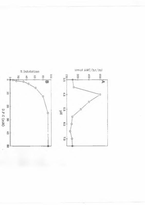


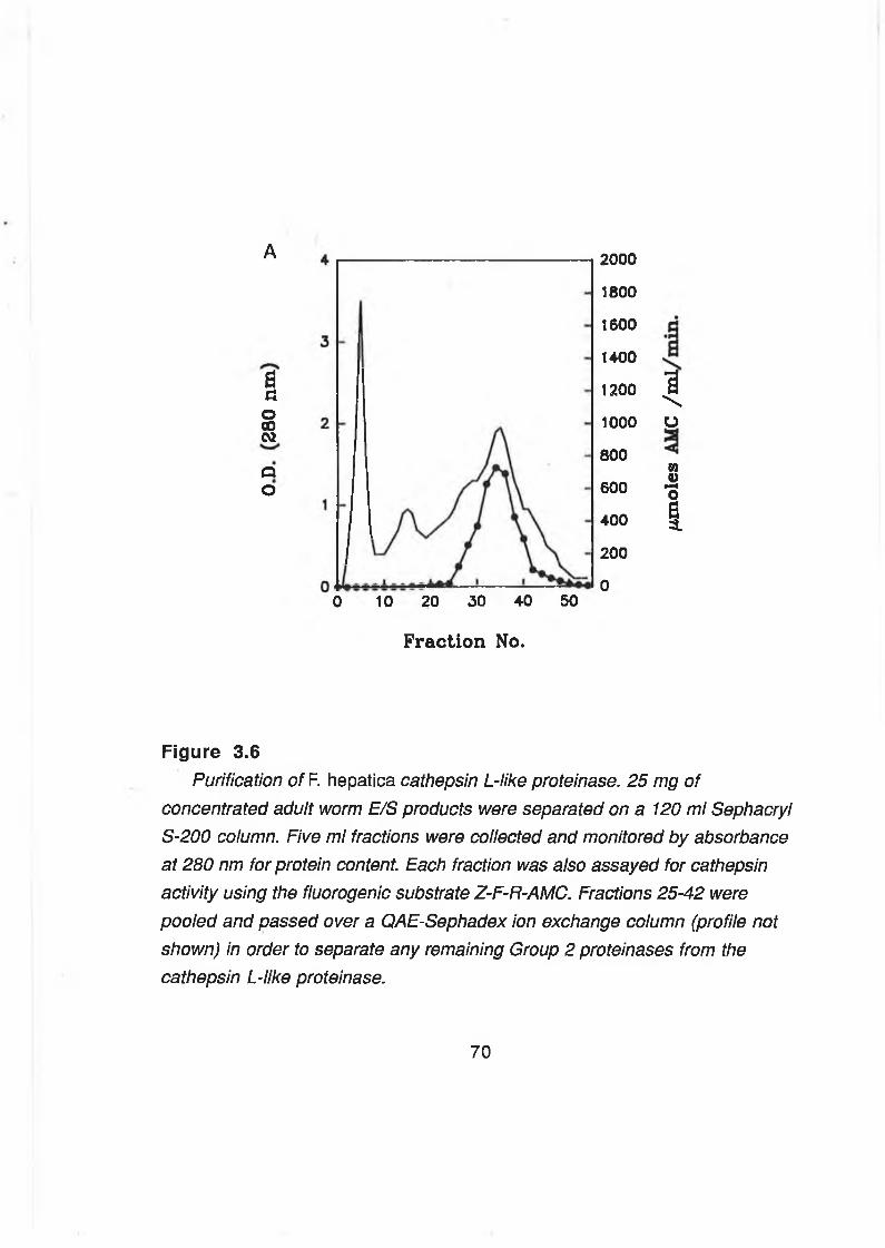


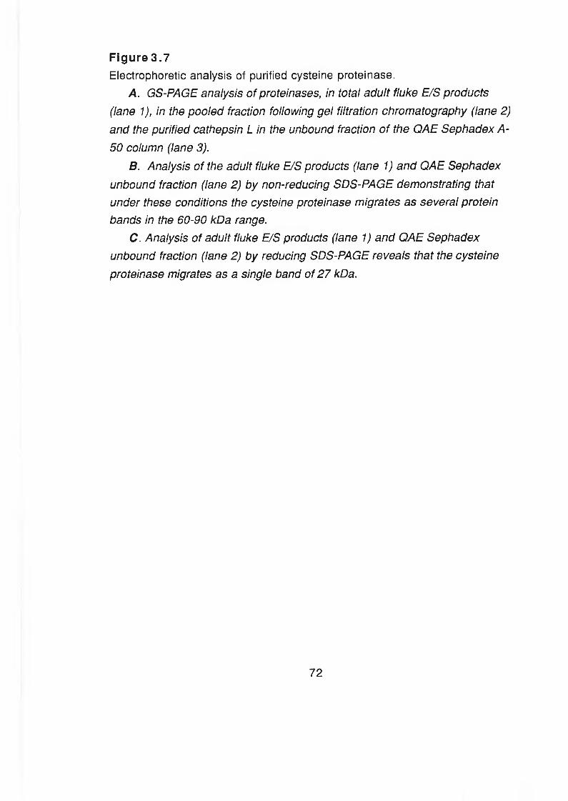


![Serum [3H]-fucose labelled glycoproteins in Fasciola hepatica](https://static.documents.pub/doc/80x56/623f6350b395777077658644/serum-3h-fucose-labelled-glycoproteins-in-fasciola-hepatica.jpg)


