Fast Calcium-Dependent Inactivation of Calcium Release-Activated Calcium Current(CRAC) in RBL-1 Cells
L. Fierro, A.B. ParekhLaboratory of Molecular and Cellular Signalling, Department of Physiology, University of Oxford, Parks Road, Oxford OX1 3PT, UK
Received: 28 August 1998/Revised: 30 November 1998
Abstract. Fast inactivation of the Ca2+ release-activatedCa2+ current (ICRAC) was studied using whole cell patch-clamp recordings in rat basophilic leukemia (RBL-1)cells. Application of hyperpolarizing voltage steps fromthe holding potential of 0 mV revealed thatICRAC de-clined in amplitude over tens of milliseconds duringsteps more negative than −40 mV. This fast inactivationwas predominantly Ca2+-dependent because first, itcould be more effectively suppressed when BAPTA wasincluded in the recording pipette instead of EGTA andsecond, replacing external Ca2+ with Sr2+ resulted in lessinactivation. Recovery from inactivation was faster inthe presence of BAPTA than EGTA. The extent of fastinactivation was independent of the whole cellICRAC
amplitude, compatible with the notion that the inactiva-tion arose from a local feedback inhibition by permeatingCa2+ ions only on the channel it permeated. Ca2+ releasefrom stores did not affect fast inactivation, nor didFC«RI receptor stimulation. Current clamp recordingsshowed that the majority of RBL cells had a membranepotential close to −90 mV following stimulation ofFC«RI receptors. Hence fast inactivation is likely to im-pact on the extent of Ca2+ influx through CRAC channelsunder physiological conditions and appears to be an im-portant negative feedback process that limits Ca2+ in-creases.
Introduction
It has now been established that the major Ca2+ entrypathway in nonexcitable cells is activated by the empty-ing of the intracellular Ca2+ store, a process that has beendubbed capacitative calcium entry (Putney, 1986). Inmast cells, jurkat T-lymphocytes and rat basophilic leu-
kemia cells, capacitative Ca2+ entry is manifestedthrough a highly selective Ca2+ current calledICRAC (re-viewed in Parekh & Penner, 1997). Hallmarks ofICRAC
include its’ voltage-independence, high selectivity forCa2+ over monovalent cations and very low single-channel conductance (<1 pS).
In order to fine-tune Ca2+ entry, cells are endowedwith mechanisms that regulate the activity of CRACchannels. In rat basophilic leukemia (RBL) cells, whichare a model system for studying store-operated Ca2+ in-flux mainly because largeICRAC can be measured, sev-eral processes exist that limit the duration of the current.These include changes in electrical driving force throughfluctuations in membrane potential (Parekh, Fleig & Pen-ner, 1997), feedback inactivation by protein kinase C(Parekh & Penner, 1995), store refilling (Parekh,inpreparation) and an intracellular Ca2+-dependent butstore-independent mechanism (Parekh, 1998). Thesepathways all operate over a time-scale of tens of secondsto minutes.
In mast cells and jurkat T-cells, a rapid inactivationmechanism has been reported that operates on a time-scale of tens of milliseconds (Hoth & Penner, 1993;Zweifach & Lewis, 1995). This has been characterizedin detail in T-cells, and arises through a negative feed-back mechanism triggered by permeating Ca2+ ions. TheCa2+ ions that permeate each channel induce rapid inac-tivation only in that particular channel, and this is ac-complished by binding to a site estimated to be within 4nm of the intracellular mouth of the pore (Zweifach &Lewis, 1995).
In RBL cells, ICRAC has been found to decline inamplitude during a hyperpolarizing voltage pulse (Hoth,1995). However, a detailed analysis of rapid inactivationhas not been carried out for any other cell-type that ex-pressesICRAC other than jurkat T-cells. This may beparticularly relevant because it has been suggested thatthere may be specialized channel subtypes within aCorrespondence to:A.B. Parekh
CRAC superfamily (Hoth, 1995). Hence extrapolationof basic features ofICRAC from one cell-type to anothermay not be valid.
Continuing our examination of feedback processesthat operate onICRAC, we have now investigated theproperties of fast inactivation in RBL cells. We find thatthis inactivation mechanism shares some striking simi-larities to that already characterized in jurkat T-cells, butthere are some notable differences. By directly measur-ing the resting membrane potential, we find that the ma-jority of the cells are sufficiently hyperpolarized to sup-port fast inactivation. We have also examined the effectsof redox potential onICRAC, since inactivation of certaintypes of K+ channel is affected by cysteine oxidation.We find that neither the activation mechanism nor thefast inactivation process are affected by redox potential.Finally we present the first report of the effects of dif-ferent divalent cations on the activation mechanism ofICRAC. The rate of activation of the current depends onthe charge carrying species.
Materials and Methods
Rat basophilic leukemia cells (RBL-1) cells, which were kindly pro-vided by Michael Pilot from the Max Planck Institute for BiophysicalChemistry in Goettingen, Germany, were cultured essentially as pre-viously described (Parekh, Fleig & Penner, 1997). Briefly, cells weremaintained in medium containing DMEM, 10% heat-inactivated fetalbovine serum and 3% streptomycin-penicillin (all from Life Technolo-gies), and kept in a 5% CO2-humidified atmosphere at 37°C. Cellswere trypsinized and then plated on glass coverslips around 24 hrbefore use. Patch-clamp experiments were conducted in the tight-sealwhole-cell configuration at room temperature (18–25°C) as previouslydescribed (Hamill et al., 1981; Parekh, 1998). Patch pipettes werepulled from borosilicate glass (Hilgenberg), sylgard coated and fire-polished. Pipettes had d.c. resistances of 2.5–4MV when filled withstandard internal solution that contained (in mM): cesium glutamate145, NaCl 8, MgCl2 1, HEPES 10, pH 7.2 with CsOH. Depending onthe experiment (described in the text), the Ca2+ chelators EGTA orBAPTA were added to this solution, as was InsP3 (0.03 mM). In someexperiments, RBL cells were sensitized to antigen (DNP-BSA) by in-cubation in IgE (500 ng/ml) for >3 hr. Antigen and IgE were kindlysupplied by Professor Israel Pecht (Weizman Institute, Rehovot, Israel).A correction of +10 mV was applied for the subsequent liquid junctionpotential that arose from this glutamate-based internal solution. Extra-cellular solution contained (in mM): NaCl 145, KCl 2.8, CaCl2 10,MgCl2 2, CsCl 10, glucose 10, HEPES 10, pH 7.2 (NaOH). In divalentsubstitution experiments, 10 mM Ca2+ was replaced by 10 mM Ba2+ orSr2+. External CsCl was present to block the activity of the inwardlyrectifying potassium channel (McCloskey & Cahalan, 1990). In somecurrent clamp experiments, CsCl was not present in the external solu-tion (pH was adjusted with NaOH), and potassium glutamate was usedinstead of caesium glutamate in the internal solution (pH with KOH).High resolution current recordings were acquired by a computer-basedpatch-clamp amplifier system (EPC 9, HEKA Electronics, Germany).Capacitative currents were canceled before each voltage ramp or stepusing the automatic compensation of the EPC 9. Series resistance wasbetween 4 and 14MV. Because of the small size of the currents (usu-ally <50 pA at −80 mV), series resistance compensation was not ap-plied. Voltage errors were therefore <1 mV. Currents were filtered
using an 8-pole Bessel filter at 2.5 kHz and digitized at 100msec.ICRAC was measured using both voltage ramps (−100 to +100 mV in 50msec) and voltage steps (described in text) applied every 2 sec usingPULSE software (HEKA Electronics) on a 9500 PowerMac, as de-scribed in Parekh, 1998. Peak currents were measured 2 msec after thestart of the pulse to minimize potential contributions from uncompen-sated capacitative current (time constant <0.5 msec). Steady-state cur-rents were measured as a 5 msecaverage at the end of the pulse. Cellswere held at 0 mV between pulses. All currents were leak subtractedby averaging the first two to four ramps or steps after breaking in andthen subtracting this from all subsequent traces. Local application wasachieved by means of a second pipette, placed within 30mm of the cell.Several parameters (capacitance, series resistance, holding current)were displayed simulataneously on a second monitor at a slower rate (2Hz) using the X-Chart display (HEKA Electronics). Data are presentedas mean ±SEM, and statistical evaluation was carried out using Studentsunpairedt-test.
Results
ICRAC INACTIVATES WITHIN MILLISECONDS DURING
HYPERPOLARIZING VOLTAGE STEPS
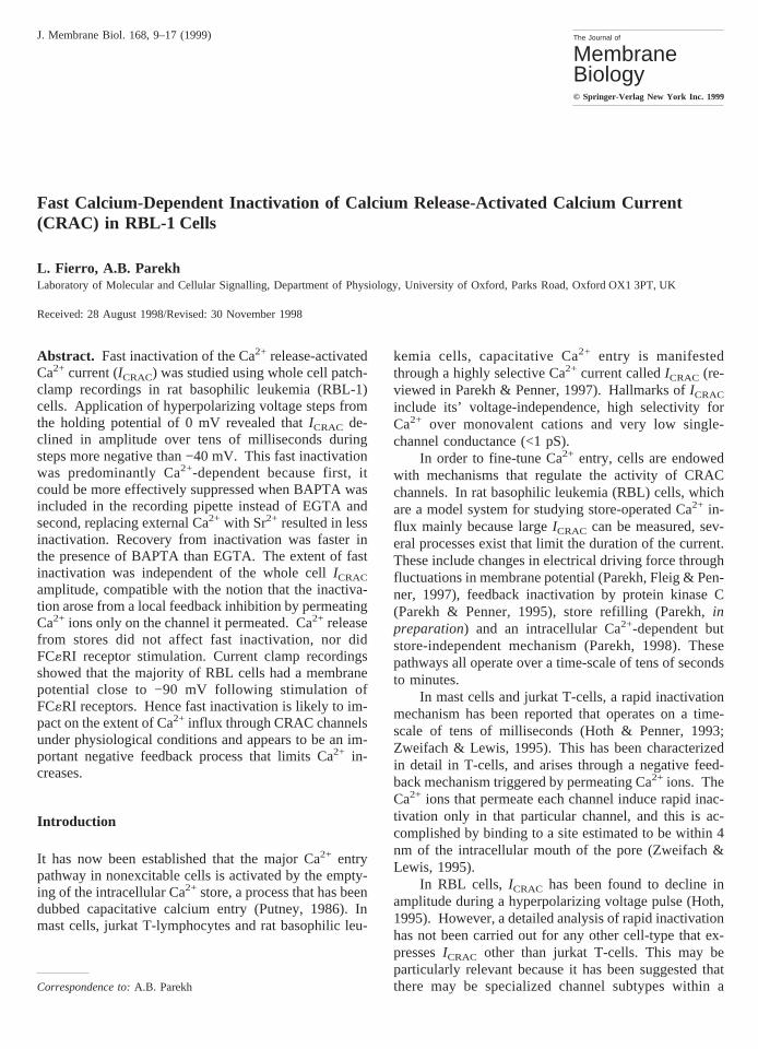
Application of hyperpolarizing pulses in mast cells andjurkat T-lymphocytes have demonstrated thatICRAC ex-hibits a rapid, Ca2+-dependent inactivation (Hoth & Pen-ner, 1993; Zweifach & Lewis, 1995). To examinewhetherICRAC inactivated during a sustained hyperpo-larizing step in RBL cells, we applied voltage pulsesfrom 0 to −100 mV. Figure 1A shows a recording thatwas taken with 10 mM EGTA in the pipette after thewhole cell CRAC current had reached a stable value.High EGTA passively depletes the stores. The leak cur-rent, obtained beforeICRAC had developed, has been sub-tracted. On stepping to −100 mV, there was an instan-taneous increase in the current which then declined toreach a steady-state amplitude that was around 50% ofthe peak. The speed of inactivation could, in most cases,be fitted with fast and slow exponential components at−100 mV (ts in the range of 10 and 120 msec, respec-tively).
To examine the voltage-dependence of this inacti-vation, we applied voltage steps up to −120 mV from theholding potential of 0 mV. Pooled data from 5 cells aresummarized in Fig. 1B. As the hyperpolarizing step in-creased in size, the extent of inactivation (steady-statecurrent divided by the initial peak value) became greater.At potentials positive to −40 mV however, inactivationbecame weak, at best.
FAST INACTIVATION IS DEPENDENT ONINTRACELLULAR
Ca2+: EFFECTS OFBAPTA
To examine whether fast inactivation in RBL cells wasdependent on intracellular Ca2+, we replaced EGTA inthe recording pipette with the faster Ca2+ chelator
10 L. Fierro and A.B. Parekh: Ca2+-Dependent Inactivation of Ca2+ Release-Activated Ca2+ Current
BAPTA. BAPTA reduced the extent of fast inactivationon stepping to −100 mV when compared with EGTA(Fig. 1A). In fact, BAPTA was substantially more effec-tive than EGTA at reducing the extent of inactivation atall those voltages (<−40 mV) where fast inactivation wasapparent (Fig. 1B, each point reflects data from 5 cells).
Further support of the notion that fast inactivation isCa2+-dependent is provided from experiments in whichCa2+ was replaced by Sr2+, and is described later.
CRAC CHANNELS INACTIVATE INDEPENDENTLY OF
EACH OTHER
To see whether Ca2+ influx through one CRAC channelcould induce fast inactivation in another CRAC channel,we examined how the extent of fast inactivation changedas the number of conducting CRAC channels increased(monitored through the increased whole cell CRAC cur-rent). The extent of inactivation stayed reasonably con-stant as the whole cell Ca2+ current increased (8 cells,data not shown). Hence, fast inactivation would appearto arise from the local buildup of Ca2+ in the vicinity ofeach specific channel, and not from overlapping micro-domains of elevated Ca2+ from several independentchannels. This is in good agreement with results fromT-cells (Zweifach & Lewis, 1995).
Ca2+ RELEASE FROM InsP3-SENSITIVE STORES DOES NOT
TRIGGER FAST INACTIVATION
Because the InsP3-sensitive Ca2+ stores that activateICRAC are thought to be close to the plasma membrane inRBL cells (Parekh & Penner, 1995), we entertained thepossibility that Ca2+ released from these stores mightenhance the fast inactivation process. To address thisissue, we dialyzed cells with 30mM InsP3 and 10 mM
EGTA, and applied voltage steps to −80 mV from theholding potential of 0 mV. Ca2+ release evoked by InsP3
concentrations >1mM occurs within seconds of obtainingthe whole-cell configuration (Parekh, Fleig & Penner,1997), and would occur before appreciable amounts ofEGTA have diffused into the cell. A typical record isshown in Fig. 1C. The extent of fast inactivation wasrelatively constant throughout the recording. If Ca2+ re-lease had contributed to fast inactivation, one would havepredicted a high extent of inactivation initially and thiswould subsequently decline as the Ca2+ released by InsP3was buffered by the high levels of EGTA diffusing fromthe recording pipette. This was clearly not the case.Similar results were obtained in 11 other cells. Figure1C also shows that the extent of inactivation was con-stant despite a fivefold increase in whole cell CRACcurrent amplitude.
KINETICS OF RECOVERY FROM FAST INACTIVATION
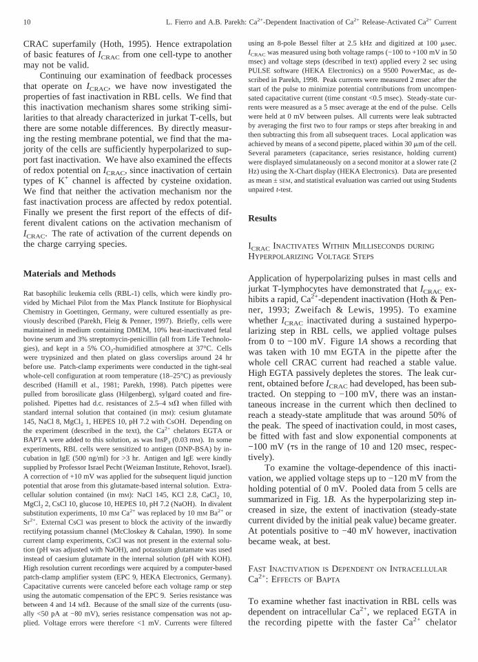
To assess how quickly CRAC channels recovered fromfast inactivation, we employed a paired-pulse protocolwhich is illustrated at the top of Fig. 2A. Cells wereinitially clamped at 0 mV and then stepped to −100 mVfor 250 msec (first pulse). The potential was then re-turned to 0 mV for a variable period of time (initially for10 msec which then increased by a factor of two) before
Fig. 1. Fast Ca-dependent inactivation ofICRAC. (A) ICRAC inactivatesmore during a voltage step in the presence of 10 mM EGTA thanBAPTA. B plots the voltage dependence of the extent of inactivation.BAPTA significantly reduced the extent of inactivation at all voltageswhere it occurred compared with EGTA. InA andB, ICRAC was acti-vated passively.C plots the extent of inactivationvs.whole cellICRAC
amplitude for a cell in whichICRAC was evoked by dialysis with InsP3.
11L. Fierro and A.B. Parekh: Ca2+-Dependent Inactivation of Ca2+ Release-Activated Ca2+ Current
stepping back to −100 mV once more (second pulse).In these experiments, we triggeredICRAC using passivestore depletion. BecauseICRAC activates slowly whenstores are depleted passively, we were able to obtain theentire set of background currents using the paired-pulseprotocol and then subtract these from the currents ob-tained onceICRAC had fully developed. Fig. 2A shows atypical leak-subtracted record, and Fig. 2B summarizesthe pooled data from 5 cells. Recovery from fast inac-tivation could be best fitted with a double-exponentialfunction yielding time-constantstrecoveryof 34 and 233msec (Fig. 2). Hence, recovery from fast inactivationappears to be a biphasic process.
When we used BAPTA instead of EGTA in the re-cording pipette, recovery from inactivation was acceler-ated at shorter time intervals between pulses (Fig. 2B, 5cells). Recovery from inactivation was best fit with adouble exponential, yielding time-constants of 9.0 and250 msec.
EFFECTS OFGROUP II DIVALENT CATIONS ON
FAST INACTIVATION
In the first set of experiments, we simply replaced Ca2+
from the extracellular solution with either Ba2+ or Sr2+
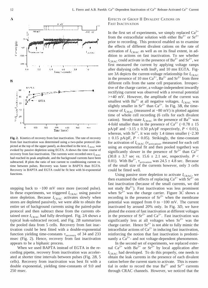
prior to recording. This protocol enabled us to examinethe effects of different divalent cations on the rate ofactivation ofICRAC as well as on its final extent, in ad-dition to actions on fast inactivation. To see whetherICRAC could activate in the presence of Ba2+ and Sr2+, wefirst measured the current by applying voltage rampsafter dialysing cells with InsP3 and 10 mM EGTA. Fig-ure 3A depicts the current-voltage relationship forICRAC
in the presence of 10 mM Ca2+, Ba2+ and Sr2+ from threedifferent cells from the same cell preparation. Irrespec-tive of the charge carrier, a voltage-independent inwardlyrectifying current was observed with a reversal potential>+40 mV. However, the amplitude of the current wassmallest with Ba2+ at all negative voltages.ICRAC wasslightly smaller in Sr2+ than Ca2+. In Fig. 3B, the time-course ofICRAC (measured at −80 mV) is plotted againsttime of whole cell recording (6 cells for each divalentcation). Steady-stateICRAC in the presence of Ba2+ was4-fold smaller than in the presence of Ca2+ (−0.78 ± 15pA/pF and −3.15 ± 0.50 pA/pF respectively,P < 0.01),whereas, with Sr2+, it was only 1.4 times smaller (−2.30± 0.15 pA/pF,P < 0.05). Strikingly, the time-constantfor activation ofICRAC (tactivation, measured for each cellusing an exponential fit and then pooled together) wassignificantly slower in the presence of Sr2+ than Ca2+
(30.8 ± 3.7 secvs. 15.6 ± 2.1 sec, respectively.P <0.01). With Ba2+, tactivationwas 24.5 ± 4.8 sec. Becauseof the small size of the currents however, only 3 cellscould be fitted well.
Using passive store depletion to activateICRAC, wethen examined the effects of replacing Ca2+ with Sr2+ onfast inactivation (because of the small currents, we didnot study Ba2+). Fast inactivation was less prominentwhen Sr2+ was the charge carrier. Figure 3C shows arecording in the presence of Sr2+ when the membranepotential was stepped from 0 to −100 mV. The currentinactivated by around 20% only. In Fig. 3D, we haveplotted the extent of fast inactivation at different voltagesin the presence of Sr2+ and Ca2+. Fast inactivation wassignificantly less at all voltages when Sr2+ was thecharge carrier. Hence Sr2+ is not able to fully mimic theintracellular actions of Ca2+ in inducing fast inactivation,reinforcing the notion that fast inactivation is predomi-nantly a Ca2+- and not voltage-dependent phenomenon.
In the second set of experiments, we replaced exter-nal Ca2+ with Ba2+ or Sr2+ by local application afterICRAC had developed. To do this properly, one needs toobtain the leak currents in the presence of each divalentcation before the current starts to activate. This is essen-tial in order to record the true Ba2+ and Sr2+ currentsthrough CRAC channels. However, we noticed that the
Fig. 2. Kinetics of recovery from fast inactivation. The rate of recoveryfrom fast inactivation was determined using a two-pulse protocol (de-picted at the top of the upper panel), as described in the text.ICRAC wasevoked by passive depletion using EGTA.A shows the time course ofrecovery from fast inactivation. The currents were recorded onceICRAC
had reached its peak amplitude, and the background currents have beensubtracted.B plots the ratio of test current to conditioning currentvs.time between pulses. Recovery was faster in BAPTA than EGTA.Recovery in BAPTA and EGTA could be fit best with bi-exponentialfunctions.
12 L. Fierro and A.B. Parekh: Ca2+-Dependent Inactivation of Ca2+ Release-Activated Ca2+ Current
leak currents changed, suggesting that Ba2+ and Sr2+
were affecting the resting cell conductance. We obtainedvery variable effects with Ba2+. In some cells, there wasa rapid fall inICRAC amplitude whereas in others a smallincrease initially occurred. In addition, different divalentcations change the surface potential to different degrees,which would alter the current-voltage relationship andcomplicate interpretation (Hagiwara & Byerly, 1981).We therefore abandoned this approach.
FAST INACTIVATION IS UNAFFECTED BY
CYSTEINE OXIDATION
A-type K+ currents exhibit a rapid N-type inactivationthat is due to oxidation of cysteine 13 on the amino ballpeptide (Ruppersberg et al., 1991). This inactivation isaccelerated by the reduced form of glutathione (GSH; themost abundant thiol in living cells) applied to the cyto-plasmic side. To see whether a similar mechanism couldaccount for the fast inactivation ofICRAC, we examinedthe effects of 5 mM GSH on the decay of the current.ICRAC was activated passively by dialysis with 10 mM
EGTA. This method was particularly suitable for ourpurposes because we were able to ensure that a signifi-cant amount of GSH had entered the cytoplasm prior tothe development of the current. Dialysis with GSH did
not affect the peak amplitude ofICRAC measured in volt-age ramps at −80 mV (−2.13 ± 0.19 pA/pF comparedwith control of −2.15 ± 0.29 pA/pF, 5 cells each), norwas there much change in the rateICRAC developed. Fig-ure 4A shows fast inactivation in the presence of GSGand Fig. 4B plots the extent of inactivation over the volt-age range −120 to −40 mV. Fast inactivation was largelyunaffected by GSH compared with controls. Underphysiological conditions, GSH is in a steady-state withoxidized glutathione (GSSG) with a maximal ratio of10:1. Dialysis with 5 mM GSH together with 0.5 mMGSSG did not alter the extent of inactivation comparedwith either GSH or control recordings (Fig. 4A andB).This mixture also did not affect the development nor thepeak amplitude ofICRAC (−1.86 ± 0.34 pA/pF, 4 cells;data not shown). Hence cysteine oxidation does notseem to play a role in the fast inactivation ofICRAC, norin the step that links store depletion to activation of thecurrent.
IS FAST INACTIVATION REGULATED BY
RECEPTORSTIMULATION ?
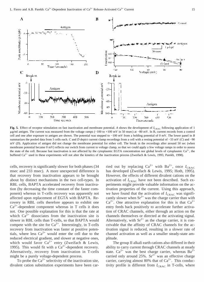
To see whether fast inactivation was regulated by cell-surface receptor stimulation, we activatedICRAC bystimulating FC«RI receptors with the antigen dinitrophe-
Fig. 3. Effects of different group II divalent cations onICRAC. (A) Current-voltage relationship of steady-stateICRAC in the presence of Ba2+, Sr2+
and Ca2+. Each trace represents a different cell.B plots the development ofICRAC (measured at −80 mV)vs. time. Note that the current developedsignificantly more slowly with Sr2+ as the charge carrier. In these experiments,ICRAC was activated by dialysis with InsP3 and 10 mM EGTA. Cshows that the extent of fast inactivation was reduced on stepping to -100 mV with Sr2+ as the charge carrier.D compares the extent of fastinactivation in the presence of Sr2+ vs.Ca2+ at different negative voltages. Fast inactivation was significantly less at all voltages in the presence ofSr2+ than Ca2+.
13L. Fierro and A.B. Parekh: Ca2+-Dependent Inactivation of Ca2+ Release-Activated Ca2+ Current
nyl-bovine serum albumin (DNP-BSA) in sensitizedRBL cells. FC«RI receptors have endogenous tyrosinekinase activity through which they phosphorylate phos-pholipase Cg1 to generate InsP3 and diacylglycerol; thelatter subsequently stimulates several different proteinkinase C isozymes expressed simultaneously in RBLcells (Ozawa et al., 1993). Stimulation of FC«RI recep-tors with antigen generatesICRAC (Zhang & McCloskey,1995; Parekh & Penner, 1995; Fig. 5A). We applied an-tigen around 50 sec after the onset of whole cell record-ing. The cell was dialyzed with a pipette solution inwhich Ca2+ was buffered at 280 nM (6.5 mM Ca-EGTA:3.5 mM EGTA) in order to prevent spontaneous activa-tion of the current. OnceICRAC had reached a stableamplitude, we applied voltage steps and measured theoverall extent of inactivation. Figure 5B shows that fastinactivation was unaffected by stimulation of FC«RI re-ceptors when compared with control cells over the volt-age range of −120 to −40 mV. Hence, the extent of fastinactivation is not regulated by receptor stimulation.
CHANGES IN MEMBRANE POTENTIAL FOLLOWING
RECEPTORSTIMULATION
For fast inactivation to be of physiological relevance, themembrane potential following receptor stimulationshould become quite hyperpolarized because fast inacti-vation becomes significant only at these negative poten-tials. To examine this, we recorded the membrane po-tential of resting RBL cells and then followed how itchanged after stimulation of FC«RI receptors in currentclamp mode. In an attempt to mimic physiological con-ditions, we bathed cells in a solution containing 1.8 mM
Ca2+ (rather than 10 mM) 1.2 mM Mg2+ and we omittedexternal Cs+. The pH of the external solution was in-creased to 7.4. The pipette solution contained K+ (in-stead of Cs+), and Ca2+ was weakly buffered with 100mM EGTA. Under these conditions, we observed twodistinct membrane potentials in RBL cells sensitized toantigen. Immediately after break in, 6 of 16 cells had apotential of −26 ± 11 mV (mean ±SD) whereas 11 cellswere more hyperpolarized (−89 ± 1 mV). The mem-brane potential was very stable and did not change withtime by more than ±5 mV. Regardless of the initialvalue, application of antigen failed to alter the membranepotential further. Typical records of a cell with a mem-brane potential of −33 mV, and one at −88 mV, areshown in Fig. 5C andD, respectively. Antigen was ap-plied as indicated, and exerted virtually no effect. Hencein the majority of cells, the membrane potential follow-ing receptor stimulation is sufficiently negative for fastinactivation to exert a significant on the extent of Ca2+
influx through CRAC channels.
Discussion
In this report, we have characterized rapid Ca2+-dependent inactivation ofICRAC in RBL cells. We findthat it shares several similarities to that described in jur-kat T-cells (Zweifach & Lewis, 1995). These include thefact that fast inactivation accounts for a modest declinein the amplitude ofICRAC unless the membrane potentialis hyperpolarized below −60 mV, that the extent ofsteady-state inactivation reached is similar in RBL andT-cells over the range −120 to −80 mV, that it is pre-dominantly Ca2+-dependent and is largely independentof the macroscopic current. It is more effectively sup-pressed by the fast chelator BAPTA than by EGTA, sug-gesting that the intracellular Ca2+ binding site(s) are inclose proximity to the channel pore.
Two striking differences between the RBL cells andthe jurkat T-cells are the kinetics of recovery from inac-tivation and the Ca2+-dependence of this process. In T-cells, recovery from inactivation in the presence ofEGTA is a biphasic process with time-constants of 9msec and 75 msec (Zweifach & Lewis, 1995). In RBL
Fig. 4. Fast inactivation is not altered by cysteine oxidation.A showscurrent traces obtained on stepping to −100 mV with a pipette solutioncontaining 5 mM GSH (upper panel) or 5 mM GSH: 0.5 mM GSSG(lower panel).B shows that neither 5 mM GSSH nor a mixture con-taining 0.5 mM GSSG: 5 mM GSH alter the relationship between in-activation and voltage compared with control recordings.ICRAC wasactivated in all three cases by passive depletion with 10 mM EGTA.
14 L. Fierro and A.B. Parekh: Ca2+-Dependent Inactivation of Ca2+ Release-Activated Ca2+ Current
cells, recovery is significantly slower for both phases (34msec and 233 msec). A more unexpected difference isthat recovery from inactivation appears to be broughtabout by distinct mechanisms in the two cell-types. InRBL cells, BAPTA accelerated recovery from inactiva-tion (by decreasing the time constant of the faster com-ponent) whereas in T-cells recovery was apparently notaffected upon replacement of EGTA with BAPTA. Re-covery in RBL cells therefore appears to exhibit oneCa2+-dependent component whereas in T cells it doesnot. One possible explanation for this is that the rate atwhich Ca2+ dissociates from the inactivation site isslower in RBL cells than T-cells, so that BAPTA wouldcompete with the site for Ca2+. Interestingly, in T-cellsrecovery from inactivation was faster at positive poten-tials, where less Ca2+ would enter the cell due to thereduced electrical gradient, and slower at negative ones,which would favor Ca2+ entry (Zweifach & Lewis,1995). This would fit with a Ca2+-dependent recovery.Alternatively, recovery from inactivation in T-cellsmight be a purely voltage-dependent process.
To probe the Ca2+ selectivity of the inactivation site,divalent cation substitution experiments have been car-
ried out by replacing Ca2+ with Ba2+, once ICRAC
has developed (Zweifach & Lewis, 1995; Hoth, 1995).However, the effects of different divalent cations on theactivation of ICRAC have not been described. Such ex-periments might provide valuable information on the ac-tivation properties of the current. Using this approach,we have found that the activation ofICRAC was signifi-cantly slower when Sr2+ was the charge carrier than withCa2+. One attractive explanation for this is that Ca2+
entry feeds back positively to accelerate further activa-tion of CRAC channels, either through an action on thechannels themselves or directed at the activating signal.Alternatively, with Sr2+ as the charge carrier, it is con-ceivable that the affinity of CRAC channels for the ac-tivation signal is reduced, resulting in a slower rate ofchannel activation as well as a smaller steady-state am-plitude.
The group II alkali earth cations also differed in theirability to carry current through CRAC channels at steadystate. Ca2+ was the best charge carrier, whereas Ba2+
carried only around 25%. Sr2+ was an effective chargecarrier, carrying almost 80% that of Ca2+. This conduc-tivity profile is different from ICRAC in T-cells, where
Fig. 5. Effect of receptor stimulation on fast inactivation and membrane potential.A shows the development ofICRAC following application of 1mg/ml antigen. The current was measured from the voltage ramps (−100 to +100 mV in 50 msec) at −80 mV. In B, current records from a controlcell and one after exposure to antigen are shown. The potential was stepped to −100 mV from a holding potential of 0 mV. The lower panel inBsummarizes the pooled data from 5 cells each.C andD depict current clamp recordings from a cell with a resting potential of −33 mV (C) and −90mV (D). Application of antigen did not change the membrane potential for either cell. The break in the recordings after around 50 sec (whenmembrane potential became 0 mV) reflects our switch from current to voltage clamp, so that we could apply a few voltage ramps in order to assessthe state of the cell. Because fast inactivation is not affected by the cytoplasmic EGTA concentration nor global levels of cytoplasmic Ca2+, thebuffered Ca2+ used in these experiments will not alter the kinetics of the inactivation process (Zweifach & Lewis, 1995; Parekh, 1998).
15L. Fierro and A.B. Parekh: Ca2+-Dependent Inactivation of Ca2+ Release-Activated Ca2+ Current
Ba2+ and Sr2+ were rather similar in that they carriedaround 40% of the current compared with Ca2+ (Zwei-fach & Lewis, 1993). The permeability profile of CRACchannels in RBL cells would therefore appear to be dis-tinct from that in T-cells, and would support the notionthat they may reflect distinct channel subtypes (Hoth,1995).
Fast inactivation of voltage-gated Ca2+ channels hasbeen well-described (Ashcroft & Stanfield 1981; Chad &Eckert, 1986) and the inactivation site is rather selectivein that Ba2+ cannot substitute for Ca2+. Ba2+ permeabil-ity through CRAC channels seems to be complex andconfusing. Hoth (1995) observed an initial large in-crease in current amplitude on replacing external Ca2+
with Ba2+, once ICRAC had reached a steady state.However, this increase was not observed by Schofieldand Mason (1996). In our hands, we observed effects ofBa2+ on the background conductance. In addition, weobserved very variable effects when we locally applied itto cells that had generatedICRAC. In T-cells, Ba2+ in-duced less inactivation than Ca2+ (Zweifach & Lewis,1995). In RBL cells, we found that the internal bindingsite was rather selective for Ca2+ in that Sr2+ was not ableto fully substitute for Ca2+ in inducing inactivation.These results are consistent with those described in T-cells with Ba2+ in that they point to a highly selectiveCa2+-binding site for fast inactivation.
Recently, a Ca2+ binding site has been described forCRAC channels in T-cells, which needs to be occupiedby Ca2+ in order to obtain the maximal channel conduc-tance (Christian et al., 1996; Zweifach & Lewis, 1996).This site is thought to be located on either the extracel-lular part of the channel or within the pore. Ion substi-tution experiments may therefore affect the number ofavailable channels in addition to conductivity. It is notclear whether such an external Ca2+-binding site is pres-ent on CRAC channels in RBL cells. If one were toexist, then it would appear that Sr2+ is better able tooccupy the site than Ba2+. In T-cells however, it hasbeen reported that Sr2+ cannot support channel activity(Christian et al., 1996).
One key question is whether Ca2+ induces fast in-activation by directly binding to a site on the channelitself (or an associated subunit) or whether it recruits anintermediate signal. This latter mechanism is thought toaccount for the Ca2+-dependent inactivation of voltage-operated, nifedipine-sensitive, Ca2+ channels in helixneurons (Chad & Eckert, 1986), where Ca2+ influxstimulates Ca2+-dependent phosphatase calcineurin todephosphorylate the channel, thereby inducing inactiva-tion. Recovery from inactivation occurs through a pro-tein kinase-mediated phosphorylation. Recent evidencehas demonstrated that protein kinases colocalize withCa2+ channels, linked by adaptor proteins of the AKAPfamily (Gao et al., 1997; Gray et al., 1997). Because fast
inactivation was remarkably constant throughout ourwhole cell recordings (>10 minutes in some experi-ments) and could occur without any ATP in the pipette,we do not think a phosphorylation reaction is fundamen-tal to the fast inactivation process.
Ca2+ release from InsP3-sensitive stores was not ableto affect the extent of fast inactivation that was inducedby Ca2+ entry. Ca2+ release starts almost immediatelyafter breaking into the cell and Ca2+ stays elevated for upto 30 sec. BecauseICRAC activates after a delay ofaround 3 sec, cytosolic Ca2+ will still be high as thecurrent starts to develop (appreciable amounts of Ca2+
buffer would not have diffused into the cell from therecording pipette at these short times). Since we findthat fast inactivation is unaffected, the subplasmalemmalCa2+ elevation in the vicinity of CRAC channels follow-ing Ca2+ release is not of sufficient amplitude to alterinactivation, and hence Ca2+ release is of little conse-quence to this process. This finding may have broadimplications for the activation mechanism ofICRAC. Inboth avian nasal gland cells and mouse pancreatic acini,a sizeable delay of several seconds between Ca2+ releaseand subsequent Ca2+ influx has been observed. BothShuttleworth (1994) and Toescu and Petersen (1995)have argued that this delay is hard to reconcile with theconformational-coupling model in which InsP3 receptorson the stores are supposed to physically attach to Ca2+
channels in the plasma membrane, but are instead com-patible with a slow biochemical step such as the genera-tion of a retrograde messenger. Berridge (1995) has ar-gued however, that the delay between Ca2+ release andentry reflects recovery from Ca2+-dependent fast inacti-vation of CRAC channels, the inactivation arising fromCa2+ release. Our finding that Ca2+ release does not af-fect fast inactivation would suggest that the delay be-tween Ca2+ release and subsequent influx does not reflectrecovery from inactivation, but instead would indicate aslow activation mechanism reminiscent of a biochemicalprocess, at least in RBL cells.
For fast inactivation to be of physiological rel-evance, two criteria need to be fulfilled. First, the mem-brane potential must be more negative than around −60mV and second, sufficient Ca2+ must enter in order toinduce the inactivation process. In our current clamp re-cordings, we found that the majority of cells had a restingpotential of around −90 mV. This would be sufficientfor significant fast inactivation ofICRAC. With physi-ological external Ca2+ concentration,ICRAC can be ob-served in T-cells although the current is small (Zweifach& Lewis, 1995). In agreement with this, we have mea-suredICRAC in RBL cells in 1.8 mM Ca2+ and fast inac-tivation can be discerned (data not shown). Hence fastinactivation is likely to impact on the extent of Ca2+
influx through CRAC channels under physiological con-
16 L. Fierro and A.B. Parekh: Ca2+-Dependent Inactivation of Ca2+ Release-Activated Ca2+ Current
ditions and appears to be an important negative feedbackprocess that limits Ca2+ increases.
The extent of fast inactivation was not modified bystimulation of FC«RI receptors. Because these receptorshave endogenous tyrosine kinase activity, stimulatephospholipase Cg1 (resulting in protein kinase C activa-tion), and activate a variety of SH2 domain-containingproteins (like Grb2 leading to Ras activation), it wouldappear that fast inactivation is not subject to regulationfrom a variety of intracellular signaling pathways. In-stead its impact is determined solely by the prevailingmembrane potential.
This work was supported by a Wellcome Trust grant to A.B.P. (grantno.: 049236/Z/96/Z). A.B.P. is the Sir Edward Abraham Research Fel-low at Keble College Oxford. We are grateful to Drs. Alison Bradingand Maike Glitsch for critical comments on the manuscript.
References
Ashcroft, F.M., Stanfield, P.R. 1982. Calcium inactivation in skeletalmuscle fibres of the stick insect,Carausius morosus. J. Physiol.330:349–372
Berridge, M.J. 1995. Capacitative calcium entry.Biochem. J.312:1–11Clapham, D.E. 1995. Calcium signalling.Cell 80:259–268Chad, J.E., Eckert, R. 1986. An enzymatic mechanism for calcium
current inactivation in dialyzedHelix neurons.J. Physiol.378:31–52
Christian, E.P., Spence, K.T., Togo, J.A., Dargis, P.G., Patel, J. 1996.Calcium-dependent enhancement of depletion-activated calciumcurrent in Jurkat T lymphocytes.J. Membrane Biol.150:63–71
Gray, P.C., Tibbs, V.C., Catterall, W.A., Murphy, B.J. 1997. Identifi-cation of a 15-kDa cAMP-dependent protein kinase-anchoring pro-tein associated with skeletal muscle of L-type calcium channels.J.Biol. Chem.272:6297–6302
Hagiwara, S., Byerly, L. 1981. Calcium channel.Annual Review ofNeuroscience4:69–125
Hamill, O., Marty, A., Neher, E., Sakmann, B., Sigworth, F. 1981.Improved patch-clamp techniques for high resolution current re-cordings from cells and cell-free membrane patches.Pfluegers.Arch. 391:85–100
Hoth, M. 1995. Calcium and barium permeation through calcium re-lease-activated calcium (CRAC) current.Pfluegers Arch.430:315–322
Hoth, M., Penner, R. 1993. Calcium release-activated calcium currentin rat mast cells.J. Physiol.465:359–386
McCloskey M.A., Cahalan, M.D. 1990. G protein regulation of potas-sium channel activity in a mast cell line.J. Gen. Physiol.95:205–227
Ozawa, K., Szallasi, Z., Kazamietz, M.G., Blumberg, P.M., Mischak,J.F., Beaven, M.A. 1993. Calcium-dependent and calcium-independent isozymes of protein kinase C mediate exocytosis inantigen-stimulated rat basophilic RBL-2H3 cells: reconstitution ofsecretory responses with calcium and purified isozymes in washedpermeabilized cells.J. Biol. Chem.268:1749–1756
Parekh, A.B. 1998. Slow feedback inhibition of Calcium release-activated Calcium current (CRAC) by Calcium entry.J. Biol.Chem.273:14925–14932
Parekh, A.B., Fleig, A., Penner, R. 1997. The store-operated calciumcurrentICRAC: nonlinear activation by InsP3 and dissociation fromcalcium release.Cell 89:973–980
Parekh, A.B., Penner, R. 1995. Depletion-activated calcium current isinhibited by protein kinase in RBL-2H3 cells.Proc. Nat. Acad. Sci.USA92:7907–7911
Parekh, A.B., Penner, R. 1995. Activation of store-operated calciuminflux at resting InsP3 levels by sensitization of the InsP3 receptorin rat basophilic leukemia cells.J. Physiol.489:377–382
Parekh, A.B., Penner, R. 1997. Store depletion and calcium influx.Physiol. Rev.77:901–930
Putney, J.W. 1986. A model for receptor-regulated calcium entry.CellCalcium7:1–12
Putney, J.W., Bird, G.S. 1993. The signal for capacitative calciumentry.Cell 75:199–202
Ruppersberg, J.P., Stocker, M., Pongs, O., Heinemann, S.H., Frank, R.,Koenem, M. 1991. Regulation of fast inactivation of cloned mam-malian I-K(A) channels by cysteine oxidation.Nature 352:711–-714
Schofield, G.G., Mason, M.J. 1996. A Ca2+ current activated by releaseof intracellular Ca2+ stores in rat basophilic leukemia cells (RBL-1). J. Membrane Biol.153:217–231
Shuttleworth, T.J. 1994. Temporal relationships between Ca2+ storemobilization and Ca2+ entry in an exocrine cell.Cell Calcium15:457–466
Toescu, E.C., Petersen, O.H. 1995. Region-specific activity of theplasma membrane Ca2+ pump and delayed activation of Ca2+ entrycharacterize the polarized, agonist-evoked Ca2+ signals in exocrinecells.J. Biol. Chem.270:8528–8535
Zhang, L., McCloskey, M.A. 1995. Immunoglobulin E receptor-activated calcium conductance in rat mast cells.J. Physiol.483:59–66.
Zweifach, A., Lewis, R.S. 1993. Mitogen-regulated Ca2+ current of Tlymphocytes is activated by depletion of intracellular Ca2+ stores.Proc. Natl. Acad. Sci USA90:6295–6299
Zweifach, A., Lewis, R.S. 1995. Rapid inactivation of depletion-activated calcium current (ICRAC) due to local calcium feedback.J.Gen. Physiol.105:209–226
Zweifach, A., Lewis, R.S. 1996. Calcium-dependent potentiation ofstore-operated calcium channels in T lymphocytes.J. Gen. Physiol.107:597–610
17L. Fierro and A.B. Parekh: Ca2+-Dependent Inactivation of Ca2+ Release-Activated Ca2+ Current


![Earthdawn - 3rd Ed [RBL] - Spell Library](https://static.documents.pub/doc/80x56/55cf9223550346f57b93f4b6/earthdawn-3rd-ed-rbl-spell-library.jpg)

