2914 Emerging Infectious Diseases • www.cdc.gov/eid • Vol. 27, No. 11, November 2021 DISPATCHES A multisystem inflammatory syndrome in chil- dren (MIS-C) and adults (MIS-A) occurring after coronavirus disease (COVID-19) has been identified; onset is ≈4–6 weeks after severe acute respiratory syndrome coronavirus 2 (SARS-CoV-2) infection (1–3). A case definition for MIS-A has been developed by the Centers for Disease Control and Prevention (CDC) (4); MIS-A after vaccina- tion is rare and remains of great clinical and public health interest (5). We report a case study and his- topathologic findings from a fatal MIS-A case after SARS-CoV-2 infection and subsequent complete COVID-19 vaccination. The Patient The patient was a healthcare worker in his 30s with no notable medical history. In December 2020, he experienced mild COVID-19–like illness symptoms, including fatigue and loss of taste and smell. He did not undergo testing for SARS-CoV-2 at that time and was unaware of the need for isolation. Six days af- ter onset of COVID-19–like symptoms, and when fully recovered, the patient received the first dose of Pfizer/BioNTech (https://www.pfizer.com) mRNA COVID-19 vaccine. He received the second dose 20 days later. After the second dose, he reported fatigue and malaise, which resolved within 2 days. Twenty-two days after receiving the second dose of the COVID-19 vaccine, he had onset of new fever, malaise, headache, and odynophagia. He was examined by an outpatient medical provider. Diag- nostic testing was notable for a negative COVID-19 test by reverse transcription PCR (RT-PCR), nega- tive rapid influenza antigen, and negative rapid an- tigen detection for group A Streptococcus. Four days later, the patient visited an emergency department because of worsening symptoms. Assessment of vital signs revealed a temperature of 37.2°C, heart rate 113 beats/min, and blood pressure of 117/66 mmHg. Physical examination identified right-sided cervical lymphadenopathy, marked bilateral con- junctival erythema, and a faint papular rash on the pelvis and left flank. Laboratory testing revealed a peripheral-blood leukocyte count of 11,000 cells/ µL, 93.5% segmented neutrophils, and thrombocy- topenia with a platelet count of 110,000/µL (Table). Portable chest radiograph results were without no- table findings. On hospital day 2, the patient remained febrile and tachycardic (heart rate 90–135 beats/min) and Fatal Multisystem Inflammatory Syndrome in Adult after SARS-CoV-2 Natural Infection and COVID-19 Vaccination Heather N. Grome, Michael Threlkeld, Steve Threlkeld, Charles Newman, Roosecelis Brasil Martines, Sarah Reagan-Steiner, Michael A. Whitt, Maria Gomes-Solecki, Nisha Nair, Mary-Margaret Fill,Timothy F. Jones, William Schaffner, John Dunn Author affiliations: Centers for Disease Control and Prevention, Atlanta, Georgia, USA (H.N. Grome, R.B. Martines, S. Reagan-Steiner); Tennessee Department of Health, Nashville, Tennessee, USA(H.N. Grome, M.-M. Fill, T.F. Jones, J. Dunn); Baptist Memorial Health Care, Memphis, Tennessee, USA (M. Threlkeld, S. Threlkeld); Methodist LeBonheur Healthcare, Memphis (M. Threlkeld, S. Threlkeld); Pathology Group of the MidSouth, Germantown, Tennessee, USA (C. Newman); Trumbull Laboratories, Germantown (C. Newman); University of Tennessee Health Science Center, Memphis (M.A. Whitt, M. Gomes-Solecki, N. Nair); Vanderbilt University School of Medicine, Nashville (W. Schaffner) DOI: https://doi.org/10.3201/eid2711.211612 We describe a fatal case of multisystem inflammatory syndrome in an adult with onset 22 days after a second dose of mRNA coronavirus disease vaccine. Serologic and clinical findings indicated severe acute respiratory syndrome coronavirus 2 infection occurred before vacci- nation. The immunopathology of this syndrome, regard- less of vaccination status, remains poorly understood.
A multisystem infl ammatory syndrome in chil-dren (MIS-C) and adults (MIS-A) occurring
after coronavirus disease (COVID-19) has been identifi ed; onset is ≈4–6 weeks after severe acute respiratory syndrome coronavirus 2 (SARS-CoV-2) infection (1–3). A case defi nition for MIS-A has been developed by the Centers for Disease Control and Prevention (CDC) (4); MIS-A after vaccina-tion is rare and remains of great clinical and public health interest (5). We report a case study and his-topathologic fi ndings from a fatal MIS-A case after SARS-CoV-2 infection and subsequent complete COVID-19 vaccination.
The PatientThe patient was a healthcare worker in his 30s with no notable medical history. In December 2020, he experienced mild COVID-19–like illness symptoms, including fatigue and loss of taste and smell. He did not undergo testing for SARS-CoV-2 at that time and was unaware of the need for isolation. Six days af-ter onset of COVID-19–like symptoms, and when fully recovered, the patient received the fi rst dose of Pfi zer/BioNTech (https://www.pfi zer.com) mRNA COVID-19 vaccine. He received the second dose 20 days later. After the second dose, he reported fatigue and malaise, which resolved within 2 days.
Twenty-two days after receiving the second dose of the COVID-19 vaccine, he had onset of new fever, malaise, headache, and odynophagia. He was examined by an outpatient medical provider. Diag-nostic testing was notable for a negative COVID-19 test by reverse transcription PCR (RT-PCR), nega-tive rapid infl uenza antigen, and negative rapid an-tigen detection for group A Streptococcus. Four days later, the patient visited an emergency department because of worsening symptoms. Assessment of vital signs revealed a temperature of 37.2°C, heart rate 113 beats/min, and blood pressure of 117/66 mmHg. Physical examination identifi ed right-sided cervical lymphadenopathy, marked bilateral con-junctival erythema, and a faint papular rash on the pelvis and left fl ank. Laboratory testing revealed a peripheral-blood leukocyte count of 11,000 cells/µL, 93.5% segmented neutrophils, and thrombocy-topenia with a platelet count of 110,000/µL (Table). Portable chest radiograph results were without no-table fi ndings.
On hospital day 2, the patient remained febrile and tachycardic (heart rate 90–135 beats/min) and
Fatal Multisystem Infl ammatory Syndrome in Adult after
SARS-CoV-2 Natural Infection and COVID-19 Vaccination
We describe a fatal case ofmultisystem inflammatorysyndrome in an adult with onset 22 days after a second doseofmRNAcoronavirusdiseasevaccine.Serologicand clinical findings indicated severe acute respiratorysyndromecoronavirus2infectionoccurredbeforevacci-nation.Theimmunopathologyofthissyndrome,regard-less of vaccination status, remains poorly understood.
had a blood pressure of 92/56 mmHg. Diagnostic evaluation revealed a negative SARS-CoV-2 RT-PCR test but a positive serologic test for SARS-CoV-2 nu-cleocapsid IgG. Additional diagnostic tests were conducted (Table). Inflammatory markers showed elevated C-reactive protein at 284.0 mg/L, serum fer-ritin at 1434.9 ng/mL, and troponin-I at 18.0 ng/mL. On the evening of hospital day 2, the patient received 75 g of intravenous immune globulin (IVIG).
Early morning on hospital day 3, the patient had an acute change in mental status, including confusion and global aphasia. An emergent computed tomogra-phy scan of the head was negative for cerebrovascular accident and showed normal brain parenchyma and
no evidence of acute infarction, mass, or hemorrhage. On completion of the scan, the patient was found nonresponsive and without a pulse. He underwent multiple rounds of advanced cardiac life support, re-sulting in return of spontaneous circulation. A chest radiograph showed an enlarged cardiac silhouette, and an echocardiogram showed severe biventricular dysfunction, severe global hypokinesis of the left ven-tricle, and left ventricular ejection fraction of 20%. The patient received a second dose of IVIg and intrave-nous steroids and extracorporeal membrane oxygen-ation support was initiated. On hospital day 4, severe multisystem organ failure continued to progress. The patient died on hospital day 4.
Table. Resultsofpertinentlaboratorytestingcompletedduringthe4-day hospitalization of a patient with fatal multisystem inflammatorysyndromeinadult,Tennessee,USA,2021*
Variable Hospitalday
Referencerange Day 1 Day 2 Day 3† Day 4† Hematologictesting
SARS-CoV-2RT-PCR,indexvalue Negative Negative SARS-CoV-2 IgG antibody,‡ index value 4.96 <1.39 AdenovirusDNAPCR,qualitative Not detected Not detected CMVPCR,quantitative Negative Negative Mononucleosis screen Negative Negative Ehrlichia chaffeensis DNAPCR Not detected Not detected HIV-1 p24 Ag Nonreactive Nonreactive Peripheral blood culture, 2 sets No growth No growth No growth No growth No growth *Laboratory values represent pertinent laboratory results during the patient’s hospitalization. Not all laboratory studies completed during hospitalization arerepresentedinthistable.Blankcellsindicatetestnotdone.ALT,alanineaminotransferase;AST,aspartateaminotransferase;aPTT,activatedpartialthromboplastintime;CMV,cytomegalovirus;ESR,erythrocytesedimentationrate;FEU,fibrinogenequivalentunits;INR,internationalnormalizedratio;PT:prothrombintime;RT-PCR,reversetranscriptionPCR;SARS-CoV-2, severe acute respiratory syndrome coronavirus 2. †Laboratory values indicate studies after cardiac arrest, which occurred at 3a.m.onhospitalday3.Notepatientwasinitiatedonextracorporealmembrane oxygenation shortly after returnofspontaneouscirculation;anticoagulationtreatments affect laboratory values. ‡SARS-CoV-2 IgG test specific for nucleocapsid protein antibody.
We reviewed the patient’s medical history and clinical chart. We assessed serum samples collected during the hospital course before and after IVIg, and we determined endpoint titers to SARS-CoV-2 nucleocapsid (IgM and IgG) and spike recep-tor binding domain with neutralization functions against spike protein (6,7). The endpoint titer was a modified protocol based on Stadlbauer et al. (8). We completed an autopsy and sent formalin-fixed, paraffin-embedded tissues to CDC. Microscopic examination of lung, airways, pulmonary lymph node, liver, heart, spleen, kidneys and stomach tis-sue samples was performed; LT-Gram stain was performed on lungs and heart. An RT-PCR assay
for SARS-CoV-2 was performed on RNA extracted from formalin-fixed, paraffin-embedded tissues from lungs, airways, and heart by methods previ-ously published (9). This activity was reviewed by CDC and was conducted consistent with applicable federal law and CDC policy.
Serum antibody results drawn before IVIg infu-sion were negative for SARS-CoV-2 IgM but positive for IgG. Serum results had a high titer of anti-spike re-ceptor binding domain antibody both before and af-ter IVIg (1:75,000) compared with a naturally infected SARS-CoV-2–positive control (1:4,000). In addition, the pre-IVIG sample serum results demonstrated neutralizing function.
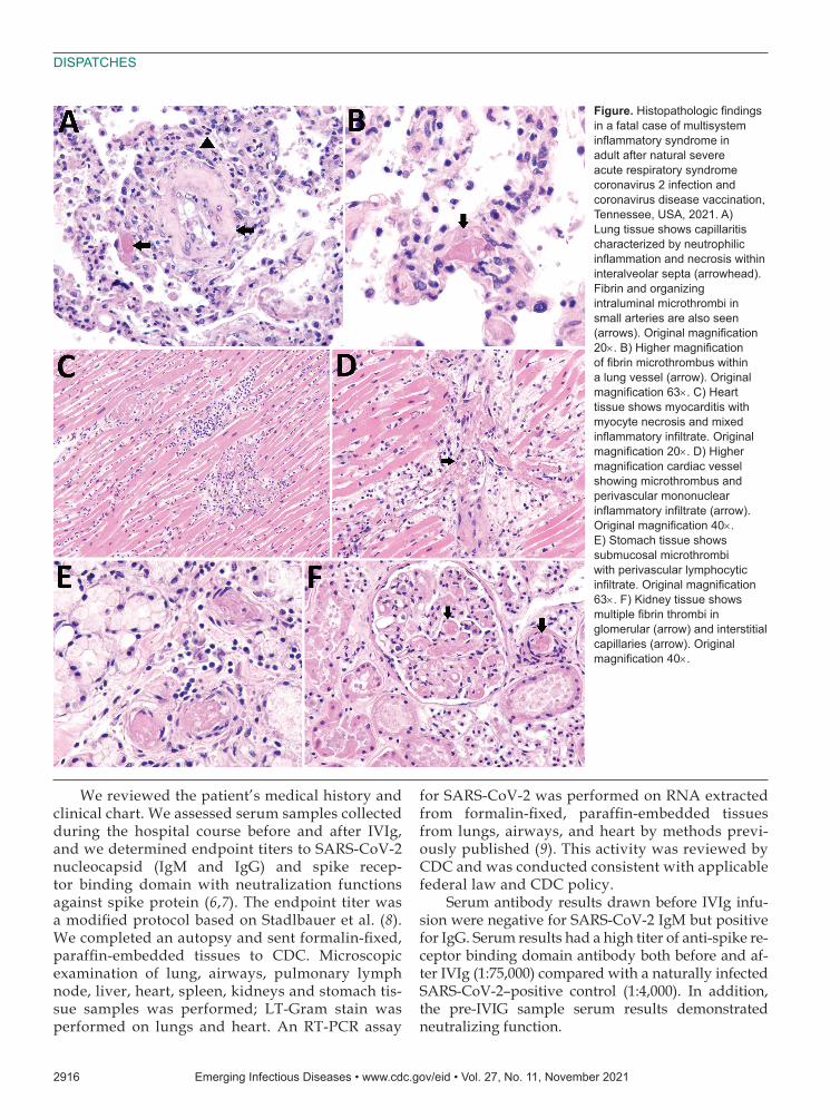
Figure.Histopathologicfindingsin a fatal case of multisystem inflammatorysyndromeinadult after natural severe acute respiratory syndrome coronavirus 2 infection and coronavirus disease vaccination, Tennessee,USA,2021.A)Lung tissue shows capillaritis characterized by neutrophilic inflammationandnecrosiswithininteralveolarsepta(arrowhead).Fibrinandorganizingintraluminal microthrombi in small arteries are also seen (arrows).Originalmagnification20×.B)Highermagnificationoffibrinmicrothrombuswithinalungvessel(arrow).Originalmagnification63×.C)Hearttissue shows myocarditis with myocyte necrosis and mixed inflammatoryinfiltrate.Originalmagnification20×.D)Highermagnificationcardiacvesselshowing microthrombus and perivascular mononuclear inflammatoryinfiltrate(arrow).Originalmagnification40×. E)Stomachtissueshowssubmucosal microthrombi with perivascular lymphocytic infiltrate.Originalmagnification63×.F)Kidneytissueshowsmultiplefibrinthrombiinglomerular(arrow)andinterstitialcapillaries(arrow).Originalmagnification40×.
Notable findings on gross internal autopsy ex-amination included a 525-mL pericardial effusion and cardiac enlargement, as well as a 5-L hemoperitoneum and a 20-cm diameter perisplenic hematoma. Micro-scopic examination of the lungs showed diffuse conges-tion, increased intra-alveolar macrophages, multifocal hemorrhage, capillaritis, and microthrombi through-out (Figure, panels A, B). We observed no viral inclu-sions or diffuse alveolar damage. Trachea and bronchi showed mild tracheobronchitis. Sections of the heart showed multifocal myocarditis with mixed inflamma-tory infiltrate, myocyte necrosis, and numerous micro-thrombi. We also identified disseminated microvascu-lar thrombosis in the heart, stomach, kidneys, and liver (Figure, panels C–F). Gram stain results were negative on lung and heart tissue. SARS-CoV-2 RT-PCR was negative on lungs, trachea, bronchi, and heart.
ConclusionsThis fatal case of MIS-A occurred after full COVID-19 vaccination in a patient with prior natural SARS-CoV-2 infection suspected 6 weeks before MIS-A symptom onset. Serum antibody results before IVIg infusion indicated the patient was previously in-fected with SARS-CoV-2 and was vaccinated with a COVID-19 vaccine. Antibodies to the nucleocapsid protein are the most sensitive target for serologic di-agnosis for natural infection (P.D. Burbelo et al., un-pub. data, https://doi.org/10.1101/2020.04.20.20071423), and these antibodies are not present following COVID-19 vaccination alone. In addition, clinical his-tory was compatible with natural infection beginning 6 days before the first mRNA vaccine dose and con-sistent with negative SARS-CoV-2 nucleocapsid IgM on testing during hospitalization.
The patient demonstrated similar clinical findings to previously reported MIS-A cases, including fever for 3 consecutive days, laboratory evidence of inflam-mation, neurologic and mucocutaneous clinical find-ings, and severe cardiac illness that included systemic hypotension progressing to cardiogenic shock (1,5). These criteria meet the CDC case definition for MIS-A, as well as a definitive case at level 1 of diagnostic cer-tainty by the Brighton collaboration case definition for MIS-A and MIS-C (10). In addition, the histopathologic findings of capillaritis and multiorgan microvascular thrombosis in association with clinical symptoms and laboratory findings are compatible with MIS-A (1,11). Substantial blood loss on gross examination may rep-resent a diffuse intravascular coagulation–type picture in which diffuse microthrombosis depleted platelets and clotting factors. The etiology for clinical deterio-ration was likely multifactorial, although considerable
cardiac compromise in the setting of high fluid vol-umes and intraperitoneal hemorrhage may have con-tributed to multiorgan failure
Whether mRNA COVID-19 vaccination con-tributed to MIS-A onset in this case is unclear, and future epidemiologic studies are needed to under-stand whether an association exists. The immunopa-thology leading to hyperinflammation causing MIS-A after SARS-CoV-2 infection remains unknown, although postinfection immune dysregulation is consistent among reported cases. Notably, MIS-A has not been reported among adult participants of COVID-19 vaccine trials (10), and no direct evidence exists to support vaccine alone as the primary etiol-ogy in this case. This article further emphasizes the importance of COVID-19 prevention, for which in-fection prevention strategies and vaccination remain our greatest defense.
This work was supported by the National Institutes of Health (grant nos. R43 AI155211 and R01 AI139267 awarded to M.G.S.).
About the AuthorDr. Grome is an infectious diseases physician and Epidemic Intelligence Service Officer for the Centers for Disease Control and Prevention, Atlanta, Georgia. She is currently assigned as a field officer at the Tennessee Department of Health in Nashville, Tennessee.
L, Balan S, et al. Case series of multisystem inflammatory syndrome in adults associated with SARS-CoV-2 infection—United Kingdom and United States, March–August 2020. MMWR Morb Mortal Wkly Rep. 2020;69:1450–6. https://doi.org/10.15585/mmwr.mm6940e1
2. Tenforde MW, Morris SB. Multisystem inflammatory syndrome in adults: coming into focus. Chest. 2021;159:471–2. https://doi.org/10.1016/j.chest.2020.09.097
3. Godfred-Cato S, Bryant B, Leung J, Oster ME, Conklin L, Abrams J, et al.; California MIS-C Response Team. COVID-19-associated multisystem inflammatory syndrome in children—United States, March–July 2020. MMWR Morb Mortal Wkly Rep. 2020;69:1074–80. https://doi.org/ 10.15585/mmwr.mm6932e2
4. National Center for Immunization and Respiratory Diseases, Centers for Disease Control and Prevention. Multisystem inflammatory syndrome in adults (MIS-A) case definition information for healthcare providers 2021 [cited 2021 Jul 1]. https://www.cdc.gov/mis/mis-a/hcp.html
5. Salzman MB, Huang CW, O’Brien CM, Castillo RD. Multisystem inflammatory syndrome after SARS-CoV-2 infection and COVID-19 vaccination. Emerg Infect Dis. 2021;27:1944–8. https://doi.org/10.3201/eid2707.210594
6. McAndrews KM, Dowlatshahi DP, Dai J, Becker LM, Hensel J, Snowden LM, et al. Heterogeneous antibodies
against SARS-CoV-2 spike receptor binding domain and nucleocapsid with implications for COVID-19 immunity. JCI Insight. 2020;5:142386. https://doi.org/10.1172/jci.insight.142386
7. Condor Capcha JM, Lambert G, Dykxhoorn DM, Salerno AG, Hare JM, Whitt MA, et al. Generation of SARS-CoV-2 spike pseudotyped virus for viral entry and neutralization assays: a 1-week protocol. Front Cardiovasc Med. 2021;7:618651. https://doi.org/10.3389/fcvm.2020.618651
8. Stadlbauer D, Amanat F, Chromikova V, Jiang K, Strohmeier S, Arunkumar GA, et al. SARS-CoV-2 seroconversion in humans: a detailed protocol for a serological assay, antigen production, and test setup. Curr Protoc Microbiol. 2020;57:e100. https://doi.org/10.1002/cpmc.100
9. Bhatnagar J, Gary J, Reagan-Steiner S, Estetter LB, Tong S, Tao Y, et al. Evidence of severe acute respiratory syndrome coronavirus 2 replication and tropism in the lungs, airways, and vascular endothelium of patients with fatal coronavirus disease 2019: an autopsy case series.
10. Vogel TP, Top KA, Karatzios C, Hilmers DC, Tapia LI, Moceri P, et al. Multisystem infl ammatory syndrome in children and adults (MIS-C/A): case defi nition & guidelines for data collection, analysis, and presentation of immunization safety data. Vaccine. 2021;39:3037–49. https://doi.org/10.1016/j.vaccine.2021.01.054
11. Magro C, Mulvey JJ, Berlin D, Nuovo G, Salvatore S, Harp J, et al. Complement associated microvascular injury and thrombosis in the pathogenesis of severe COVID-19 infection: a report of fi ve cases. Transl Res. 2020;220:1–13. https://doi.org/10.1016/j.trsl.2020.04.007
Address for correspondence: Heather N. Grome, Tennessee Department of Health, Communicable Diseases and Emergency Preparedness Division, 710 James Robertson Pkwy, Nashville, TN 37243, USA; email: [email protected]
Among the 1.2 million cases of nontyphoidal Salmonellainfections in the United States each year, only 23,000
patients are hospitalized. Although most Salmonella cases resolve on their own, patients with severe illness might
require treatment with antimicrobial drugs.
But what happens when treatment doesn’t work? Antimicrobial resistance among Salmonella is a growing threat, and public health officials at CDC and beyond are
on a mission to curb its spread before it is too late.
In this EID podcast, Dr. Felicita Medalla, a CDC epidemiologist, investigates the rising incidence
of AMR nontyphoidal Salmonella in the United States.
EID Podcast: AMR Nontyphoidal Salmonella Infec� ons,
United States
Visit our website to listen: h� ps://go.usa.gov/xFZyx