Feasibility of Using a Computer Modeling Approach to Study SUI Induced by Landing a Jump YINGCHUN ZHANG, 1 SEOGGWAN KIM, 2 ARTHUR G. ERDMAN, 2 KENNETH P. ROBERTS, 1 and GERALD W. TIMM 1 1 Department of Urologic Surgery, University of Minnesota, 725 Mayo Memorial Building, 420 Delaware Street S.E., Minneapolis, MN 55455, USA; and 2 Department of Mechanical Engineering, University of Minnesota, Minneapolis, USA (Received 20 October 2008; accepted 17 April 2009; published online 5 May 2009) Abstract—Stress urin ary incontin ence (SUI ) occur s due to ana tomi c and/ or neur olog ic fact ors invo lvin g conn ectiv e tissues, muscles and nerves. Although SUI is more common in post -men opau sal and mult ipar ous women, stud ies have also shown a high prevalence of SUI in young, physically fit female athletes. With a goal toward dynamic subject-specific mechanical characterization of the interaction between ana- tomical structures during physical activities that elicit SUI in females during physical or daily activities, a computer aided design (CAD)-based computer model of the female pelvis has been developed to test the fe asibil it y of the comput er modeling approach in understanding the measurable differ- ences between stress-cont inent and stress-inc ontinent women. In the pres ent stud y, a fluid–str uctur e interactio n analy sis was conducted by using the finite element (FE) analysis technique based on the CAD-ba sed comput er mod el of the fema le pel vis to investigate the urine leakage in females during jumping. To the best of our knowledge, this is the first application of a fluid–str uc ture interact ion FE anal ys is appr oa ch in understanding the mechanisms of SUI in females. Through a series of computer simulations, the effects of varying impact forc es deter mine d by jump ing heig ht and blad der volu me were investigated. The dynamic computer simulation results revealed that jumping heights have a significant influence on the volume of urine leakage caused by the landing impact ofjumping. Bladder volume did not have a significant influence on leakage when the jumping heights were smaller than 1 ft, which ind ica tes that normal walki ng (co rre spond s to a jumping height smaller than 0.1 ft) is not the primary cause of uri ne lea kag e for hea lth y femal es. The comput er simul ati on results also showed that the deformation difference between the anterior and posterior portion of the female pelvis causes ope nin g of the ure thr a and res ult ant uri ne lea kag e. The pres ent stud y demo nstr ates the feasi bili ty of usin g a computer modeling approach to study female SUI during physical and daily activities . Keywords—Finite element analysis, Fluid–structure interac- tion analysis, Female athletes, Stress urinary incontinence, Urologic pelvic floor disorder, Physical activity. INTRODUCTION Stress urinary incontin ence (SUI) occurs because ofproblems with connective tissues, muscles, and nerves that help to hold or release urine. It currently affects ove r 13 million Americ ans with the majority being females. Although SUI is more common in post-men- opausal and multiparous women, 13 studies have also shown a high prevalence of SUI in young, physically fit femal e athletes. 4,6,9,14,16,19,20,22 Regarding the associa- tion of exercise type with female athletic SUI rate, it is not ed that exe rcis es that inv olv e chr oni c, repeti tive motion and involve high impact landing from jumping and running cause high SUI rates. 6,16,20,22 New data als o suggests a cor rel ati on bet ween severi ty of SUI symptoms and physical inactivity. 4,18 The symptoms ofSUI tha t cause women to avo id sports par tic ipa tion may be one etiologic factor for physical inactivity in women. 4,10,14,15 Howeve r, the mec hanism of SUI in young female athletes remains unclear. Uro dynamic studie s provid e a met hod for obj ective ly ass ess ing how the bla dde r and ure thra l coordi nat ed fun cti ons of storing and rel easi ng urin e are ope rat ing in ord er to gai n a bet ter und erst anding of the occurrenc e ofSUI and to assist with choosing an effective treatment. The complete urodynamic study consists of cystometry with electromyography, urethral profilometry and fre e uro flowmetry using a dir ect mea suring approa ch. 1 These techniques, however, cannot provide the detailed dynami c informat ion of force , str ess, st rain, and resulting organ displacements of the bladder and ure- thr a whi ch are cri tic al in understanding , dia gnosing, and treati ng SUI probl ems. A compu ter model ing approa ch, whic h can provide us the dynami c bi omechani cal respo nses of the bladd er, urethr a, rhabdo sphin cter, and al l the ot he r organs and ti ssues insi de the pelvis as wellas thei r neural control, has the capabi li ty of fixing this gap. A fe w st ud ies have been attempted in this field, 3,7,8,10,24,25 in which the finit e el ement (FE) me thod Address corresponden ce to Ying chun Zhan g, Dep artment ofUro logi c Surg ery, Univ ersi ty of Minneso ta, 725 Mayo Memoria l Building, 420 Delaware Street S.E., Minneapolis, MN 55455, USA. Electroni c mail: zhang320 @umn.edu Annals of Biomedical Engineering, Vol. 37, No. 7, July 2009 (Ó 2009) pp. 1425–1433 DOI: 10.1007/s10 439-009-9705 -2 0090-6964/09/0700-1425/0Ó 2009 Biomedical Engineering Society 1425
Transcript
8/8/2019 Feasibility of Using a Computer Modeling Approach to Study SUI
Feasibility of Using a Computer Modeling Approach to Study SUIInduced by Landing a Jump
Y INGCHUN Z HANG ,1
SEOGGWAN K IM ,2
ARTHUR G. E RDMAN ,2
K ENNETH P. R OBERTS ,1
and G ERALD W. T IMM1
1Department of Urologic Surgery, University of Minnesota, 725 Mayo Memorial Building, 420 Delaware Street S.E.,Minneapolis, MN 55455, USA; and 2Department of Mechanical Engineering, University of Minnesota, Minneapolis, USA
(Received 20 October 2008; accepted 17 April 2009; published online 5 May 2009)
Abstract —Stress urinary incontinence (SUI) occurs due toanatomic and/or neurologic factors involving connectivetissues, muscles and nerves. Although SUI is more commonin post-menopausal and multiparous women, studies havealso shown a high prevalence of SUI in young, physically tfemale athletes. With a goal toward dynamic subject-specicmechanical characterization of the interaction between ana-tomical structures during physical activities that elicit SUI infemales during physical or daily activities, a computer aideddesign (CAD)-based computer model of the female pelvis hasbeen developed to test the feasibility of the computermodeling approach in understanding the measurable differ-ences between stress-continent and stress-incontinent women.In the present study, a uid–structure interaction analysis wasconducted by using the nite element (FE) analysis techniquebased on the CAD-based computer model of the female pelvisto investigate the urine leakage in females during jumping. Tothe best of our knowledge, this is the rst application of auid–structure interaction FE analysis approach inunderstanding the mechanisms of SUI in females. Througha series of computer simulations, the effects of varying impactforces determined by jumping height and bladder volumewere investigated. The dynamic computer simulation resultsrevealed that jumping heights have a signicant inuence onthe volume of urine leakage caused by the landing impact of jumping. Bladder volume did not have a signicant inuenceon leakage when the jumping heights were smaller than 1 ft,which indicates that normal walking (corresponds to a jumping height smaller than 0.1 ft) is not the primary causeof urine leakage forhealthy females.The computer simulationresults also showed that the deformation difference betweenthe anterior and posterior portion of the female pelvis causesopening of the urethra and resultant urine leakage. Thepresent study demonstrates the feasibility of using a computer
modeling approach to study female SUI during physical anddaily activities.
Keywords —Finite element analysis, Fluid–structure interac-tion analysis, Female athletes, Stress urinary incontinence,Urologic pelvic oor disorder, Physical activity.
INTRODUCTION
Stress urinary incontinence (SUI) occurs because of problems with connective tissues, muscles, and nerves
that help to hold or release urine. It currently affectsover 13 million Americans with the majority beingfemales. Although SUI is more co mmon in post-men-opausal and multiparous women, 13 studies have alsoshown a high pr eva lence of SU I in young, physically tfemale athletes. 4,6 ,9 ,14 ,16 ,19 ,20 ,22 Regarding the associa-tion of exercise type with female athletic SUI rate, it isnoted that exercises that involve chronic, repetitivemotion and involve high impact lan ding fro m jumpingand running cause high SUI rates. 6 ,16 ,20 ,22 New dataalso suggests a correlation betw een severity of SUIsymptoms and physical inactivity. 4,18 The symptoms of SUI that cause women to avoid sports participationmay be one etiologic factor for physical inactivity inwomen. 4,10 ,14 ,15 However, the mechanism of SUI inyoung female athletes remains unclear.
Urodynamic studies provide a method for objectivelyassessing how the bladder and urethral coordinatedfunctions of storing and releasing urine are operating inorder to gain a better understandingof the occurrence of SUI and to assist with choosing an effective treatment.The complete urodynamic study consists of cystometrywith electromyography, urethral prolometry and fre euroowmetry using a direct measuring approach. 1
These techniques, however, cannot provide the detailed
dynamic information of force, stress, strain, andresulting organ displacements of the bladder and ure-thra which arecritical in understanding, diagnosing, andtreating SUI problems.A computer modeling approach,which can provide us the dynamic biomechanicalresponses of the bladder, urethra, rhabdosphincter, andall the other organs and tissues inside the pelvis as wellastheir neural control, has the capability of xing this gap.A few studi es have been attempted in thiseld, 3 ,7,8,10 ,24 ,25 in which the nite element (FE) method
Address correspondence to Yingchun Zhang, Department of Urologic Surgery, University of Minnesota, 725 Mayo MemorialBuilding, 420 Delaware Street S.E., Minneapolis, MN 55455, USA.Electronic mail: [email protected]
Annals of Biomedical Engineering , Vol. 37, No. 7, July 2009 ( Ó 2009) pp. 1425–1433DOI: 10.1007/s10439-009-9705-2
0090-6964/09/0700-1425/0 Ó 2009 Biomedical Engineering Society
1425
8/8/2019 Feasibility of Using a Computer Modeling Approach to Study SUI
was employed to build a pelvis model. These previouspelvis models either did not include sufcient anatomi-cal parts of the pelvis which are closely related to thelower urinary tract function, or used mechanical prop-erties of the tissues from either dated literatureor animalexperimental data.
With a goal toward the development of a dynamicsubject-specic mechanical characterization the inter-action between anatomical structures during physicalactivities that elicit SUI, a computer aided design(CAD)-based FE model of female pelvis has beendeveloped to understand female SUI induced by thelanding impact of jumping. CAD-based modeling of acomplex structure, such as the female pelvis, requiressome simplifying assumptions regarding its geometry.This modeling approach, however, has an advantageof much easier modication of the geometry andconsequently makes a feasibility and parameter studyfor an advanced subject-specic computer model. Thisstudy describes a computer modeling approach that
has the capability of characterizing the interactionbetween anatomical structures during physical activi-ties that elicit SUI.
MATERIALS AND METHODS
Anatomy of the Female Pelvis
The female pelvis is created by two innominatebones and the sacrum 2,12 ,17 as shown in Fig. 1a. Theinferior aperture, or pelvic outlet, is covered by thepelvic diaphragm; a set of thin broad muscles isattached to pelvi c sidewall by vascular and connectivetissue mesentery. 5 The muscles of the pelvic diaphragminclude the levator ani, a combined muscle sheetincluding the iliococcygeus, pubococcygeus, andpuborectalis muscles, and the coccygeus muscle. Justinferior to the pelvic diaphragm the anterior half of thepelvic outlet is covered by the urogenital (UG) dia-phragm. The UG diaphragm is composed of a deeptransverse perineal muscle, spanning from one ischialtuberosity to the other, and the external urethralsphincter covered by the perineal membrane. Thesupercial transverse perineal muscle lies superior to
the perineal membrane which is directly above the deeptransverse perineal muscle, and separates the super-cial and deep perineal compartments. Visceral struc-tures supported by the UG and pelvic diaphragmsinclude the uterus, the vagina, the bladder, and theurethra. The rectum lies behind the UG diaphragmand is supported primarily by the levator ani portionof the pelvic diaphragm. There are several sets of fas-cial ligaments that stabilize the pelvic viscera. Thedeepest and strongest of these are formed from the
endopelvic fascia. 23 These include the pubovesicle lig-aments that support the bladder neck and urethra, andthe pubocervical, transverse cervical (Cardinal), andureterosacral ligaments that support the uterus. Inaddition, the peritoneal broad ligament supports thebody of the uterus and the adnexa.
The female pelvic viscera include the bladder,vagina, uterus, fallopian tubes, ovaries, and rectum.Each of these structures, with the exception of theovaries, is composed primarily of smooth muscle. Thepelvic and UG diaphragms support these structuresand participate in their function by providing striatedsphincter musculature.
CAD-Based FE Model Building Procedure
CAD-Based Geometry Model and Hexahedral ElementMesh
Based on the understanding of the anatomicalstructure of the female pelvis, a three-dimensional (3D)FE model was built using the CAD technique as shownin Fig. 1b. The 3D FE model of female pelvis was builtto include the pelvic bone, uterus, vagina, rectum,pelvic diaphragm, UG diaphragm, abdominal muscles,intestine, bladder, urethra, urine, etc., to model theactual anatomical structure of the female pelvis. Forthe ordinary static structural analyses, 3D FE model-ing of structures with complex shapes can be done
FIGURE 1. Anatomical structure of the female pelvis and thecorresponding CAD-based FE model with half of the modelabout sagittal plane except urine. (a) Anatomical structure ofthe female pelvis; (b) 3D FE model of the female pelvis whichconsists of the pelvic bone, uterus, vagina, rectum, pelvicdiaphragm, uro-genital diaphragm, abdomen muscle, intes-tine, bladder, urethra, urine etc.
Z HANG et al.1426
8/8/2019 Feasibility of Using a Computer Modeling Approach to Study SUI
without much simplication of the geometry usingeither a solid modeler or image bas ed so lid constructiontechnique and tetrahedron mesh. 3 ,7 ,10 Simulating thebiomechanical responses of the human body during thelanding impact of jumping requires dynamic, largedeformation analysis with large mesh distortions.Tetrahedron elements without strong distortion resis-tance are known to be much less accurate than hexa-hedral elements in this case. In order to achieve thesame accuracy of analysis results, the number of tet-rahedron elements that will be required is 4 to 5 timesmore than the number of hexahedral elements. Thiswill increase the model size and require more com-puting resource. In the present study, the female pelvismodel was meshed with 8-noded hexahedral elements,as shown in Fig. 1b, to achieve the satised accuracywhile keep the relative small model size (with the ele-ment number of 325,531 and node number of 82,918).
The urethra was modeled with shell elements insteadof solid elementsto avoid low-quality elements.The urine
inside the bladder was modeled using uid elements inorder to investigate urine leakage during jumping.
Tissue Mechanical Properties
Mechanical properties of the tissues involved in thisstudy are listed in Table 1.7,26 Human t issues showvisco-hyperelastic material characteristics, 26 however,a quasi-linear material property was found in humanurological soft tissues from our soft tissue tensiletesting experiments when the stress level is below 70%of the maximal stress value. An example of thisobservation is shown in Fig. 2, which is the stress–
strain curve obtained by performing the soft tissuetesting procedure on a bladder wall tissue specimenfrom a fresh 18-year-old female cadaver. It was foundin our preliminary FE analysis results that the peakstress values developed in the tissues during jumpingwere in this quasi-linear range of the stress strainproles. Consequently linear material models wereimplemented in the present study to approximate theessential visco-hyperelastic material models.
In order to rene the pelvis model, a method tocharacterize the visco-hyperelastic material propertiesof human soft tissues by performing soft tissue testing
procedures on urological tissue specimens harvestedfrom fresh cadavers within 24 h of the time of deathhas been undertaken in our lab. Three soft tissuetesting procedures (tensile tests, creep tests, and stressrelaxation tests) were performed on urological softtissue specimens to develop this urological tissueproperties database. The completion of the database isstill in process and the computer model of the femalepelvis will be continuously rened by using updatedvisco-hyperelastic material properties of urologicaltissues involved in the model.
Contacts and Interaction Conditions
Both contact pairs and tie constraints were set up inthe present pelvis model to describe the interactionconditions along the boundaries between differentorgans. The tie constraints were set to boundaries onwhich two neighboring organ surfaces would not haveany relative sliding and/or disconnection during jumping. For example, the boundaries between pelvicbones and pelvic muscles as well as pelvic bones andpelvic ligaments were under this tie constraint. Thecontact pairs were set to boundaries on which twoneighboring organ surfaces would slide and/or move
apart during jumping. For example, the boundariesbetween pelvic muscles and fat tissues, pelvic musclesand pelvic ligaments, pelvic ligaments and fat tissues,bladder and uterus, uterus and colon, etc., all were ableto slide under such contact conditions.
Load Modulus
This model assumed that the pelvic bones supportedthe superincumbent body during jumping and theentire pelvis model had an initial velocity V initial . Then
TABLE 1. Mechanical properties of the tissues involvedin this study.
Tissue Modulus of e las ticity (MPa) Density (kg/m 3 )
FIGURE 2. A Stress–Green Strain curve obtained by per-forming soft tissue testing procedure on a bladder wall tissuespecimen from a fresh 18-year-old female cadaver.
Feasibility of Using a Computer Modeling Approach to Study SUI 1427
8/8/2019 Feasibility of Using a Computer Modeling Approach to Study SUI
the velocity of the bones was assumed to drop to zeroin a very short time period T impact after the subject’sfeet touch the ground. Choices for V initial and T impact ,which describe the landing impact effects on the femalepelvis and the organs inside caused by jumping, arecritical in forming the load modulus which is a neces-sary part of the computer model.
An ambulatory device was specically developed fordetermining the real initial velocity V initial and impactperiod T impact for each subject. The device consists of three sensors; the rst sensor is an accelerometer(CXL25LP3 accelerometer, Crossbow Technology,Inc., San Jose, CA) for measuring the time-historyacceleration of pelvis in three directions in an orthog-onal coordinate system, the second sensor is an incli-nometer (SQ-SI2X-360DA inclinometer, SignalQuest,Inc., Lebanon, NH) for measuring the pitch and rollangles of pelvis, and the third sensor is a urinaryleakage detector for quantitatively measuring subjects’urine leakage during jumping. The measurements from
the accelerometer and inclinometer directly contrib-uted to calculating the initial velocity V initial andimpact period T impact , while the measurements fromthe urinary leakage detector were used to evaluate UIduring jumping.
In human subject experiments, the accelerometerand inclinometer were xed over the lower back at thelevel of the posterior iliac crest or lumbar spine of subject as shown in Fig. 3. All the measurements werecollected wirelessly by a PC during subject’s physicalor daily activities. The initial velocity V initial was esti-mated from the temporal acceleration recordingsthrough the integration algorithm and the impactperiod T impact was estimated from the temporal accel-eration recordings in the impact parameters database.Those subject-specic landing impact parameters were
used to form the load modulus of the subject-specicpelvis model.
Dynamic Finite Element Analysis
A commercial ni te element (FE) analysis softwarepackage LS-DYNA 11 which has the capability of uid–structure interaction analysis was chosen for thisdynamic study. The Eulerian type of elements werechosen as uid elements to model the urine in whichthe mesh was xed in space and only material wouldmove around over the mesh to avoid large elementdistortions. Consequently, there was no mesh distor-tion and the motion of uids, such as urine, wereadequately modeled. The interaction between thestructure and uid, i.e. the bladder wall and the urineinside, were detected by overlapping the solid and uidelements. Note that the uid elements must cover theentire volume of space where the solid elements reachwhile they are under deformation.
A uid like media, such as urine, was modeled as theviscosity material with no yield strength, no shearstiffness, and an equation of state relates the uidpressure to the neighboring structures. LS-DYNAprovides a viscosity material model in which theequations of state were dened and erosion in tensionand compression was allowed. Viscosity of 0.87 9
10À3 N s, wave speed of 4.58 m/s and density of 1020
kg/m 3 were used for the physical properties of urine.
RESULTS
The dynamic biomechanical responses of the entirefemale pelvis and the urinary leakage informationcaused by the landing impact of jumping were achieved
FIGURE 3. Placement of the ambulatory device including a tri-axial accelerometer, a bi-axial inclinometer and a urinary leakagedetector. (a) Frontal view; (b) back view.
Z HANG et al.1428
8/8/2019 Feasibility of Using a Computer Modeling Approach to Study SUI
by performing dynamic FE analysis by means of LS-DYNA based on the computer models. Consideringthis is a feasibility study rather than a subject-specicstudy to simulate the real biomechanical response of the female pelvis during jumping, it was assumed in thecomputer simulations that the pelvic bones supportedthe superincumbent body and the pelvic bones stoppedimmediately and completely as soon as the feet tou-ched the ground. The bladder was assumed to be fullylled with urine, and the effects of jumping heights andbladder volumes on the amount of urinary leakagewere investigated. The results of three jumping heights(1, 2, and 3 ft) were compared to that of a normal dailywalking height (0.1 ft) and the bladder volumes werechosen to be 50, 100 and 200 mL.
The 1st computer simulation of a female subject jumping from a 3-feet high table with 100 mL urineinside her bladder was conducted and it took around900 s of CPU time for a single run on an IBM super-computer with 312 Power4 processors in the Minne-
sota Supercomputing Institute (MSI) at the Universityof Minnesota. Dynamic computer simulation resultsshowed that it took approximately 7 ms for the lowerportion of the pelvis to reach its lowest vertical posi-tion and come back to its normal position after thepelvic bones completely stopped. Figure 4 shows theresults from this computer simulation. Figure 4ashows the initial pelvis geometry model without anydeformation at 0.0 ms. Figure 4b shows the modeldeformation caused by the jumping impact at 2.7 msafter the pelvic bones completely stopped. Here we canclearly see the model geometry deformation caused theopening of the urethra and the urine was traveling intothe urethra through the urethro-vesical junction.Figure 4c shows the model deformation 7 ms after thepelvic bones completely stopped. We found that thelower portion of the pelvis almost reached its lowestpoint at this time instant and the model geometrydeformation caused by the jumping impact is muchlarger than that at 2.7 ms. The interesting phenome-non here was that the urethra already closed at thistime instant although the bladder deformation is verylarge.
Figure 5 shows the dynamic status of urine owinside of the bladder and urethra observed from thecomputer simulation results. Figure 5a shows urineow at 0.0 ms when the pelvic bones just completelystopped. The bladder deformation has not yet startedand there was no urethra opening and urine leakage.Figure 5b shows the urine ow at 4.3 ms after thepelvic bones completely stopped, where the urethraopened widely in the region near the bladder neck, anda remarkable amount of urine owed into the urethrafrom the bladder. Figure 5c shows that the urethrabegan to close in the region near the bladder neck
5.6 ms after the pelvic bones completely stopped, butthe volume of urine was already pushed into the mid-dle part of the urethra. Figure 5d shows that at 7 msafter the pelvic bones completely stopped, the urethrahas closed, but urine has already been pushed out of the body and generated the urine leakage althoughthere was still a very small amount of residual urine inthe middle portion of the urethra.
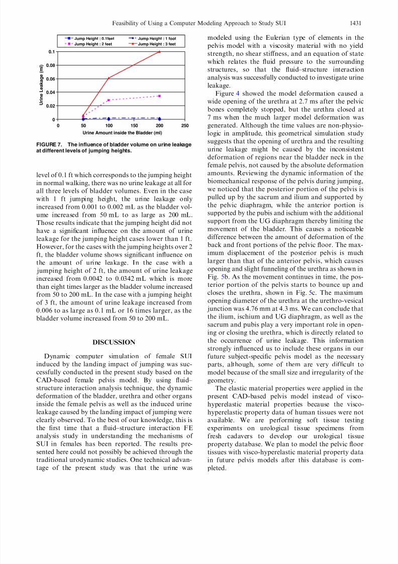
The inuence of jumping heights on the amount of urine leakage at different levels of bladder volumes of 50, 100, and 200 mL was investigated through a seriesof computer simulations based on the present CAD-based female pelvis computer model. The computersimulation results in Fig. 6 show that the amount of
FIGURE 4. Deformation of the bladder and opening of theurethra for a case of simulating a female subject jumping froma 3-feet high table with 100 mL urine inside the bladder with-out urine in the bladder. The model deformation caused ure-thra opening at (a) 0.0 ms, (b) 2.7 ms, and (c) 7 ms, after thepelvic bones completely stopped.
Feasibility of Using a Computer Modeling Approach to Study SUI 1429
8/8/2019 Feasibility of Using a Computer Modeling Approach to Study SUI
urine leakage increases signicantly as the jumpingheight increases when the bladder volume is greaterthan 100 mL. In the case with the bladder volume of
100 mL, the amount of urine leakage was increasedover 30 times, from 0.002 to 0.061 mL as the jumpingheight increased from 1 to 3 ft. In the case with thebladder volume of 200 mL, the amount of urine leak-age was increased by 50 times, from 0.002 mL to ashigh as 0.1 mL as the jumping height increased from 1to 3 ft. Even for the case with only 50 mL urine in thebladder, an increase of urinary leakage was alsoobserved when the jumping height increased, theamount of urine leakage, which increased from 0.001to 0.006 mL as the jumping height increased from 1 to3 ft, is increased by six times.
Similarly, the inuence of the bladder volume on theamount of urine leakage at jumping heights of 0.1, 1.0,2.0, and 3.0 ft was investigated through a series of computer simulations based on the CAD-based femalepelvis computer model (see Fig. 7). Notice that thebladder volume did not have noticeable inuence onthe amount of urine leakage when the jumping heightwas smaller than 1 ft. In the lowest jumping height
FIGURE 5. Urine ow in the case simulating a female subject jumping from a 3-feet high table with 100 mL urine inside thebladder. The urethra opening and resulting urine ow at (a) 0.0 ms, (b) 4.3 ms, (c) 5.6 ms, and (d) 7 ms, after the pelvic bonescompletely stopped.
0
0.02
0.04
0.06
0.08
0.1
0 0.5 1 1.5 2 2.5 3 3.5
Jumping Height (ft)
U r i n e
L e a
k a g e
( m l )
Bla dder Volum e : 5 0ml Bla dder Volum e : 1 00m lBladder Volume : 200m l
FIGURE 6. The inuence of jumping heights on urine leak-age at various bladder volumes.
Z HANG et al.1430
8/8/2019 Feasibility of Using a Computer Modeling Approach to Study SUI
level of 0.1 ft which corresponds to the jumping heightin normal walking, there was no urine leakage at all forall three levels of bladder volumes. Even in the casewith 1 ft jumping height, the urine leakage onlyincreased from 0.001 to 0.002 mL as the bladder vol-ume increased from 50 mL to as large as 200 mL.Those results indicate that the jumping height did nothave a signicant inuence on the amount of urineleakage for the jumping height cases lower than 1 ft.However, for the cases with the jumping heights over 2ft, the bladder volume shows signicant inuence onthe amount of urine leakage. In the case with a jumping height of 2 ft, the amount of urine leakageincreased from 0.0042 to 0.0342 mL which is morethan eight times larger as the bladder volume increased
from 50 to 200 mL. In the case with a jumping heightof 3 ft, the amount of urine leakage increased from0.006 to as large as 0.1 mL or 16 times larger, as thebladder volume increased from 50 to 200 mL.
DISCUSSION
Dynamic computer simulation of female SUIinduced by the landing impact of jumping was suc-cessfully conducted in the present study based on theCAD-based female pelvis model. By using uid– structure interaction analysis technique, the dynamicdeformation of the bladder, urethra and other organsinside the female pelvis as well as the induced urineleakage caused by the landing impact of jumping wereclearly observed. To the best of our knowledge, this isthe rst time that a uid–structure interaction FEanalysis study in understanding the mechanisms of SUI in females has been reported. The results pre-sented here could not possibly be achieved through thetraditional urodynamic studies. One technical advan-tage of the present study was that the urine was
modeled using the Eulerian type of elements in thepelvis model with a viscosity material with no yieldstrength, no shear stiffness, and an equation of statewhich relates the uid pressure to the surroundingstructures, so that the uid–structure interactionanalysis was successfully conducted to investigate urineleakage.
Figure 4 showed the model deformation caused awide opening of the urethra at 2.7 ms after the pelvicbones completely stopped, but the urethra closed at7 ms when the much larger model deformation wasgenerated. Although the time values are non-physio-logic in amplitude, this geometrical simulation studysuggests that the opening of urethra and the resultingurine leakage might be caused by the inconsistentdeformation of regions near the bladder neck in thefemale pelvis, not caused by the absolute deformationamounts. Reviewing the dynamic information of thebiomechanical response of the pelvis during jumping,we noticed that the posterior portion of the pelvis is
pulled up by the sacrum and ilium and supported bythe pelvic diaphragm, while the anterior portion issupported by the pubis and ischium with the additionalsupport from the UG diaphragm thereby limiting themovement of the bladder. This causes a noticeabledifference between the amount of deformation of theback and front portions of the pelvic oor. The max-imum displacement of the posterior pelvis is muchlarger than that of the anterior pelvis, which causesopening and slight funneling of the urethra as shown inFig. 5b. As the movement continues in time, the pos-terior portion of the pelvis starts to bounce up andcloses the urethra, shown in Fig. 5c. The maximumopening diameter of the urethra at the urethro-vesical junction was 4.76 mm at 4.3 ms. We can conclude thatthe ilium, ischium and UG diaphragm, as well as thesacrum and pubis play a very important role in open-ing or closing the urethra, which is directly related tothe occurrence of urine leakage. This informationstrongly inuenced us to include these organs in ourfuture subject-specic pelvis model as the necessaryparts, although, some of them are very difcult tomodel because of the small size and irregularity of thegeometry.
The elastic material properties were applied in thepresent CAD-based pelvis model instead of visco-hyperelastic material properties because the visco-hyperelastic property data of human tissues were notavailable. We are performing soft tissue testingexperiments on urological tissue specimens fromfresh cadavers to develop our urological tissueproperty database. We plan to model the pelvic oortissues with visco-hyperelastic material property datain future pelvis models after this database is com-pleted.
As a feasibility study, some simplifying assumptions
were made to use a CAD-based pelvis modelingapproach to model a complex structure such as thefemale pelvis. For example, the urethra was modeled asthin layer with shell elements although the urethralwall actually consists of four layers from lumen toouter wall including the vascular plexus, the longitu-dinal and circul ar smooth muscle and circumferentialstriated muscle. 21 This modeling approach, however,has an advantage of much easier modication of thegeometry and is consequently suitable for a feasibilityand parameter study for future advanced subject-spe-cic pelvis modeling studies.
In order to overcome this limitation to develop asubject-specic pelvis model, female athletes with andwithout SUI were recruited to participate in the studyunder the University of Minnesota InstitutionalReview Board (IRB) guidelines. The subject-specicgeometry models of their pelvis and the correspondingFE meshes were reconstructed from subject-specichigh resolution contrast MR images. A generatedrealistic geometry FE model of a 20-year-old femalesubject’s pelvis is shown as an example in Fig. 8. Themodel consists of 35 anatomical parts including 10pelvic muscles, 10 pelvic ligaments, 6 pelvic bones,skin, fat tissues, bladder, urethra, uterus, vagina and
colon, rectum, anus, etc. The ambulatory device wasused on the participants to characterize their speciclanding impact parameters including the accelerationand inclination of their pelvis during jumping. Thusthe initial velocity V initial and impact period T impact
were calculated from the time-history measurements toform the subject-specic load modulus of their specicpelvis models. The visco-hyperelastic material proper-ties of urological tissues involved in the pelvis modelwill be used to rene the model after the database is
completed. The future plan is to conduct dynamic FE
analysis based on the subject-specic pelvis model, sothat dynamic mechanical behavior of the integratedlower urinary tract system can be correlated with thedynamic biomechanical response of pelvis caused byphysical or daily activities, further advancing ourunderstanding of the mechanisms of SUI in females.
CONCLUSIONS
The present study demonstrated the feasibility of using a computer modeling approach to study femaleSUI by correlating dynamic mechanical behavior of the
integrated lower urinary tract system with the dynamicbiomechanical response of pelvis, and suggested thecomputer modeling approach has the capability toadvance our understanding of the mechanisms of SUI.
ACKNOWLEDGMENTS
This work was supported by the National ScienceFoundation Grant #0646818, MIMTeC (an NSFI/UCRC), the Minnesota Medical Foundation, theUniversity of Minnesota Supercomputing Institute,
and the Medical Devices Center of the Institutefor Engineering in Medicine at the University of Minnesota.
REFERENCES
1Abrams, P., J. G. Blaivas, S. L. Stanton, and J. T.Andersen. The standardization of terminology of low
FIGURE 8. Subject-specic FE model of the female pelvis from a 20-year-old subject’s specic high resolution MR images. Themodel consists of 35 anatomical parts in total including 10 pelvic muscles, 10 pelvic ligaments, 6 pelvic bones, skin, fat tissues,bladder, urethra, uterus, vagina and colon, rectum, anus, etc.
Z HANG et al.1432
8/8/2019 Feasibility of Using a Computer Modeling Approach to Study SUI
urinary tract function recommended by the InternationalContinence Society. Int. Urogynecol. J. 1:45–58, 1990.doi: 10.1007/BF00373608 .
2Agur, A. M. R., and A. F. Dalley. Grant’s Atlas of Anatomy. Philadelphia: Lippincott Williams and Wilkins,2005.
3Anderson, A. E., C. L. Peters, B. D. Tuttle, and J. A.Weiss. Subject-specic nite element model of the pelvis:development, validation and sensitivity studies. Trans.
ASME J. Biomech. Eng. 127:364–373, 2005.4Bø, K., R. Hagen, B. Kvarstein, and S. Larsen. Femalestress urinary incontinence and participation in differentsport and social activities. Scand. J. Sports Sci. 11(3):117– 127, 1989.
5DeLancey, J. O. L. Structural support of the urethra as itrelates to stress urinary incontinence: the hammockhypothesis. Am. J. Obstet. Gynecol. 170:1713–1720, 1994.
6Eliasson, K., T. Larsson, and E. Mattson. Prevalence of stress incontinence in nulliparous elite trampolinists.Scand. J. Med. Sci. Sports 12:106–110, 2002. doi: 10.1034/ j.1600-0838.2002.120207.x .
7Haridas, B., H. Hong, R. Minoguchi, S. Owens, andT. Osborn. PelvicSim—A computational-experimentalsystem for biomechanical evaluation of female pelvic oor
organ disorders and associated minimally invasive inter-ventions. In: Medicine Meets Virtual Reality, vol. 14, pp.182–187, 2006.
8Hubener, U., and R. Van Mastrigt. Computer simulationsof micturition. Urodinamica 4:81–90, 1994.
9Hunskaar, S., E. P. Arnold, K. Burgio, A. C. Diokno,A. P. Herzog, and V. T. Mallett. Epidemiology and naturalhistory of urinary incontinence. Int. Urogynecol. J. PelvicFloor Dysfunct. 11:301–319, 2000. doi: 10.1007/s001920070021 .
10 Lien, K. C., B. Mooney, J. O. L. DeLancey, and J. A.Ashton-Miller. Levator ani muscle stretch induced bysimulated vaginal birth. Obstet. Gynecol. 103(1):31–40,2004.
11 LS-DYNA. A program for nonlinear dynamic analysis of
structures in three dimensions. Livermore Software Tech-nology Corporation, 2005.
12 Netter, F. H. Atlas of Human Anatomy. ICON LearningSystems LLC, 2003.
13 Nygaard, I., M. D. Barber, K. L. Burgio, K. Kenton,S. Meikle, J. Schaffer, C. Spino, W. E. Whitehead, J. Wu,and D. J. Brody. Prevalence of symptomatic pelvic oordisorders in US women. J. Am. Med. Assoc. 300(11):1311– 1316, 2008. doi: 10.1001/jama.300.11.1311 .
14 Nygaard, I., J. O. L. DeLancey, L. Arnsdorf, andE. Murphy. Exercise and incontinence. Obstet. Gynecol.75:848–851, 1990.
15 Nygaard, I., T. Girts, N. H. Fultz, K. Kinchen, G. Pohl,and B. Sternfeld. Is urinary incontinence a barrier toexercise in women. Obstet. Gynecol. 106(2):307–314, 2005.
16 Nygaard, I. E., F. L. Thompson, S. L. Svengalis, and J. P.Albright. Urinary incontinence in elite nulliparous athletes.Obstet. Gynecol. 84:183–187, 1994.
17
Platzer, W., and H. Monsen. Pernkopf Anatomy, Atlas of Topographic and Applied Human Anatomy. Urban andSchwarzenberg, 1989.
18 Salvatore, S., M. Serati, R. M. S. Laterza, S. Uccella,M. Torella, and P. Bolis. The impact of urinary stressincontinence in young and middle-age women practicingrecreational sport activity: an epidemiological study. Br. J.Sports Med . 2008. doi: 10.1136/bjsm.2008.049072 .
19 Sherman, R. A., G. D. Davis, and M. F. Wong. Behavioraltreatment of exercise-induced urinary incontinence amongfemale soldiers. Mil. Med. 162(10):690–694, 1997.
20 Steiger, M. M., G. W. Timm, and A. G. Erdman. Lowerurinary tract symptoms and incontinence in collegiate elitefemale athletes and age matched controls. Int. Urogyn. J .,in press.
21
Strohbehn, K., and J. O. L. DeLancey. The anatomy of stress incontinence. Oper. Tech. Gynecol. Surg. 2:5–16,1997.
22 Thyssen, H. H., L. Clevin, S. Olesen, and G. Lose. Urinaryincontinence in elite female athletes and dancers. Int.Urogynecol. J. Pelvic Floor Dysfunct. 13:15–17, 2002.doi: 10.1007/s001920200003 .
23 Tunn, R., J. O. L. DeLancey, and E. E. Quint. Visibility of pelvic organ support system structures in magnetic reso-nance images without an endovaginal coil. Am. J. Obstet.Gynecol. 184(6):1156–1163, 2001. doi: 10.1067/mob.2001.112972 .
24 Van Duin, F., P. F. W. M. Rosier, B. L. H. Bemelmans,F. M. J. Debruyne, and H. Wijkstra. A computer model fordescribing the effect of urethral afferents on simulated
25 Van Duyl, W. A. Urodynamics of the lower urinary tract.In: Biomechanical Modeling and Simulation on a PC: AWorkbench for Physiology and Biomedical Engineering,edited by R. P. Van Wijk van Brievingh and D. P. F.Moeller. New York: Springer-Verlag, 1993.
26 Yamada, H. Strength of Biological Materials. Baltimore:Williams & Wilkins, 1970.
Feasibility of Using a Computer Modeling Approach to Study SUI 1433