_____________________ Received: January 2021; in final form January 2021. ROMANIAN J. BIOPHYS., Vol. 31, No. 3, P. 121–135, BUCHAREST, 2021 FEATURE EXTRACTION OF BRAIN TUMOR FROM MRI IMAGE USING MATLAB GRAPHICAL USER INTERFACE T. JEMAL, N. FIKADU, B.S. GOSHU # Department of Physics, Dire Dawa University, Ethiopia, # e-mail: [email protected]Abstract. The medical image processing is the most emergent field these days and processing of images scanned through the magnetic resonance images (MRI) are the part of it. The main purpose of this study is to extract and detect brain tumor from magnetic resonance scanned images. This study was using MATLAB by developing graphical user interface to extract the brain tumor from MRI scan image. Raw image converted to gray, some noise removal, morphological operation, segmentation using the threshold and region growing was done during coding. All 56 selected brain tumors were tested. About 94.64 % (53) were detected correctly and the rest of 5.36 % (3) were partially detected, since the images have complex nature in their composition. Those cystic (fluid filled) appear dark in T-W MRI which was difficult to detect as tumor in this algorithm. Image feature extraction from brain MRI using the Graphic User Interface (GUI) is detecting the tumor and its geometrical feature extraction. Key words: Brain tumor, MRI, threshold segmentation, MATLAB, GUI. INTRODUCTION The brain is the central processing unit (CPU) of the world’s most complicated bio-computing machinery, which acts as the center of thoughts, emotions, wisdom, communication, coordination of muscular movements from sense organs [19, 28, 29]. In this modern time, biomedical science enabled the detection, segmentation, identification and classification of brain tumors instead of using visual inspection. The visual inspection method is prone to human error and it is time consuming. The brain is composed of two tissue types: the outer part, gray matter (GM) and inner part, white matter (WM) [15]. GM is made of neuronal and glial cells, also known as neuroglia or glia that controls brain activities. White matter fibers consist of many myelinated axons which connect the cerebral cortex with other brain regions. The brain tumor is abnormal growth of cells within the brain [13]. The exact cause of the brain tumors is not clear, and neither is an exact set of symptoms
Transcript
_____________________ Received: January 2021; in final form January 2021.
ROMANIAN J. BIOPHYS., Vol. 31, No. 3, P. 121–135, BUCHAREST, 2021
FEATURE EXTRACTION OF BRAIN TUMOR FROM MRI IMAGE USING MATLAB GRAPHICAL USER INTERFACE
T. JEMAL, N. FIKADU, B.S. GOSHU#
Department of Physics, Dire Dawa University, Ethiopia, #e-mail: [email protected]
Abstract. The medical image processing is the most emergent field these days and processing of images scanned through the magnetic resonance images (MRI) are the part of it. The main purpose of this study is to extract and detect brain tumor from magnetic resonance scanned images. This study was using MATLAB by developing graphical user interface to extract the brain tumor from MRI scan image. Raw image converted to gray, some noise removal, morphological operation, segmentation using the threshold and region growing was done during coding. All 56 selected brain tumors were tested. About 94.64 % (53) were detected correctly and the rest of 5.36 % (3) were partially detected, since the images have complex nature in their composition. Those cystic (fluid filled) appear dark in T-W MRI which was difficult to detect as tumor in this algorithm. Image feature extraction from brain MRI using the Graphic User Interface (GUI) is detecting the tumor and its geometrical feature extraction.
The brain is the central processing unit (CPU) of the world’s most complicated bio-computing machinery, which acts as the center of thoughts, emotions, wisdom, communication, coordination of muscular movements from sense organs [19, 28, 29]. In this modern time, biomedical science enabled the detection, segmentation, identification and classification of brain tumors instead of using visual inspection. The visual inspection method is prone to human error and it is time consuming.
The brain is composed of two tissue types: the outer part, gray matter (GM) and inner part, white matter (WM) [15]. GM is made of neuronal and glial cells, also known as neuroglia or glia that controls brain activities. White matter fibers consist of many myelinated axons which connect the cerebral cortex with other brain regions.
The brain tumor is abnormal growth of cells within the brain [13]. The exact cause of the brain tumors is not clear, and neither is an exact set of symptoms
T. Jemal, N. Fikadu, B.S. Goshu 2 122
defined, thus, people may be suffering from it without realizing the risk [13]. Brain tumors can be categorized as follows [3]:
● A benign brain tumor, which originates from cells within, or neighboring the brain, which do not comprise cancerous cells. It grows slowly, and typically have clear edges that do not blowout into other tissue. Significantly, it can affect neurological symptoms depending on its size, and place near other structures of the brain [12, 20].
● A malignant brain tumor contains cancerous cells and often does not have clear borders. It is considered to be life-threatening because it grows rapidly and invades surrounding brain tissue. It can be treated with surgery, chemotherapy and radiation therapy, but it may recur after treatment.
Whether cancerous or benign, tumors that start in cells of the brain are called primary brain tumors [12, 20]. Primary brain tumor is localized in the brain. It may spread to other parts via blood vessels. The tumor originates from the tissues of the brain and its immediate surroundings. It can be categorized as glial or non-glial and benign or malignant [5, 12].
Metastatic or secondary brain tumors begin in other part of the body and spread to the brain. The name is given from the place where it begins. It can be treated from the place where it originates [5, 12].
Medical image processing is an innovative field. MRI imaging technique is the best for the brain tumor detection due to its high resolution and ability to show the brain structure clearly. Therefore, the main purpose of this study is to extract brain tumor images from MRI scan images using a computer code and graphical user interface visualization.
MATERIALS AND METHODS
Brain tumor detection is a serious issue in imaging science [21]. It is performed by dividing the whole process into three stages such as pre-processing stage, segmentation stage, and output stage. Pre-processing stage consists of three steps. The first one is accession of MRI input image, second step is conversion into a gray scale image, and third step is enhancement by passing it through various processes [22]. In the segmentation stage we use threshold segmentation algorithm for segmentation of the tumor from the brain [11]. The third stage is the output stage where the tumor regions are segmented and shown in output image. Finally, developing GUI to make easier for health professional to analyze and interpret the detected health problem.
3 Brain tumor feature extraction 123
STUDY DESIGN
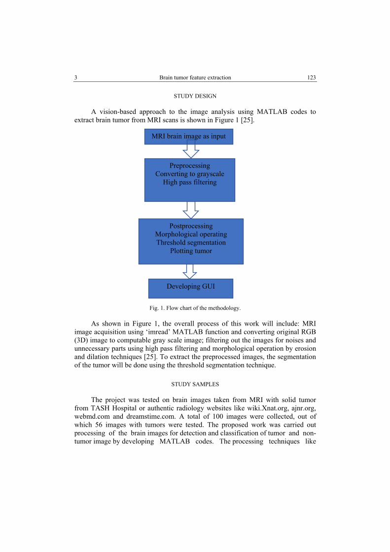
A vision-based approach to the image analysis using MATLAB codes to extract brain tumor from MRI scans is shown in Figure 1 [25].
Fig. 1. Flow chart of the methodology.
As shown in Figure 1, the overall process of this work will include: MRI image acquisition using ‘imread’ MATLAB function and converting original RGB (3D) image to computable gray scale image; filtering out the images for noises and unnecessary parts using high pass filtering and morphological operation by erosion and dilation techniques [25]. To extract the preprocessed images, the segmentation of the tumor will be done using the threshold segmentation technique.
STUDY SAMPLES
The project was tested on brain images taken from MRI with solid tumor from TASH Hospital or authentic radiology websites like wiki.Xnat.org, ajnr.org, webmd.com and dreamstime.com. A total of 100 images were collected, out of which 56 images with tumors were tested. The proposed work was carried out processing of the brain images for detection and classification of tumor and non-tumor image by developing MATLAB codes. The processing techniques like
histogram equalization, image enhancement, image segmentation and feature extraction are aimed to detect the portion of the image that represents the brain tumor.
DIGITAL IMAGE PROCESSING
Nowadays, the image analysis is gaining importance with an increase in the demand for automated, reliable, fast and efficient diagnosis, which can give insight quality image better than human eyes observation [18]. The process includes processes of image analysis and also, includes processes that extract qualities images, up to and including recognition of objects. There are different aspects of image processing:
● Image enhancement: Processing an image so that the result is more suitable for a particular application. It is used for sharpening or deblurring an out-of-focus image, highlighting edges, improving image contrast, or brightening an image, removing noise [11, 14].
● Image restoration: It may be considered as reversing the damage done to an image by a known cause. It was used for removing of blur caused by linear motion, or it may be removal of optical distortions [11].
● Image segmentation: It involves subdividing an image into constituent parts, or isolating certain aspects of an image, finding lines, circles, or particular shapes in an image such as in an aerial photograph, identifying cars, trees, buildings, or roads [11, 14].
IMAGE ENHANCEMENT
Image enhancement fundamentally improves image information for human viewers and generates a better image as an input for further automatic image processing methods. The basic objective of image enhancement is to manipulate the characteristics and features of an image to improve it for the desired application.
The aim of image enhancement is to improve the interpretability or perception of information in images for human viewers, or to provide better input for automated image processing techniques. Image enhancement techniques can be divided into two broad categories:
Spatial-domain techniques operate directly on the pixels of an image. It is denoted by the expression:
( , ) ( , )g x y T f x y= (1)
where f (x, y) is the input image, g(x, y) is the output (processed) image, and T is an operator on defined over a specified neighborhood about the point (x,y). In
5 Brain tumor feature extraction 125
addition, T can operate on a set of images, such as performing the addition of K images for noise reduction.
Frequency-domain methods are more widely applied than the spatial-domain methods. Here embedding is done in the frequency component of the image pixel in frequency-domain the human visual system is more sensitive to low frequency components than the high frequency component [6, 9]. Frequency-domain processing procedures are based on reforming the Fourier transform of an image.
HISTOGRAM PROCESSING
A histogram is an accurate graphical representation of the distribution of numerical data. It is an estimate of the probability distribution of a continuous variable (quantitative variable) and was first introduced by Karl Pearson. The histogram of a digital image with intensity levels in the range [0, L-1] is a discrete function.
( )k kh r n= (2)
where rk is the kth intensity value, nk is the number of pixels in the image intensity rk. Histograms are frequently normalized by the total number of pixels in the image, assuming an M X N image, a normalized histogram.
( ) kk
np rMN
= (3)
where p(rk) is related to probability of occurrence of rk in the image.
NOISE DETECTION AND FILTERING TECHNIQUE
Noise is defined as random variations in the intensity of an image. Noise is generated in sensors or through transmission channel during the acquisition process. Noise should be removed from the image in order to recover the meaningful information from the image. It is important to remove noise while preserving edges.
HIGH PASS FILTER
A high pass filter is method for sharpening methods. An image is sharpened when contrast is enhanced between adjoining areas with variation in brightness or darkness. The kernel array of the high pass filter is designed to increase the brightness of the central pixels relative to neighboring pixels. The kernel array contains a positive value surrounded by negative values [26].
T. Jemal, N. Fikadu, B.S. Goshu 6 126
MORPHOLOGICAL OPERATIONS
The term morphology means deforming or reconstructing the structure or shape of an object [28]. Morphological operations are applied on binary images to get representation of the shape of an object. They are used in pre- or post-processing, such as filtering, thinning or pruning or for getting a representation or description of the shape of objects or regions. The major morphological operations are erosion, dilation, opening and closing.
Erosion is a morphology operation applied on the binary images, which erode away the boundaries of regions of foreground pixels [28]. Areas of foreground pixels shrink in size and holes within. The erosion operator takes two inputs: the first is the image to be eroded, whereas the second is the structuring element. To compute the erosion of a binary image by this structuring element, we superimpose the structuring element on top of the input image so that the origin of the structuring element coincides with the input pixel.
Dilation is the morphological operation that adds pixels to the boundaries of objects in image to fill the holes. The dilation operator takes two pieces of data as inputs [28]. The first is the image to be dilated, the second is the structuring element. For each background pixel, the structuring element is superimposed on the input image so that the origin of the structuring element coincides with the input pixel position. The basic effect of dilation on a binary image is to enlarge the area of foreground pixels [27].
A median filter is widely used in digital image processing because, under certain conditions, it preserves edge while removing noise. The task of filtering is performed by the median filter by use of window that is a pattern of neighbors. The window pattern slides, entry by entry, over the entire signal. The middle value of the window is decided by the median value of all the entries at a time [4].
IMAGE SEGMENTATION
Image segmentation is the process of partitioning a digital image into multiple segments (sets of pixels, also known as super pixels). This involves subdividing an image into constituent parts, or isolating certain aspects of an image, finding lines, circles, or particular shapes in an image [8, 23].
Thresholding is a simple technique for edge detection and image segmentation. In thresholding approach, image segmentation is based on gray level intensity value of pixels. However, thresholding is often used as an initial step in a sequence of image segmentation process. Its main limitation is that only two classes are generated, and it does not work when confronted with structures that lack clear borders [8, 23].
7 Brain tumor feature extraction 127
In this technique, first a threshold value (T) is chosen in the gray-level range of the image and pixels over and under the threshold value are marked differently [24].
( , ) ( , ) 1Tl x y T I x y> → = (5)
( , ) ( , ) 0Tl x y T I x y≤ → = (6)
where IT(x,y) is the output image after thresholding. Hence, all pixels with intensities larger than T are set to a certain number, i.e., 1, and the rest are set to zero [24]. Consequently, the result of thresholding is a binary image [18].
RESULTS AND DISCUSSION
To confirm the performance of our algorithm for the extraction of brain tumor images, we developed a code written in the high-level programming language MATLAB (The MathWorks, Natick, MA, USA). Since a tumor in an MRI image usually has a higher intensity than its background, it is easy to locate it and extract it from the MRI image. The results we obtained in this process will be presented in the following sections.
GRAY SCALE IMAGE
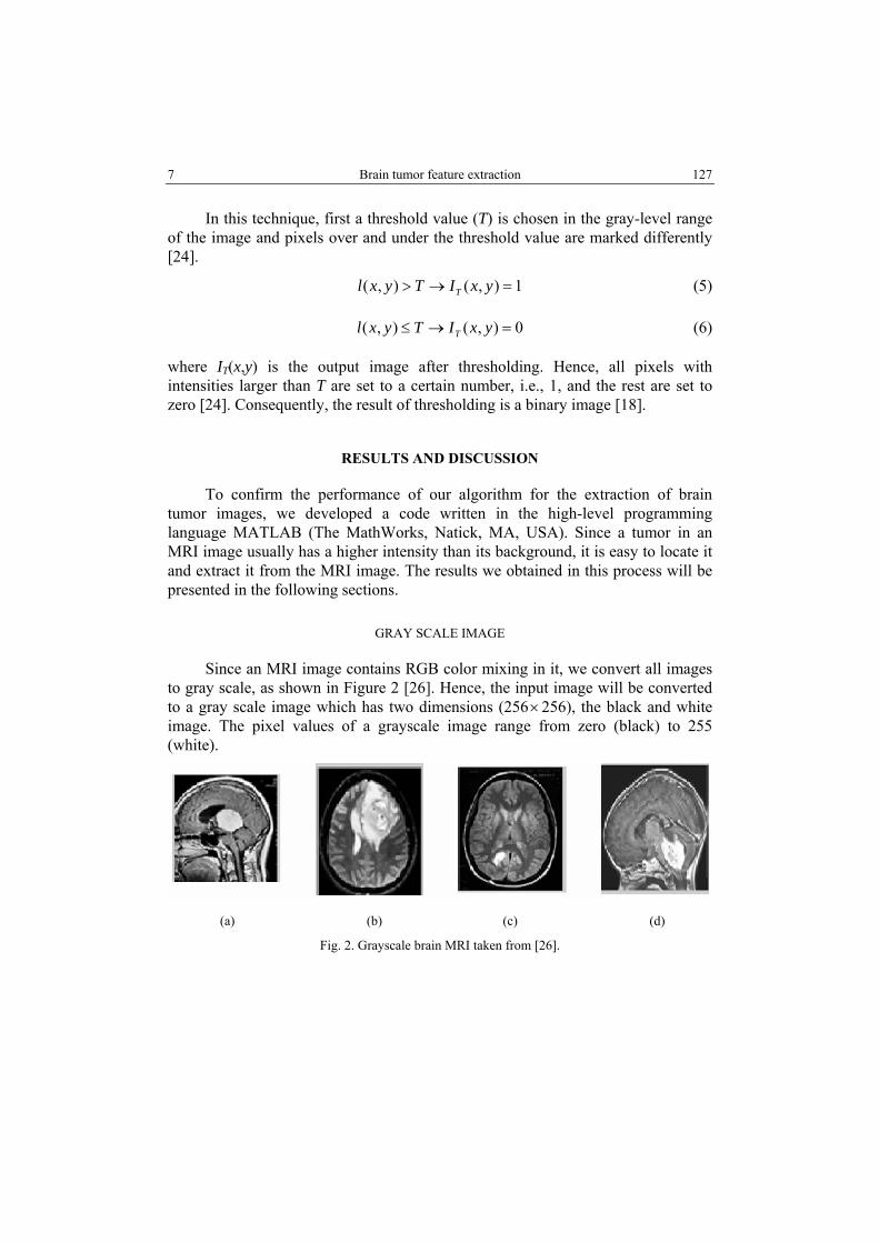
Since an MRI image contains RGB color mixing in it, we convert all images to gray scale, as shown in Figure 2 [26]. Hence, the input image will be converted to a gray scale image which has two dimensions (256×256), the black and white image. The pixel values of a grayscale image range from zero (black) to 255 (white).
(a) (b) (c) (d)
Fig. 2. Grayscale brain MRI taken from [26].
T. Jemal, N. Fikadu, B.S. Goshu 8 128
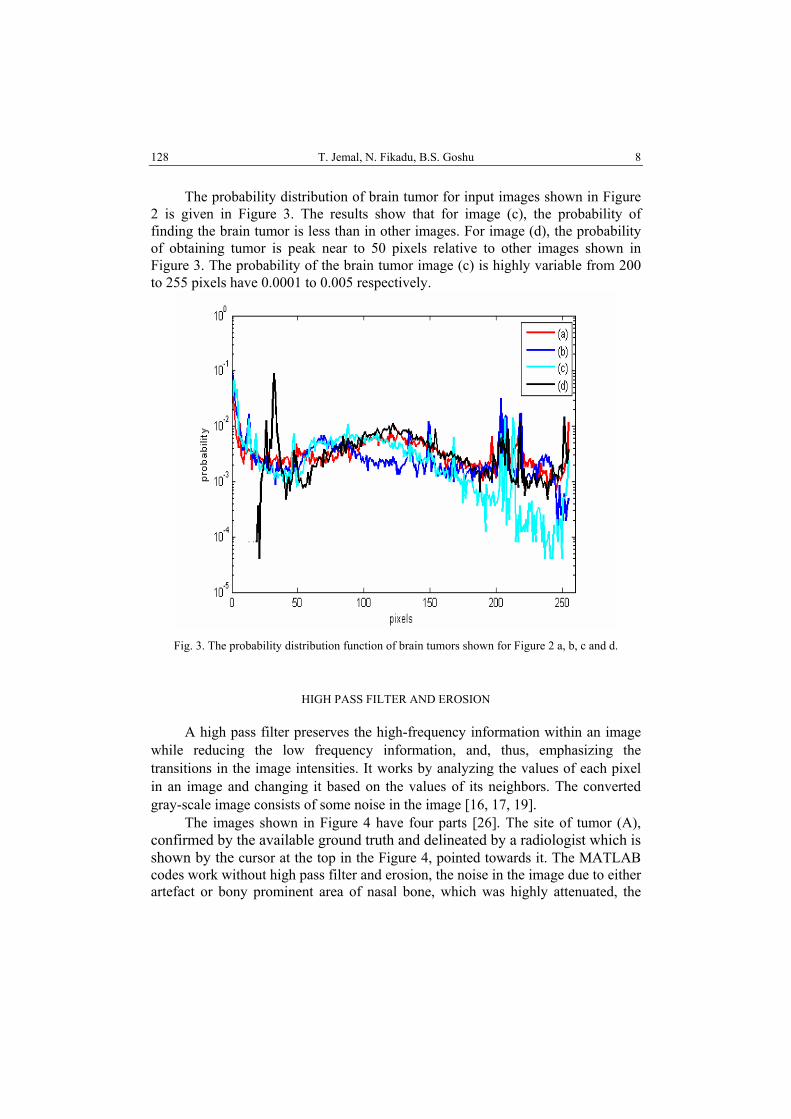
The probability distribution of brain tumor for input images shown in Figure 2 is given in Figure 3. The results show that for image (c), the probability of finding the brain tumor is less than in other images. For image (d), the probability of obtaining tumor is peak near to 50 pixels relative to other images shown in Figure 3. The probability of the brain tumor image (c) is highly variable from 200 to 255 pixels have 0.0001 to 0.005 respectively.
Fig. 3. The probability distribution function of brain tumors shown for Figure 2 a, b, c and d.
HIGH PASS FILTER AND EROSION
A high pass filter preserves the high-frequency information within an image while reducing the low frequency information, and, thus, emphasizing the transitions in the image intensities. It works by analyzing the values of each pixel in an image and changing it based on the values of its neighbors. The converted gray-scale image consists of some noise in the image [16, 17, 19].
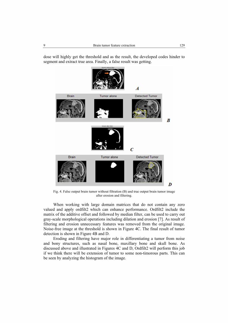
The images shown in Figure 4 have four parts [26]. The site of tumor (A), confirmed by the available ground truth and delineated by a radiologist which is shown by the cursor at the top in the Figure 4, pointed towards it. The MATLAB codes work without high pass filter and erosion, the noise in the image due to either artefact or bony prominent area of nasal bone, which was highly attenuated, the
9 Brain tumor feature extraction 129
dose will highly get the threshold and as the result, the developed codes hinder to segment and extract true area. Finally, a false result was getting.
Fig. 4. False output brain tumor without filtration (B) and true output brain tumor image
after erosion and filtering.
When working with large domain matrices that do not contain any zero valued and apply ordfilt2 which can enhance performance. Ordfilt2 include the matrix of the additive offset and followed by median filter, can be used to carry out gray-scale morphological operations including dilation and erosion [7]. As result of filtering and erosion unnecessary features was removed from the original image. Noise-free image at the threshold is shown in Figure 4C. The final result of tumor detection is shown in Figure 4B and D.
Eroding and filtering have major role in differentiating a tumor from noise and bony structures, such as nasal bone, maxillary bone and skull bone. As discussed above and illustrated in Figures 4C and D, Ordfilt2 will perform this job if we think there will be extension of tumor to some non-timorous parts. This can be seen by analyzing the histogram of the image.
T. Jemal, N. Fikadu, B.S. Goshu 10 130
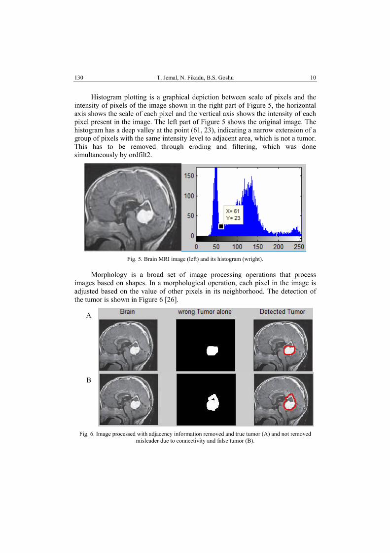
Histogram plotting is a graphical depiction between scale of pixels and the intensity of pixels of the image shown in the right part of Figure 5, the horizontal axis shows the scale of each pixel and the vertical axis shows the intensity of each pixel present in the image. The left part of Figure 5 shows the original image. The histogram has a deep valley at the point (61, 23), indicating a narrow extension of a group of pixels with the same intensity level to adjacent area, which is not a tumor. This has to be removed through eroding and filtering, which was done simultaneously by ordfilt2.
Fig. 5. Brain MRI image (left) and its histogram (wright).
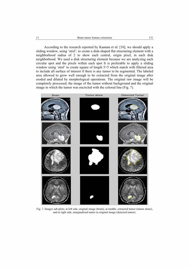
Morphology is a broad set of image processing operations that process images based on shapes. In a morphological operation, each pixel in the image is adjusted based on the value of other pixels in its neighborhood. The detection of the tumor is shown in Figure 6 [26].
Fig. 6. Image processed with adjacency information removed and true tumor (A) and not removed
misleader due to connectivity and false tumor (B).
A
B
11 Brain tumor feature extraction 131
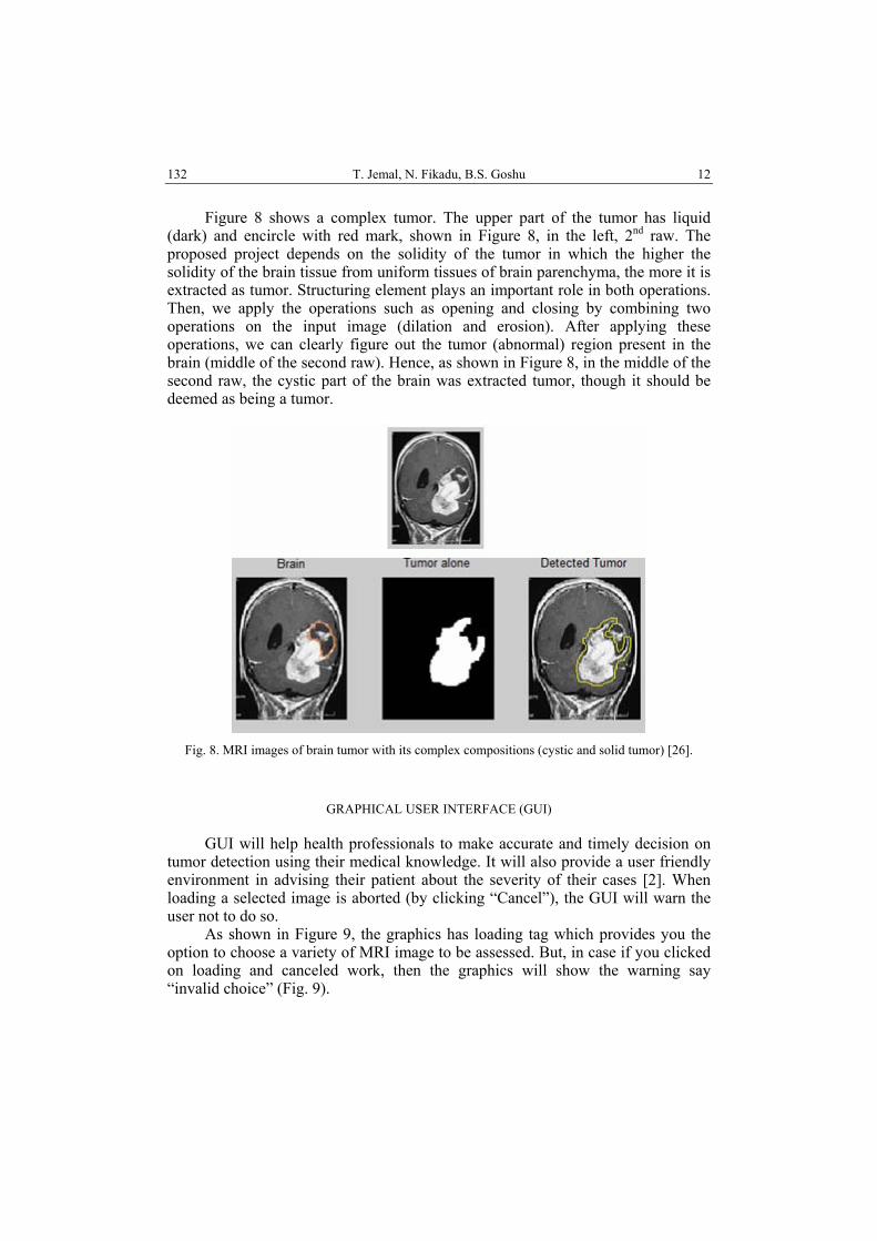
According to the research reported by Kannan et al. [10], we should apply a sliding window, using ‘strel’, to create a disk-shaped flat structuring element with a neighborhood radius of 2 to show each central, origin pixel, in each disk neighborhood. We used a disk structuring element because we are analyzing each circular spot and the pixels within each spot It is preferable to apply a sliding window using ‘strel’ to create square of length 5×5 which match with filtered area to include all surface of interest if there is any tumor to be segmented. The labeled area allowed to grow well enough to be extracted from the original image after eroded and dilated by morphological operations. The original raw image will be completely processed, the image of the tumor without background and the original image in which the tumor was encircled with the colored line (Fig. 7).
Fig. 7. Images sub-plots; at left side, original image (brain), at middle, extracted tumor (tumor alone),
and at right side, marginalized tumor in original image (detected tumor).
T. Jemal, N. Fikadu, B.S. Goshu 12 132
Figure 8 shows a complex tumor. The upper part of the tumor has liquid (dark) and encircle with red mark, shown in Figure 8, in the left, 2nd raw. The proposed project depends on the solidity of the tumor in which the higher the solidity of the brain tissue from uniform tissues of brain parenchyma, the more it is extracted as tumor. Structuring element plays an important role in both operations. Then, we apply the operations such as opening and closing by combining two operations on the input image (dilation and erosion). After applying these operations, we can clearly figure out the tumor (abnormal) region present in the brain (middle of the second raw). Hence, as shown in Figure 8, in the middle of the second raw, the cystic part of the brain was extracted tumor, though it should be deemed as being a tumor.
Fig. 8. MRI images of brain tumor with its complex compositions (cystic and solid tumor) [26].
GRAPHICAL USER INTERFACE (GUI)
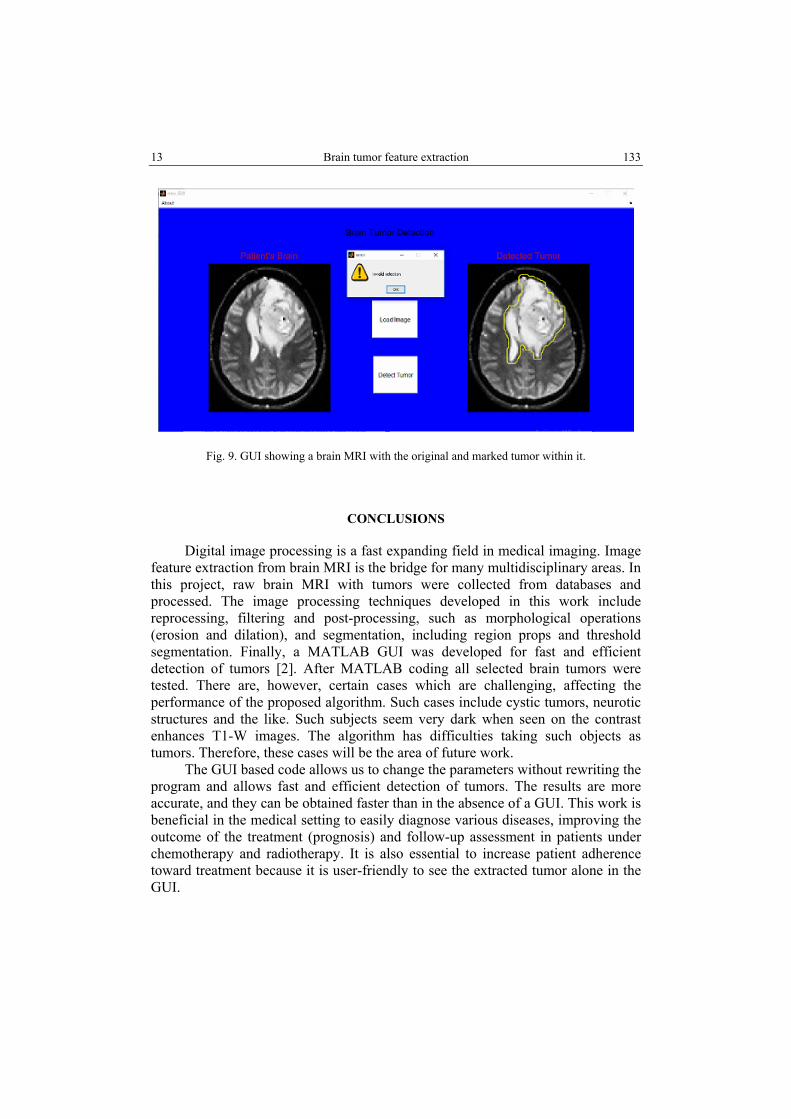
GUI will help health professionals to make accurate and timely decision on tumor detection using their medical knowledge. It will also provide a user friendly environment in advising their patient about the severity of their cases [2]. When loading a selected image is aborted (by clicking “Cancel”), the GUI will warn the user not to do so.
As shown in Figure 9, the graphics has loading tag which provides you the option to choose a variety of MRI image to be assessed. But, in case if you clicked on loading and canceled work, then the graphics will show the warning say “invalid choice” (Fig. 9).
13 Brain tumor feature extraction 133
Fig. 9. GUI showing a brain MRI with the original and marked tumor within it.
CONCLUSIONS
Digital image processing is a fast expanding field in medical imaging. Image feature extraction from brain MRI is the bridge for many multidisciplinary areas. In this project, raw brain MRI with tumors were collected from databases and processed. The image processing techniques developed in this work include reprocessing, filtering and post-processing, such as morphological operations (erosion and dilation), and segmentation, including region props and threshold segmentation. Finally, a MATLAB GUI was developed for fast and efficient detection of tumors [2]. After MATLAB coding all selected brain tumors were tested. There are, however, certain cases which are challenging, affecting the performance of the proposed algorithm. Such cases include cystic tumors, neurotic structures and the like. Such subjects seem very dark when seen on the contrast enhances T1-W images. The algorithm has difficulties taking such objects as tumors. Therefore, these cases will be the area of future work.
The GUI based code allows us to change the parameters without rewriting the program and allows fast and efficient detection of tumors. The results are more accurate, and they can be obtained faster than in the absence of a GUI. This work is beneficial in the medical setting to easily diagnose various diseases, improving the outcome of the treatment (prognosis) and follow-up assessment in patients under chemotherapy and radiotherapy. It is also essential to increase patient adherence toward treatment because it is user-friendly to see the extracted tumor alone in the GUI.
2. ABOSHGIFA, A., E. HASSAN, Detecting brain tumour from MRI image using MATLAB GUI programme, International Journal of Computer Science & Engineering Survey (IJCSES), 2015, 6(6), 47–60.
3. ASLAM, M.A., D, CUI, Brain tumor detection from medical images: A survey, Nano Biomedical Eng., 2017, 9(1), 72–81
4. BOUNTRIS, P., E. FARANTATOS, N. APOSTOLOU, Advanced image analysis tool development for the early stage bronchial cancer detection, PWASET, 2005, 9, 151–156.
5. BUCKNER, J.C., Central nervous system tumors, Mayo Clinic Proceedings, 2007, 82(10), 1271–1286.
6. CANCER.NET, EDITORIAL BOARD, Brain tumor: statistics, National Cancer Institute, Jan., 2019.
7. HARALICK, R.M., Computer & Robot Vision, Addison-Wesley, New York, 1992. 8. HENDEE, W.R., E.R. RITENOUR, Medical Imaging Physics, 4th ed., Wiley~Liss. inc., New
York, 2002. 9. HUSSIEN, R.A., S. EKO, Digital processing for computed tomography images: brain tumor
extraction and histogram analysis, Mathematics and Computers in Contemporary Science, 2013, 4(1) 119–128.
10. KANNAN, M., H. NGUYEN, A.U. AVILA, M. JIN, Brain tumor MRI detection using MATLAB, Instructables Circuits, https://www.instructables.com/Brain-Tumor-MRI-Detection-Using-Matlab/.
11. KUMAR, D., S. VANDHANA, Brain tumour image segmentation using MATLAB, International Journal for Innovative Research in Science & Technology, 2015, 12(1), 447–451.
12. LOUIS, D.N., H. OHGAKI, O.D. WIESTLER, W.K CAVENEE, WHO classification of tumors of the central nervous system, International Agency for Research on Cancer (IARC), Lyon, France, 2007, 114(2), 97–10.
13. MUTHAMIL, P.S, B. ASHADEVI, Analysis of image segmentation techniques with morphological operation in detecting brain tumor, IJRAR, 2019, 6(2), 181–189.
14. MUSTAQEEM, A., J. ALI, T. FATIMA, An efficient brain tumor detection algorithm using watershed and thresholding based segmentation, International Journal Graphics and Signal Processing, 2012, 10, 34–39.
15. NIDHI, P. KUMARI, Brain tumor and edema detection using MATLAB, International Journal of Computer Engineering and Technology (IJCET), 2014, 5(3), 122–131.
16. NOBACK, C.R., N.L. STROMINGER, R.J. DEMAREST, D. RUGGIERO, The Human Nervous System: Structure and Function, 6th ed., Humana Press, Totowa, New Jersey, 2005.
17. PITAS, I., Digital Image Processing Algorithms And Applications, John Wiley & Sons, Inc., 2000.
18. POP, S., C. A.C. DUFOUR, J.-F. LE GARREC, C.V. RAGNI, C. CIMPER, S.M. MEILHAC, J.-C. OLIVO-MARIN, Extracting 3D cell parameters from dense tissue environments: application to the development of the mouse heart, Bioinformatics, 2013, 29, 772–779.
19. RAFAEL, C., R.F. GONZALEZ, Digital Image Processing Using MATLAB, 3rd ed., Pearson Prentice-Hall, London, 2004.
20. RANA, R., P. SINGH, Brain tumor detection through MR images: a review of literature, IOSR Journal of Computer Engineering (IOSR-JCE), 2015, 17(5), 7–18.
21. REDDY, B.V., P.S. KUMAR, P.B. REDDY, B.N. REDDY, Identifying brain tumor from MRI image using modified FCM and support vector machine, International Journal of Computer Engineering & Technology (IJCET), 2013, 4(1), 244–262.
22. RODRIGUEZ, A.O., Principles of magnetic resonance imaging, Revista Mexicana de Fisica, 2004, 50(3), 272–286.
15 Brain tumor feature extraction 135
23. SANJAY, D.M., B.B., GITE, Brain tumor segmentation using fuzzy C-Means and K-Means clustering and its area calculation and disease prediction using Naive-Bayes algorithm, International Journal of Innovative Research in Science, Engineering and Technology, 2017, 6(11), 21342.
24. SEZGIN, M., B. SANKUR, Survey over image thresholding techniques and quantitative performance evaluation, Journal of Electronic Imaging, 2004, 13(1), 146–165.
25. SUHAG, S., L.M. SAINI, Automatic detection of brain tumor by image processing in MATLAB, International Journal of Advances in Science Engineering and Technology, 2015, 3(3), 114–117.
26. TIGENEH, W., A. MOLLA, A. ABREHA, M. ASSEFA, Pattern of cancer in Tikur Anbessa specialized hospital oncology center in Ethiopia from 1998 to 2010, International Journal of Cancer Research and Molecular Mechanisms, 2015, 1.1: doi http://dx.doi.org/10.16966/2381- 3318.103.
27. VARUNA, N.S, T.N.R. KUMAR, Identification and classification of brain tumor MRI images with feature extraction using DWT and probabilistic neural network, Brain Informatics, 2018, 5, 23–30.
28. XIA, Y., K. BETTINGER, L. SHEN, A. REISS, Automatic segmentation of the caudate nucleus from human brain MR images, IEEE Transactions on Medical Imaging, 2007, 26(4), 509–517.
29. ZHANG, Z., N.V. KUZMIN, M.L. GROOT, J.C. DE MUNCK, Extracting morphologies from third harmonic generation images of structurally normal human brain tissue, Bioinformatics, 2017, 33(11), 1712–1720.