79
Introduction to NEOPLASIA (Part 1) Rex Bentley, M.D. Department of Pathology DUMC

Introduction to NEOPLASIA
(Part 1)
Rex Bentley, M.D.Department of Pathology
DUMC

NEOPLASIAToday’s Goals and Objectives
1. Define neoplasm
2. Define benign and malignant
3. Differentiate benign from malignant neoplasmsbased on histologic appearance
4. Explain how neoplasms are named and infer properties of a neoplasm from its name
5. Explain what grade is, and how it impacts prognosis

1. What is a Neoplasm?• NEOPLASM = “New growth”
• Synonym: TUMOR = “swelling”– Originally used for inflammation, but
now used as synonym for neoplasm
• Oncology = the study of tumors (Greek “oncos” = tumor)
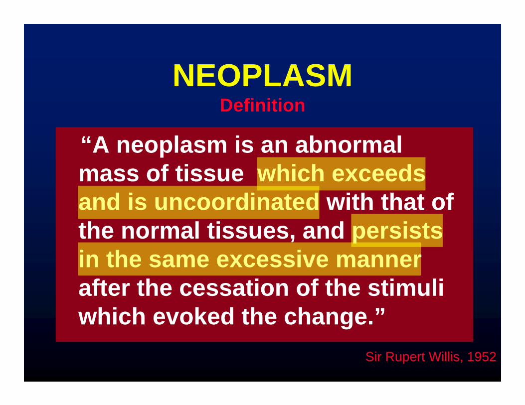
NEOPLASMDefinition
“A neoplasm is an abnormal mass of tissue which exceeds and is uncoordinated with that of the normal tissues, and persists in the same excessive manner after the cessation of the stimuli which evoked the change.”
Sir Rupert Willis, 1952

Two Fundamental Features of Neoplasms
1. Unregulated growth2. Clonal genetic defects
Subject of later lectureNeoplasia III (Dr. Yan)

Mount Sacagawea, Montana

2. What Do “Benign” and “Malignant” Mean?
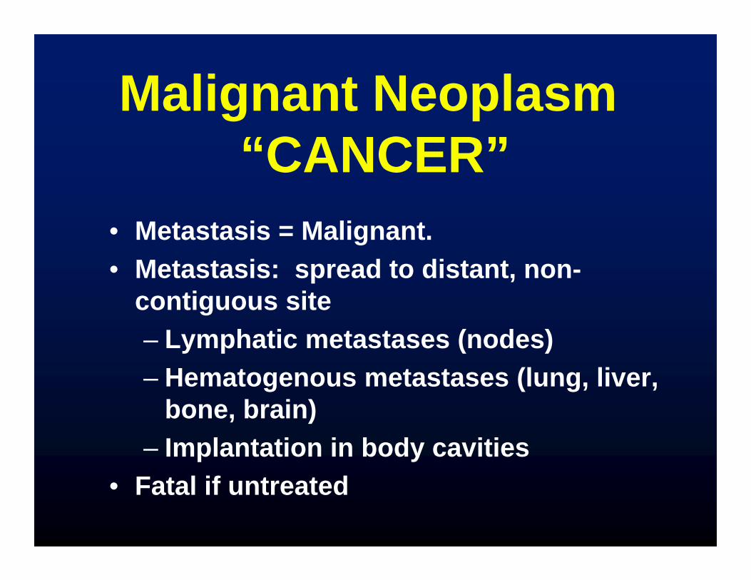
Malignant Neoplasm“CANCER”
• Metastasis = Malignant.• Metastasis: spread to distant, non-
contiguous site– Lymphatic metastases (nodes)– Hematogenous metastases (lung, liver,
bone, brain)– Implantation in body cavities
• Fatal if untreated

Lymph Node Metastasis
Cancer
Normal
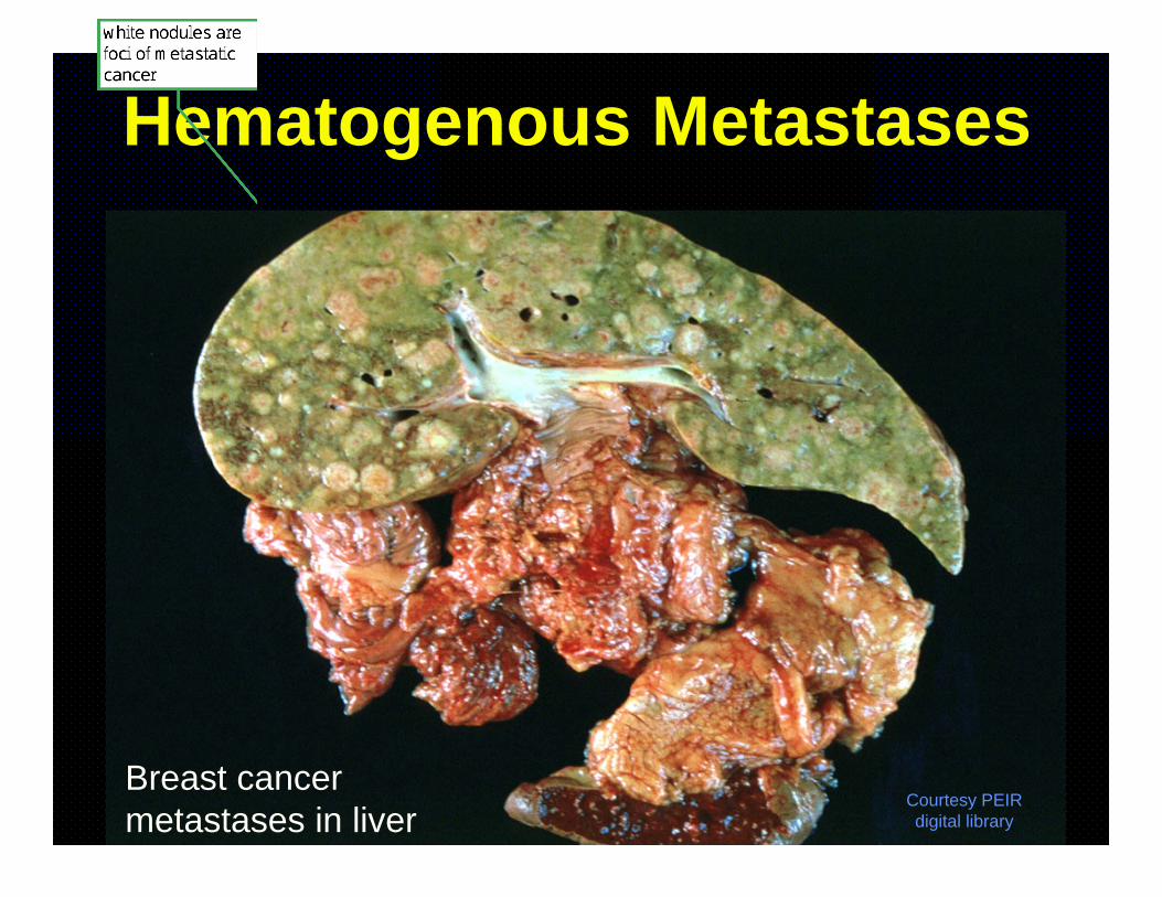
Hematogenous Metastases
Breast cancer metastases in liver
Courtesy PEIR digital library

Hematogenous Metastases
Breast cancer metastases in vertebra Courtesy PEIR digital library
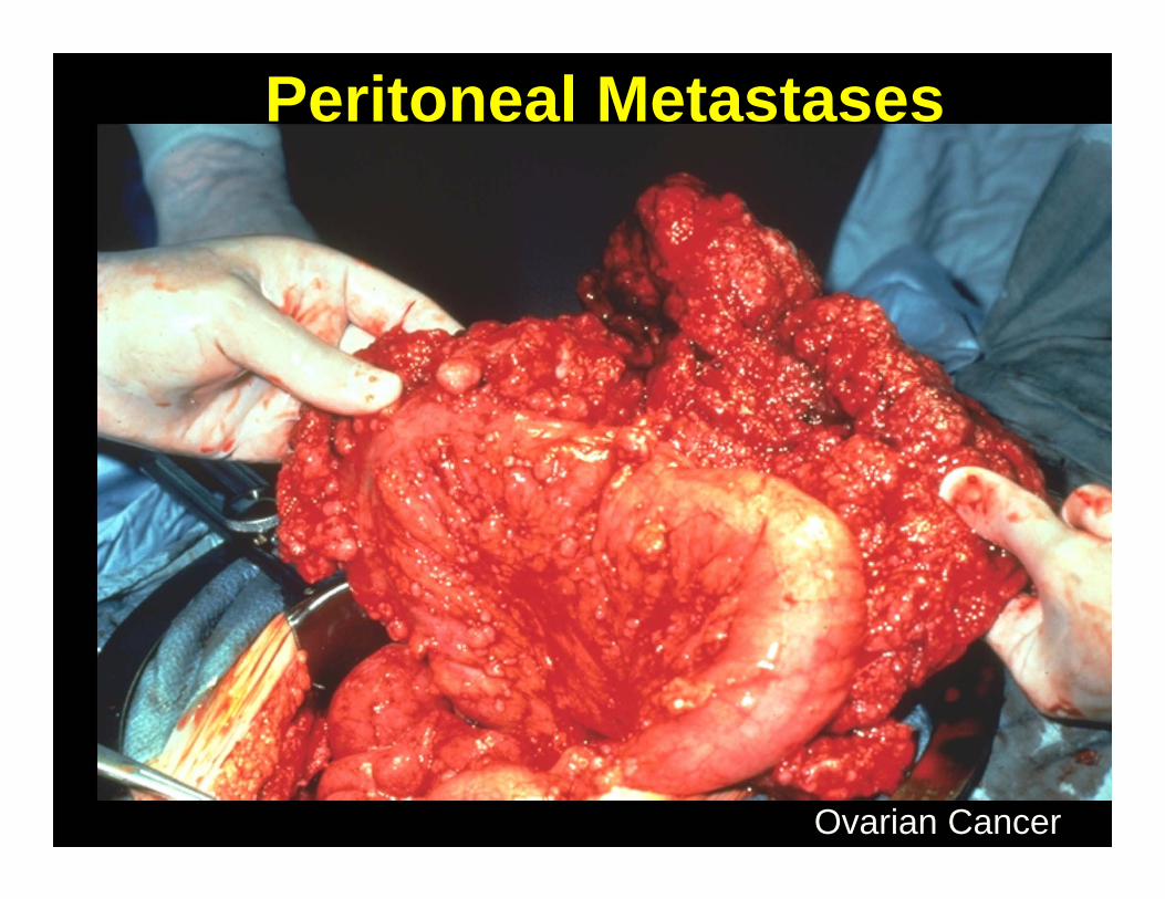
Peritoneal Metastases
Ovarian Cancer

Benign Neoplasms• Do not metastasize• In general, do not result in
death of the patient–Location, location, location!–Secretory products can be
lethal (e.g. endocrine tumors)
From a practical standpoint, benign neoplasms often can be cured by simple surgical
excision while malignant neoplasms often cannot be
cured by surgery alone

Benign vs. Malignant
Benign Malignant
Distant Metastases?
No Yes
Life-threatening
No (usually)
Yes
Malignant neoplasms have the potential for metastasis
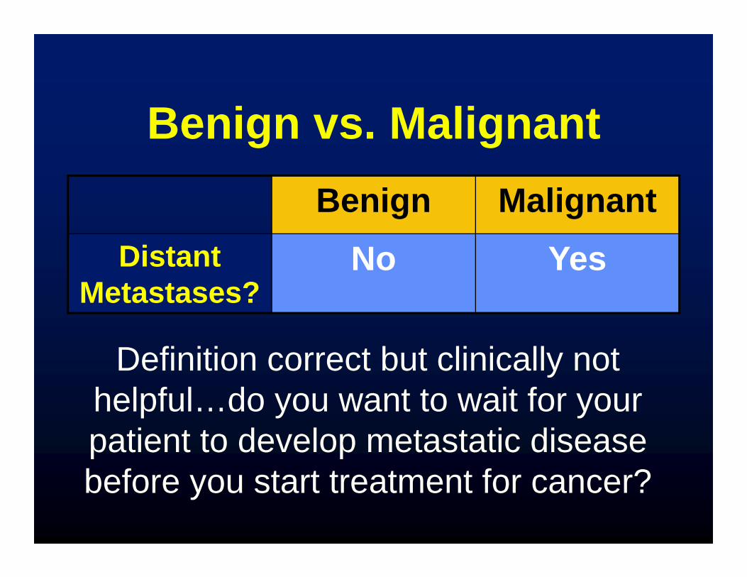
Benign vs. MalignantBenign Malignant
Distant Metastases?
No Yes
Definition correct but clinically not helpful…do you want to wait for your patient to develop metastatic disease before you start treatment for cancer?

Cham Museum, Danang, Viet Nam
3. How can we tell if a neoplasm is malignant
BEFORE it metastasizes?
Histopathology!

Histologic Features Distinguishing Benign vs. Malignant
a) Bordersb) Growth
ratec) Anaplasia
Is this cancer or not?
Courtesy PEIR digital library
Benign Neoplasms• Encapsulated (pushing borders)
–Do not invade locally• Slow growth• Mild anaplasia (well differentiated)
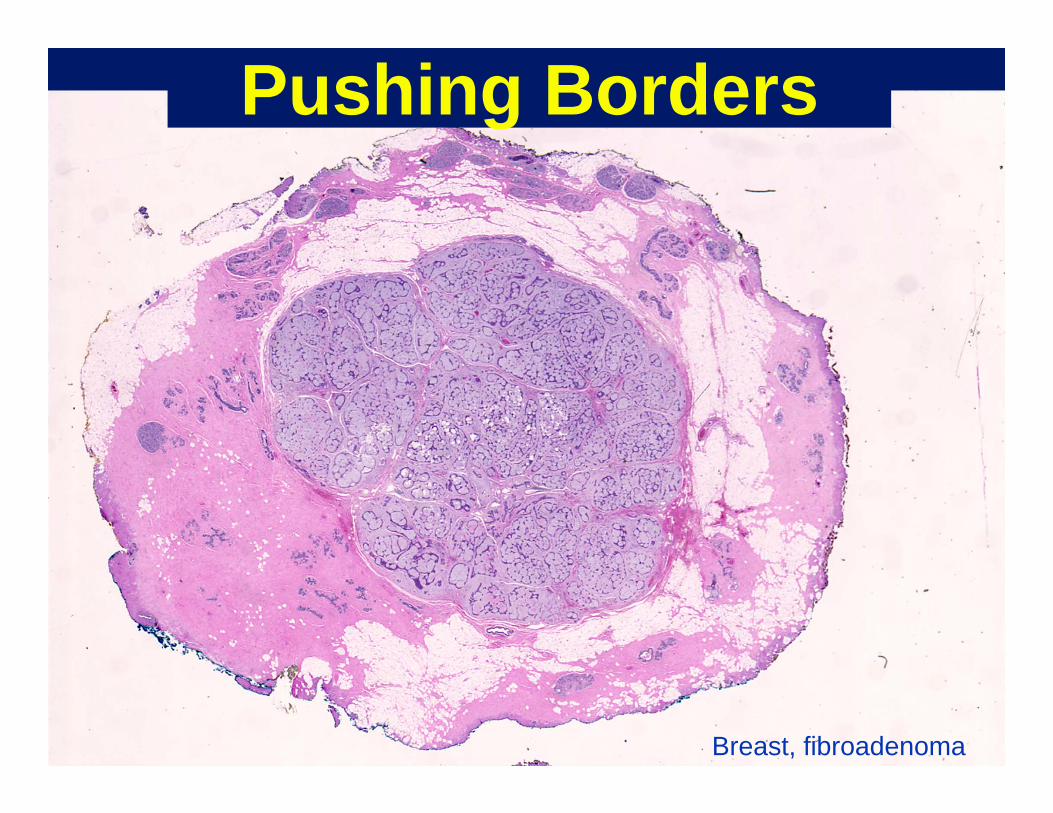
Breast, fibroadenoma
Pushing Borders

Pushing Borders
Breast, fibroadenoma
Malignant Neoplasms
• Local Invasion–Infiltrative borders–“Stellate” or “spiculated”

Local Invasion
Lung Cancer

Local Invasion
Breast cancer
Malignant Neoplasms• Local Invasion• Rapid growth rate
– Histology: Mitotic figures numerous
– Not unique to malignancies, many normal tissues grow rapidly (GI mucosa, endometrium, bone marrow)
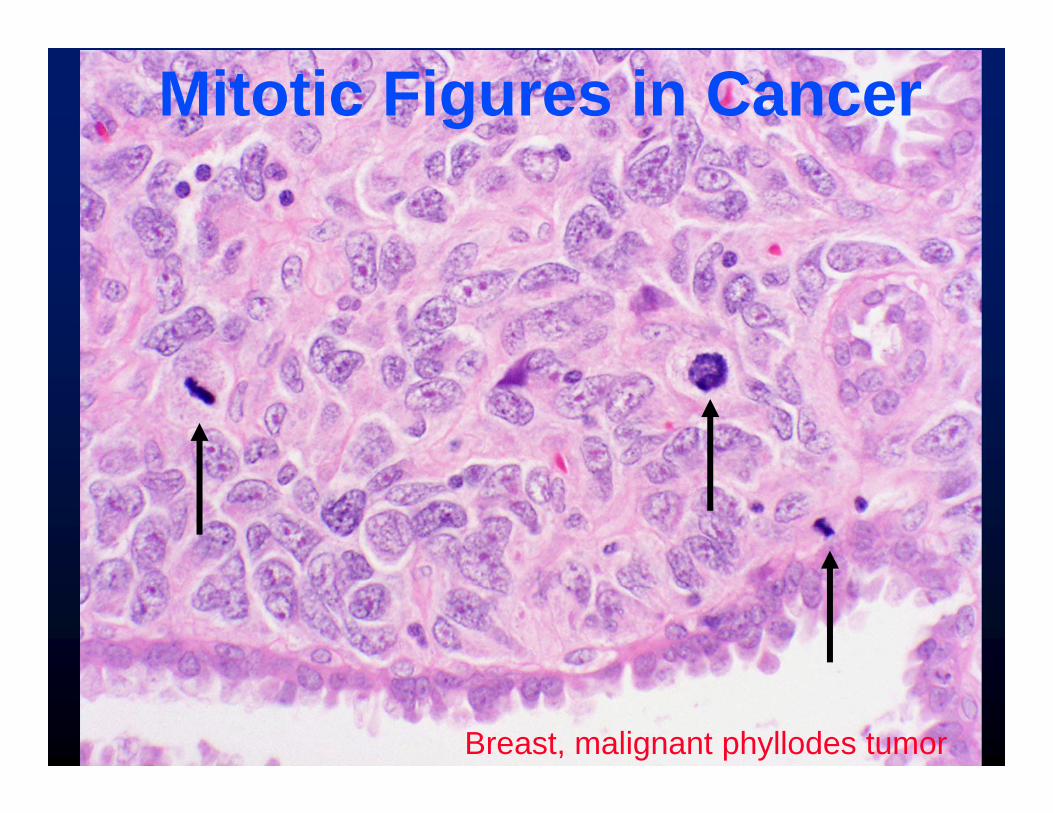
Mitotic Figures in Cancer
Breast, malignant phyllodes tumor

Malignant Neoplasms
• Local Invasion• Rapid growth rate• Anaplasia
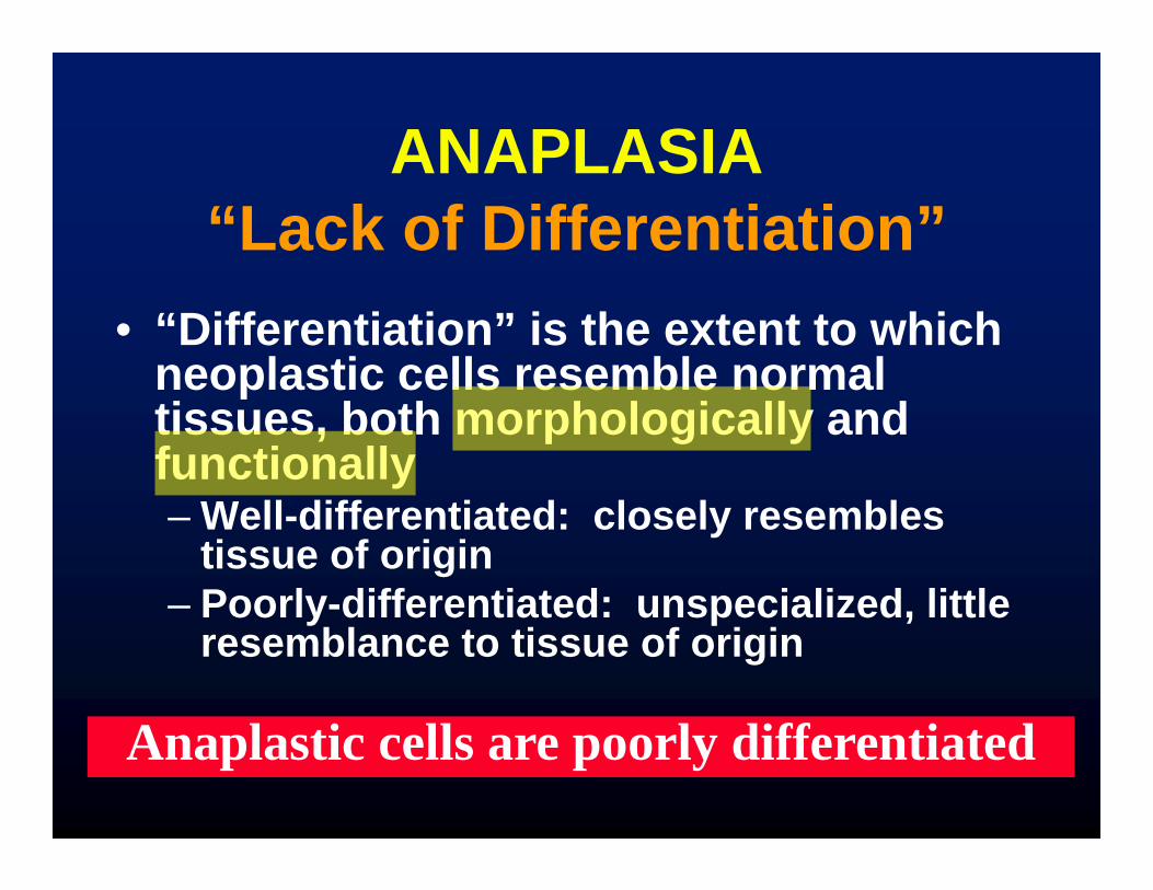
ANAPLASIA“Lack of Differentiation”
• “Differentiation” is the extent to which neoplastic cells resemble normal tissues, both morphologically and functionally– Well-differentiated: closely resembles
tissue of origin– Poorly-differentiated: unspecialized, little
resemblance to tissue of origin
Anaplastic cells are poorly differentiated
ANAPLASIA“Lack of Differentiation”
–Anaplastic skeletal muscle cells make little actin and myosin (lose cross striations)
–Anaplastic colonic epithelial cells make little or no mucin
–Anaplastic glandular cells make only few glands

Benign: No Anaplasia
Note microscopic similarity to normal smooth muscle
Uterus, leiomyoma

Normal
Benign: Mild Anaplasia
Neoplastic glands still resemble normal endometrial gland

Cancer: Moderate Anaplasia
Neoplastic squamous cells still make abundant keratin (arrows)
Normal Skin Squamous cell carcinoma
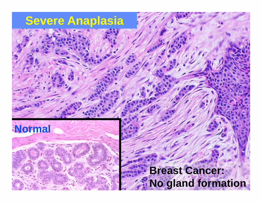
Normal
Breast Cancer: No gland formation
Severe Anaplasia
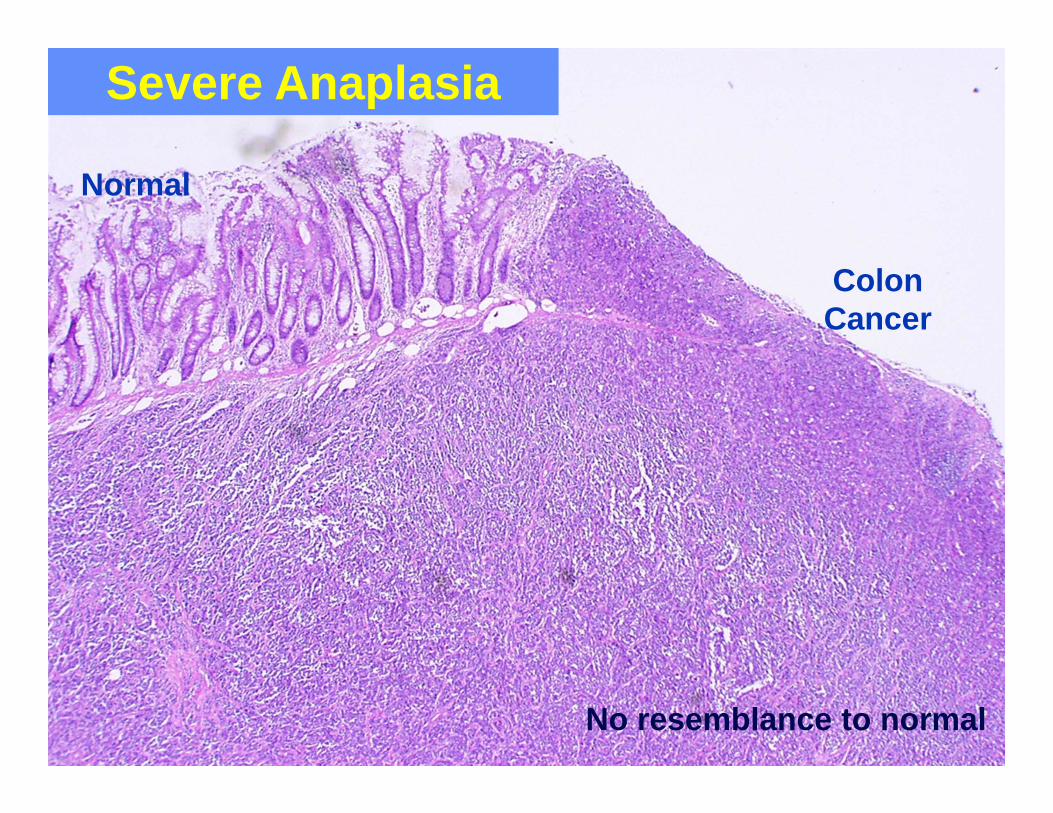
Severe Anaplasia
Normal
Colon Cancer
No resemblance to normal

– High ratio of nucleus to cytoplasm– Nuclear hyperchromasia.– Clumped chromatin.– Prominent nucleoli.
ANAPLASIA: Abnormal Nuclei
“Blue is BAD”

– Pleomorphism• Variation in size and shape • Nuclear and cytoplasmic• Tumor giant cells
– Frequent and sometimes abnormal mitoses
ANAPLASIA:
Other Nuclear Features
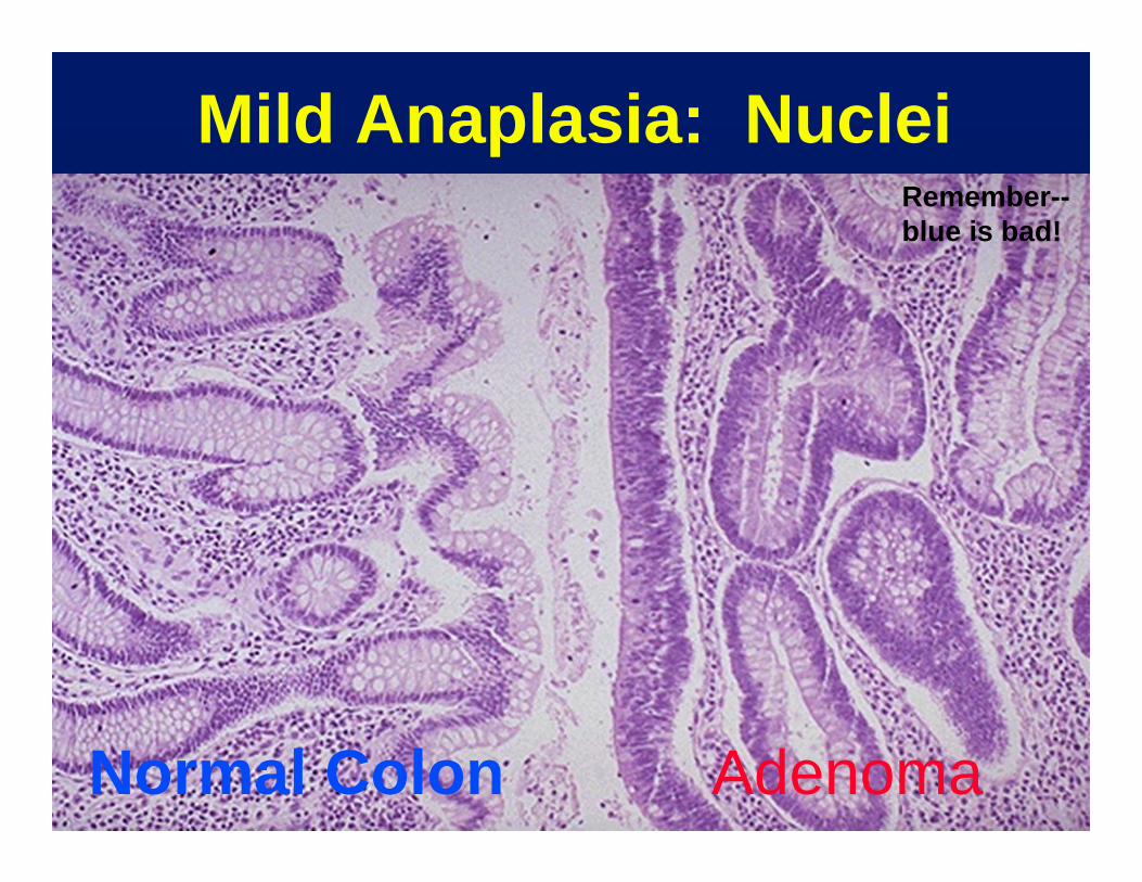
Mild Anaplasia: Nuclei
Normal Colon Adenoma
Remember--blue is bad!
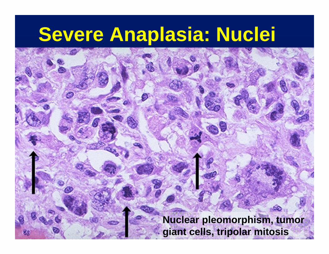
Severe Anaplasia: Nuclei
Nuclear pleomorphism, tumor giant cells, tripolar mitosis

Histologic Diagnosis Of Malignancy
There is no single parameter (other than metastasis) which always allows recognition of a malignant neoplasm microscopically. However, the presence of severe anaplasia and a pattern of invasiveness are the criteria which are most generally useful.
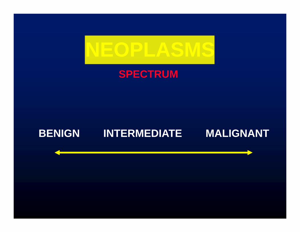
NEOPLASMS
BENIGN INTERMEDIATE MALIGNANT
SPECTRUM

Quick Review: Which of these is malignant?
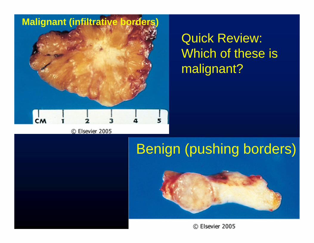
Quick Review: Which of these is malignant?
Benign (pushing borders)
Malignant (infiltrative borders)

Quick Review: Which of these thyroid tumors is malignant?

Quick Review: Which of these thyroid tumors is malignant?
Malignant (severe anaplasia!)
Benign (no anaplasia!)

Duke University, North Carolina
4. How do we name neoplasms?

Nomenclature
Neoplasms are composed of proliferating neoplastic cells but also contain non-neoplastic supportive stroma of connective tissue and blood vessels.
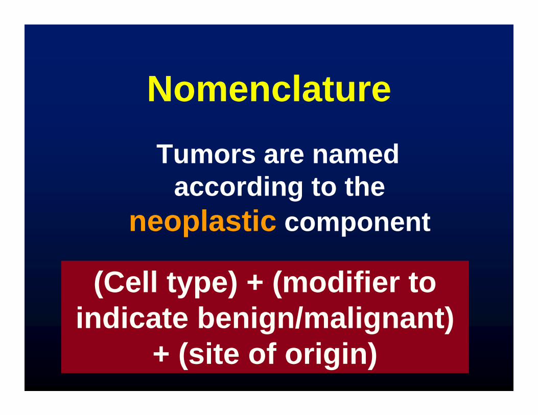
NomenclatureTumors are named
according to the neoplastic component
(Cell type) + (modifier to indicate benign/malignant)
+ (site of origin)

Benign Neoplasms: Nomenclature
• Benign tumors are often designated by the suffix -“oma”.
• Prefix designates the cell of origin
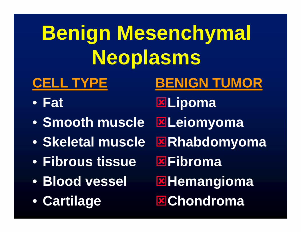
Benign Mesenchymal Neoplasms
CELL TYPE• Fat• Smooth muscle• Skeletal muscle• Fibrous tissue• Blood vessel • Cartilage
BENIGN TUMORLipomaLeiomyomaRhabdomyomaFibromaHemangiomaChondroma

Benign Epithelial Neoplasms
• ADENOMA: benign neoplasm derived from glandular epithelium
• CYSTADENOMA: benign epithelial neoplasm with cystic or fluid-filled cavity
• PAPILLOMA: benign epithelial neoplasm producing finger-like or papillary projections (think sea anemone)

Interior of tumor
Papillary growth inside cyst
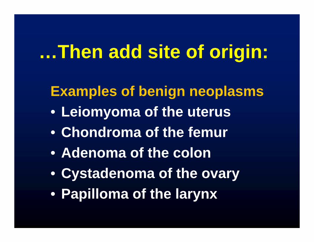
…Then add site of origin:
Examples of benign neoplasms• Leiomyoma of the uterus• Chondroma of the femur• Adenoma of the colon• Cystadenoma of the ovary• Papilloma of the larynx
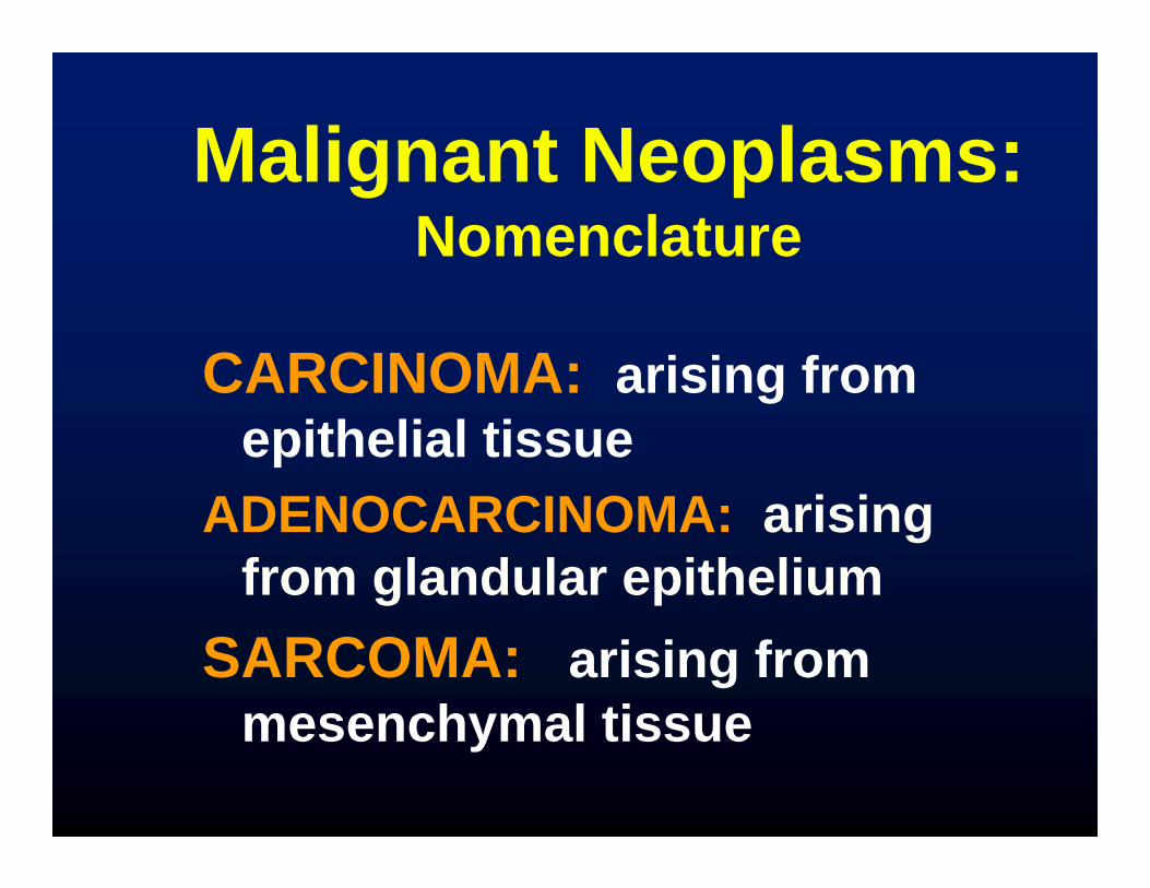
Malignant Neoplasms:Nomenclature
CARCINOMA: arising from epithelial tissue
ADENOCARCINOMA: arising from glandular epithelium
SARCOMA: arising from mesenchymal tissue

Malignant NeoplasmsNomenclature
LYMPHOMA = arising from lymphoid tissue
LEUKEMIA = arising from blood or bone marrow elements
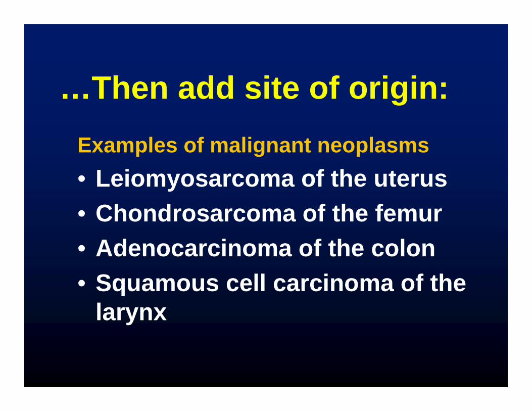
Examples of malignant neoplasms• Leiomyosarcoma of the uterus• Chondrosarcoma of the femur• Adenocarcinoma of the colon• Squamous cell carcinoma of the
larynx
…Then add site of origin:
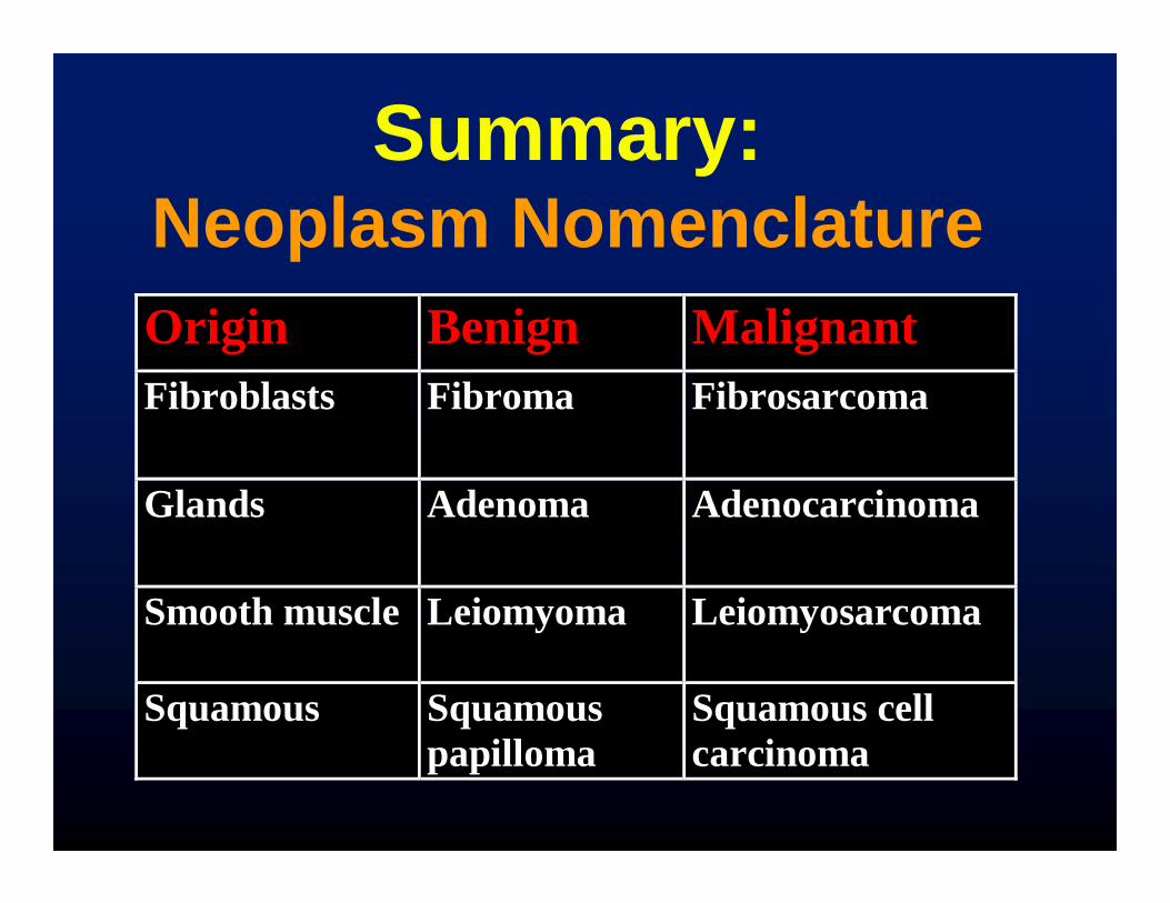
Summary:Neoplasm NomenclatureOrigin Benign Malignant Fibroblasts Fibroma Fibrosarcoma
Glands Adenoma Adenocarcinoma
Smooth muscle Leiomyoma Leiomyosarcoma
Squamous Squamous papilloma
Squamous cell carcinoma
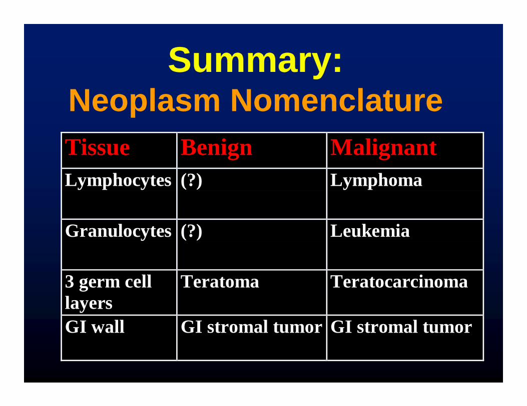
Tissue Benign MalignantLymphocytes (?) Lymphoma
Granulocytes (?) Leukemia
3 germ celllayers
Teratoma Teratocarcinoma
GI wall GI stromal tumor GI stromal tumor
Summary:Neoplasm Nomenclature
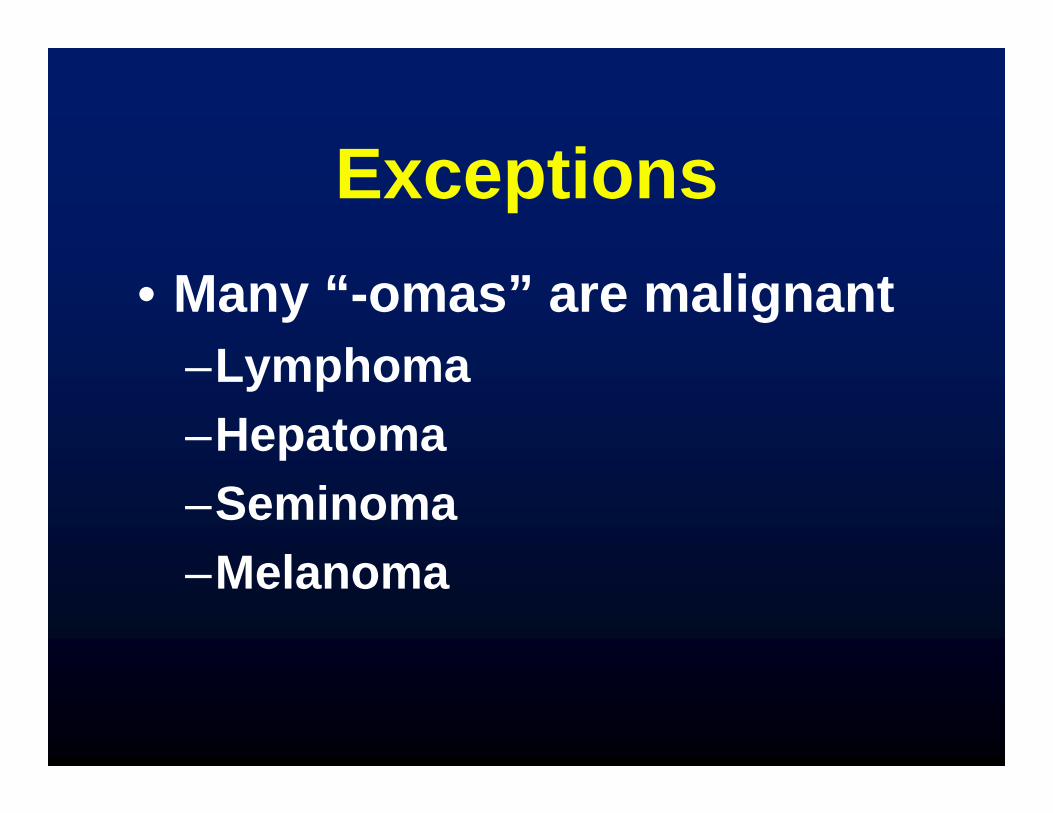
Exceptions• Many “-omas” are malignant
–Lymphoma–Hepatoma–Seminoma–Melanoma

Exceptions• Some “carcinomas” or
“sarcomas” are benign–Basal cell carcinoma of skin–Cystosarcoma phyllodes of
breast–Well differentiated
liposarcoma of skin

Name that tumor!
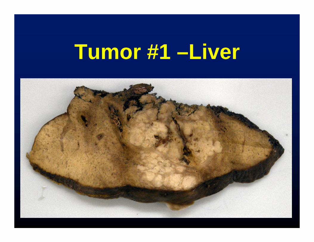
Tumor #1 –Liver

Tumor #1Tumor #1 –Liver
Mitoses

Tumor #1• Dx: Adenocarcinoma of the
bile duct • Malignant features
– Infiltrative borders, many mitoses
– Gland forming neoplasm• aka “Cholangiocarcinoma”
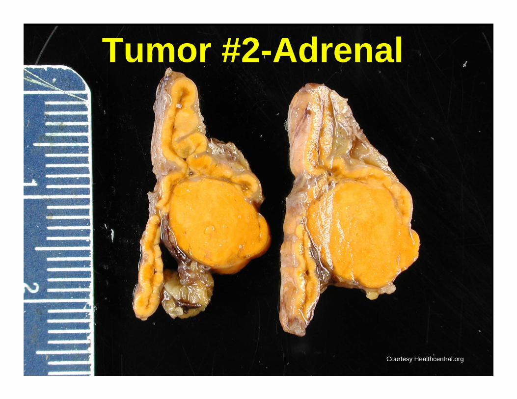
Tumor #2-Adrenal
Courtesy Healthcentral.org
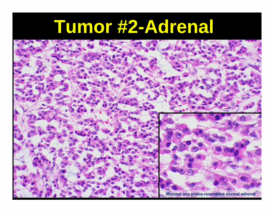
Tumor #2-Adrenal
Minimal ana plasia-resembles normal adrenal
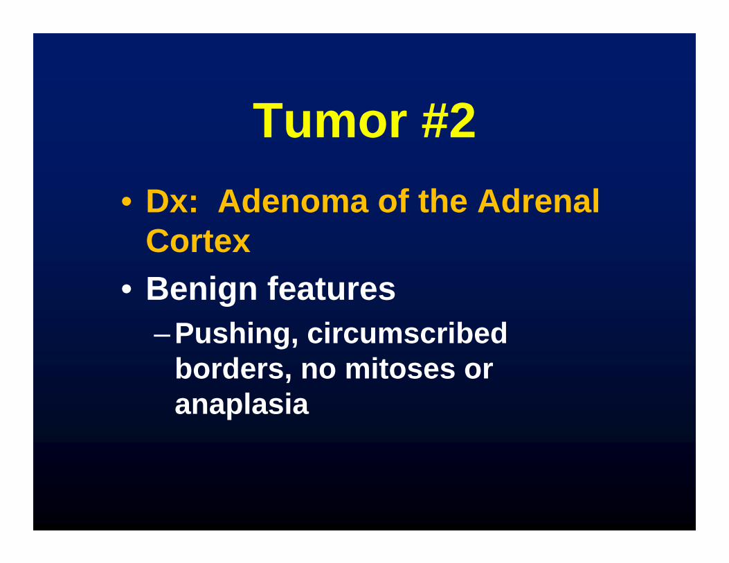
Tumor #2• Dx: Adenoma of the Adrenal
Cortex• Benign features
– Pushing, circumscribed borders, no mitoses or anaplasia

5. What is Grade?

Grading Of Cancer
Grade: A histologic parameter quantitating the degree of differentiation of the cancer cells.
Differentiation• Well-differentiated (“low grade”)
tumors resemble mature normal cells of the tissue of origin.
• Poorly differentiated (“high grade”) tumors show little resemblance to the tissue of origin.
Grading of Cancer• Many tumors graded according
to a three-tiered scheme: well, moderately, and poorly differentiated (grade 1, 2, 3).
• Grading systems vary by different tumor type.
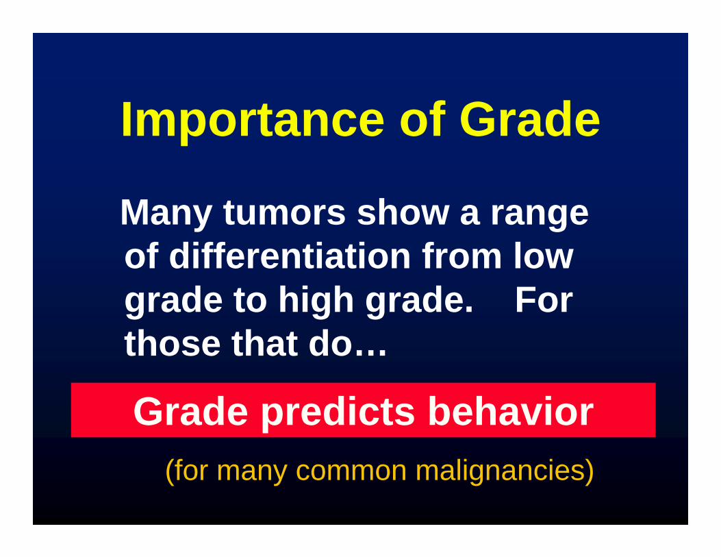
Importance of Grade
Many tumors show a range of differentiation from low grade to high grade. For those that do…
Grade predicts behavior(for many common malignancies)
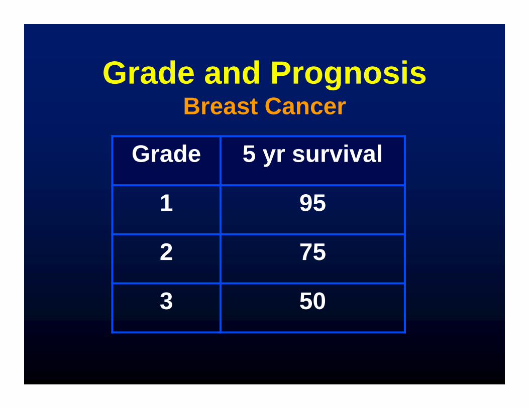
Grade and PrognosisBreast Cancer
Grade 5 yr survival
1 95
2 75
3 50
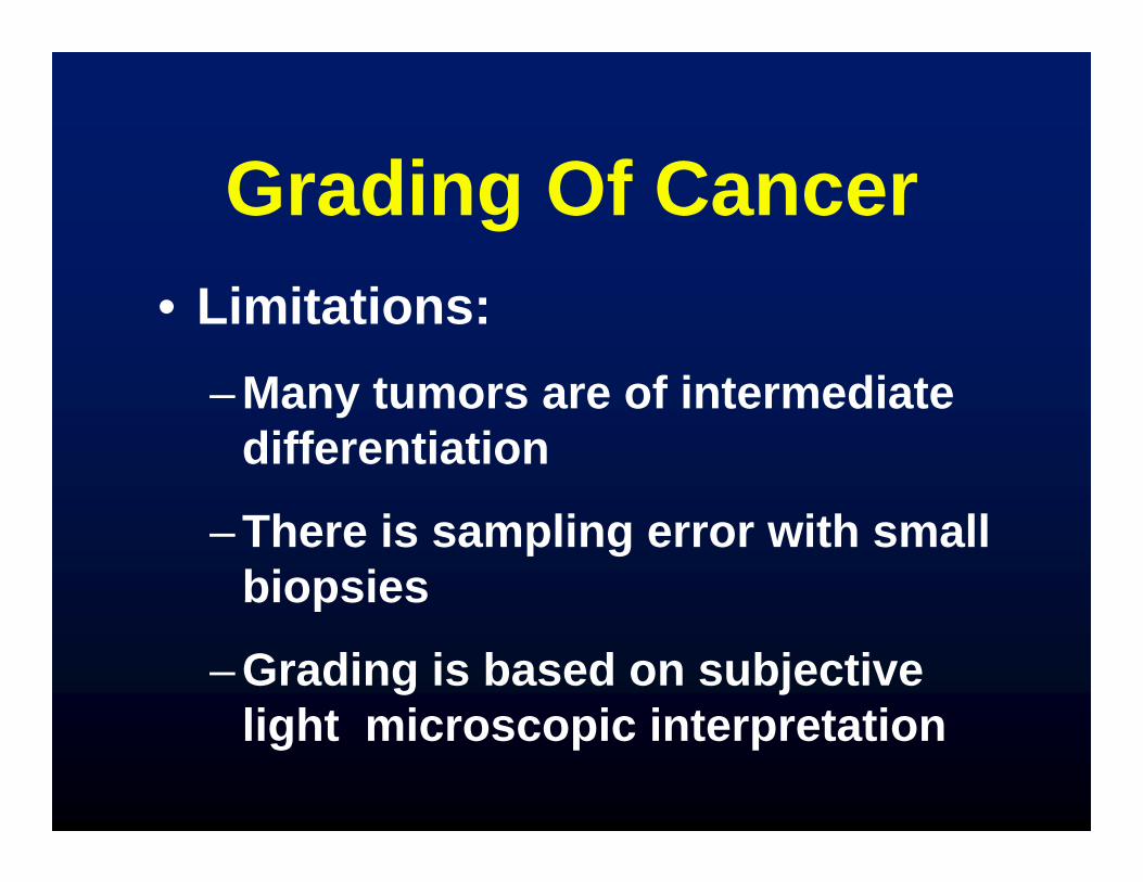
Grading Of Cancer• Limitations:
– Many tumors are of intermediate differentiation
– There is sampling error with small biopsies
– Grading is based on subjective light microscopic interpretation

Factors that would influence whether a surgical resection would be curative include:
A. Whether it is benign or malignant
B. Location of the neoplasm
C. Cell type of the neoplasm
D. Degree of anaplasia of the neoplasm
E. All of the above
Quick Review

Factors that might influence whether surgery for a neoplasm will be curative include:
A. Whether the neoplasm is benign or malignant
B. Location of the neoplasm
C. Cell type of the neoplasm
D. Degree of anaplasia of the neoplasm
E. All of the above
Quick Review

The EndIntroduction to Neoplasia
(Part I)
Rex Bentley684-6423
[email protected] Duke South
Green Zone


