Page 1
1
Metal corrosion and biological H2S cycling in closed systems
Fernanda Abreu*
Instituto de Microbiologia Paulo de Góes, Universidade Federal do Rio de Janeiro, Rio de
Janeiro, Brazil; [email protected] *
Abstract Sulfate reducing bacteria produce H2S during growth. This gas is toxic and is associated
with corrosion in industrial systems. In the environment purple sulfur bacteria, green
sulfur bacteria and sulfur oxidizing bacteria use the H2S produced by sulfate reducing
bacteria as electron donors. The major aim of this project is to evaluate the possibility
of using H2S consuming bacteria to lower H2S concentration and prevent corrosion.
Introduction
In metal corrosion process the surface of the metal is destroyed due to certain external
factors that lead to its chemical or electrochemical change to form more stable
compounds. The simplified explanation of the corrosion process is the oxidation of at
an anode (corroded end releasing electrons) and the reduction of a substance at a
cathode. Corrosion mechanisms are very diverse and can be based on inorganic
physicochemical reactions and/or biologically influenced. Microbiologically influenced
corrosion (MIC) is a natural process that occurs in the environment as a result of
metabolic activity of microorganisms. Microbial colonization and biofilm formation on
metal surfaces modify the electrochemical conditions at the metal–solution interface,
which usually have positive influence on corrosion process. MIC of steel generates
approximately US$ 100 million financial losses per annum in the United States (Muyzer
and Stams, 2008). In industrial settings, especially in petroleum, gas and shipping
industries, sulfate reducing bacteria (SRB) are a major concern.
SRB are ubiquitous in anoxic habitats and have an important role in both the sulfur and
carbon cycles (Muyzer and Stams, 2008). According to rrs gene phylogenetic analysis,
Page 2
2
the majority of sulfate reducing bacteria are grouped in Bacteria domain within
Deltaproteobacteria subgroup; there are some SRB species localized within the
Clostridia (Desulfotomaculum, Desulfosporosinus and Desulfosporomusa genera) and
within three of thermophilic SRB lineages (Nitrospirae, Thermodesulfobacteria and
Thermodesulfobiaceae). Sulfate reduction is also described for Archaea domain and
belong to the genus Archaeoglobus (Euryarchaeota) and to the genera Thermocladium
and Caldirvirga (Crenarchaeota) (Muyzer and Stams, 2008). During growth SRB
produce corrosive metabolic products that will increase corrosion rates (Videla and
Herrera, 2005). Hydrogen sulfide gas (H2S) is the product of sulfate reduction by SRB in
anaerobic respiration to obtain energy. This gas is extremely toxic and corrosive.
Inhalation of low concentrations of hydrogen sulfide cause irritation to mucous
membranes, headaches, dizziness, and nausea; inhalation of higher concentrations
(200-300 ppm) result in respiratory arrest leading to coma, unconsciousness,
pulmonary paralysis, collapse and death. Hydrogen sulfide removal in industrial
systems is usually done by chemical methods, which are expensive and energy
consuming (Sayed et al., 2006). In this way, biological methods are considered a
desirable as an alternative to chemical treatment.
Clues for the development of hydrogen sulfide removal methods using biological
methods are based on sulfur cycle. SRB play a huge hole in sulfur cycling and are
responsible for the consumption of the most significant oxidation state of sulfur
specimen in nature, the +6 oxidation state (sulfate). In anaerobic regions in aquatic
environment, for example, SRB promotes the conversion of sulfate to sulfide, using the
first sulfur specimen as an electron acceptor in metabolic pathways (Tang et al., 2009).
In anaerobic regions where light is available, the H2S generated by SRB is used by
anaerobic phototrophic bacteria (ANP) as electron donors in order to obtain energy.
Chemolitotrophic sulfur-oxidizing bacteria (SOB) also utilize H2S during growth and are
located in the interface between oxygen and sulfide. They can use sulfide as an
electron donors and oxygen as an electron acceptor. In these sob the oxidation of
sulfide usually leads to the production of phase-bright globules of elemental sulfur in
the periplasm. In the environment aerobic regions, chemotrophic bacteria can obtain
their energy from oxidation of H2S and S0 to form SO42- (Sayed et al., 2006). Therefore,
Page 3
3
interesting candidates to be used in biological H2S removal are the ANP and SOB,
which can consume H2S during growth.
Here H2S concentration and metal corrosion were evaluated in environmental samples
and also SRB, ANP and SOB relation was studied in vitro to determine the possible
effect of sulfide consumption in (1) H2S concentration in a closed system, (2) SRB
growth (i.e. inhibition or stimulation) and (3) metal corrosion process. Community
analysis on a sulfide rich environmental sample was performed in order to determine
the diversity of ANP, SOB and SRB.
Materials and Methods
Sampling
Samples of sediment and water were collected in Trunk River using cores. The
structure of the sediment layers were observed during sampling. Only samples which
had visible differentiation of layers were kept and stored in the laboratory.
Micro sensor measurement
Oxygen, H2S and pH profiles in the Trunk River core sample were done using
Uniscience electrodes. Calibrations were performed according to each sensor manual.
Corrosion of steel in the different layers of sediment sample
Three stainless steel nails (GripRite Fas’Ners) were introduced in the pink and black
layers of the sediment in a core of approximately 6 cm in diameter. The core was
maintained in the hood under artificial light illumination. After 8 days the two nails of
each sediment layer were removed from the core and fixed in formaldehyde 1% for
CARD-FISH analysis. There other nail of each layer was inoculated in the gradient
medium for iron oxidizing bacteria (Emerson and Floyd, 2005).
Corrosion assay in microcosms
A core of approximately 4 cm in diameter was also sampled in Trunk River and used in
microcosms corrosion assays. The pink and black layers of the sediment were carefully
separated and 5g of each layer was transferred to 50 mL glass vials. The pink layer of
Page 4
4
the sediment was used for purple sulfur bacteria (PSB) enrichment; the sediment just
below pink layer was used for green sulfur bacteria (GSB) and sulfur-oxidizing bacteria
(SOB) enrichment. The black layer of the sediment was used for SRB enrichment.
Marine phototrophic base (MPB) was used for bacterial enrichment and especial
conditions were defined for each type of enrichment. The following substances were
added to MPB according to the enrichment: (1) PSB enrichment sodium thiosulfate (5
mM) and sodium sulfide (1 mM) was added to MPB; (2) GSB sodium sulfide (3 mM); (3)
SOB sodium nitrate (2 mM) and sodium sulfide (1 mM) was added; (4) SRB sodium
sulfate (20 mM) and sodium acetate (5 mM) was added. Controls were done by the
addition of filtered sterilized water from the sampling site. All enrichments and
respective control (except for SOB) were incubated at 30oC. SOB enrichment and
control were incubated at room temperature. PSB enrichment and control were
illuminated at 850 nm and GSB enrichment and control at 770 nm. Corrosion control
was performed adding MPB or filtered sterilized water from the sampling site to 50 mL
glass vials.
The remaining pink and black layers of the sediment were mixed and 10g of the mixed
sediment was added to a 50 mL glass vial. The enrichment conditions and controls
described above were maintained for PSB, GSB, SOB and SRB enrichments.
DNA extraction, PCR amplification and 454 Pyrosequencing
DNA extraction was done from pink and black layers of the sediment collected at Trunk
River. DNA extraction was performed with the PowerSoilTM DNA isolation kit (Mo BIO
laboratories, Inc.) according to the kit instructions. 454 barcode 16S PCR was
performed for both pink and black layers for the sediment according to Microbial
diversity laboratory manual instructions.
PSB, GSB, SOB and SRB isolation and growth
The bacteria used in these work have been isolated during the first weeks of the 2012
Microbial diversity summer course. ANP, SOB and BRS were gently provided by Brian
Brigham, Stefan Thiele and Florence Schubotz, respectively. A colony of plates
containing each bacterium was obtained and transferred to specific liquid medium
Page 5
5
described in the laboratory manual. For SOB liquid medium was developed based on
the overlay medium for SOB described in the laboratory manual; sodium sulfide (1
mM) and sodium nitrate (2 mM) were added to the overlay medium.
Corrosion experiment
The medium used in corrosion experiment in liquid and in gradient tubes was
developed based on the media described for each bacterium in the laboratory manual.
For the semisolid medium the sulfide agarose plug used was prepared as described in
the laboratory manual (page 7.4). The overlay medium is described below:
Overlay medium for SRB, PSB, GSB and SOB:
1x Sea water base 400 mL
100x NH4Cl 0.4 mL
100x Kphosphate 0.04 mL
1% resazurin 2 mL
Na thiosulfate 0.48 g
Na bicarbonate 2,32 g
Na acetate 0.4 g
Na2SO4 1,5 g
NaNO3 0.2 g
Cystein 0.24g
1000x Trace elements 500 l
1000x Vitamin solution 500 l
Agarose 0.08%
Autoclave
Page 6
6
Add Na2S 1M 400 l
Check pH. It should be around 7. If needed add sterile HCl or NaOH.
For 400 mL of the liquid medium: the overlay medium described above was used, but
agarose was omitted. Glass vials were used to add 5 mL of the liquid medium; the
medium was flushed with nitrogen gas for 5 minutes, caped with butyl caps and the
head space was flushed for 2 minutes with the same gas.
Growth was monitored by checking turbidity and by phase-contrast microscopy.
Before inoculation in liquid and semisolid medium cell quantification per mL of each
culture was performed in order to add know cell concentration of H2S producing and
consuming bacteria.
One of each liquid and gradient medium was inoculated as follow: (1) only 2.8 x 10⁷
SRB cells; (2) 1.9 x 10⁷ cell of PSB and 0.9 x 10⁷ cell of SOB (PSB + SOB will be called
PSSOB); (3) PSSOB and SRB in the relation 1:1; (4) PSSOB and SRB in the relation 1:3.
Controls which were maintained in dark were prepared. All cultures were maintained
at 30oC at 850 nm. Controls were incubated at the same condition, but were covered
with aluminum foil.
Evaluation of H2S concentration and SRB growth in the medium
The colorimetric method described in the laboratory manual was used to determine de
H2S concentration on selected microcosms and on liquid cultures. For estimation of
SRB growth CARD-FISH was performed according to the laboratory manual using
deltabacteria probes and its competitors (probes: Delta495-a; Delta495-B; Delta495-c).
Results and Discussion
Microcosms observations
Steel nail corrosion in the core
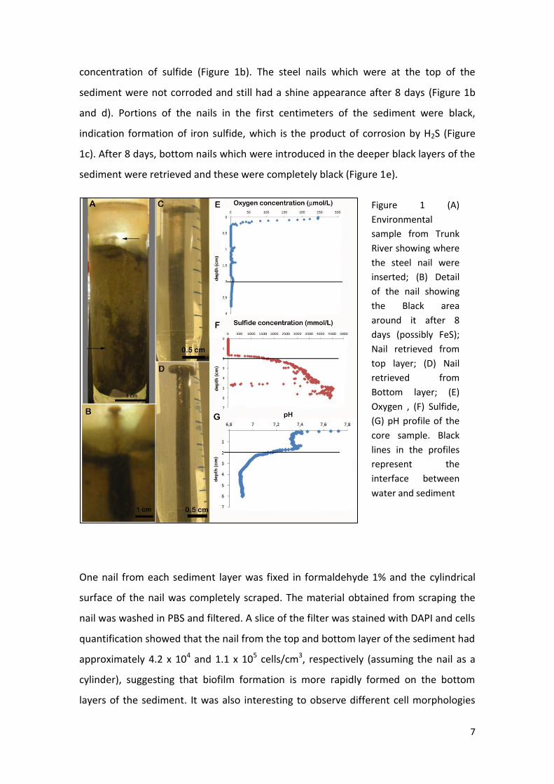
During core storage it was possible to see the growth of a green layer on the surface of
the sediment (Figure 1a). Oxygen and hydrogen sulfide profile showed that the
interface between water and sediment is an anaerobic environment with high
Page 7
7
concentration of sulfide (Figure 1b). The steel nails which were at the top of the
sediment were not corroded and still had a shine appearance after 8 days (Figure 1b
and d). Portions of the nails in the first centimeters of the sediment were black,
indication formation of iron sulfide, which is the product of corrosion by H2S (Figure
1c). After 8 days, bottom nails which were introduced in the deeper black layers of the
sediment were retrieved and these were completely black (Figure 1e).
One nail from each sediment layer was fixed in formaldehyde 1% and the cylindrical
surface of the nail was completely scraped. The material obtained from scraping the
nail was washed in PBS and filtered. A slice of the filter was stained with DAPI and cells
quantification showed that the nail from the top and bottom layer of the sediment had
approximately 4.2 x 10⁴ and 1.1 x 10⁵ cells/cm3, respectively (assuming the nail as a
cylinder), suggesting that biofilm formation is more rapidly formed on the bottom
layers of the sediment. It was also interesting to observe different cell morphologies
Figure 1 (A)
Environmental
sample from Trunk
River showing where
the steel nail were
inserted; (B) Detail
of the nail showing
the Black area
around it after 8
days (possibly FeS);
Nail retrieved from
top layer; (D) Nail
retrieved from
Bottom layer; (E)
Oxygen , (F) Sulfide,
(G) pH profile of the
core sample. Black
lines in the profiles
represent the
interface between
water and sediment
Page 8
8
that could be observed exclusively on the top layers (Figure 2). In the nail sample from
bottom layer of the sediment several cells are attached to metal pieces, supporting the
idea that biofilm formation was more efficiently formed in this sediment layer (Figure
2d).
Figure 2 Fluorescence microscopy of nail samples from the (A and B) pink and (C and D)
black layers of the core. In the first sample numerous filamentous bacteria could be
observed (A); in the sample from deeper regions several bacteria were attached to
metal particles, indicating biofilm formation (D).
Despite the fact that lost of weight was not observed in nails from neither layers,
results indicate that corrosion is favored in the bottom layers of the sediment, where
H2S concentration is higher and were the SRB population is probably more abundant
and forming biofilms.
Nails from both the top and bottom layers were washed in sterile PBS and inoculated
on iron oxidizing bacteria gradient medium (Emerson and Floyd, 2005). After one week
it was possible to see a white band formation in the nail from the top layer (arrow).
5 m
Page 9
9
Rust was observed in both cases at the flat portion of the nail (arrowhead, Figure 3).
Phase contrast microscopy of the top nail culture showed a variety in cell
morphotypes; in the bottom nail cultured rod-shaped bacteria were the dominant
(Figure3).
Figure 3 (A) Gradient medium for iron oxidizing bacteria (Emerson and Floyd, 2005); (B)
Tubes which where inoculated with the nails from the top and bottom layers of the
core; (C) Microscopy image of the culture inoculated with the top nail; (D) Microscopy
image of the culture inoculated with the top nail.
Steel nail corrosion in enrichment microcosms
After 14 days of incubation the appearance of the steel nail and the color and turbidity
of the microcosms were different among each other depending on the type of
enrichment. For PSB enrichment in microcosms with the mixed sediment and with the
pink layer the nail characteristics were preserved and the liquid and sediment layers
were pink, indicating the growth of the target bacteria. PSB microcosms controls (that
means no enrichment) with mixed sediment layers and pink sediment presented
B C
D
5 m
Page 10
10
different results in relation to nail preservation and microcosms characteristics. The
nail inside the microcosm containing the mixed sediment layers was very damaged by
corrosion and rust formation was observed (Figure 4). The sulfide quantification in the
microcosms with mixed sediment layers (control and enrichment for PSB) indicates
similar levels of the gas, suggesting that the corrosion observed in the control might be
caused by the action of other bacteria. An aliquot of the control microcosm where rust
was observed was inoculated on iron oxidizing gradient medium described by Emerson
and Floyd (2005). After 7 days a white band was formed in the tube and microscopy
observation confirmed the presence of bacterial growth (cocci and spirilla) (Figure 5).
In the microcosm containing the pink layer sediment corrosion was not observed for
both enrichment and control. The presence of pink pigmentation was only observed in
the enrichment microcosm.
Figure 4 (A and B) Different microcosms used to evaluate metal corrosion and possible
prevention by PSB; (C) Comparison between medium and water controls and PSB
enrichment and control.
Page 11
11
Figure 5 (A) Gradient tube for iron oxidizing bacteria isolation; (B) Phase-contrast
microscopy of the white band showed in (A).
In the microscosms for GSB enrichment turbidity and green color in the liquid phase
was observed for both mixed sediment layers and sediment fraction below pink layer,
suggesting GSB growth. Corrosion in the nail and rust was only detected on the GSB
enrichment microcosm containing mixed sediment layers. However this corrosion
might not be related to SRB H2S production because H2S concentration was low (Figure
9). Controls for GSB enrichments showed similar results; both with liquid phase clear
and no apparent corrosion (Figure 6).
5 m
Page 12
12
Figure 6 (A and B) Different microcosms used to evaluate metal corrosion and possible
prevention by GSB; (C) Comparison GSB enrichment and control from mixed sediment
layers samples.
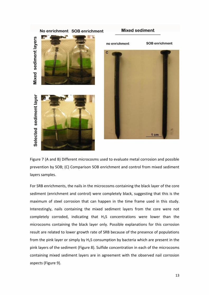
In the microcosms for SOB enrichment nail changes in appearance were similar, they
had some black portions along the nail, but no rust formation was observed. Nails from
both enrichments were completely black, suggesting the production of H2S, which
indicates SRB growth. In the mixed sediment microcosms a green color was observed
in both control and enrichment. Possibly GSB grew in these microcosms because the
inoculated sample had some H2S and this sample was maintained at room
temperature in the bench, so there was light availability (Figure 7).
Page 13
13
Figure 7 (A and B) Different microcosms used to evaluate metal corrosion and possible
prevention by SOB; (C) Comparison SOB enrichment and control from mixed sediment
layers samples.
For SRB enrichments, the nails in the microcosms containing the black layer of the core
sediment (enrichment and control) were completely black, suggesting that this is the
maximum of steel corrosion that can happen in the time frame used in this study.
Interestingly, nails containing the mixed sediment layers from the core were not
completely corroded, indicating that H2S concentrations were lower than the
microcosms containing the black layer only. Possible explanations for this corrosion
result are related to lower growth rate of SRB because of the presence of populations
from the pink layer or simply by H2S consumption by bacteria which are present in the
pink layers of the sediment (Figure 8). Sulfide concentration in each of the microcosms
containing mixed sediment layers are in agreement with the observed nail corrosion
aspects (Figure 9).
Page 14
14
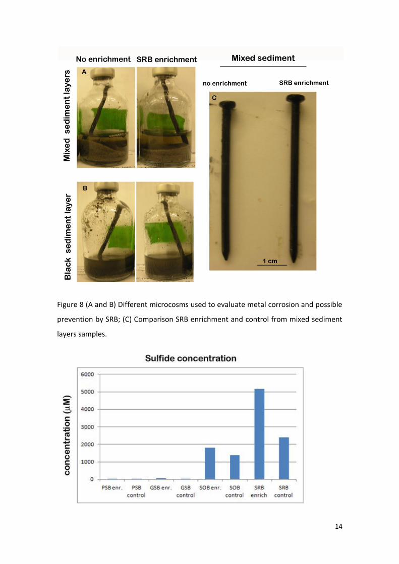
Figure 8 (A and B) Different microcosms used to evaluate metal corrosion and possible
prevention by SRB; (C) Comparison SRB enrichment and control from mixed sediment
layers samples.
Page 15
15
Figure 9 Sulfide concentrations in microcosms with mixed sediment layers.
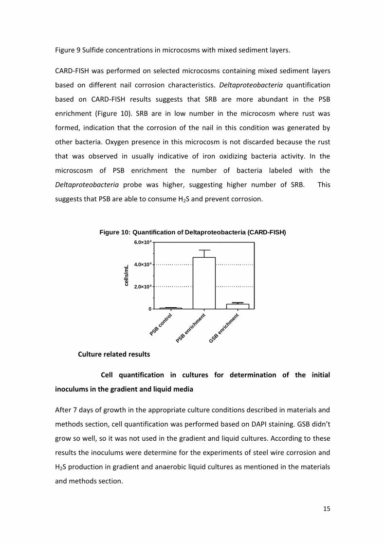
CARD-FISH was performed on selected microcosms containing mixed sediment layers
based on different nail corrosion characteristics. Deltaproteobacteria quantification
based on CARD-FISH results suggests that SRB are more abundant in the PSB
enrichment (Figure 10). SRB are in low number in the microcosm where rust was
formed, indication that the corrosion of the nail in this condition was generated by
other bacteria. Oxygen presence in this microcosm is not discarded because the rust
that was observed in usually indicative of iron oxidizing bacteria activity. In the
microscosm of PSB enrichment the number of bacteria labeled with the
Deltaproteobacteria probe was higher, suggesting higher number of SRB. This
suggests that PSB are able to consume H2S and prevent corrosion.
Culture related results
Cell quantification in cultures for determination of the initial
inoculums in the gradient and liquid media
After 7 days of growth in the appropriate culture conditions described in materials and
methods section, cell quantification was performed based on DAPI staining. GSB didn’t
grow so well, so it was not used in the gradient and liquid cultures. According to these
results the inoculums were determine for the experiments of steel wire corrosion and
H2S production in gradient and anaerobic liquid cultures as mentioned in the materials
and methods section.
Figure 10: Quantification of Deltaproteobacteria (CARD-FISH)
PSB c
ontrol
PSB e
nrichm
ent
GSB
enri
chm
ent
0
2.0×10⁴
4.0×10⁴
6.0×10⁴
cell
s/m
L
Page 16
16
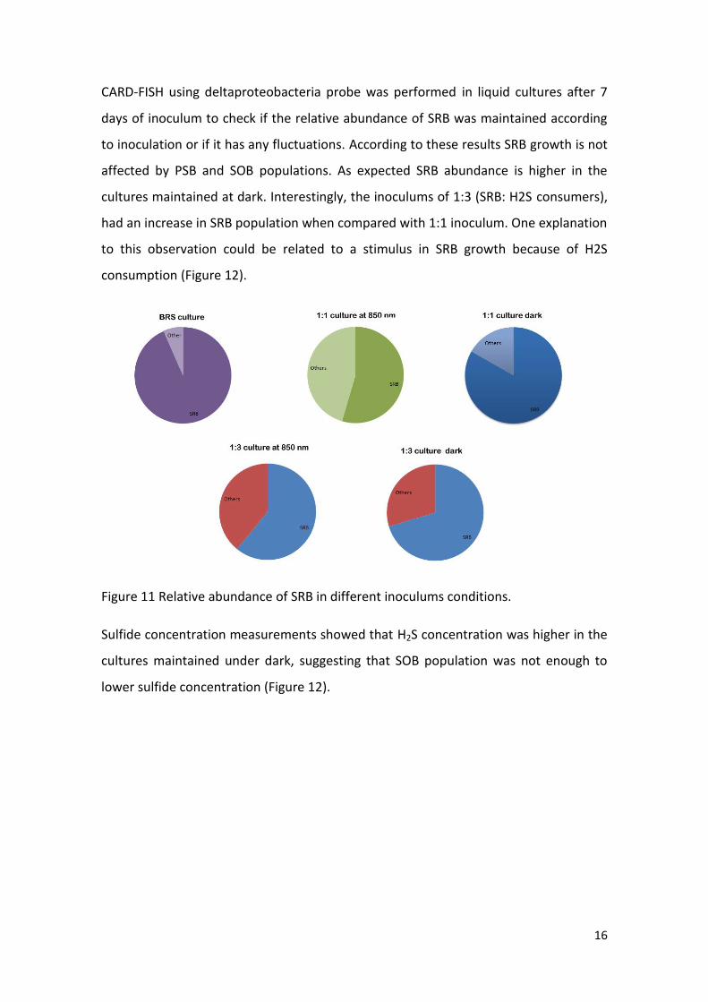
CARD-FISH using deltaproteobacteria probe was performed in liquid cultures after 7
days of inoculum to check if the relative abundance of SRB was maintained according
to inoculation or if it has any fluctuations. According to these results SRB growth is not
affected by PSB and SOB populations. As expected SRB abundance is higher in the
cultures maintained at dark. Interestingly, the inoculums of 1:3 (SRB: H2S consumers),
had an increase in SRB population when compared with 1:1 inoculum. One explanation
to this observation could be related to a stimulus in SRB growth because of H2S
consumption (Figure 12).
Figure 11 Relative abundance of SRB in different inoculums conditions.
Sulfide concentration measurements showed that H2S concentration was higher in the
cultures maintained under dark, suggesting that SOB population was not enough to
lower sulfide concentration (Figure 12).
Page 17
17
Figure 13 Sulfide concentrations in the liquid cultures.
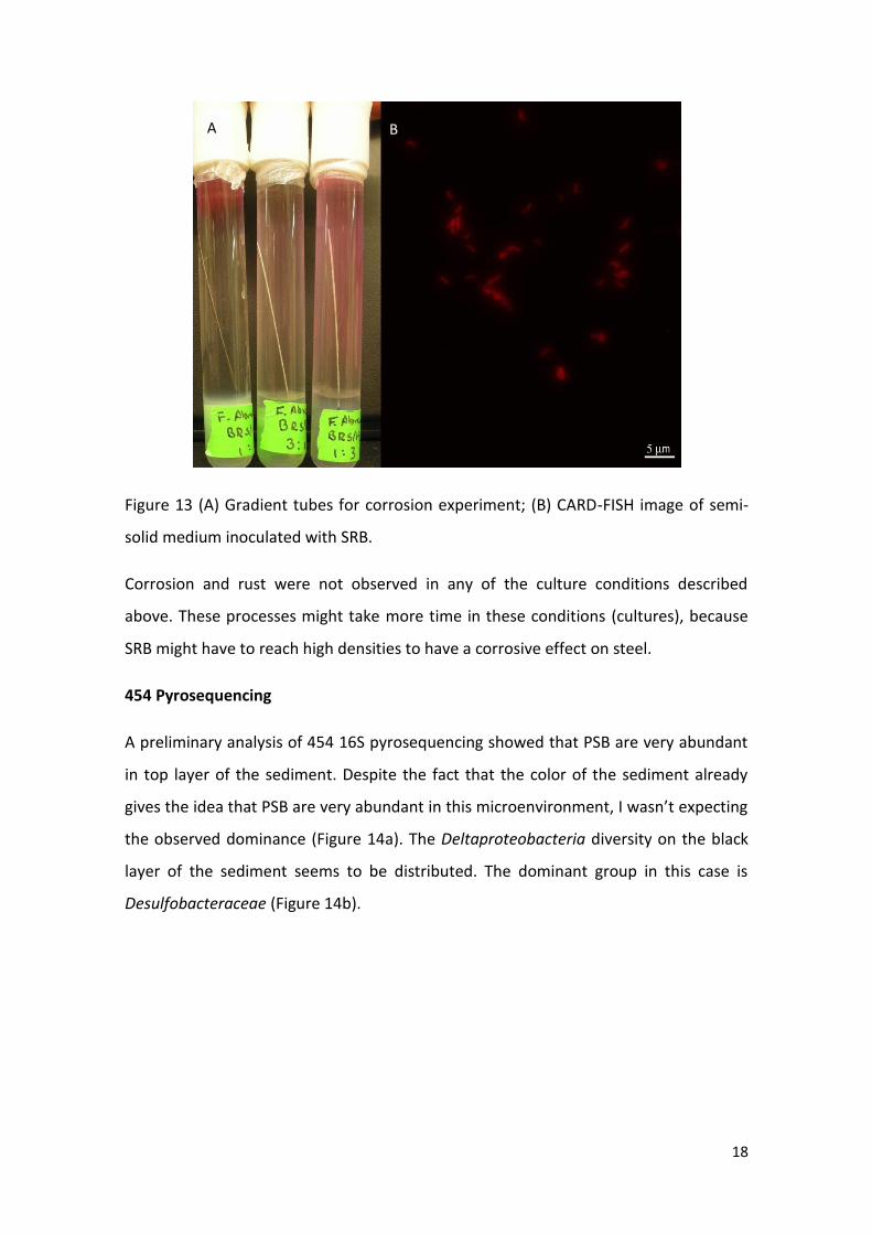
In the gradient tubes inoculated only with SRB the oxidized fraction on the top of the
medium was completely reduce in a few days, indicating SRB growth and H2S
production. Culture tubes with 1:1 and 3:1(SRB: PSSOB) were similar in growth and
turbidity. No band formation was observed in any of these tubes, but an irregular
growth was seen around the steel wire (Figure 13). CARD-FISH using
deltaproteobacteria probe indicate that PSSOB and SRB are growing on the gradient
tubes (Figure 13). Unfortunately total and SRB quantification was not possible because
of the high background of DAPI on the images. Cultures which were maintained in
dark conditions had to be transferred to light conditions because those that were
illuminated were accidentally autoclaved, so H2S and oxygen measurements along the
gradient media were not compared between light and dark conditions.
Page 18
18
Figure 13 (A) Gradient tubes for corrosion experiment; (B) CARD-FISH image of semi-
solid medium inoculated with SRB.
Corrosion and rust were not observed in any of the culture conditions described
above. These processes might take more time in these conditions (cultures), because
SRB might have to reach high densities to have a corrosive effect on steel.
454 Pyrosequencing
A preliminary analysis of 454 16S pyrosequencing showed that PSB are very abundant
in top layer of the sediment. Despite the fact that the color of the sediment already
gives the idea that PSB are very abundant in this microenvironment, I wasn’t expecting
the observed dominance (Figure 14a). The Deltaproteobacteria diversity on the black
layer of the sediment seems to be distributed. The dominant group in this case is
Desulfobacteraceae (Figure 14b).
A B
Page 19
19
Conclusions
According to the results observed in this preliminary study, PSB and GSB are capable to
decrease H2S concentration in the environment. In experiments related to PSB
enrichment metal corrosion was also prevented. Many research groups are aiming in
the use of these bacteria to decrease sulfide concentration in industrial systems. A
good review on the subject is Sayed et al., 2006.
Acknowledgments
I would like to thank the course co-directors Dan Buckley and Steve Zinder, the course
coordinator Asheley Campbell and all the instructors, especially Sara Kleindienst,
Verena Salman. I also would like to thank Suzanna Brauer for helping me with media
development and all my course friends. MBL, Daniel S. and Edith T. Grosch
Scholarship Fund, The Gordon & Betty Moore Foundation and CNPq are
acknowledged.
References
Emerson, D. and Floyd, M.M. Enrichment and isolation of iron-oxidizing bacteria at
neutral pH. Methods Enzymol. 397:112-23.
Muyzer, G. and Stams, A. J. M. 2008. The ecology and biotechnology of sulphate-
reducing bacteria. Nature Reviews Microbiology, vol. 6, no. 6, pp. 441–454.
Page 20
20
Sayed, M., Soreanu, G., Falleta, P. and Beland, M. 2006. Removal of hydrogen sulfide
from gas streams using biological processes-A review, Canadian Biosystems
Engineering, 48, 2.2-2.14.
Tang, K., Baskaran, V. and Nemati. M. 2009. Bacteria of the sulphur cycle: An overview
of microbiology, biokinetics and their role in petroleum and mining industries.
Biochemical Engineering Journal, vol. 44, no1, pp. 73–94.