Page 1
International Journal of Scientific and Research Publications, Volume 5, Issue 4, April 2015 1 ISSN 2250-3153
www.ijsrp.org
Ferromagnetic Properties of Zn substituted Spinel
Ferrites for High Frequency Applications
K.Praveena1,2*
, K. Sadhana3
1Materials Research Center, Indian Institute of Science, Bangalore-560012, India. 2School of Physics, Eternal University, Baru Sahib-173101, Himachal Pradesh, India
3Department of Physics, University College of Science, Osmania University, Saifabad, Hyderabad – 500 004, India
Research Type: Review Article
Abstract- This review focuses on two systems, nanocrystalline Zn substituted Co1-xZnxFe2O4 (0≤x≥1) and
Ni0.4Zn0.2Mn0.4Fe2O4, were synthesized using microwave hydrothermal and auto-combustion method respectively. The synthesized
powders were characterized by X-ray diffraction (XRD), transmission electron microscope (TEM), thermo-gravimetric-
differential thermal analysis (TG-DTA) and FTIR. The average particle size was obtained from TEM and it is found to be 17 nm.
Zero field cooled (ZFC) and Field cooled (FC) measurements for Co1-xZnxFe2O4 reveal that samples with 0.6≤x≥1 have
superparamagentic behavior at room temperature, which confirms weak interaction between magnetic particles. The blocking
temperatures obtained form ZFC-FC curves decreases as Zn concentration increases. Lower reduced remnant magnetization
(Mr/Ms) values (x<0.5) suggest that all the samples have uniaxial anisotropy. For Ni0.4Zn0.2Mn0.4Fe2O4 bulk densities of the
samples were increased with an increase of sintering temperature. The grain sizes of all the samples vary in between 18 nm to 30
nm. The hysteresis loops show high saturation magnetization and low coercivity, indicates that it is a soft material. The
incremental permeability (permeability with magnetic field superposition) was influenced by both ΔM and Hc. So these ferrites
has the properties and are widely used as electromagnetic wave absorbing materials in the VHF/UHF region and as radar
absorbing materials in C-band frequencies.
Index Terms- Spinel Ferrites, Magnetic Properties, Saturation Magnetization, Zero Field Cooling, Blocking Temperature,
Permeability, Q-factor, DC-bias-superposition
I. INTRODUCTION
Ferrites have the general formula (M1 − 𝑥Fe𝑥)[M𝑥Fe2 − 𝑥]O4. The divalent metal element M (Mg, Zn, Mn, Fe, Co, Ni, or mixture of
them) can occupy either tetrahedral eight (A) or sixteen octahedral [B] sites of a cubic mineral spinel (MgAl2O4) structure as
depicted by the parentheses or brackets, respectively. For example the structural formula of Co-ferrite is usually written as (Co1−
𝑥Fe𝑥)[Mg𝑥 Fe2−𝑥]O4, where 𝑥 represents the degree of inversion (defined as the fraction of (A) sites occupied by Fe3+
cations).
Depending on distribution of cations in (A) and [B] sites, ferrites may exist in two extreme states, normal (𝑥 = 0) and inverse (𝑥 =
1) or in an intermediate mixed state [1].
Among these materials, spinel ferrite nanoparticles have special importance. They show various magnetic properties depending on
the composition and cation distribution. Various cations can be placed in A site and B site to tune its magnetic properties.
Depending on A site and B site cations, it can exhibit ferrimagnetic, antiferromagnetic, spin (cluster) glass, and paramagnetic
behaviour.
Nanosized ferrites may have extraordinary electric and magnetic properties that are comparatively different from microstructured
materials, tailoring them to modern technologies, as well as providing novel applications such as ferrofluids [2], magnetic drug
delivery [3], high density information storage [4], photocatalysis [5], gas sensors [6], etc.
Among those spinel ferrites we are interested in Zn substituted mixed ferrites are useful for low and high frequency applications
generally for for power transformers, power inductors, microwave devices, read and write heads for high speed digital tape, etc.
because of their high resistivity, low losses, mechanical hardness, high Curie temperature and chemical stability [7-12].
CoFe2O4 has attracted considerable interest because of its large magnetic multi-axial anisotropy, high saturation magnetization,
high Curie temperature and extraordinary chemical stability [13-16]. CoFe2O4 is predominantly an inverse spinel oxide with Co2+
ions mainly on B sites and Fe3+
ions distributed, almost equally, between A and B sites [17]. Cobalt ferrite is ferrimagnetic below
790 K (TN) which suggests that the magnetic interactions in this ferrite are very strong. When Co2+
is replaced by Zn2+
in Co1-
xZnxFe2O4, Zn2+
preferentially occupies the tetrahedral site and the Fe3+
ions are displaced to the octahedral sites. Thus, with
increasing x, the FeA–O–FeB interaction becomes weak and TN is expected to decrease. ZnFe2O4 is a normal spinel, i.e.,
ZnA[Fe2]BO4 , and it is not ferrimagnetic, but antiferromagnetic due to FeB–FeB interactions only. TN of ZnFe2O4 is very small, 9 K
[18]. The absence of magnetic ions on the A site does not permit the antiferromagnetic A–O–B interaction and hence the magnetic
interaction in this ferrite is only on the octahedral (B) site. Now as the overall interaction in a ferrite is antiferromagnetic, the only
magnetic interaction in ZnFe2O4, i.e., FeB–FeB is antiferromagnetic [19].
Page 2
International Journal of Scientific and Research Publications, Volume 5, Issue 4, April 2015 2 ISSN 2250-3153
www.ijsrp.org
Cobalt zinc ferrite is one of the promising soft ferrite used in electronic devices such as transformer cores, electric motors and
generators. Cobalt ferrite nanoparticles are suitable for the isolation and refining of genomic DNA, the parting of polymerase
chain reaction ready DNA [20, 21] and especially in hyperthermia treatment. Variation in the concentration of the third metal ion
like Zn in Co ferrite can easily alter the distribution of Fe3+
ion [22]. Zn substituted mixed ferrites (Co-Zn) are chosen due to their
high sensitivity of magnetization to temperature for some applications like self-controlled hyperthermia [23-25].
Till now, low-temperature-fired Ni-Cu-Zn ferrites are the most important materials subjected to study the DC-bias-superposition
characteristics [26-28] due to their relatively low sintering temperature, high resistivity and good magnetic properties in the high
frequency range [29, 30]. But these investigations were mainly focused how to lower sintering temperature of the NiCuZn ferrites
by adding various sintering aids or changing the preparation process, improving the electromagnetic properties [31-37] but there
are no reports present on pure NiCuZn ferrite which was studied for DC bias superposition. And, moreover, they are the most
important soft ferrites used in multilayer chip inductors (MLCIs) and relevant inductive devices [38-40, 29]. In this work, we
chose Mn element to replace Cu element in the NiCuZn ferrites, and investigated the effects of ‘Mn’ on the magnetic properties,
especially DC-bias-superposition characteristic.
Studies on spinel ferrites synthesis methods have led to the development of different chemical synthesis techniques, which have a
common feature that all reagents are mixed in atomic or molecular level. Most popular methods of bottom-up synthesis approach
mentioned above are co-precipitation [41], sol–gel method [42], microemulsion method [43], hydrothermal [44], spray pyrolysis
[45], reverse micelle [46], precursor method [47], etc. Complex schedules and low production rate are common problems of these
wet-chemical methods [48].
The main advantages of this process over conventional-hydrothermal process are: (a) the rapid heating to treatment temperature
saves time and energy, (b) the kinetics of the reaction are enhanced by one to two orders of magnitude, (c) lead to the formation of
novel phases, and (d) lead to selective crystallization. Praveena et al. [49–52] have used the microwave hydrothermal (M–H)
method to prepare nano-sized powders of various ferrites with large surface area.
Sol–gel auto-combustion synthesis method (also called low-temperature self-combustion, auto-ignition or self-propagation, as
well as gel-thermal decomposition), where the chemical sol–gel and combustion process are combined, has shown great potential
in the preparation of spinel type ferrite nanomaterials. Generally, this method can be considered as solution combustion technique
[53].
II. EXPERIMENTAL
Synthesis of Co1-xZnxFe2O4 Ferrites: Nanocrystalline powders with compositions Co1-xZnxFe2O4 (0≤x≤1) were prepared using
microwave hydrothermal route. High purity (sigma, 99.99%) of cobalt nitrate [Co(NO3)2.6H2O], zinc nitrate [Zn(NO3)2.6H2O] and
iron nitrate [Fe(NO3)2.9H2O] were dissolved in double de-ionized water. An aqueous NaOH was added dropwise to the solution
until pH (~13) was obtained. The mixture was then transferred into Teflon lined vessel and kept in microwave digestion system
(Model MDS-2000, CEM Corp., Mathews, NC). This system uses 2.45 GHz microwaves and can operate at 0-100% full power
(1200±50W). The system is controlled by pressure and can attain maximum pressure of 200 psi, which is equivalent to 194°C.
The time, pressure and powder were computer controlled. The products obtained were filtered and then washed repeatedly with
de-ionized water, followed by freeze drying overnight. The prepared powders were weighed and the percentage yields were
calculated from the actual weight of the salts taken and the amount that was actually crystallized. The particle sizes of as-
synthesized powders were estimated by Transmission Electron Microscope (TEM, JEOL). FTIR spectra’s were taken on Bruker
Tensor 27 model from 4000 to 400 cm-1
. The samples were characterized by Phillips PANalytical X’pert powder X-ray diffraction
(XRD) with Cu-Kα (λ=1.5406A) radiation. The lattice constant (a) for the cubic crystal system was calculated using the equation
2 2 2a d h k l where (h k l) are the Miller indices of the diffraction peak, and ‘d’ is the inter-planar spacing. The average
crystallite size was calculated from Scherer formula cosm
KD
where K is a constant, λ radiation wavelength, β is full
width at half maxima (FWHM) and θ is the diffraction angle. The room temperature complex permeability (μ′ and μ″) were
measured over a wide frequency range from 1 MHz - 1.8 GHz using Agilent RF impedance analyser 4291B. Magnetic
measurements were obtained from a super conducting quantum interference device (SQUID) at room temperature (400 K).
III. RESULTS AND DISCUSSIONS
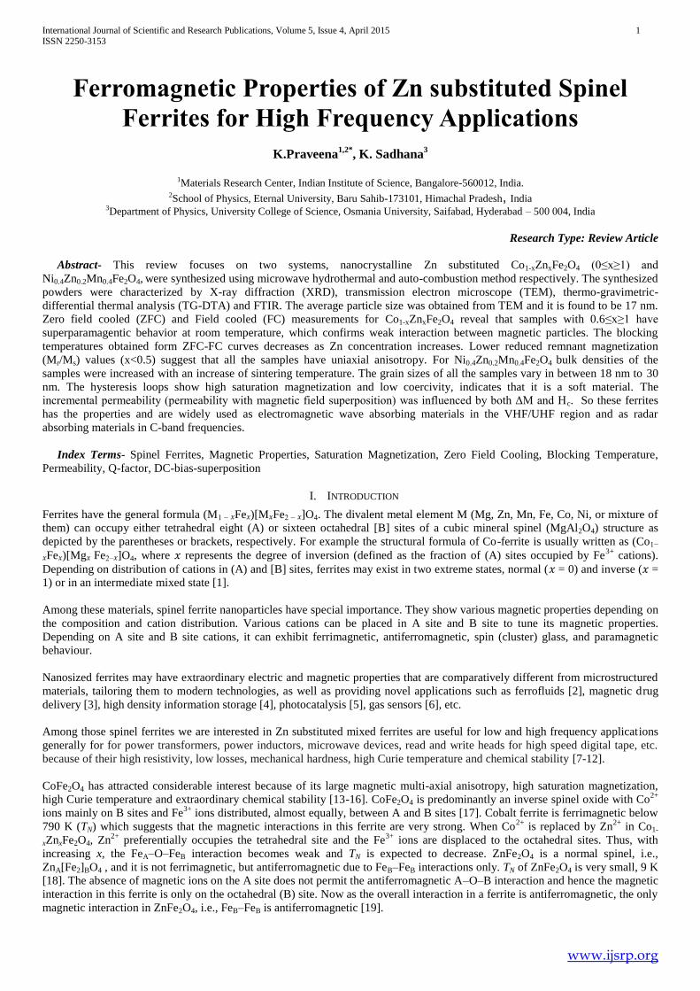
Structural and morphological studies: Fig.1 shows powder X-ray diffraction patterns of as-synthesized nanopowders. It shows
single-phase cubic spinel structure, without any impurity phases. It is interesting to observe that as Zn concentration is increasing
the diffraction peaks become broader indicating the nanosize of the crystals with space group symmetry, 3fd m
[54]. The
experimental values of lattice constants are compared with the standard JCPDS-ICDD Cards, for Co ferrite (22-1086 ICDD) and
for Zn ferrite (89-1009 ICDD).
Using XRD results, the average crystallite size of the samples was calculated as given in Table 1. When the crystallite size drops
from bulk to nanosize, the cation distributions as well as the magnetic properties of the compounds would be altered drastically.
Page 3
International Journal of Scientific and Research Publications, Volume 5, Issue 4, April 2015 3 ISSN 2250-3153
www.ijsrp.org
As the Zn2+
concentration increases crystallite size decreased from 20 to 15 nm, this result conveys that the introduction of Zn in
CoFe2O4 obstructs the crystal growth, which is based on the entropy stabilization which forms disorder in the spinel structure
reported by Sharifi and Shokrollahi [55]. The formation of free energy will be comparable to enthalpy of formation for normal
spinels and somewhat more negative for spinels with intermediate or inverse cation distributions [56]. As expected the
introduction of zinc in the system, more heat will be liberated, decreasing the molecular concentration at the crystal surface and
there by obstructing the crystal growth [57, 41]. It is interesting to note that Zn2+
ions in the spinel structure have a very strong
preference for tetrahedral sites and Co2+
ions have a similar strong preference for octahedral sites. Also Fe3+
ions have a stronger
preference for the tetrahedral sites as compared to the octahedral sites. As Zn is introduced in the system, it forces Fe3+
to occupy
octahedral sites and the situation becomes less favourable. The decrease in particle size by increase in Zn content may be
explained by the electronic configuration of Co2+
(3d7) and its more tendency to interact with ligands and oxygen anions, as
compared to Zn2+
(3d10
), which has a complete electronic configuration. The lack of ‘d’ electron is important because it is very
weak in covalent interaction and there is a tendency towards extension between Zn2+
and ligand. Furthermore, it is reported by
some researchers [58] that the smaller particle sizes of the samples doped with Zn ions are due to the lower bond energy of Zn2+
-
O2-
(159 kJ/mol) as compared with that of Co2+
- O2-
(384kJ/mol).
The lattice constants of the present samples vary from 8.388 to 8.44 Å and are given in Table 1. It is observed that lattice constant
‘a’ increases as zinc content (x) increases. This variation can be explained on the basis of difference in ionic radii of the
substituted ions. The ionic radii of Zn2+
(0.82Å) [59] is larger than that of Co2+
(0.78Å) [60] ions. In the present system Zn2+
ions
are substituted in place of Co2+
ions and hence lattice constant increases with Zn content (x).
Using the cation distribution data the mean ionic radius of tetrahedral (A) site (rA) and octahedral [B] site (rB) was calculated. It is
observed from Fig. 3 that rA increases and rB decreases with zinc substitution. The increase in rA is due to the replacement of Fe3+
ions at the tetrahedral A-site by the larger radius Zn2+
ions. The decrease in rB may be due to the increased migration of the larger
Co2+
ions to the octahedral B-site instead of Fe3+
ions.
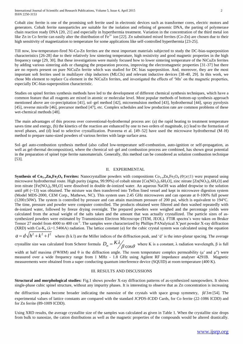
Transmission electron microscope (TEM) analysis: The Selected Area Electron Diffraction (SAED) ring pattern (Fig. 2a) of
the nanoparticles of Co0.4Zn0.6Fe2O4 is well resolved at (220), (311), (400), (422), (511), (440) reflections, as in XRD pattern. The
samples exhibited more or less spherical morphology with uniform size. The lattice spacing of 0.24 nm corresponding to the
(311) plane confirms the presence of crystalline Co0.4Zn0.6Fe2O4 particles (Fig. 2b). From Fig. 2c, TEM images of the particles are
well separated from each other. The particle sizes of these samples are compared with those calculated from XRD and are listed in
Table 1.
Fig. 1. XRD pattern of the Co1-xZnxFe2O4 (0≤ x ≤1) nanoparticles synthesized at temperature 160°C for 15 min.
Page 4
International Journal of Scientific and Research Publications, Volume 5, Issue 4, April 2015 4 ISSN 2250-3153
www.ijsrp.org
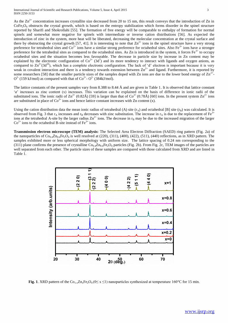
Table 1. Variation of particle size and lattice constant (a) of Co1-xZnxFe2O4 (0≤ x ≤1)
(a) (b)
Fig. 2 (a). SAED pattern of Co0.4Zn0.6Fe2O4. (b) High magnification HRTEM image of Co0.4Zn0.6Fe2O4
showing the (311) oriented lattice planes.
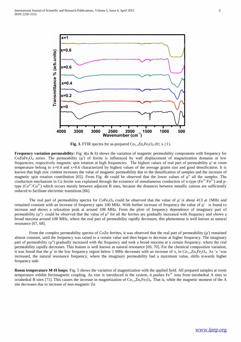
Fourier transform infrared spectroscopy (FTIR): FTIR spectra of as-synthesized nanopowders are presented in Fig. 3. Two
main broad metal-oxygen bands are seen in the FTIR spectra of all spinels, and ferrites in particular. The highest one, υ1, is
generally observed in the range 600–500 cm-1
, and it corresponds to intrinsic stretching vibrations of the metal at the tetrahedral
site (Td), Mtetra↔O, whereas the υ2 lowest band is usually observed in the range 430–385 cm-1
, is assigned to octahedral metal
stretching (Oh), Mocta↔O [61-63, 4, 1]. We observed the band at 600 cm-1
is assigned as υ1 (Mtetra↔O), near 400 cm-1
, υ2 (430–
385 cm-1
) and below 400 cm-1
. The absorption bands observed at ~3450 and ~1630 cm-1
prove the presence of adsorbed water on
the surface of the ferrite nanopowders. The small absorption band at around 1380 and 1720 cm-1
in the as-prepared material may
be assigned to the unreacted metallic salts and carbonyl respectively. Consequently, the υ1 band observed at 561 cm-1
for ZnFe2O4
can be assigned to tetrahedral Zn2+
stretching and the υ2 band observed at 425 cm-1
, involves the Fe3+
vibration at the octahedral
site. The broad band (3600–2500 cm-1
) centered at 3430 cm-1
can be assigned to hydrogen-bonded O–H stretching vibration
arising from surface hydroxyl groups on nanoparticles and adsorbed water. It seems that the υ1 band shifts slightly toward the
lower wave numbers with increase in ‘x’ over the composition range and indicating weakening of the metal–oxygen bonds in the
tetrahedral sites due to the transition between the extent of normal spinel and inverse structures [64].
Composition
‘x'
Particle size
(nm)
TEM
Particle size
(nm)
XRD
a (Å)
0 15.2 12 8.388
0.2 14.5 15 8.39
0.4 13.8 18 8.41
0.6 19.4 20 8.42
0.8 8.43
1 8.44
Page 5
International Journal of Scientific and Research Publications, Volume 5, Issue 4, April 2015 5 ISSN 2250-3153
www.ijsrp.org
Fig.2c. TEM images of Co1-xZnxFe2O4 (0≤x≤1) nanoparticles.
50 nm x=0 x=0.2
x=0.4
50 nm
50 nm 50 nm x=0.6
50 nm
x=0.8 x=1 50 nm
Page 6
International Journal of Scientific and Research Publications, Volume 5, Issue 4, April 2015 6 ISSN 2250-3153
www.ijsrp.org
Fig. 3. FTIR spectra for as-prepared Co1-xZnxFe2O4 (0≤ x ≥1).
Frequency variation permeability: Fig. 4(a & b) shows the variation of magnetic permeability components with frequency for
CoZnFe2O4 series. The permeability (μ') of ferrite is influenced by wall displacement of magnetization domains at low
frequencies, respectively magnetic spin rotation at high frequencies. The highest values of real part of permeability μ' at room
temperature belong to x=0.4 and x=0.6 characterized by highest values of the average grains size and good densification. It is
known that high zinc content increases the value of magnetic permeability due to the densification of samples and the increase of
magnetic spin rotation contribution [65]. From Fig. 4b could be observed that the lower values of μ" all the samples. The
conduction mechanism in Co ferrite was explained through the existence of simultaneous conduction of n-type (Fe3+
/Fe2+
) and p-
type (Co2+
/Co3+
) which occurs mainly between adjacent B sites, because the distances between metallic cations are sufficiently
reduced to facilitate electronic transitions [66].
The real part of permeability spectra for CoFe2O4 could be observed that the value of µ' is about 413 at 1MHz and
remained constant with an increase of frequency upto 100 MHz. With further increase of frequency the value of µ' is found to
increase and shows a relaxation peak at around 108 MHz. From the plots of frequency dependence of imaginary part of
permeability (μ'') could be observed that the value of μ'' for all the ferrites are gradually increased with frequency and shows a
broad maxima around 108 MHz, where the real part of permeability rapidly decreases, this phenomena is well known as natural
resonance [67, 68].
From the complex permeability spectra of CoZn ferrites, it was observed that the real part of permeability (µ') remained
almost constant, until the frequency was raised to a certain value and then began to decrease at higher frequency. The imaginary
part of permeability (μ'') gradually increased with the frequency and took a broad maxima at a certain frequency, where the real
permeability rapidly decreases. This feature is well known as natural resonance [69, 70]. For the chemical composition variation,
it was found that the µ' in the low frequency region below 1 MHz decreases with an increase of x, in Co1-xZnxFe2O4. As ‘x ‘was
increased, the natural resonance frequency, where the imaginary permeability had a maximum value, shifts towards higher
frequency side.
Room temperature M-H loops: Fig. 5 shows the variation of magnetization with the applied field. All prepared samples at room
temperature exhibit ferrimagnetic coupling. As zinc is introduced in the system, it pushes Fe3+
ions from tetrahedral A sites to
octahedral B sites [71]. This causes the increase in magnetization of Co1-xZnxFe2O4. That is, while the magnetic moment of the A
site decreases due to increase of non-magnetic Zn
Page 7
International Journal of Scientific and Research Publications, Volume 5, Issue 4, April 2015 7 ISSN 2250-3153
www.ijsrp.org
Fig 4a. Frequency variation of real part of permeability of Co1-xZnxFe2O4 (0≤x≤1).
Fig 4b. Frequency variation of imaginary part of permeability of Co1-xZnxFe2O4 (0≤x≤1).
ions in the A site, the magnetic moment of the B site increases due to increase of Fe3+
ions in B site. Therefore, in the present
study, when the Zn concentration increases from 0 to 0.2, total magnetization (Moct - Mtet) of Co1-xZnxFe2O4 increases due to the
increase of inter-sublattice A–B super-exchange interaction between the magnetic ions of the sublattices A and B. This increase in
saturation magnetization with Zn concentration from x = 0.0 to x = 0.2 is in good agreement with Neel’s collinear two-sublattice
model [72]. Further, with increase in the Zn concentration from x = 0.2 to x = 1.0, the saturation magnetization gradually
decreases from 79.04 emu/g to 26.04 emu/g. The results obtained are in well accordance with the earlier reported values [73, 74].
The coercivity also decreases with Zn concentration which is attributed to the non-magnetic character of Zn ion. Since more Zn
ions replace with Co ions by increasing ‘x’, the saturation magnetization and coercivity both decrease.
Page 8
International Journal of Scientific and Research Publications, Volume 5, Issue 4, April 2015 8 ISSN 2250-3153
www.ijsrp.org
According to Neel’s two sublattice model of ferromagnetism the magnetic moments of ions on the tetrahedral (A) and octahedral
(B) sites are aligned antiparallel to each other and spins have collinear structure. Therefore, the theoretic magnetic moment per
formula unit in μB, ntB, is described as
ntB(x) = MB(x)-MA(x) (1)
where MB(x) and MA(x) are the B and A sublattice magnetic moments in μB, respectively. The cation distribution of the Co1-
xZnxFe2O4 can be written as
2 3 2 3 2_
(1 x) A (1 x) (1 x) B 4(Zn Fe ) [Co Fe ] Ox
(2)
As a function of Zn concentration, the theoretical magnetic moment values, ntB, of the Co1-xZnxFe2O4 were calculated using the
cation distribution given in Eq. 2 and the ionic magnetic moments of Fe3+
, Co2+
, and Zn2+
as 4.85μB, 2.78μB and 0 μB,
respectively. The experimental values of the magnetic moments (neB), per unit formula in Bohr magnetron (μB), were calculated
according to the relation [75]:
.
5585
e w s
B
M Mn (3)
where Mw is the molecular weight, Ms is the saturation magnetization and 5585 is the magnetic factor. The variation of the
theoretic magnetic moment and experimental magnetic moment (ntB and n
eB, respectively) with respect to Zn composition for Co1-
xZnxFe2O4 is represented in Table 2. It is observed from the table that the theoretical magnetic moment increases linearly as a
function of Zn concentration, and also the experimental magnetic moment, increases with Zn concentration of up to x = 0.2, and
then gradually decreases with further increase in Zn concentration. This increase in neB with Zn concentration of up to x = 0.2 can
be attributed to the Neel’s collinear two-sublattice model. However, this model is unable to explain the decrease of neB (and hence
Ms) with Zn concentration for x>0.2. The decrease in ntB (and hence Ms) for x>0.2 which results from the existence of non-
collinear spin arrangement in the system could be explained on the basis of the three-sublattice model suggested by Yafet and
Kittel [76].
When higher Co2+
ions are substituted by Zn2+
ions in the Co1-xZnxFe2O4, the magnetic ions of tetrahedral A site are so much
decreased, the dominant inter-sublattice A–B super-exchange interaction becomes weaker and hence the intra-sublattice B–B
super-exchange interaction strengthens, which in turn results in occurrence of random spin canting on the B site with respect to
the direction of spins of the A site. Therefore, it is reasonable to conclude that the canted (non-collinear) spins lead to a decrease
in the values of the experimental magnetic moment (and thus the magnetization) with Zn concentration for x>0.2.
Fig. 5. The magnetic hysteresis loops of Co1-xZnxFe2O4 (0≤ x ≥1) at room temperature.
Page 9
International Journal of Scientific and Research Publications, Volume 5, Issue 4, April 2015 9 ISSN 2250-3153
www.ijsrp.org
Table 2. Data of saturation magnetization (Ms) for RT, magnetic moment (experimental and theoretical), Y–K angle (αYK),
blocking temperature (TB) for Co1-xZnxFe2O4 system.
M-T analysis: Fig. 6 shows zero field cooled (ZFC) and field cooled (FC) curves of Co1-xZnx Fe2O4 (0≤ x ≤1) measured in the
temperature range of 10 - 400 K under an applied field of 50 Oe. Firstly, the samples were cooled down without any external
magnetic field and then the magnetization of samples was recorded during heating up to 400 K under an applied field of 50 Oe.
Later, the samples were cooled down again in an applied field of 50 Oe and then it was recorded during heating up to 400 K under
an applied field of 50 Oe. As the temperature increases in ZFC measurement, firstly, the magnetic zero field cooling (MZFC)
increases and then reaches a maximum value at specific critical temperature, which is called the blocking temperature (TB). Above
TB in the unblocked region, the MZFC monotonically decreases with increasing temperature. This is the characteristic behaviour
of super-paramagnetic materials.
It is clear from Fig. 6, only the samples with Zn concentration of 0.6≤ x ≤1.0 show super-paramagnetic behaviour at RT indicating
that the magnetic interactions between the particles are weak and these samples can be regarded as good candidates for biomedical
applications. The ZFC and FC measurements of all the samples except for the samples with Zn concentration (x = 0.0 and 0.2)
show an irreversible magnetic behaviour below the temperature, called irreversibility (Tirr) temperature. Tirr is defined as the
temperature at which the ZFC and FC curves split from each other and corresponds to the blocking temperature of the largest
nanoparticles in the super-paramagnetic systems [77].
Fig. 6. Zero field cooled (ZFC) and field cooled (FC) curves of the Co1-xZnxFe2O4 under an applied magnetic field of 50 Oe.
‘x’ Ms(emu/g) RT neB(μB) n
tB (μB) TB(K)
0 68.71 3.12 2.98 -
0.2 79.04 3.25 4.2 -
0.4 57.7 2.56 5.9 350
0.6 50.82 2.12 7.2 245
0.8 39.12 0.98 8.8 110
1 26.04 0.2 10.05 49.5
Page 10
International Journal of Scientific and Research Publications, Volume 5, Issue 4, April 2015 10 ISSN 2250-3153
www.ijsrp.org
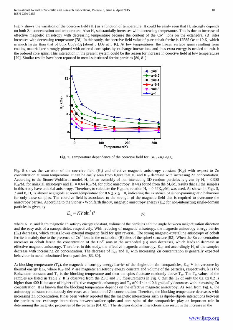
Fig. 7 shows the variation of the coercive field (Hc) as a function of temperature. It could be easily seen that Hc strongly depends
on both Zn concentration and temperature. Also Hc substantially increases with decreasing temperature. This is due to increase of
effective magnetic anisotropy with decreasing temperature because the content of the Co2+
ions on the octahedral (B) sites
increases with decreasing temperature [78]. In this study, the coercive field value of pure cobalt ferrite is 12585 Oe at 10 K, which
is much larger than that of bulk CoFe2O4 (about 5 kOe at 5 K). At low temperatures, the frozen surface spins resulting from
coating material are strongly pinned with ordered core spins by exchange interactions and thus extra energy is needed to switch
the ordered core spins. This interaction in the present system could be the reason for increase in coercive field at low temperatures
[79]. Similar results have been reported in metal-substituted ferrite particles [80, 81].
Fig. 7. Temperature dependence of the coercive field for Co1-xZnxFe2O4.
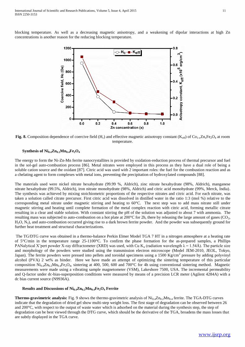
Fig. 8 shows the variation of the coercive field (Hc) and effective magnetic anisotropy constant (Keff) with respect to Zn
concentration at room temperature. It can be easily seen from figure that Hc and Keff decrease with increasing Zn concentration.
According to the Stoner-Wohlfarth model, Hc for an assembly of non-interacting 3D random particles is given by Hc = 0.985
Keff/Ms for uniaxial anisotropy and Hc = 0.64 Keff/Ms for cubic anisotropy. It was found from the Mr/Ms results that all the samples
in this study have uniaxial anisotropy. Therefore, to calculate the Keff, the relation Hc = 0.64Keff/Ms was used. As shown in Figs. 5,
7 and 8, Hc is almost negligible at room temperature for 0.6 ≤ x ≤ 1.0, indicating the existence of super-paramagnetic behaviour
for only these samples. The coercive field is associated to the strength of the magnetic field that is required to overcome the
anisotropy barrier. According to the Stoner - Wohlfarth theory, magnetic anisotropy energy (EA) for non-interacting single-domain
particles is given by
2sinAE KV (5)
where K, V, and θ are magnetic anisotropy energy constant, volume of the particles and the angle between magnetization direction
and the easy axis of a nanoparticles, respectively. With reducing of magnetic anisotropy, the magnetic anisotropy energy barrier
(EA) decreases, which causes lower external magnetic field for spin reversal. The strong magneto-crystalline anisotropy of cobalt
ferrite is mainly due to the presence of Co2+
ions in the octahedral (B) sites of the spinel structure [82]. When the Zn concentration
increases in cobalt ferrite the concentration of the Co2+
ions in the octahedral (B) sites decreases, which leads to decrease in
effective magnetic anisotropy. Therefore, in this study, the effective magnetic anisotropy, Keff and accordingly Hc of the samples
decrease with increasing Zn concentration. The decrease of Keff and Hc with increasing Zn concentration is generally expected
behaviour in metal-substituted ferrite particles [83, 80].
At blocking temperature (TB), the magnetic anisotropy energy barrier of the single-domain nanoparticles, Keff V is overcome by
thermal energy kTB, where Keff and V are magnetic anisotropy energy constant and volume of the particles, respectively, k is the
Boltzmann constant and TB is the blocking temperature and then the spins fluctuate randomly above TB. The TB values of the
samples are listed in Table 2. It is observed from the ZFC and FC measurements in Fig. 6 that the TB of only the 0≤ x≤ 0.2 is
higher than 400 K because of higher effective magnetic anisotropy and TB of 0.4 ≤ x ≤ 0.6 gradually decreases with increasing Zn
concentration. It is known that the blocking temperature depends on the effective magnetic anisotropy. As seen from Fig. 6, the
anisotropy constant continuously decreases as a function of Zn concentration. Therefore, the blocking temperature decreases with
increasing Zn concentration. It has been widely reported that the magnetic interactions such as dipole–dipole interactions between
the particles and exchange interactions between surface spins and core spins of the nanoparticles play an important role in
determining the magnetic properties of the particles [84, 85]. The stronger dipolar interactions also result in the increase in the
Page 11
International Journal of Scientific and Research Publications, Volume 5, Issue 4, April 2015 11 ISSN 2250-3153
www.ijsrp.org
blocking temperature. As well as a decreasing magnetic anisotropy, and a weakening of dipolar interactions at high Zn
concentrations is another reason for the reducing blocking temperature.
Fig. 8. Composition dependence of coercive field (Hc) and effective magnetic anisotropy constant (Keff) of Co1-xZnxFe2O4 at room
temperature.
Synthesis of Ni0.4Zn0.2Mn0.4Fe2O4
The energy to form the Ni-Zn-Mn ferrite nanocrystallites is provided by oxidation-reduction process of thermal precursor and fuel
in the sol-gel auto-combustion process [86]. Metal nitrates were employed in this process as they have a dual role of being a
soluble cation source and the oxidant [87]. Citric acid was used with 2 important roles: the fuel for the combustion reaction and as
a chelating agent to form complexes with metal ions, preventing the precipitation of hydroxylated compounds [88].
The materials used were nickel nitrate hexahydrate (99.99 %, Aldrich), zinc nitrate hexahydrate (98%, Aldrich), manganese
nitrate hexahydrate (99.5%, Aldrich), iron nitrate monohydrate (98%, Aldrich) and citric acid monohydrate (99%, Merck, India).
The synthesis was achieved by mixing stoichiometric proportions of the respective nitrates and citric acid. For each nitrate, was
taken a solution called citrate precursor. First citric acid was dissolved in distilled water in the ratio 1:3 (mol %) relative to the
corresponding metal nitrate under magnetic stirring and heating to 60°C. The next step was to add mass nitrate still under
magnetic stirring and heating until complete formation of the metal complex reaction with citric acid, forming metallic citrate
resulting in a clear and stable solution. With constant stirring the pH of the solution was adjusted to about 7 with ammonia. The
resulting mass was subjected to auto-combustion on a hot plate at 200°C for 2h, there by releasing the large amount of gases (CO2,
H2O, N2), and auto-combustion occurred giving rise to a dark brown ferrite powder. And the powder was subsequently ground for
further heat treatment and structural characterizations.
The TG/DTG curve was obtained in a thermo-balance Perkin Elmer Model TGA 7 HT in a nitrogen atmosphere at a heating rate
of 5°C/min in the temperature range 25-1100°C. To confirm the phase formation for the as-prepared samples, a Phillips
PANalytical X’pert powder X-ray diffractometer (XRD) was used, with Cu Kα (radiation wavelength λ = 1.54Å). The particle size
and morphology of the powders were studied using the transmission electron microscope (Model JEM-2010, JEOL, Tokyo,
Japan). The ferrite powders were pressed into pellets and toroidal specimens using a 1500 Kg/cm2 pressure by adding polyvinyl
alcohol (PVA) 2 wt% as binder. Here we have made an attempt of optimizing the sintering temperature of this particular
composition Ni0.4Zn0.2Mn0.4Fe2O4, sintering at 400, 500, 600 and 700°C for 4h using conventional sintering method. Magnetic
measurements were made using a vibrating sample magnetometer (VSM), Lakeshore 7500, USA. The incremental permeability
and Q-factor under dc-bias-superposition conditions were measured by means of a precision LCR meter (Agilent 4284A) with a
dc bias current source (N9936A).
Results and Discussions of Ni0.4Zn0.2Mn0.4Fe2O4 Ferrite
Thermo-gravimetric analysis: Fig. 9 shows the thermo-gravimetric analysis of Ni0.4Zn0.2Mn0.4 ferrite. The TGA-DTG curves
indicate that the degradation of dried gel show multi-step weight loss. The first stage of degradation can be observed between 26
and 200°C, with respect to the output of waste water which is adsorbed on the material during the synthesis step, the step of
degradation can be best viewed through the DTG curve, which should be the derivative of the TGA, broadens the mass losses that
are subtly displayed in the TGA curve.
Page 12
International Journal of Scientific and Research Publications, Volume 5, Issue 4, April 2015 12 ISSN 2250-3153
www.ijsrp.org
The second stage of degradation occurs between 200-550°C, which is related to structural water and traces of NO, NO2, CO and
CO2. The third stage of degradation occurs between 550 and 1000°C, this loss is related to the output of volatile organics still
remaining in the material after 1000°C. A continuous line in the TGA curve is related to the end of the degradation of organic
matter and the formation of oxides can be observed indicating formation of 100% crystalline phase and ceramic yield of about
90%.
In aqueous chemistry, the degree of condensation varies with different ligand types and the formation of ligands in turn depends
on the pH region [89]. As the pH value of the aqueous solution was modified to 7, better condensation process during hydrolysis
provided single step decomposition to form the nanocrystallites. The weight loss for precursor occurs in two steps. In fact, O-H
groups, carboxyl group and NO3-
ions exist in the dried gel [90]. The first weight loss represents the water vaporization of O-H
groups. In the second step, a large weight loss represents the decomposition of carboxyl, NO3-
ions and NH4NO3, and at the same
time, auto-combustion occurred. The decomposition of NH4NO3 supplied oxygen to accelerate the combustion reaction [91]. As
the decomposition of NH4NO3 is endothermic process, the combustion temperature is decreased and the amount of heat generated
in the combustion process is also decreased. Therefore, the decomposition of NH4NO3 favours the formation of small size
crystallites in this synthesis.
Fig. 9. TGA-DTG of Ni0.4Zn0.2Mn0.4Fe2O4 of dried gel.
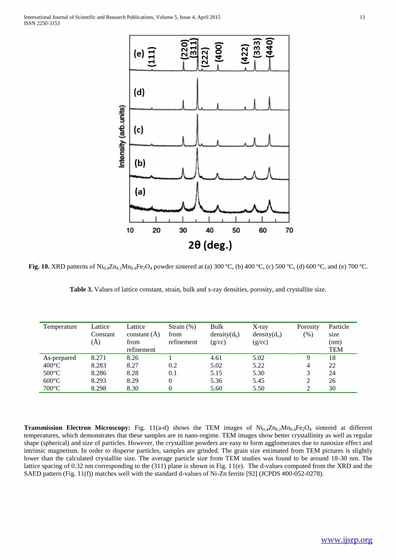
X-ray diffraction and Rietveld refinement: Fig.10. shows the X-ray diffraction of ferrite Ni0.4Zn0.2Mn0.4Fe2O4 sintered at 300ºC,
400 ºC, 500ºC, 600ºC and 700ºC for 4h. The peaks indicate the formation of single phase spinel. The formation of single phase
spinel type was achieved by controlling the atmosphere using argon. By analyzing only the ferrite phase, an increase in peak
intensity with increasing calcination temperature can be noted. This is due to the fact that diffusion occurs more readily at higher
temperatures, resulting in better organization of the system, which increases the amount of crystalline phase formation observed in
the diffraction peaks with better definition and more intensity.
It could be seen from the table 3 that as the sintering increases there is increase in the average crystallite size (D) from 30 nm to 40
nm. Increasing the sintering temperature causes a greater diffusion between grains, increasing the average size of the crystals. The
value of lattice constant increases from 8.271 Å to 8.298 Å with an increase of sintering temperature and the lattice parameters are
matching well with the reported values.
Page 13
International Journal of Scientific and Research Publications, Volume 5, Issue 4, April 2015 13 ISSN 2250-3153
www.ijsrp.org
Fig. 10. XRD patterns of Ni0.4Zn0.2Mn0.4Fe2O4 powder sintered at (a) 300 ºC, (b) 400 ºC, (c) 500 ºC, (d) 600 ºC, and (e) 700 ºC.
Table 3. Values of lattice constant, strain, bulk and x-ray densities, porosity, and crystallite size.
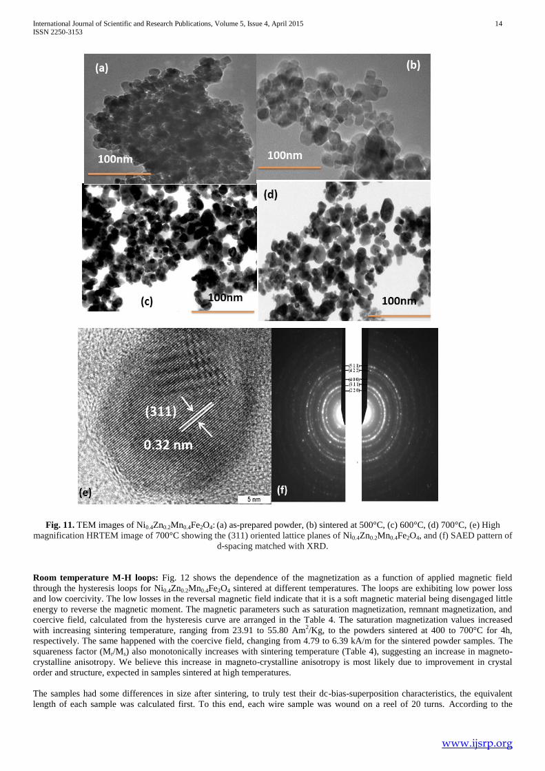
Transmission Electron Microscopy: Fig. 11(a-d) shows the TEM images of Ni0.4Zn0.2Mn0.4Fe2O4 sintered at different
temperatures, which demonstrates that these samples are in nano-regime. TEM images show better crystallinity as well as regular
shape (spherical) and size of particles. However, the crystalline powders are easy to form agglomerates due to nanosize effect and
intrinsic magnetism. In order to disperse particles, samples are grinded. The grain size estimated from TEM pictures is slightly
lower than the calculated crystallite size. The average particle size from TEM studies was found to be around 18-30 nm. The
lattice spacing of 0.32 nm corresponding to the (311) plane is shown in Fig. 11(e). The d-values computed from the XRD and the
SAED pattern (Fig. 11(f)) matches well with the standard d-values of Ni-Zn ferrite [92] (JCPDS #00-052-0278).
Temperature Lattice
Constant
(Å)
Lattice
constant (Å)
from
refinement
Strain (%)
from
refinement
Bulk
density(db)
(g/cc)
X-ray
density(dx)
(g/cc)
Porosity
(%)
Particle
size
(nm)
TEM
As-prepared 8.271 8.26 1 4.61 5.02 9 18
400°C 8.283 8.27 0.2 5.02 5.22 4 22
500°C 8.286 8.28 0.1 5.15 5.30 3 24
600°C 8.293 8.29 0 5.36 5.45 2 26
700°C 8.298 8.30 0 5.60 5.50 2 30
Page 14
International Journal of Scientific and Research Publications, Volume 5, Issue 4, April 2015 14 ISSN 2250-3153
www.ijsrp.org
Fig. 11. TEM images of Ni0.4Zn0.2Mn0.4Fe2O4: (a) as-prepared powder, (b) sintered at 500°C, (c) 600°C, (d) 700°C, (e) High
magnification HRTEM image of 700°C showing the (311) oriented lattice planes of Ni0.4Zn0.2Mn0.4Fe2O4, and (f) SAED pattern of
d-spacing matched with XRD.
Room temperature M-H loops: Fig. 12 shows the dependence of the magnetization as a function of applied magnetic field
through the hysteresis loops for Ni0.4Zn0.2Mn0.4Fe2O4 sintered at different temperatures. The loops are exhibiting low power loss
and low coercivity. The low losses in the reversal magnetic field indicate that it is a soft magnetic material being disengaged little
energy to reverse the magnetic moment. The magnetic parameters such as saturation magnetization, remnant magnetization, and
coercive field, calculated from the hysteresis curve are arranged in the Table 4. The saturation magnetization values increased
with increasing sintering temperature, ranging from 23.91 to 55.80 Am2/Kg, to the powders sintered at 400 to 700°C for 4h,
respectively. The same happened with the coercive field, changing from 4.79 to 6.39 kA/m for the sintered powder samples. The
squareness factor (Mr/Ms) also monotonically increases with sintering temperature (Table 4), suggesting an increase in magneto-
crystalline anisotropy. We believe this increase in magneto-crystalline anisotropy is most likely due to improvement in crystal
order and structure, expected in samples sintered at high temperatures.
The samples had some differences in size after sintering, to truly test their dc-bias-superposition characteristics, the equivalent
length of each sample was calculated first. To this end, each wire sample was wound on a reel of 20 turns. According to the
Page 15
International Journal of Scientific and Research Publications, Volume 5, Issue 4, April 2015 15 ISSN 2250-3153
www.ijsrp.org
equation: e
e
NINI Hl H
l , where N is the number of wire turns, I is the current, H is the magnetic field, and le is the
equivalent length of the cores, we could change to compensate the difference in equivalent length of each sample and to ensure
that all samples were subject to the same dc-bias-superposition magnetic field.
Fig. 12. Magnetic Hysteresis loops of Ni0.4Zn0.2Mn0.4Fe2O4
Table 4. Remnant magnetization (Mr), saturation magnetization (Ms) and coercive field (Hc) of Ni0.4Zn0.2Mn0.4Fe2O4 sintered at
different temperatures. As temperature increases Mr, Ms and Hc increase.
Magnetic field dependent permeability: Fig. 13 shows the variation of incremental permeability (tested at 100 kHz) with
superposition magnetic field. Permeability first increased for all the samples as the sintering temperature increased, without dc-
bias-superposition, which was mainly due to the increase of sintered density and average grain size. With increasing dc-bias-
superposition magnetic field, the permeability of all samples decreased continuously.
Fig. 13. Variation of permeability with superposition
magnetic field.
Temperature Ms
(Am2/Kg)
Mr
(Am2/Kg)
Hc
(kA/m)
ΔM(Ms-Mr)
(Am2/Kg)
Mr/Ms
400°C 23.91 9.10 4.79 14.81 0.38
500°C 35.87 13.43 5.58 22.44 0.37
600°C 44.26 19.01 5.90 25.25 0.42
700°C 55.80 24.44 6.39 31.36 0.43
Page 16
International Journal of Scientific and Research Publications, Volume 5, Issue 4, April 2015 16 ISSN 2250-3153
www.ijsrp.org
Magnetic field dependent quality factor: Fig. 14 shows the variation of Q-factor (tested at 100 kHz) with superposition
magnetic field for the four samples. The Q-factor first increased with the superposition magnetic field, attaining a maximum at
600 A/m, and then it continuously decreased upon further increasing the superposition magnetic field for all samples. Hence, it
could be concluded that a sample that was well sintered and had an even and relatively small average grain size was favourable for
attaining better performance in terms of dc-bias-superposition characteristics [93].
To demarcate the intrinsic relationship between microstructure and dc-bias-superposition characteristics, Mr, Ms, Hc and density of
the samples were measured. The sample sintered at 400°C has low values of Ms and Mr owing to the fact that it was not well
sintered. As the sintering temperature increases, all the parameters Mr, Ms and Hc increase as well as the density also rises,
minimizing the porosity of the samples.
It is known that higher Ms and ΔM favour a higher incremental permeability [94]. As the temperature increases, permeability
increased and the behaviour is explained above. The Q-factor is determined by density and microstructure. Higher density favours
a higher Q-factor. The microstructure with an even and single domain grain size also favours a higher Q-factor. According to
Snoek’s law, permeability is inversely proportional to the cut-off frequency of ferrites. And the frequency at which the Q-factor
attained the maximum value is proportional to the cut-off frequency of ferrites.
In this study, the Q-factor was tested at 100 kHz, although this was not the frequency at which the peak Q-factor appeared. The
samples could attain higher Q-factor values at less than 100 kHz. With the increase of superposition magnetic field, incremental
permeability decreases; the cut-off frequency and the frequency at which the peak Q-factor appeared gradually shifted to higher
values. Hence, with appropriate superposed magnetic field, the frequency at which the peak Q-factor appeared just shifted to 100
kHz, so the Q-factor increased. Thereafter, the frequency at which the peak Q-factor appeared shifted to higher values, so the Q-
factor at 100 kHz also gradually decreased.
Fig. 14. Variations of Q-factor with superposition magnetic field.
IV. Conclusions
The presence of Zinc ions causes appreciable changes in the structural and magnetic properties of Zn-substituted CoFe2O4
synthesized by microwave hydrothermal route. All the nanocrystals exhibit cubic spinel structure. This extensive study of
magnetic properties of Co-Zn ferrites confirms that these materials are suitable and good candidates for hyperthermia applications.
Zero field cooled (ZFC) and Field cooled (FC) measurements reveal that samples with 0.6≤ x ≥1 have super-paramagnetic
behaviour at room temperature, which shows weak interaction between magnetic particles. The blocking temperature obtained
form ZFC-FC curves decreases as Zn concentration increases. Lower reduced remnant magnetization (Mr/Ms) values (x<0.5)
suggest that all the samples have uniaxial anisotropy. It was found that the effective magnetic anisotropy, the coercivity and
remnant magnetization continuously decrease with increasing Zn concentration. The blocking temperature decreases with Zn
concentration.
The results specify that the auto-combustion method was effective in getting the single phase of Ni0.4Zn0.2Mn0.4Fe2O4. The average
crystallite size increased linearly with the calcination temperature. Increasing the crystallite size is related to increased diffusion
effect caused by the increase of the sintering temperature. The hysteresis loops showed soft magnetic behaviour. Consequently,
Page 17
International Journal of Scientific and Research Publications, Volume 5, Issue 4, April 2015 17 ISSN 2250-3153
www.ijsrp.org
the sample that was well sintered and had a microstructure that consisted of relatively small and even grains favoured the
attainment of better dc-bias-superposition characteristics, including permeability and Q-factor. With the addition of Mn to Ni-Zn
ferrites, there is not much change in its structure, but it increases its permeability and it is stable up to 600 kA/m of dc-bias
superposition magnetic field; so these materials can be used in high frequency power applications as they have very high resonant
frequency.
References
[1] J. Smit, H.P.J. Wijn. Ferrites, Philips Technical Library, Eindhoven, 1959.
[2] V.G. Harris, A. Geiler, Y. Chen, et al. Recent advances in processing and applications of microwave ferrites, Journal of
Magnetism and Magnetic Materials, 321(14) (2009) 2035–2047.
[3] Y. Qu, H. Yang, N. Yang, et al. The effect of reaction temperature on the particle size, structure and magnetic properties of
co-precipitated CoFe2O4 nanoparticles, Materials Letters, 60(29–30) (2006) 3548–3552.
[4] N. Kasapoglu, B. Birsoz, A. Baykal, Y. Koseoglu, M.S. Toprak, Synthesis and magnetic properties of octahedral ferrite
NixCo1 – xFe2O4 nanocrystals. Central European Journal of Chemistry, 5(2) (2007) 570– 580.
[5] S.W. Cao, Y.J. Zhu, G.F. Cheng, et al, ZnFe2O4 nanoparticles: microwave-hydrothermal ionic liquid synthesis and
photocatalytic property over phenol, Journal of Hazardous Materials, 171 (1–3) (2009) 431–435.
[6] Y.L. Liu, Z.M. Liu, Y. Yang, et.al, Simple synthesis of MgFe2O4 nanoparticles as gas sensing materials. Sensors and Actuators
B: Chemical, 107(2) (2005) 600–604.
[7] H. Igarash, K. Okazaki, Effects of porosity and grain size on the magnetic properties of NiZn ferrites, J. Am. Ceram. Soc, 60
(1977) 51-54.
[8] K. Kulikowski, Soft magnetic ferrites — Development or stagnation, J. Magn. Magn. Mater. 41 (1984) 56-62.
[9] P. Ravindranathan, K.C. Patil, Novel solid solution precursor method for the preparation of ultrafine Ni-Zn ferrites, J. Mater.
Sci, 22 (1987) 3261-3264.
[10] T. Abraham, Economics of ceramic magnets, Am. Ceram. Soc. Bull. 73 (1994) 62-65.
[11] P.I. Slick, in Ferromagnetic Materials, vol. 2, ed. by E.P. Wohlforth North-Holland, Amsterdam, 1980, p. 196.
[12] B.V. Bhise, M.B. Dongare, S.A. Patil, S.R. Sawant, X-ray infrared and magnetization studies on Mn substituted Ni-Zn
ferrites, J. Mater. Sci. Lett. 10 (1991) 922-924.
[13] S.A. Saafan, S.T. Assar, B.M. Moharram, M.K. El Nimr, Comparison study of some structural and magnetic
properties of nano-structured and bulk Li–Ni–Zn ferrite samples, J. Magn. Magn. Mater. 322 (2010) 628-632.
[14] X. Lu, G. Liang, Q. Sun, and C. Yang, High-frequency magnetic properties of Ni-Zn ferrite nanoparticles synthesized by a
low temperature chemical method, Mater. Lett, 65 (2011) 674-676.
[15] B. Zhou, Z. Liu, X. Wang, Y. Sui, X. Huang, Z. Lu, W. Su, Effect of SiO2 coating on the magnetic properties of Ni–Zn
ferrite, Physica B: Cond. Matt, 405 (2010) 374-378.
[16] S. Rohilla, S. Kumar, P. Aghamkar, S. Sunder, and A. Agarwal, Investigations on structural and magnetic properties of
cobalt ferrite/silica nanocomposites prepared by the co-precipitation method, J. Magn. Magn. Mater. 323 (2011) 897-902.
[17] G. A. Sawatzky, F van der Woude, and A. H. Morrish, Mössbauer Study of Several Ferrimagnetic Spinels, Phys. Rev. 187
(1969) 747-757.
[18] G. A. Pettit and D. W. Forester, Mossbauer study of Co-Zn ferrites, Phys. Rev. B 4 (1971) 3912-3923.
[19] T. Sato, K. Haneda, M. Seki, and T. Iijima, Morphology and magnetic properties of ultrafine ZnFe2O4 particles, Appl. Phys.
A: Mater. Science and Processing. 50 (1990) 13-16.
[20] D .Hork, B. Rittich, A. Spanov, D. Horak, and A. Spanova, Carboxyl-functionalized magnetic microparticle carrier for
isolation and identification of DNA in dairy products, Journal of Magnetism and Magnetic Materials 311 (2007) 249–254.
Page 18
International Journal of Scientific and Research Publications, Volume 5, Issue 4, April 2015 18 ISSN 2250-3153
www.ijsrp.org
[21] J. Prodelalova, B. Rittich, A. Spanova, K. Petrova, and M. J. Benes, Isolation of genomic DNA using magnetic cobalt ferrite
and silica particles, J. Chromatography A1056 (2004) 43- 48.
[22] Y. Koseoglu, M. Bay, M.Tan, A. Baykal, H. Sozeri, R. Topkaya, and N. Akdogan, Magnetic and dielectric properties of
Mn0.2Ni0.8Fe2O4 nanoparticles synthesized by PEG-assisted hydrothermal method, J. Nanoparticle Res, 13 (2010) 2235-
2244.
[23] G. Vaidyanathan, S. Sendhilnathan, Characterization of Co1-xZnxFe2O4 nanoparticles synthesized by co-precipitation method,
Physica B: Condensed Matter 403 (2008) 2157-2167.
[24] I. Sharifi, H. Shokrollahi, S. Amiri, Ferrite-based magnetic nanofluids used in hyperthermia applications, J. Magn. Magn.
Mater, 324 (2012) 903-915.
[25] V.S.S.P. Gaikwad, H.S. Potdar, V. Ravi, and S.R. Dhage, Co-precipitation method for the preparation of nanocrystalline
ferroelectric SrBi2Nb2O9 ceramics J. Electro cera. 14 (2005) 83-87.
[26] Hua Su, Xiaoli Tang, Huaiwu Zhang, Yulan Jing, and Zhiyong Zhong, Effects of Nb2O5 on DC-Bias-Superposition
Characteristic of the Low-Temperature-Fired NiCuZn Ferrites IEEE Trans. Magn. 49 (2013) 4222-4225.
[27] Shuoqing Yan, Li Dong, Zhongyan Chen, Xian Wang, Zekun Feng, Shuoqing Yan, Li Dong, Zhongyan Chen, Xian
Wang, Zekun Feng, The effect of the microstructure on the DC-bias superposition characteristic of NiCuZn ferrite, J.
Magn. Magn. Mate, 353 (2014) 47-50.
[28] Hua Su, Huaiwu Zhang, Xiaoli Tang, Baoyuan Liu, Zhiyong Zhong, Effects of Co-substitution on DC-bias-
superposition characteristic of the NiCuZn ferrites, Physica B, 405 (18) (2010) 4006-4009.
[29] Hai-Bo Wang, Jin Hong Liu, Wen Fing Li, Jian-Bo Wang, Li Wang, Li-Jing Song, Shi-Jun Yuan, Fa-Shen Li, and F.S. Li,
Structural, dynamic magnetic and dielectric properties of Ni0.15Cu0.2Zn0.65Fe2O4 ferrite produced by NaOH co-precipitation
method, J. Alloys Compd, 461 (1-2) (2008) 373-377.
[30] Hua Su, Xiaoli Tang, Huaiwu Zhang, Yulan Jing, Zhiyong Zhong, Low-temperature-fired NiCuZn ferrites with BBSZ
glass, J. Magn. Magn. Mater, 323(5) (2011) 592-595.
[31] J. Murbe and J. Topfer, Ni-Cu-Zn Ferrites for low temperature firing: II. Effects of powder morphology and Bi2O3 addition
on microstructure and permeability, J. Electroceramics, 16 (2006) 199-205.
[32] T. Krishnaveni, B. Rajini Kanth, V. Seetha Rama Raju and S. R. Murthy, Fabrication of multilayer chip inductors
using Ni–Cu–Zn ferrites, J. Alloys Compd, 414 (1-2) (2006) 282-286.
[33] O. F. Caltun, L. Spinu, A. Stancu, L. D. Thung, and W. Zhou, Study of the microstructure and of the permeability
spectra of Ni–Zn–Cu ferrites, J. Magn. Magn. Mater, 242 (2002) 160-162.
[34] Wei-Chih Hsu, S.C Chen, P.C Kuo, C.T Lie, W.S Tsai, Preparation of NiCuZn ferrite nanoparticles from chemical co-
precipitation method and the magnetic properties after sintering, Mater. Sci. Eng. B. 111 (2004) 142-149.
[35] Yao Li, Jiupeng Zhao, Jiecai Han, Xiaodong He, Combustion synthesis and characterization of NiCuZn ferrite powders,
Mater. Res. Bull, 40 (2005) 981-989.
[36] P. K. Roy and J. Bera, Effect of Mg substitution on electromagnetic properties of (Ni0.25Cu0.20Zn0.55)Fe2O4 ferrite
prepared by auto combustion method, J. Magn. Magn. Mater, 298 (1) (2006) 38-42.
[37] Hua Su, Xiaoli Tang, Huaiwu Zhang, Lijun Jia, Zhiyong Zhong, Influences of Fe- deficiency on electromagnetic
properties of low-temperature-fired NiCuZn ferrites, J. Magn. Magn. Mater, 322(13) (2010) 1779-1783.
[38] S.H. Hong, J.H. Park, Y.H. Choa, J. Kim, Magnetic properties and sintering characteristics of NiZn (Ag, Cu) ferrite for
LTCC applications, J. Magn. Magn. Mater. 290-291 (2005) 1559-1562.
[39] Lingjuan Ma, Linshen Chen, Songying Chen, Study on the characteristics and activity of Ni–Cu–Zn ferrite for
decomposition of CO2, Mater. Chem. Phys. 114 (2009) 692-696.
[40] Hua Su, Huaiwu Zhang, Xiaoli Tang, Yulan Jing, Effects of nanocrystalline ferrite particles on densification and magnetic
properties of the NiCuZn ferrites, J. Mater. Sci. 42(8) (2007) 2849-2853.
Page 19
International Journal of Scientific and Research Publications, Volume 5, Issue 4, April 2015 19 ISSN 2250-3153
www.ijsrp.org
[41] I.H. Gul, W. Ahmed, A. Maqsood, Electrical and magnetic characterization of nanocrystalline Ni–Zn ferrite synthesis by
coprecipitation route, Journal of Magnetism and Magnetic Materials, 320(3–4) (2008) 270–275.
[42] S. Zahi, M. Hashim, A.R. Daud, Synthesis, magnetic properties and microstructure of Ni–Zn ferrite by sol–gel technique,
Journal of Magnetism and Magnetic Materials, 308(2) (2007) 177–182.
[43] A. Košak, D. Makovec, A. Žnidaršič A, et al, Preparation of Mn-Zn ferrite with microemulsion technique, Journal of the
European Ceramic Society, 24(6) (2004) 959–962.
[44] X. Jiao, D. Chen, Y. Hu, Hydrothermal synthesis of nanocrystalline Mx(Zn1 – x)Fe2O4 (M = Ni, Mn, Co; x = 0.40–0.60)
powders, Materials Research Bulletin, 37(9) (2002) 1583–1588.
[45] A. Takayama, M. Okuya, S. Kaneko, Spray pyrolysis deposition of NiZn ferrite thin films, Solid State Ionics, 172(1–4)
(2004) 257– 260.
[46] S.Thakur, S.C. Katyal, M. Singh, Structural and magnetic properties of nano nickel–zinc ferrite synthesized by reverse
micelle technique. Journal of Magnetism and Magnetic Materials, 321(1) (2009) 1–7.
[47] P.P. Sarangi, S.R. Vadera, M.K. Patra, et al. Synthesis and characterization of pure single phase Ni–Zn ferrite nanopowders
by oxalate based precursor method. Powder Technology, 203(2) (2010) 348–353.
[48] S. Balaji, K. Kalai Selvan, L. John Berchmans, et al. Combustion synthesis and characterization of Sn4+
substituted
nanocrystalline NiFe2O4. Materials Science and Engineering B, 119(2) (2005) 119–124.
[49] Praveena Kuruva, Uma Maheshwara Singh Rajaputra, Srinath Sanyadanam, Ramana Murthy Sarabu, Effect of microwave
sintering on grain size and dielectric properties of barium titanate, Turkish Journal of Physics, 37 (2013) 312-321.
[50] K. Praveena, K. Sadhana, S. Srinath, S.R. Murthy, Effect of TiO2 on electrical and magnetic properties of
Ni0.35Cu0.12Zn0.35Fe2O4 synthesized by the microwave– hydrothermal method, J. Phys. Chem. Solids 74 (2013) 1329–1335.
[51] K. Praveena, K. Sadhana, S.R. Murthy, Microwave-hydrothermal synthesis of Ni0.53Cu0.12Zn0.35Fe2O4/SiO2 nanocomposites
for MLCI Integrated Ferroelectrics, 119(1) (2010) 122-134.
[52] Sadhana Katlakunta, Sher Singh Meena, S. Srinath, M. Bououdina, R. Sandhya, K. Praveena, Improved magnetic properties
of Cr3+
doped SrFe12O19 synthesized via microwave hydrothermal route, Materials Research Bulletin 63 (2015) 58–66.
[53] S.T. Aruna, A.S. Mukasyan, Combustion synthesis and nanomaterials. Current Opinion in Solid State and Materials, Science,
12(3–4) (2008) 44–50.
[54] S. Kumar, V. Singh, S. Aggarwal, U.K. Mandal, and R.K. Kotnala, J. Phys. Chem. C, Influence of processing methodology
on magnetic behavior of multicomponent ferrite nanocrystals 114 (2010) 6272-6280.
[55] Ibrahim Sharifi, and H.Shokrollahi, Nanostructural, magnetic and M¨ossbauer studies of nanosized Co1-xZnxFe2O4
synthesized by co-precipitation, J. Magn. Magn. Mater, 324 (2012) 2397-2403.
[56] A. Navrotsky, and O.J.Kleppa, Thermodynamics of Formation of Simple Spinels J. Inorg. Nucl. Chem. 30 (1968) 479-498.
[57] C. Upadhyay, H.C. Verma, S. Anand, Cation distribution in nanosized Ni–Zn ferrites, J. Appl. Phys, 95 (2004) 5746-5751.
[58] D. Domide, O. Walter, S. Behrens, E. Kaifer, and H.J. Himmel, Synthesis of Heterobimetallic Zn/Co Carbamates: Single-
Source Precursors to Nanosized Magnetic Oxides under Mild Condiditons Eur. J. Inorg. Chem, 2011(6) (2011) 860-867.
[59] M. Sertkol, Y. Koseoglua, A. Baykal, H. Kavas, A. Bozkurt, and M.S. Toprak, Cation distribution and magnetic properties
of Zn doped NiFe2O4 nanoparticles synthesized by PEG- assisted hydrothermal route, J. Alloys Compd, 479 (2009) 49-55.
[60] R.C. Kambale, P.A. Shaikh, S.S. Kamble, and Y.D. Kolekar, Effect of cobalt substitution on structural, magnetic and electric
properties of nickel ferrite, J. Alloys Compd, 478 (2009) 599-603.
[61] A. Baykal, N. Kasapoglu, Y. Koseoglu, A.C. Basaran, H. Kavas, and M.S. Toprak, Microwave- induced combustion
synthesis and characterization of NixCo1−xFe2O4 nanocrystals (x = 0.0, 0.4, 0.6, 0.8, 1.0), Cent. Eur. J. Chem, 6 (2008) 125-
130.
Page 20
International Journal of Scientific and Research Publications, Volume 5, Issue 4, April 2015 20 ISSN 2250-3153
www.ijsrp.org
[62] Y. Koseoglu, A. Baykal, M.S. Toprak, F. Gozuak, A.C. Basaran, and B. Aktas, Synthesis and characterization of ZnFe2O4
magnetic nanoparticles via a PEG-assisted route, J. Alloy Compd. 462 (2008) 209-213.
[63] A. Baykal, N. Kasapoglu, Y. Koseoglu, M.S. Toprak, H. Bayrakdar, CTAB-assisted hydrothermal synthesis of NiFe2O4 and
its magnetic characterization, J. Alloys Compd. 464 (2008) 514-518.
[64] M. Sertkol, Y. Koseoglu, A.Baykal, H. Kavas, A. Bozkurt, and M.S. Toprak, Microwave synthesis and characterization of
Zn-doped nickel ferrite nanoparticles J. Alloys Comp 486 (2009) 325-329.
[65] M.M. Haque, M. Huq, M.A. Hakim, Physica B 404 (2009) 3915–3921.
[66] J.S. Ghodake, R.C. Kambale, S.V. Salvi, S.R. Sawant, S.S. Suryavanshi, Journal of Alloys and Compounds, 486 (2009)
830–834.
[67] K.Praveena, B.Radhika and S.Srinath, Dielectric and magnetic properties of NiFe2-xBixO4 nanoparticles at
microwave frequencies prepared via co-precipitation method, Procedia Engineering, 76 (2014) 1-7.
[68] I.L. Snoek, Physica 4 (1948) 207.
[69] J.L. Snoek, Physica XIV 4 (1948) 207.
[70] T.George, Tado, Rev. Mod. Phys., 25 (1953) 81.
[71] C. Upadhyay, H.C. Verma, S. Anand, Cation distribution in nanosized Ni–Zn ferrites, J. Appl. Phys. 95 (2004) 5746-5751.
[72] L. Neel, C. R. Acad. Sci. Paris, 230 (1950) 375-377.
[73] G. Vaidyanathan, S. Sendhilnathan, R. Arulmurugan, Structural and magnetic properties of Co1−xZnxFe2O4 nanoparticles by
co-precipitation method, J. Magn. Magn. Mater. 313(2) (2007) 293-299.
[74] Misbah Ul Islam, Mazhar Uddin Rana, Tahir Abbas, Study of magnetic interactions in Co–Zn–Fe–O system, Mater. Chem.
Phys. 57 (1998) 190-193.
[75] Y. Yafet, and C. Kittel, Antiferromagnetic Arrangements in Ferrites, Phys. Rev. 87 (2) (1952) 290-294.
[76] A.K.M. Akther Hossain, and M.L. Rahman, Enhancement of microstructure and initial permeability due to Cu substitution in
Ni0.50−xCuxZn0.50Fe2O4 ferrites, J. Magn. Magn. Mater. 323(15) (2011) 1954-1962.
[77] A. Franco Jr and F.C. Silva, High temperature magnetic properties of cobalt ferrite nanoparticles, Appl. Phys. Lett. 96 (17)
(2010) 172505–172508.
[78] B. Martinez, X. Obradors, L. Balcells, A. Rouanet, and C. Monty, Low Temperature Surface Spin-Glass Transition in γ-
Fe2O3 Nanoparticles, Phys. Rev. Lett. 80(1) (1998) 181-184.
[79] Y. Koseoglu, F. Alan, M. Tan, R. Yilgin, and M. Ozturk, Low Temperature Hydrothermal Synthesis and Characterization of
Mn Doped Cobalt Ferrite Nanoparticles, Ceram. Int. 38 (2012) 3625-363.
[80] Y. Melikhov, J.E. Snyder, D.C. Jiles, A.P. Ring, J.A. Paulsen, C.C.H. Lo, and K.W. Dennis, Temperature dependence of
magnetic anisotropy in Mn-substituted cobalt ferrite, J. Appl. Phys. 99 (2006) 08R102-3.
[81] J.H. Fendler, Nanoparticles and Nanostructured Films, Wiley, New York, 1998, p. 81.
[82] S.S. Jadhav, S.E. Shirsath, S.M. Patange, and K.M. Jadhav, Effect of Zn substitution on magnetic properties of
nanocrystalline cobalt ferrite, J. Appl. Phys. 108 (2010) 093920-6.
[83] J. Gao, W. Zhang, P. Huang, B. Zhang, X. Zhang, and B. Xu, Intracellular spatial control of fluorescent magnetic
nanoparticles, J. Am. Chem. Soc. 130(12) (2008) 3710-3711.
[84] S. Pal, S. Chandra, M.H. Phan, P. Mukherjee, and H. Srikanth, Carbon nanostraws: nanotubes filled with superparamagnetic
nanoparticles, Nanotechnology IOP, 20 (48) (2009) 485604 (7 pages).
[85] K. Bhattacharjee, C. K. Ghosh, M. K. Mitra, G. C. Das, S. Mukherjee, Kalyan Kumar Chattopadhyay, Novel synthesis of
NixZn1-xFe2O4(0≤x≤1) nanoparticles and their dielectric properties, J. Nanoparticle Res. 13 (2011) 739-750.
Page 21
International Journal of Scientific and Research Publications, Volume 5, Issue 4, April 2015 21 ISSN 2250-3153
www.ijsrp.org
[86] K. H. Wu, T. H. Ting, M. C. Lia and W. D. Ho, Sol–gel auto-combustion synthesis of SiO2- doped NiZn ferrite by using
various fuels, J. Magn. Magn. Mater, 298 (2006) 25-32.
[87] H. Chyi Ching, W. Tsung Yung, W. Jun, T. Jih Sheng, Development of a novel combustion synthesis method for
synthesizing of ceramic oxide powders, Mater. Sci. Eng. B 111 (2004) 49-56.
[88] J Livage, C. Sanchez, M.Henry and S. Doeuff, The chemistry of the sol-gel process, Solid State Ionics, 32/33 (1989) 633-
638.
[89] C. J. Brinker and G. W. Scherer, Sol-Gel Science, Academic Press, San Diego, 1990, pp. 21-78.
[90] M. Epifani, E. Melissano, G. Pace, M. Schioppa, Precursors for the combustion synthesis of metal oxides from the sol–gel
processing of metal complexes, J. Eur. Ceram. Soc, 27 (2007) 115-123.
[91] C.A. Jackobson (Ed.) Encylopedia of Chemical Reactions, Reinhold Publishing, New York, 1953, vol. V, pp. 175.
[92] Xiaoli Tang, Huaiwu Zhang, Hua Su, Zhiyong Zhong, and Feiming Bai, Influence of Microstructure on the DC-Bias-
Superposition Characteristics of NiZn Ferrites, IEEE Trans. Magn. 47 (2011) 4332-4335.
[93] www.fair-rite.com/newfair/pdf/Directcurrent.pdf for document 'The Effect of Direct Current on the Inductance of a Ferrite
Core', Fair-Rite Product's Catalog, Part Data Sheet, 9477016002, Printed: 2010-11-09.
[94] Sea-Fue Wang, Yuh-Ruey Wang, Thomas C.K Yang, Po-Jeng Wang, Chun-An Lu, Densification and properties of fluxed
sintered NiCuZn ferrites, J. Magn. Magn. Mater. 217(1-3) (2000) 35-43.


![Co2+ substituted Mg–Cu–Zn ferrite: Evaluation of ... · absorbing properties [12]. However, Ni–Cu–Zn ferrites are still facing certain inherent problems like sensitivity to](https://static.documents.pub/doc/80x56/5e57e3e5ae37012e0401be1d/co2-substituted-mgacuazn-ferrite-evaluation-of-absorbing-properties-12.jpg)


