Page 1
Fidei et Veritatis: The Liberty University Fidei et Veritatis: The Liberty University
Journal of Graduate Research Journal of Graduate Research
Volume 2 Issue 1 Article 1
2018
Orotic Aciduria Orotic Aciduria
Aliah L. Fonteh Liberty University, [email protected]
Follow this and additional works at: https://digitalcommons.liberty.edu/fidei_et_veritatis
Part of the Congenital, Hereditary, and Neonatal Diseases and Abnormalities Commons, and the
Nutritional and Metabolic Diseases Commons
Recommended Citation Recommended Citation Fonteh, Aliah L. (2018) "Orotic Aciduria," Fidei et Veritatis: The Liberty University Journal of Graduate Research: Vol. 2 : Iss. 1 , Article 1. Available at: https://digitalcommons.liberty.edu/fidei_et_veritatis/vol2/iss1/1
This Article is brought to you for free and open access by Scholars Crossing. It has been accepted for inclusion in Fidei et Veritatis: The Liberty University Journal of Graduate Research by an authorized editor of Scholars Crossing. For more information, please contact [email protected] .
Page 2
OROTIC ACIDURIA
By Aliah Fonteh
CASE REPORT I1
Overview
Uridine monophosphate synthase (UMPS) is a bifunctional enzyme with the catalytic
sites for orotate phosphoribosyl pyrophosphate transferase (OPRT) and orotidine monophosphate
decarboxylase (OMPDC). The conversion of orotic acid to uridine-5’PO4 is catalyzed by UMPS.
Orotic aciduria can occur whether one or both of these enzymes are impaired. In 1968 when this
case was presented, only three previous patients homozygous for a defect in the enzymes OPRT
and OMPDC had been reported. A pedigree analysis was utilized to focus on the genotype of
family members four generations before the patient. The pedigree revealed family members
heterozygous for hereditary orotic aciduria type I. The pattern of inheritance for hereditary orotic
aciduria type I is autosomal recessive. The enzyme activity for the patient’s father, siblings, and
niece revealed “borderline” enzyme activity.
History, Initial Symptoms, and Laboratory Examination
On April 1, 1965, a 6 lb. 7 ¾ oz. female patient was born to normal 21-year old parents.
Abnormalities in patient hemoglobin leading to anemia were first evidenced after two months of
life. Administration of 30 milligrams (mg) of iron and 50 mg of Vitamin C daily did not improve
the patient’s anemic conditions. After hospital admission to North Carolina Baptist Hospital in
Winston-Salem, NC, the six-month old patient was found to have pallor and bilateral strabismus.
Laboratory analysis, bone marrow analysis, and a peripheral blood smear revealed erythrocyte
dysfunction. Erythroid hyperplasia was also discovered after the bone marrow analysis. The
peripheral blood smear revealed anisocytosis, poikilocytosis, hypersegmentation of granulocytes,
and mild hypochromia. The initial treatment plan was intramuscular administration of 15 mg and
7.5 mg of folic acid for five days each, respectively. Oral administration of folic acid for two
weeks, then 30 microgram (μg) rotations of Vitamin B12 every other day were administered.
Vitamin B12 supplementation was repeated three times.
Diagnostics
Diagnostic exams included erythrocyte enzyme assays and urinary screening tests. The
patient’s OMPDC and OPRT activity was 0.02 and 0.96, respectively. Control values for
OMPDC activity were 0.79 and 1.9, while control values for OPRT were 5.4, 4.1, and 3.6. The
measure of the conversion of orotic acid to uridine-5’PO4 was also studied. Measurement of
these values was 0.02 compared to control values of 0.83, 1.5, and 2.4. Overall, the patient’s
enzyme activity for both OMPDC and OPRT was very low compared to controls.
Symptoms
1
Fonteh: Orotic Aciduria
Published by Scholars Crossing, 2018
Page 3
The patient’s main symptoms included megaloblastic anemia, orotic aciduria and orotic
acid crystallization, growth delays, and developmental delays. Early patient symptoms required
an erythrocyte transfusion twice, and by early 1966, the patient was first observed to have orotic
acid crystalluria. The crystalluria became observable following dehydration and gastroenteritis.
Orotic acid excretion for the patient was 3.89 gm per gram of creatinine, compared to other cases
which may have up to 1,000 times the orotic acid excretion quantity compared to normal adults.
The patient was experiencing growth and length delay because the patient’s weight and length
were in the 10th and 50th percentiles, respectively.
Management and Results of Treatment
At eleven months, the administration of 1.5 gm/day of oral uridine began. Reticulocyte
development spiked within two days, hemoglobin concentration rose from 7.8 gm/100 mL at the
first hospital admission to 12.3 gm/100 mL on the fifth day of therapy, and the leukocyte count
increased to a range of 3900 to 12000 per m3 during therapy. Bone marrow analysis after two
months did not reveal any abnormalities. In addition, the child’s development was evident due to
increased activity and appetite. Compared to the weight and height percentiles at six months, by
eighteen months the patient was in the 50th and 90th percentiles, respectively. Mental delays
were not present when the patient was assessed at age 2.5.
The daily dosage of 1.5 gm was significant for the reduction in orotic acid output, and
this measure helped to reduce the risk of urinary tract obstruction due to the crystallized orotic
acid. Oral uridine is responsible for reticulocyte development and correction of megaloblastic
changes of the bone marrow. Upon recognition that Vitamin B12 and iron were not correcting
the anemia, the administration of oral uridine may have occurred early enough to prevent further
damage such as mental retardation. Physician’s knowledge of the patient’s history also provided
an earlier diagnosis for this rare disease. Rogers et al. speculate that early therapeutic
management is important for future cases as uridine administration may have a role in reducing
the abnormal effects that can occur in patients. They hypothesize that such effects as mental
retardation and physical growth disturbances can be reversed with early treatment. The patient
clearly exemplifies the benefit of early therapy because by the age of 2.5, the patient had
achieved all developmental milestones for her age. Other cases do not always generate these
results, even with oral uridine treatment.
CASE REPORT II2
Overview
Imaeda et al. was the first to identify a case of hereditary orotic aciduria in Japan. Though
previous patients heterozygous for the condition were phenotypically “normal” except for orotic
aciduria, the case report for the patient (introduced below) exhibited severe symptoms.
Pyrimidines can be found in mother’s milk and are crucial in the neonatal period and in the
duration of breast-feeding. For normal patients, de novo pyrimidine synthesis will provide
sufficient nucleotides important in cellular function and development. However, in a
homozygote for hereditary orotic aciduria, pyrimidine therapy must be administered. This patient
2
Fidei et Veritatis: The Liberty University Journal of Graduate Research, Vol. 2 [2018], Iss. 1, Art. 1
https://digitalcommons.liberty.edu/fidei_et_veritatis/vol2/iss1/1
Page 4
remained deficient in pyrimidines from breast milk and by de novo synthesis for over a week.
After nine days, administration of dietary pyrimidine was added to the milk of the patient.
History, Initial Symptoms, and Laboratory Examination
The family history of the patient in this case includes members heterozygous for
hereditary orotic aciduria (type not specified). The patient’s father demonstrated normal enzyme
activity and was not found to be a carrier for a mutation in UMPS. However, though the patient’s
mother was phenotypically normal, she was found to be a heterozygote carrier. The patient was
also found to be a heterozygote carrier. The patient is a three-year old male born to
nonconsanguineous parents. Born prematurely, the patient was admitted into the NICU (newborn
intensive care unit). Asphyxia and low blood sugar were determined after Apgar test score
results and laboratory examination, respectively. The newborn had low birth weight and was
difficult to feed. Initial computed tomography of the cranium did not signify any abnormal
mental developments.
Symptoms
The patient’s symptoms included orotic aciduria, neurological defects, and
developmental delays. In some cases of orotic aciduria, megaloblastic anemia is affiliated with
orotic aciduria. In the case of this patient, megaloblastic anemia was not identified. By eight
months old, the patient began to experience developmental abnormalities and impaired motor
milestones. At thirteen months, the patient was diagnosed with cerebral palsy (spastic
quadriplegia). In addition, mental retardation progressed in this time frame, becoming most
apparent at age 3. Imaeda et al proposes that insufficient pyrimidine nucleosides in the neonatal
period may have led to severe neurological symptoms in this patient.
Diagnostics
Upon urinary analysis, high levels of orotic acid and orotidine were discovered.
Creatinine excretion is based on 20 mg per kg of body weight. Control patients have orotic acid
levels of 1.1-1.9 μmol per mmol of creatinine, but the patient had levels of 10.5 μmol.
Compared to control values of 0.3 to 1.5 for orotidine, the patient had a value of 2.6 μmol. Both
OPRT and OMPDC activity was very low with a value of 8.3%. Though the father had higher
levels of activity for both enzymes (94.3% and 88.6% respectively), the heterozygous mother
had very low activity. Her activity was documented as 4.4% for OPRT and 7.9% for OMPDC.
Therapy/Management
Since the newborn had difficult feeding and low birth weight, therapy included
intravenous infusion of glucose and minerals. Imaeda et al proposes that for future treatment of
similar cases, oral uridine therapy must be introduced as soon as possible. If possible, treatment
should begin from birth and leading up to the month after birth for the ability to mitigate the
effects of insufficient pyrimidine levels in the cell.
3
Fonteh: Orotic Aciduria
Published by Scholars Crossing, 2018
Page 5
INTRODUCTION
The organic acid 2,6-dioxo-1,2,3,6-tetrahydropyrimidine-4-carboxylic acid is most
commonly known as Orotic acid or “Vitamin B13”. The body can obtain orotic acid exogenously
from dairy products and root vegetables (i.e. carrots, beets) or synthesize orotic acid
endogenously through the pyrimidine synthetic pathway commonly found in hepatocytes,
erythrocytes, and the kidney (See Figure 1).3-6 The mitochondrial enzyme dihydroorotate
dehydrogenase (DHODH) converts dihydroorotate into orotic acid so that uridine
monophosphate synthase (UMPS) can catalyze the conversion of orotic acid to uridine
monophosphate (UMP).6 Orotic acid is mainly found within the cytoplasm of cells.
As an intermediate in pyrimidine synthesis, orotic acid is important for deoxyribonucleic
acid (DNA) and ribonucleic acid (RNA) production, diphosphosugar formation within basement
membranes, and the formation of uridine nucleotides. Though the body can attain pyrimidines
from the diet, a sufficient amount of pyrimidines to regulate the above cellular processes is
required. Moreover, it is the endogenous production of pyrimidines that generates the quantity of
pyrimidines needed to meet the cellular requirement. Thus, when orotic acid is present but not
able to continue on in the pathway for the synthesis of pyrimidines, DNA/RNA production,
basement membrane stability, and uridine nucleotide formation is compromised.
Orotic acid plays a role in the stability of tissues because it is a precursor for uridine
diphosphate formation. The link between orotic acid and uridine diphosphate is due to the role
that diphosphosugars have in the abundant connective tissue, collagen. Collagen, which is an
important protein for strength within connective tissues, requires glycosylation of uridine
diphosphate to form UDP sugar moieties. Thus, uridine diphosphosugars, which are important
components of collagen, require sufficient pyrimidine synthesis to provide normal structural
stability for tissues in the body. Additionally, membranes within the kidney depend on orotic
acid for a similar reason. Both the glomerular and tubular basement membrane depend on the
formation of uridine nucleotides for the formation of thicker membranes. If orotic acid (the
precursor to uridine nucleotides), is not able to enter the pyrimidine pathway, then the strength of
these membranes is reduced. Furthermore, orotic acid is also involved in other key metabolic
pathways, some of which are implicated in certain disease states. Orotic acid is a compound
found to be associated with the pathways linked to β-Ureidopropionase deficiency,
Mitochondrial Neurogastrointestinal Encephalopathy (MNGIE) and dihydropyrimidinase
deficiency.3 Orotic acid may also connected with the following diseases: Canavan disease,
colorectal cancer, Crohn’s disease, and ulcerative colitis.3
Though normal amounts of orotic acid are essential for pyrimidine synthesis and other
crucial biochemical processes, excessive amounts of orotic acid can lead to metabolic acidosis
and have toxic effects on tissues. Excess orotic acid causes harm to tissues because of the role
that orotic acid plays as an acidogen and a metabotoxin.3 An acidogen is a compound that
increases acid concentration, which in large quantities can lead to metabolic acidosis.3 An
increase in hydrogen ion concentration correlates with a decrease in blood pH. If pH levels
decrease, then the range necessary for normal enzyme function is altered. Thus, acidosis will
lead to a compromise of enzyme function and also an impairment of cellular function. Based on
the composition of cells, tissues, organs, and organ systems, when function is impaired at the
enzymatic level, it will also affect the body’s ability to maintain homeostasis and thus normal
function. Acidosis is dangerous because it will change the environment for optimal function of
enzymes within many bodily systems, particularly the cardiovascular and the nervous system. As
4
Fidei et Veritatis: The Liberty University Journal of Graduate Research, Vol. 2 [2018], Iss. 1, Art. 1
https://digitalcommons.liberty.edu/fidei_et_veritatis/vol2/iss1/1
Page 6
a metabolite, an accumulation of orotic acid suggests the role that it can play as a metabotoxin. A
metabotoxin is a compound that releases metabolites that could be damaging or toxic to the cell.3
When orotic acid donates a proton to a conjugate base, it can form a salt and water in this
neutralization reaction. Nonetheless, as an acid, the release of protons will contribute to the
decrease of pH and the increase of toxicity within the plasma.
OROTIC ACID TRANSPORTERS
The solute carrier gene 22 family codes for a subfamily of at least ten transmembrane
organic anion transporters (OATs), organic cation transporters(OCTs), and organic carnitine
transporters(OCTNs).7 The structural components of this subfamily of transporters includes
twelve transmembrane domains, and approximately 540-560 amino acids (See Figure 2).7OATs
are transport proteins responsible for influx of anions between epithelial barriers and fluids
throughout the body. Previously, orotic acid transport in the liver and kidney was unknown.
Evidence that the transporters OAT1, OAT2, OAT4, and hURAT1 play a role in orotic acid
transport has been discovered.7-9
OAT1 is found primarily on the basolateral surface of the proximal tubule of the kidney
as well as in the choroid plexus. Substrates of OAT1 include numerous small xenobiotics,
polycyclic aromatic hydrocarbon (PAH), cyclic nucleotides, α-ketoglutarate, folate, diuretics,
nonsteroidal anti-inflammatory drugs (NSAIDs), indoxyl sulfate, prostaglandin E2, toxins, and
mercurials.7 The mechanism of transport for OAT1 appears to function mainly as an efflux
transporter in drug removal from the plasma. Metabolomic analysis of wild-type OAT and
knock-out OAT mice were extracted and further investigated by reverse-phase liquid
chromatography and mass spectrophotometry.7 The purpose of this research was to determine the
substrates of OAT in vivo. Findings revealed that if OAT1(SLC22A6) is affected, then there is a
reduction in the urinary excretion or orotic acid and orotate.7
OAT2 is found in greatest concentration in the liver, however, it is also found in the
kidney. The substrates for OAT2 includes antivirals, cGMP, acetylsalicylate, prostaglandin E2,
dicarboxylates, glutamate, PAH, and salicylate.7 Orotic acid has also been identified as a specific
substrate for OAT2 (SLC22A7).7 Results from Fork et al. suggest that OAT2 has the following
roles: 1) it is responsible for the efflux of hepatic glutamate from the cytosol 2) it allows the liver
to maintain homeostasis for plasma concentrations of glutamate, and 3) transports orotic acid by
influx (See Figure 3).10 Research studies have confirmed that this function of glutamate efflux
from cells is conserved across the following organisms: human, rat, pig, and mouse.10 Orotic acid
may also be effluxed from hepatocytes by OAT2, and this efflux may be accelerated by
glutamate. If liver failure is occurring, it can impact ureagenesis and the maintenance of
glutamate concentrations in the plasma. OAT2 may be important to investigate for therapeutic
intervention in acute glutamate brain toxicity or to increase glutamate efflux by orotic acid
infusion.
Two other isoforms are of interest to researchers for their role in orotic acid transport-
OAT4 and hURAT1.9 OAT4 can be found in the placenta, kidney, and brain and uses estrone
sulfate, dehydroepiandrosterone sulfate, prostaglandin E2, urate, NSAIDs, antihypertensives,
uric acid, and ochratoxin A as a substrate.7 However, Anzai et al also discovered that the isoform
OAT4 (SLC22A11) transports orotic acid into renal proximal tubular cells.7-9 Nigam et al.
proposes that OAT4 may allow reabsorption of organic acids on the apical membrane of these
proximal tubular cells.7,8 Regulation of OAT4 transport can be mediated by strong inhibitors
5
Fonteh: Orotic Aciduria
Published by Scholars Crossing, 2018
Page 7
such as estrone sulfate, DHEA sulfate, probenecid and benzbromarone.8 Clinically, such
inhibitors must be further investigated due to the impact that they have on the transport of the
substrates listed above.
Lastly, the human urate transporter 1 (hURAT1) can be found in the kidney and utilizes
urate and orotate as a substrate. The gene SLC22A12 codes for the hURAT1, formerly known as
renal specific transporter (Rst).7,9 This transporter was first reported to facilitate the transport of
oratate in renal proximal tubular cells by Miura et al.9 The hURAT1 can be compared to OAT1
due to functional similarities.7Studies on orotic acid transport are relevant for understanding the
mechanism for orotic acid excretion. Particularly, since orotic acid crystal formation could lead
to urinary obstruction, these transporters may be further investigated as possible drug targets in
cases of orotic aciduria.1
ETIOLOGY OF OROTIC ACIDURIA
The causes of orotic aciduria can be divided into four main categories. First, orotic
aciduria caused by a defect in pyrimidine synthesis; second, orotic aciduria caused by a defect in
the urea cycle; third, drug-induced orotic aciduria; and lastly, idiopathic orotic aciduria. The
primary cause of orotic aciduria is also known as hereditary orotic aciduria. Hereditary orotic
aciduria, which has three subtypes (type I, II, and III), is due to a genetic defect which
significantly impairs uridine monophosphate synthetase (UMPS) activity. Reduction in this
activity impairs the production of pyrimidines, important biological macromolecules that are
essential for life. Urea cycle defects which cause orotic aciduria are due to enzymes and
transporters within the urea cycle which are not functioning properly. The urea cycle is the
process used to remove nitrogenous substances from the body. Since an accumulation of
nitrogenous substances will impair the brain and other organ systems, the urea cycle is a very
important process for homeostasis. Drugs such as allopurinol, 5-fluorouracil, and 6-azauridine
are main inhibitors of the pyrimidine pathway and will also induce orotic aciduria. Idiopathic
orotic aciduria occurs when other diseases cause an increase in orotic acid excretion in the urine.
Regardless of the cause generating orotic aciduria, similar symptoms such as motor and mental
impairment may result. Thus, early diagnosis of the condition and the appropriate therapy can
reduce the damage which may be caused by sustained orotic aciduria.
OROTIC ACIDURIA DUE TO DEFECTS IN PYRIMIDINE METABOLISM
A pyrimidine is a six-membered heterocyclic ring containing carbon, nitrogen, and
oxygen. DNA contains pyrimidines such as the nitrogenous bases cytosine (2-hydroxy-4-
aminopyrimidine) and thymine (2, 4-dihydroxy-5-methyl pyrimidine). RNA contains mainly
cytosine and uracil (2, 4-dihydroxypyrimidine). The combination of the nitrogenous base, a
deoxyribose sugar, and a phosphate group form the backbone of DNA. Ribonucleic acid is a
linear structure which contains a nitrogenous base, ribose sugar, and a phosphate group.
Phosphorylation of these bases will generate a nucleotide. A nucleotide is incorporated into the
double helical structure within DNA, versus a single, linear strand for RNA.These bases can also
be mono, di, and triphosphorylated for chemical reactions in the body. Many tissues need to
synthesize pyrimidines for the regulation of cellular identity and function.
Biochemical Significance of Pyrimidines
6
Fidei et Veritatis: The Liberty University Journal of Graduate Research, Vol. 2 [2018], Iss. 1, Art. 1
https://digitalcommons.liberty.edu/fidei_et_veritatis/vol2/iss1/1
Page 8
The cellular machinery within the body maintains a universal code that is essential for the
proper development of organ systems, organs, tissues, cells, and the cellular organelles
governing cell activity. Ultimately, this universal code is affiliated with the macromolecules
deoxyribonucleic acid (DNA) and ribonucleic acid (RNA) which are mandatory for life. The
central dogma of life orchestrates how the instructions for life contained in DNA can be
converted into a message that the cell uses. First, DNA is important for making more copies of
the same cell, and DNA to DNA copying occurs through a process called replication. Second, the
DNA must be converted to RNA through a process known as transcription. Transcription allows
the genetic information (genes) to be prepared for translation to proteins. The translation of RNA
to gene products is vital for the regulation of processes involved in metabolism, growth,
development, aging, disease, etc.
Pyrimidines are important compounds necessary within the cell cycle for the synthesis of
genetic information. Genetic information regulates the activity of all cells in the body because it
contains the manuscript for cellular machinery, is regulated to maintain homeostasis within the
body, and allows for cellular growth and development. Defects in the synthesis of these
macromolecules will impact the quantity of bases available for DNA and RNA synthesis. When
DNA and RNA synthesis is not sufficient, cellular growth and stability is impacted. The
phosphate groups on these nucleotides may also serve to be used in phosphate transfer reactions.
The release of phosphate from adenine triphosphate (ATP) and other nucleotides is an important
source of cellular energy which is required for various biochemical reactions. Biochemical
reactions including the active transport of substances across the cell membrane, the activation of
enzymes, the contraction of skeletal muscle, and the inhibition of certain pathways depend on
phosphate transfer. Specifically, guanine triphosphate (GTP) is a main regulator in protein
synthesis, uridine triphosphate (UTP) provides energy of activation in the reaction of glucose and
galactose, and cytidine triphosphate (CTP) plays a role for energy in lipid metabolism. 11,12
Disruption of the pathway causes problems because the body cannot synthesize these
compounds, and the normal recycling of these compounds for re-incorporation into DNA or
RNA (“salvage pathway”) may also be disrupted or insufficient to provide the needs of the cell.
In addition, β-alanine and β-aminoisobutyrate are formed from the catabolism of
pyrimidines. Both compounds can be converted into substrates in the energy-producing
tricarboxylic acid (TCA) cycle.3,11,12 The TCA cycle produces GTP, NADH, and FADH2. The
TCA cycle is a main producer of 3 molecules of the reducing agents NADH and 2 molecules of
the compound FADH2 which serve as donors to the electron transport enzymes. For each
molecule of NADH, the electron transport chain (ETC) can generate 2.5 moles of ATP. For each
mole of FADH2, about 1.5 moles of ATP. The electron transport enzymes function to generate
ATP through the electrochemical gradient generated in the mitochondrial matrix. ATP, as
discussed previously, is the energy investment required for many biochemical processes to
proceed.
Pyrimidine synthesis is not only important for DNA and RNA production, but also for
other biochemical processes. Pyrimidines are used as nucleotide-lipid cofactors to generate stable
erythrocyte membranes, for cell proliferation, and for the biosynthesis of glycogen and
phospholipids. The role of pyrimidines can also be identified in cell-mediated immunity.6,13 In
early development, rapid pyrimidine synthesis is required for neural maturation and
organogenesis. Thus, mutations with a severe effect on the activity of UMPS can lead to death.14
7
Fonteh: Orotic Aciduria
Published by Scholars Crossing, 2018
Page 9
OVERVIEW OF TYPE I, TYPE II, AND TYPE III OROTIC ACIDURIA
A mutation that disrupts the bifunctional activity of uridine 5’- monophosphate synthase
(UMPS) is the most frequent inborn error of pyrimidine nucleotide synthesis.5 Orotic aciduria
(type I, II, or III) occurs due to this defect in pyrimidine synthesis. This disease was discovered
as a rare autosomal recessive disorder in 1959.5,15,16 Hereditary orotic aciduria can be divided
into three subtypes, two of which are clinically indistinguishable (type I and II) and a third that
does not present with macrocytic hypochromic megaloblastic anemia (See Table 1).16 In type I,
both orotate phosphoribosyl pyrophosphate transferase (OPRT) and orotidine monophosphate
decarboxylase (OMPDC) are defective, whereas in type II, only OMPDC is impacted.16 OPRT
and OMPDC have been found defective in erythrocytes, leukocytes, fibroblasts, and saliva due to
a mutation in the individual catalytic sites of UMPS.13 UMPS activity also plays an important
role in the brain, liver, skeletal muscle, and spleen.14 Defects in pyrimidine synthesis commonly
lead to mental retardation.
Epidemiology of Disease
Hereditary orotic aciduria is a rare autosomal recessive disorder with a prevalence in
infants and children of 1:1,000,000.17 There have been only about twenty identified cases
worldwide.14 Out of these cases, point mutations were discovered in three Japanese families.18
Further information about this rare disorder is limited.
Biochemical Pathology
There are three subtypes of hereditary orotic aciduria. In the most common type, type I
orotic aciduria, 90% of patients experience severe loss of both OPRT and OMPDC activities due
to a deficiency in UMPS.19,20 Studies show that deficient quantities of wild-type UMPS activity
are responsible for the variation in levels of orotate to orotidine in type I orotic aciduria.
Erythrocyte activity of OPRT is absent in type I.13 Type II is clinically indistinguishable from
type I but includes specific inactivation of OMPDC which occurs through an unknown
mechanism.16,19 Cells will have less than 1% activity of OMPDC and an increase in OPRT
activity with the type II subtype.20 The orotate levels are comparable to that of type I, but there is
an increase in the excretion of orotidine. Type III, or hereditary orotic aciduria without
megaloblastic anemia (OAWA), is correlated with a deficiency in OMPDC. The case where this
subtype first presented revealed a homozygous missense mutation within a conserved region of
the OMPDC domain of the UMPS gene (c.928 T4>G; p. Phe310Val).13 In a type III patient, the
levels of OPRT activity are present but remain low.
Carbamoyl phosphate is formed intracellularly from glutamine, bicarbonate, and ATP by
CAD, a cytoplasmic enzyme with active sites for carbamoyl phosphate synthetase II (CPS II),
aspartate transcarbamoylase, and dihydroorotase (See Figure 5).5 The reaction proceeds with
CPS II forming carbamoylaspartate, with enzyme-catalyzed cyclization by aspartate
transcarbamoylase (ATC) to form dihydroorotate, and with dihydroorotate dehydrogenase
(DHODH) oxidation of dihydroorotate to orotic acid. Orotic acid is converted to uridine
8
Fidei et Veritatis: The Liberty University Journal of Graduate Research, Vol. 2 [2018], Iss. 1, Art. 1
https://digitalcommons.liberty.edu/fidei_et_veritatis/vol2/iss1/1
Page 10
monophosphate (UMP) by UMPS.21 The formation of UMP can also occur via a “salvage”
pathway from uracil phosphoribosyltransferase using uracil and 5-phosphoribosyl-1-
pyrophosphate ([PRPP] See Figure 4).22
The enzyme uridine monophosphate synthase (UMPS) is essential in this process because
of its bifunctional regulation of pyrimidine synthesis. This enzyme is involved in the ribosylation
of orotate to orotidine monophosphate (OMP) via orotate phosphoribosyl pyrophosphate
transferase (OPRT). UMPS also decarboxylates OMP to uridine monophosphate with OMPDC.
When the enzyme uridine monophosphate synthase is impaired, there will be a buildup of orotate
(OA) or orotidine monophosphate (OMP). Though OA is permeable, once it crosses the cell
membrane it does not undergo any modifications and is excreted. OMP is impermeable and is
dephosphorylated to form the compound orotidine prior to secretion. Normally, the UMPS
reaction promotes UMP formation and only generates small amounts of cellular OA and urinary
OA. In hereditary orotic aciduria, though more cellular orotate and urinary orotate are being
produced, OA is not able to complete the pathway of pyrimidine synthesis.13
Consecutive phosphorylation of UMP to UDP and UTP occurs by the enzymes uridylate
kinase and nucleoside diphosphate kinase (NDPK) (See Figure 4). Cytidine triphosphate (CTP)
and thymidine triphosphate (TTP) can be formed from uridine triphosphate (UTP). Pyrimidine
synthesis is important for the provision of high energy molecules for biochemical processes and
for DNA and mRNA synthesis. The regulatory processes that govern pyrimidine synthesis
include negative feedback by UTP and positive regulation by PRPP upon the CPS II active site
of CAD (See Figure 4).5,23 Low pyrimidine concentration due to a defect in UMPS will impact
erythrocyte formation and cause megaloblastic anemia.20 Megaloblastic anemia is usually the
first manifestation that will occur for this condition because a defect in synthesis of the
pyrimidine UTP will decrease the synthesis of the other pyrimidines, CTP and TTP as well.18
This condition results in an inadequate amount of nucleotides for DNA synthesis, causing the
bone marrow to produce large cells that are unable to undergo cell division. Since cell division is
impaired, the patient will not be able to develop normal shaped erythrocytes.6,17
Genetic Basis of Disease
Orotic aciduria is a rare, autosomal recessive disorder that can occur due to an inborn
error in metabolism. Hereditary orotic aciduria occurs with an autosomal recessive mutation of
3q21.17,24 The UMPS gene is 480 amino acids long with six exons. While the amino acid
sequence for the UMPS gene has an N-terminal of 214 amino acids and contains OPRT, the C-
terminal of the enzyme is 258 amino acids and contains OMPDC. The underlying genetic defect
may occur due to biallelic missense mutations which lead to amino acid substitutions.24 These
mutations decrease the steady-state levels of UMPS, impair UMPS binding ability, and reduce
UMPS catalytic activity. Variants may include null alleles and missense changes. Upon
investigation of two type I families, researchers discovered that polymorphisms within exon
regions may not reduce the function of OPRT or OMPDC, as discovered with G231A and
440Gpoly.14
Clinical Presentation
9
Fonteh: Orotic Aciduria
Published by Scholars Crossing, 2018
Page 11
Patients with hereditary orotic aciduria may present with symptoms primarily before
twelve months of life.13 Though an alteration in pyrimidine synthesis disrupts many biological
processes, clinical presentation of UMPS defects mainly includes phenotypic expression of
mental retardation and neurological dysfunction.13,25 Deficient quantities of pyrimidines will
impact the stability of erythrocytes, causing macrocytic hypochromic megaloblastic anemia in
type I and II.19 Neurological deficits occur in type I, II, and III because of a decrease in
pyrimidine nucleosides which are converted into nucleotides (CTP, UTP, and TTP). Nucleotides
are important in metabolic reactions and DNA and mRNA synthesis.14 Both type I and II
hereditary orotic aciduria will present with megaloblastic anemia. Type III does not present with
megaloblastic anemia, but it is still unclear as to why the bone marrow does not produce
abnormal erythrocytes in type III patients.22 Heterozygous UMPS-mutations can lead to orotic
aciduria that appears mild or isolated and does not cause clinical abnormalities.24
Signs/Symptoms
When nucleotide synthesis is impaired, as for patients with UMPS-mutations, then the
functions of the nervous system, the renal system, the immune system, and the integumentary
system become compromised. First, insufficient pyrimidines will lead to the manifestation of
symptoms within the nervous system which include growth retardation, developmental delay,
intellectual disability, strabismus, motor impairment, and hypotonia.13,24 Symptoms that may
occur include delayed growth and developmental/psychomotor retardation (type I, II, III),
congenital malformations (type II), and immune deficiencies (type I,II).20 A patient with type III
may often suffer from neurological abnormalities, congenital development concerns, and
developmental delay. However, neurological abnormalities and developmental delays are also
possible in type I and II. Besides neurological symptoms, the abnormal function of the renal,
immune, and integumentary system will also generate symptoms within some patients.
For the renal system, orotic acid crystallizes in the urine because there are large amounts
of insoluble orotic acid present in the kidneys.2,26,27 Moreover, crystallization can lead to renal
failure due to the obstruction of the ureters and urethra by crystallized orotic acid. Other signs
affiliated with this disease include hematuria and splenomegaly.13,27,28 Defects in the immune
system will occur because nucleotides promote the synthesis of cell types that are necessary in
cell-mediated immunity and for defense from bacterial and fungal invasion.29 If nucleotide
synthesis is impaired, then immune function will also become impaired and lead to a reduced
ability to fight infection. Defective pyrimidine synthesis can vary from normal to low T cell
number and T-cell mediated cell death, defects in the delayed type hypersensitivity response, and
reduced T levels of serum IgG and IgA.28 Immunological involvement may also cause
neutropenia, leukopenia, and lymphopenia (particularly due to infections of candidiasis, fatal
varicella, and meningitis).13,22,24,30 In regards to the integumentary system, impaired pyrimidine
synthesis will lead to the generation of scant hair and reduced nail growth. Heterozygous
individuals exemplify orotic aciduria but are asymptomatic.2
Diagnosis
Most inherited defects of pyrimidine metabolism are difficult to diagnose due to the rarity
of this condition, heterogeneous presentations which may appear similar to other disorders, and
their varied phenotypic spectrum which is still under investigation.9 Type I, II, and III orotic
10
Fidei et Veritatis: The Liberty University Journal of Graduate Research, Vol. 2 [2018], Iss. 1, Art. 1
https://digitalcommons.liberty.edu/fidei_et_veritatis/vol2/iss1/1
Page 12
aciduria can be differentiated from a urea cycle disorder, as it is not characterized by
hyperammonemia or an altered amino acid profile. Definitive diagnosis of type I or type II orotic
aciduria is based on megaloblastic anemia without a deficiency in B12 or folic acid as well as
abnormal OMPDC/OPRT activity and hyperoroticaciduria.4 Type III diagnosis will be due to
motor or neurological impairment, as well as to low enzyme activity and hyperoroticaciduria.
Since pyrimidines will be quickly cleared from blood and CSF by the renal system, the
best diagnostic marker for pyrimidine deficiencies is through urine analysis.20 Upon urinary
evaluation, homozygotes may present with more than one millimole(mmol) of orotic acid per
millimole creatinine, whereas disease free individuals have ∼1 micromole (μmol) orotic acid
excreted per mmol creatinine.5 Elevated orotic acid levels include concentrations greater than 60
nanomoles (nmoles) of orotic acid per milligram (mg) of creatine.31 Urinary orotic acid
concentrations of 500-1000x normal in homozygotes with a UMPS mutation will lead to orotic
acid crystals, especially during dehydration.2,5,18 Orotate (OA) will be great in concentration
when the wild-type UMPS is functional. If OMPDC is inhibited, then this factor indicates that
orotidine monophosphate (OMP) is great in concentration. Such tests determine the
concentration of both intracellular amounts of OA and OMP.16An orotate to orotidine ratio above
10 will occur in type I patients. Type II presents with a orotate to orotidine ratio that is lower
than type I due to increased orotidine levels. The urinary analyses of patients with type III
include equimolar orotate to orotidine concentrations, or a ratio of about 1. It is best to diagnose
this disease within the neonatal or infancy period because if pyrimidine nucleoside supply is
reduced, it may be the main cause of neurological symptoms.13,28
Prognosis
If hereditary orotic aciduria is detected early, treatment can be administered for the life-
time of the patient. The prognosis for a patient with type I, II, or III orotic aciduria is good if the
UMPS defect is detected early and if oral uridine treatment is also started early. In addition, due
to early uridine treatment, then normal psychomotor development may be attainable (See Case
Study 1).18 Pyrimidine replacement therapy can lead also to remission and reduced urinary orotic
acid levels.13 Late detection of the disorder may not allow neurological deficits to be reversed.
Late detection may lead to coma, seizures, or death.
Management
Early diagnosis and a distinction between type I and II orotic aciduria, vitamin B12, and
folate deficiency must be made for proper treatment. Hereditary orotic aciduria type I and II
causes hypochromic erythrocytes or megaloblastic bone marrow that are not responsive to
common hematinic treatments of pyridoxine, iron, folic acid, or vitamin B12.13,22 One of the
early cases of congenital orotic aciduria was managed with an oral cytidylic-uridylic compound.
Case Study I discussed earlier also revealed that oral administration of uridine can reduce
symptoms and restore the patient to normal developmental milestones.1 In 2015, the United
States Food & Drug Administration approved the drug Uridine triacetate (Xuriden), which was
developed by Wellstat Therapeutics, Inc. for treatment of hereditary orotic aciduria.32 Xuriden is
the current drug for lifelong treatment of orotic aciduria type I and II.13,33 Oral uridine is an
effective therapeutic agent because it can be ingested in granules with milk, formula, applesauce,
pudding, or yogurt, so it is in a usable form for pyrimidine synthesis.32 The prescription is a
11
Fonteh: Orotic Aciduria
Published by Scholars Crossing, 2018
Page 13
single daily dosage of 60 mg/kg, though as long as the dosage is not more than 8g, the patient
may take up to 120 mg/kg daily.33 Other research discusses the administration of the nucleoside
uridine in doses of 50–300 mg/kg/ daily as effective therapy. Xuriden provides uridine as the
substrate for tissue kinase to form UMP. UMP is the precursor for uracil, cytosil, and thymine
nucleoside synthesis.19 The effectiveness of uridine triacetate to accomplish the completion of the
de novo pyrimidine synthesis pathway is due to its ability to be transported across the blood-
brain-barrier and into cells throughout the body. Additionally, pyrimidine replacement reduces
urinary excretion of orotic acid and generates clinical and hematological remission. Treatment
has not yet been established for type III.
OROTIC ACIDURIA DUE TO UREA CYCLE DEFECTS
Defects in Urea Cycle Enzymes
Orotic aciduria can occur in urea cycle disorders when there is a defect in an enzyme
such that carbamoyl phosphate builds up. The most common defect in ureagenesis is an X-linked
deficiency which correlates with impaired activity of the mitochondrial matrix enzyme ornithine
transcarbamoylase (OTC). 34,35 The prevalence of OTC deficiency has been reported as
1:40,000.34 A defect in ornithine transcarbamoylase (OTC) will generate orotic aciduria because
within the urea cycle, OTC is normally responsible for coupling carbamoyl phosphate with
ornithine to form citrulline.35 If there is a defect in this enzyme, then the levels of carbamoyl
phosphate build up and are shuffled to an alternate pathway, the pathway for orotic acid
synthesis (See Figure 6).23 The more carbamoyl phosphate accumulates, the more carbamoyl
phosphate is pushed into the pathway for the formation of orotic acid. With a deficiency in OTC,
upon a liver biopsy there is low enzyme activity of OTC after enzymatic assay, low levels of
citrulline in serum, and hyperammonemia.35,36 In addition, orotic acid can be found in the urine
of OTC deficient patients.34 The clinical manifestations of OTC deficiency also include lethargy
and neurological deficits such as coma.37
With early diagnosis, particularly if family history is revealed, and treatment of the
hyperammonemia occurs early, then the life expectancy of the patient may increase. When
knowledge of the family history reveals affected individuals, patients should be monitored in the
neonatal period because of an increased risk for coma or death.35 Mental retardation may present
for boys with OTC deficiency forms milder than others. Girls heterozygous for the mutation may
present with milder symptoms compared to boys.34,35,37 Hyperammonemia can generate coma,
ataxia, seizures, and cerebral edema in more serious cases.37 Since hyperglutaminemia may also
accompany the hyperammonemia, it may also be used as a diagnostic tool.35,37 In the case of a 2-
day-old infant whose mother was heterozygous for OTC deficiency, death of the child occurred
due to the impacts of hyperammonemia.34 Symptoms that can confirm OTC deficiency after
birth include lethargy, difficulty feeding, respiratory alkalosis, and encephalopathy.34 Treatment
for this disorder includes the intravenous administration of sodium benzoate, sodium
phenylacetate, or arginine hydrochloride.37
There is a slight elevation in urinary orotic acid levels when the urea cycle enzymes
argininosuccinate lyase (AL), arginase, argininosuccinic synthetase (AS), and arginase are
defective. (See Figure 6).23 The OTC gene is found on chromosome Xp21.1, primarily in the
liver. The AS gene is found on chromosome 9q34 and is expressed in the liver and the skin. AL
can be found on the AL gene of chromosome 7cen-q11.2. Expression of the AL gene is
12
Fidei et Veritatis: The Liberty University Journal of Graduate Research, Vol. 2 [2018], Iss. 1, Art. 1
https://digitalcommons.liberty.edu/fidei_et_veritatis/vol2/iss1/1
Page 14
predominant in the liver, skin, and erythrocytes. The ARG1 gene of chromosome 6q23 codes for
Arginase.
AS is an autosomal recessive deficiency which is characterized by both orotic aciduria and
citrullinemia. With normal AL activity, AL breaks arginosuccinate to fumarate (for the TCA
cycle) and arginine. Abnormal activity will also lead to an accumulation of orotic acid, due to an
accumulation of the precursor products in the pathway (See Figure 6).23 Orotic aciduria also
occurs with the autosomal recessive defect in arginase. Instead of hydrolyzing arginine to
ornithine and urea in the completion of the urea cycle, this rare disorder leads to an accumulation
of ammonia, arginine, and orotic acid.38 Therapy for AS and AL deficiency includes
supplementation of arginine. Arginine supplementation will promote the removal of amino acids
and nitrogen waste products because arginine will allow citrulline and arginosuccinate to be
synthesized.39
In conclusion, it is important to determine whether orotic aciduria is due to a primary or
secondary factor. To distinguish the specific cause of orotic aciduria, defects in urea cycle
enzymes may initially be considered. However, laboratory measurements of orotate and
orotidine can be used in the diagnosis of type I, II, and III hereditary orotic aciduria. Diagnosis
of primary orotic aciduria (type I, II, III) can be confirmed when the patient does not have
hyperammonemia. A lack of hyperammonemia eliminates disorders of urea cycle enzymes such
as ornithinine transcarbamoylase (OTC), argininosuccinate lyase (AL), argininosuccinic
synthetase (AS), and arginase as the precipitating cause for excess orotic acid in the urine.
Defects in Urea Cycle Transporters
A functional deficiency in the transporters Citrin (aspartate-glutamate transporter) and
ORNT-1 (ornithine transporter) will also cause elevated orotic acid levels. There will be normal
arginosuccinic acid (ASA) levels and increased citrulline and arginine plasma levels if either
transporter is defective. The aspartate-glutamate transporter is coded by the gene CITRIN, found
on 7q21.3, and is expressed mainly in the liver. ORNT-1 is coded by the ORNT1 gene which is
found on chromosome 13q14 and expressed in the liver and skin. A second ORNT transporter,
the hepatic ORNT-2, may also impact orotic acid excretion if it is also defective.
DRUG-INDUCED OROTIC ACIDURIA
Patients should avoid the following pharmacogenetic drugs which further affect
pyrimidine metabolism: nifedipine and nimodipine. Nifedipine and nimodipine are calcium
channel blockers that competitively inhibit uridine kinase and OMPDC (See Figure 4).20 Drugs
such as allopurinol, 5-fluorouracil, and 6-azauridine will lead to an increase in urinary orotic acid
content by inhibiting the final steps of the pyrimidine pathway. Allopurinol will compete with
orotic acid for binding to orotate phosphoribosyltransferase. Uracil and de novo pyrimidine
levels reduce because orotate phosphoribosyl transferase generates the oxypurinol nucleotide
from allopurinol, and this nucleotide inhibits orotidylate decarboxylase. The influence of
allopurinol causes a reduction in pyrimidine levels and simultaneous accumulation of orotic acid
and orotidine in the cell, thus spilling over in high concentration to the urine. The anticancer drug
5-fluorouracil will worsen orotic aciduria because it is phosphoribosylated by orotate
phosphoribosyl transferase.
13
Fonteh: Orotic Aciduria
Published by Scholars Crossing, 2018
Page 15
The anticancer drug 6-azauridine (6-AZUR) is converted to 6-azauridylate, and 6-
azauridylate will inhibit orotidylate decarboxylase and promote increased orotic acid and
orotidine excretion (See Figure 7,8). 19,40-42 ATP and Mg2+ are required for phosphorylation of 6-
AZUR.41 Phosphorylated 6-AZUR is responsible for the competitive inhibition of OMPDC.41
Fallon et al. performed urine analyses of orotic acid on six leukemia patients receiving
intravenous 6-AZUR dosages for treatment.42 Their investigation of plasma and urine levels of
orotic acid suggest that highest orotic acid excretion occurs with high dosage of 6-AZUR. These
dosages are 120-200 mg per kg.42 A second discovery was that orotic acid excretion reduced
after three days, whereas orotidine excretion in the urine was still evidenced one to three days
after orotic acid excretion stopped.42 With administration of 6-AZUR, orotidine levels did not
decline constantly as the orotic acid levels, but instead continued at the same maximum excretion
value, even when 6-AZUR was removed.42 Since feedback pathways regulate pyrimidine
synthesis, if 6-AZUR is administered as an anticancer agent, it blocks pyrimidine synthesis and
increases orotic acid production (See Figure 7,8).41,42 Excess orotic acid may be converted to
orotidylic acid. As a result, after conversion to orotidylic acid (orotidine 5’-monophosphate),
orotidylic acid pyrophosphorylase will generate the product orotidine.
IDIOPATHIC OROTIC ACIDURIA
Mitochondrial disorders, hepatic fibrosis, lysinuric protein intolerance, Reye syndrome,
malignancies, and trauma can also induce orotic aciduria.21 Reye syndrome correlates with
damaged mitochondria such that carbamoyl phosphate is channeled into orotic acid synthesis.
Mother’s milk consists of pyrimidine nucleosides, and if feeding is delayed or if an infant
formula is given without nucleosides to an infant with hereditary orotic aciduria, this could
correlate with neurological symptoms (See Case Study II).2
CONCLUSION
Though the first identified cases for this disease were in 1959, laboratory examinations,
early diagnosis, analysis of family history, and prompt treatment with oral uridine is crucial for
reducing potential motor and neurological impairment. For case study I and II presented above,
since both the enzymes OPRT and OMPDC had very low activity compared to controls, this is
why the physicians could propose an inborn error of metabolism. Reflecting on the patients in
case I and case II, both newborns had low birth weight, difficulty feeding, and low enzyme
activity. In case I, the presence of megaloblastic anemia suggests a type I or type II hereditary
orotic aciduria (See Table I). Interestingly, studies have shown that not all cases will present
with megaloblastic anemia (type III orotic aciduria). Though not specified, this may have been
the subtype for the male patient in case II (See Table I). Other considerations may be needed
when discussing the phenotypic severity of disease. For example, when comparing a female to a
male patient with this disorder, whether or not lyonization has inactivated an X chromosome for
the female and thus reduced the phenotypic severity of the disease should also be considered.
Dangers of excess orotic aciduria include acidosis in three forms: organic acidemia,
organic aciduria, and metabolic acidosis. Acidosis causes mental and physical impairment, so a
delay in diagnosis and treatment could lead to abnormalities in key organ systems such as the
brain or heart, seizures, coma or death.3 Children who are treated and live may have
developmental and intellectual delays. However, as exhibited in case study I, some patients are
14
Fidei et Veritatis: The Liberty University Journal of Graduate Research, Vol. 2 [2018], Iss. 1, Art. 1
https://digitalcommons.liberty.edu/fidei_et_veritatis/vol2/iss1/1
Page 16
able to attain normal developmental milestones, birth weight, and birth height. If therapy with
oral uridine is not administered after an early diagnosis, then in patients with severe phenotypes,
coma or death may ensue. Early diagnosis of moderate phenotypes can reduce significant mental
abnormalities. However, whether early diagnosis can reverse any neurological impairment in
severe phenotypes may need to be studied further. Though hereditary orotic aciduria is a rare
disease, the impact on de novo pyrimidine synthesis embodies the reason why early symptoms
should be investigated, why an early diagnosis and analysis of family history is crucial for proper
therapy, and how combinations of both may increase the potential of life for the patient.
Several other inborn errors of metabolism besides hereditary orotic aciduria can also lead
to excess orotic acid in the urine. These inborn errors include the following: argininemia,
lysinuric protein intolerance/lysurinic protein intolerance syndrome, hyperornithinemia-
hyperammonemia-homocitrullinuria (HHH), ornithine transcarbamoylase (OTC) deficiency,
citrullinemia type I, and purine nucleoside phosphorylase deficiency.3 However, hereditary
orotic aciduria (type I, II, III) is principally investigated because it is the most common of these
errors. Impacts of anticancer drugs on the pyrimidine synthesis pathway could be studied further,
since addition of these compounds impairs pyrimidine synthesis. Treatment for type III orotic
aciduria, which is still unknown, should be investigated. Improved databases for identification of
genetic mutations and categorization of these mutations based on phenotypic severity are needed
for earlier detection of future cases. Currently, databases are not up to date with the genomic
deletions involving UMPS. Different databases record deletion and missense variations but
provide clinicians with little to no information on which mutation is pathogenic or benign. Future
research should focus on finding better screening methods and thus improved detection of type I,
II, and III orotic aciduria. Researchers should explore treatment options for type III orotic
aciduria and further analyze the biochemistry affiliated with the question “Why does type III not
cause megoblastic anemia?”. Studies of the role of the OAT transporters may help in designing a
therapy to remove excess orotic acid and to reduce crystalluria.
15
Fonteh: Orotic Aciduria
Published by Scholars Crossing, 2018
Page 17
FIGURES
Figure 1. Orotic Acid (C5H4N2O4) Structure.3
Orotic acid is a precursor to pyrimidine synthesis. It is a 156.1 g/mol compound that functions as
an acid and a metabolite. In excess, orotic acid can spill over into the urine and form orotic acid
crystals.
16
Fidei et Veritatis: The Liberty University Journal of Graduate Research, Vol. 2 [2018], Iss. 1, Art. 1
https://digitalcommons.liberty.edu/fidei_et_veritatis/vol2/iss1/1
Page 18
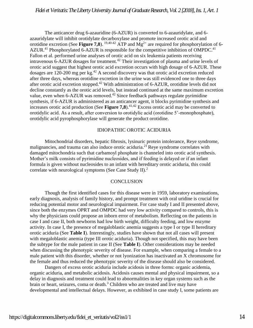
Figure 2. Structural Composition of OAT and proposed transport mechanism from blood
to urine.7
The above figure is the proposed structure of organic anion transporters (OAT). A total of twelve
transmembrane loops (two pairs of six domains) are present. Two major loops are formed in the
transporter- the first loop is located extracellularly while the second loop is located
intracellularly. The first loop contains sites for glycosylation, whereas the second loop between
sites 6-7 represents the substrate for protein kinase C (PKC) phosphorylation. The termini of the
protein include an amino (NH2) and carboxy (COOH) terminus which is located within the cell.
17
Fonteh: Orotic Aciduria
Published by Scholars Crossing, 2018
Page 19
Figure 3. Renal Proximal Tubule Transport of Organic Anions.7
The above illustration attempts to explain the mechanism of secondary active transport of
organic anions from the blood to the urine by renal proximal tubule cells. As organic anions
enter the basolateral side of the tubule from the plasma, the OAT transport system exports
dicarboxylates from the intracellular space to the plasma. A second transporter, a
Na+/dicarboxylate symporter, brings both Na+ and carboxylates in from the plasma to the
tubule. The concentration of dicarboxylate and Na+ couple to allow OAT transport of organic
anions, but since dicarboxylates are transported against their concentration gradient, the energy
for this process is also dependent upon the electrochemical gradient established by Na+-K+-
ATPase. Organic anions can be transported into the urine across the apical membrane.
18
Fidei et Veritatis: The Liberty University Journal of Graduate Research, Vol. 2 [2018], Iss. 1, Art. 1
https://digitalcommons.liberty.edu/fidei_et_veritatis/vol2/iss1/1
Page 20
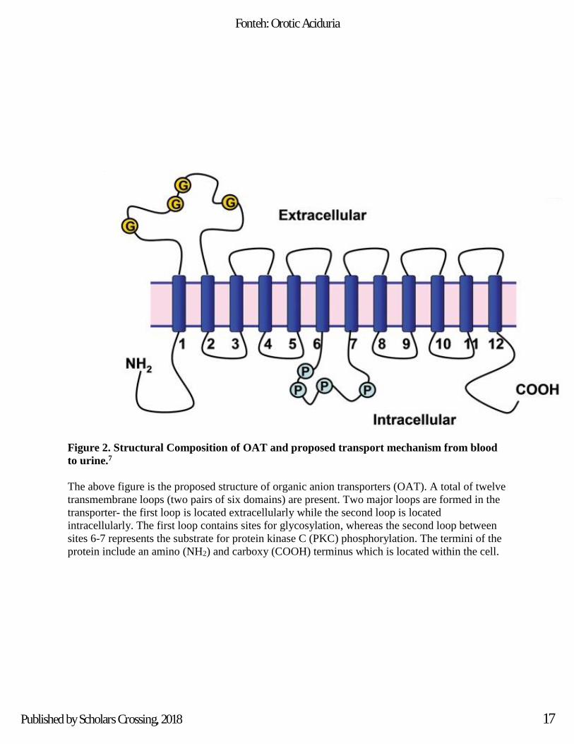
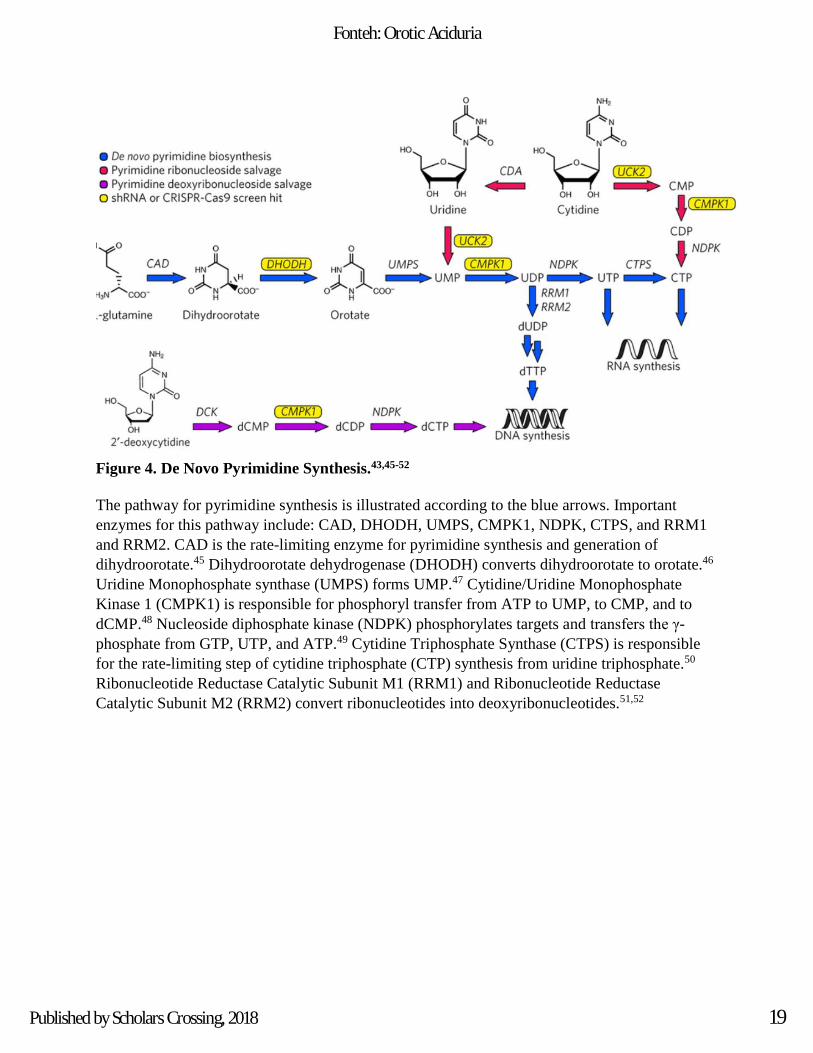
Figure 4. De Novo Pyrimidine Synthesis.43,45-52
The pathway for pyrimidine synthesis is illustrated according to the blue arrows. Important
enzymes for this pathway include: CAD, DHODH, UMPS, CMPK1, NDPK, CTPS, and RRM1
and RRM2. CAD is the rate-limiting enzyme for pyrimidine synthesis and generation of
dihydroorotate.45 Dihydroorotate dehydrogenase (DHODH) converts dihydroorotate to orotate.46
Uridine Monophosphate synthase (UMPS) forms UMP.47 Cytidine/Uridine Monophosphate
Kinase 1 (CMPK1) is responsible for phosphoryl transfer from ATP to UMP, to CMP, and to
dCMP.48 Nucleoside diphosphate kinase (NDPK) phosphorylates targets and transfers the γ-
phosphate from GTP, UTP, and ATP.49 Cytidine Triphosphate Synthase (CTPS) is responsible
for the rate-limiting step of cytidine triphosphate (CTP) synthesis from uridine triphosphate.50
Ribonucleotide Reductase Catalytic Subunit M1 (RRM1) and Ribonucleotide Reductase
Catalytic Subunit M2 (RRM2) convert ribonucleotides into deoxyribonucleotides.51,52
19
Fonteh: Orotic Aciduria
Published by Scholars Crossing, 2018
Page 21
Figure 5. Pyrimidine Synthesis from Carbamoyl Phosphate 5,20,44
This figure illustrates that carbamoyl phosphate will be translocated to the cytosol for the
formation of carbamoylasparate by aspartate transcarbamoylase. The amide group of glutamine
will be used to donate nitrogen and the cyclization of carbamoylasparate forms dihydroorotate.
Dihydroorotate dehydrogenase(DHODH) will convert dihydroorotate to orotic acid by oxidation
with NAD+. 5-phosphoribosyl-1-pyrophosphate (PRPP) will donate a ribose phosphate group to
orotic acid to form orotidylate by pyrimidine phosphoribosyltransferase. The decarboxylation of
orotidylate generates uridylate by the activity of orotidylate decarboxylase. Uridine
monophosphate synthase (UMPS) will convert orotic acid to uridine monophosphate (UMP). Not
shown is the conversion of UMP to UDP by UMP kinase. Next, UDP is converted to UTP, then
CTP. The active forms of nucleotides exist as triphosphates, so the activity of a monophosphate
kinase (uridylate kinase) and nucleoside diphosphate kinase is necessary.22
20
Fidei et Veritatis: The Liberty University Journal of Graduate Research, Vol. 2 [2018], Iss. 1, Art. 1
https://digitalcommons.liberty.edu/fidei_et_veritatis/vol2/iss1/1
Page 22
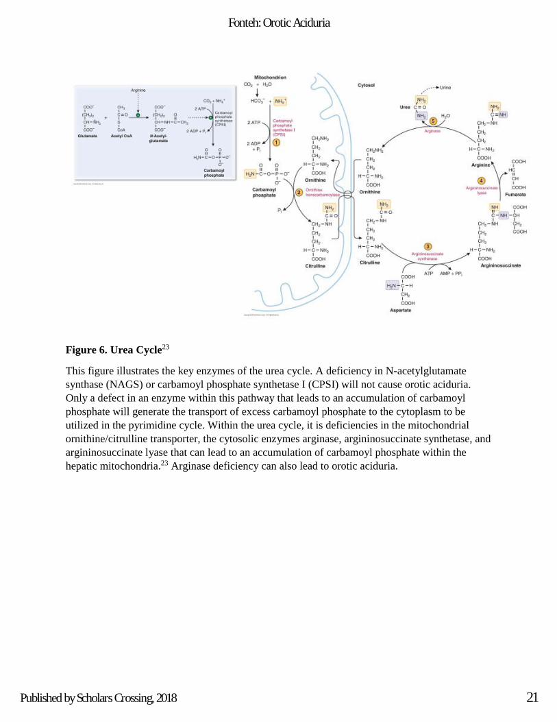
Figure 6. Urea Cycle23
This figure illustrates the key enzymes of the urea cycle. A deficiency in N-acetylglutamate
synthase (NAGS) or carbamoyl phosphate synthetase I (CPSI) will not cause orotic aciduria.
Only a defect in an enzyme within this pathway that leads to an accumulation of carbamoyl
phosphate will generate the transport of excess carbamoyl phosphate to the cytoplasm to be
utilized in the pyrimidine cycle. Within the urea cycle, it is deficiencies in the mitochondrial
ornithine/citrulline transporter, the cytosolic enzymes arginase, argininosuccinate synthetase, and
argininosuccinate lyase that can lead to an accumulation of carbamoyl phosphate within the
hepatic mitochondria.23 Arginase deficiency can also lead to orotic aciduria.
21
Fonteh: Orotic Aciduria
Published by Scholars Crossing, 2018
Page 23
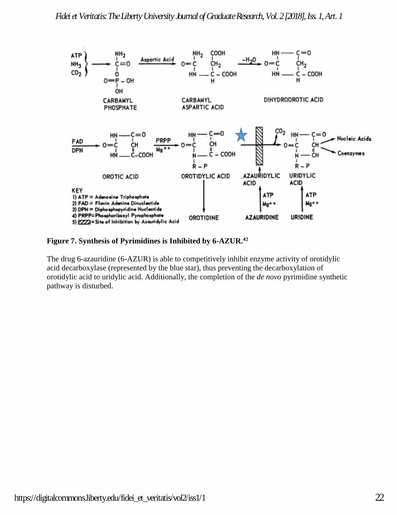
Figure 7. Synthesis of Pyrimidines is Inhibited by 6-AZUR.42
The drug 6-azauridine (6-AZUR) is able to competitively inhibit enzyme activity of orotidylic
acid decarboxylase (represented by the blue star), thus preventing the decarboxylation of
orotidylic acid to uridylic acid. Additionally, the completion of the de novo pyrimidine synthetic
pathway is disturbed.
22
Fidei et Veritatis: The Liberty University Journal of Graduate Research, Vol. 2 [2018], Iss. 1, Art. 1
https://digitalcommons.liberty.edu/fidei_et_veritatis/vol2/iss1/1
Page 24
Figure 8. Lineweaver-Burk Plot of Competitive Inhibition of Orotidylic decarboxylase by
Azauridine 5’ Phosphate. 41
A kinetic study conducted with 0.05 M Tris Buffer at pH 8 and 25° C generated enzyme units for
the enzyme that was 0.05/mL. Earlier pH studies determined that at pH 8, the enzyme activity
was interrupted most significantly by 6-AZUR. The active form of 6-AZUR occurs when the
compound is in a solution at a pH above 7, and ionized at the carboxylic position.
23
Fonteh: Orotic Aciduria
Published by Scholars Crossing, 2018
Page 25
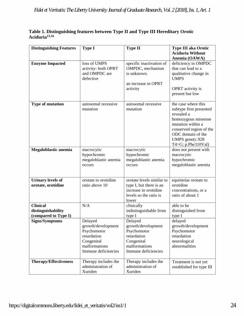
Table 1. Distinguishing features between Type II and Type III Hereditary Orotic
Aciduria13,16
Distinguishing Features Type I Type II Type III aka Orotic
Aciduria Without
Anemia (OAWA)
Enzyme Impacted
loss of UMPS
activity- both OPRT
and OMPDC are
defective
specific inactivation of
OMPDC, mechanism
is unknown.
an increase in OPRT
activity
deficiency in OMPDC
that can lead to a
qualitative change in
UMPS
OPRT activity is
present but low
Type of mutation autosomal recessive
mutation
autosomal recessive
mutation
the case where this
subtype first presented
revealed a
homozygous missense
mutation within a
conserved region of the
ODC domain of the
UMPS gene(c.928
T4>G; p.Phe310Val)
Megaloblastic anemia macrocytic
hypochromic
megaloblastic anemia
occurs
macrocytic
hypochromic
megaloblastic anemia
occurs
does not present with
macrocytic
hypochromic
megaloblastic anemia
Urinary levels of
orotate, orotidine
orotate to orotidine
ratio above 10
orotate levels similar to
type I, but there is an
increase in orotidine
levels so the ratio is
lower
equimolar orotate to
orotidine
concentrations, or a
ratio of about 1
Clinical
distinguishability
(compared to Type I)
N/A clinically
indistinguishable from
type I
able to be
distinguished from
type I
Signs/Symptoms Delayed
growth/development
Psychomotor
retardation
Congenital
malformations
Immune deficiencies
Delayed
growth/development
Psychomotor
retardation
Congenital
malformations
Immune deficiencies
delayed
growth/development
Psychomotor
retardation
neurological
abnormalities
Therapy/Effectiveness Therapy includes the
administration of
Xuriden
Therapy includes the
administration of
Xuriden
• Treatment is not yet
established for type III
24
Fidei et Veritatis: The Liberty University Journal of Graduate Research, Vol. 2 [2018], Iss. 1, Art. 1
https://digitalcommons.liberty.edu/fidei_et_veritatis/vol2/iss1/1
Page 26
References
1. Rogers LE, Warford LR, Patterson RB, Porter FS. Hereditary orotic aciduria. I. A
new case with family studies. Pediatrics. 1968;42(3):415-422.
2. Imaeda M, Sumi S, Imaeda H, et al. Hereditary orotic aciduria heterozygotes
accompanied with neurological symptoms. Tohoku J Exp Med. 1998;1:67-70.
https://www.ncbi.nlm.nih.gov/pubmed/9710947. Accessed July 30, 2018.
3. Orotic acid. National Center for Biotechnology Information. PubChem Compound
Database. https://pubchem.ncbi.nlm.nih.gov/compound/967. Accessed August 2, 2018.
4. Gerhardt V, Tutughamiarso M, Bolte M. Conformational studies of hydantoin-5-
acetic acid and orotic acid. Acta Crystallogr C. 2012;68(Pt 2):92-98.doi:
10.1107/S0108270112001151.
5. Brosnan ME, Brosnan JT. Orotic acid excretion and arginine metabolism. J Nutr.
2007;6:1656S–1661S. doi: 10.1093/jn/137.6.1656S.
6. Löffler M, Carrey E, Zameitat E. Orotic acid, more than just an intermediate of
pyrimidine de novo synthesis. J Genet Genome. 2015;42(5):207-219. doi:
10.1016/j.jgg.2015.04.001.
7. Nigam SK, Bush KT, Martovetsky G, et al. The organic anion transporter (OAT)
family: a systems biology perspective. Physiol Reviews. 2015;95(1):83-123. doi:
10.1152/physrev.00025.2013.
8. Anzai N, Miura D, Endou H. Orotic acid transport via organic anion transporters
OATs. Japanese Soc of Gout and Nucleic Acid Metab. 2008;32(2):141-146.
https://www.jstage.jst.go.jp/article/gnam1999/32/2/32_141/_article. Accessed August 2, 2018.
9. Miura D, Anzai N, Jutabha P, et al. Human urate transporter 1 (hURAT1)
mediates the transport of orotate. J Physiol Sci. 2011;61(3): 253-257. doi: 10.1007/s12576
011-0136-0.
10. Fork C, Bauer T, Golz S, et al. OAT2 catalyses efflux of glutamate and uptake of
orotic acid. Biochem J. 2011:436(2);305-312.
https://pdfs.semanticscholar.org/1b2d/b383a3ba085ad194bd50231523f0161b3b88.pdf. Accessed
August 2, 2018.
11. Angstadt CN. Purine and pyrimidine metabolism.
https://library.med.utah.edu/NetBiochem/pupyr/pp.htm. Published December 4, 1997. Accessed
August 2, 2018
25
Fonteh: Orotic Aciduria
Published by Scholars Crossing, 2018
Page 27
12. Somerville R. Pyrimdine. Encyclopedia of Genetics. 2001:1586.
www.sciencedirect.com/science/article/pii/B0122270800017250?via%3Dihub. Accessed August
2, 2018.
13. Debnath SK, Aggarwal A, Mittal H. Megaloblastic anemia--a rare cause. Indian
J Pediatr. 2011;78(10):1293–1295. doi: 10.1007/s12098-011-0461-6.
14. Suchi M, Mizuno H, Kawai Y, et al. Molecular cloning of the human UMP synthase
gene and characterization of point mutations in two hereditary orotic aciduria families. Am J of
Hum Genet. 1997;60(3):525-539. https://www.ncbi.nlm.nih.gov/pubmed/9042911. Accessed
July 27, 2018.
15. Chakraborty P, Geraghty MT. Inborn errors of urea synthesis. In: Glew RH,
Rosenthal MD, eds. Clinical Studies in Medical Biochemistry. 3rd ed. Oxford, NY: Oxford
University Press; 2007.195-203.
16. Bailey CJ. Orotic aciduria and uridine monophosphate synthase: a reappraisal. J
Inherit Metab Dis. 2009;32:227-233. doi: 10.1007/s10545-009-1176-y.
17. In Brief: Uridine Triacetate (Xuriden) for hereditary orotic aciduria. The Medical
Letter. https://secure.medicalletter.org/w1491g. Published March 28, 2016. Accessed March 8,
2018.
18. Berghe G, Vincent M, Springer MS. Disorders of purine and pyrimidine
metabolism: UMP synthase deficiency (hereditary orotic aciduria).
http://eknygos.lsmuni.lt/springer/223/VIII%20Part/433-449.pdf. Accessed March 13,2018.
19. Chhabra N. Orotic Acidura-causes, clinical manifestations, diagnosis and treatment.
Biochemistry for Medics. http://www.namrata.co/orotic-aciduria-causes-clinical-manifestations
diagnosis-and-treatment/. Published February 19, 2013. Accessed March 8, 2018.
20. Balasubramaniam S, Duley J, & Christodoulou J. Inborn errors of pyrimidine
metabolism: clinical update and therapy. J Inherit Metab Dis. 2014;37(5):687-698. doi:
10.1007/s10545-014-9742-3.
21. Choi YJ, Yoon Y, Lee KY, et al. Orotic acid induces hypertension associated with
impaired endothelial nitric oxide synthesis. Toxicol Sci. 2015;144(2):307-317. doi:
10.1093/toxsci/kfv003.
22. Bhagavan NV, Ha CE. Nucleotide Metabolism. Essentials of Medical
Biochemistry. San Diego, CA: Academic Press; 2011:333-354.
23. Kalu B. Amino Acid Metabolism. 2018.
24. Wortmann SB, Chen MA, Colombo R, et al. Mild orotic aciduria in UMPS
heterozygotes: a metabolic finding without clinical consequences. J Inherit Metab Dis.
26
Fidei et Veritatis: The Liberty University Journal of Graduate Research, Vol. 2 [2018], Iss. 1, Art. 1
https://digitalcommons.liberty.edu/fidei_et_veritatis/vol2/iss1/1
Page 28
2017;40(3):423-431. doi: 10.1007/s10545-017-0015-9.
25. Micheli V, Camici M, Tozzi MG, et al. Neurological disorders of purine and pyrimidine
metabolism. Curr Top Med Chem. 2011;11(8):923-947.
https://www.ncbi.nlm.nih.gov/pubmed/21401501. Accessed March 13, 2018.
26. Lanzkowsky P. Megaloblastic Anemia. In: Lanzkowsky P, Lipton J, Fish J, eds. 6th
ed. Lanzkowsky’s Manual of Pediatric Hematology and Oncology. San Diego, CA: Academic
Press; 2016:84-101. https://www.sciencedirect.com/book/9780128013687/lanzkowskys-manual
of-pediatric-hematology-and-oncology. Accessed August 10, 2018.
27. Schramm V, Grubmeyer C. Phosphoribosyltransferase mechanisms and roles in
nucleic acid metabolism. Prog Nucleic Acid Res Mol Biol. 2004;78:261-304. doi:
10.1016/S0079-6603(04)78007-1.
28. Orotic Aciduria Type 1. National Center for Advancing Translational
Sciences: Genetic and Rare Diseases Information Center.
https://rarediseases.info.nih.gov/diseases/5429/orotic-aciduria-type-1. Updated September 13,
2017. Accessed March 7, 2018.
29. Curtis CS, Kudsk KA. Do immunonutrients improve outcome in the critically ill? In:
Deutschman CS, Neligan PJ, eds.1st ed. Evidence-Based Practice of Critical Care. Philadelphia,
PA: Saunders; 2010:467-472. https://www.sciencedirect.com/book/9781416054764/evidence
based-practice-of-critical-care. Accessed August 10, 2018.
30. Ming JE, Graham.JM. Genetic syndromes with evidence of immune deficiency.In:
Sullivan KE, Stiehm ER, eds. 1st ed. Stiehm's Immune Deficiencies; 2014:281-324.
https://www.sciencedirect.com/science/article/pii/B9780124055469000121. Accessed August
10, 2018.
31. Scaglia F, Scheuerle AE, Towbin JA, Armstrong DL, Sweetman L, Wong L-JC.
Neonatal presentation of ventricular tachycardia and a reye-like syndrome episode associated
with disturbed mitochondrial energy metabolism. BMC Pediatr. 2002;2:12. doi:10.1186/1471
2431-2-12.
32. Rautio J, Kärkkäinen J, Sloan KB. Prodrugs-recent approvals and a glimpse of the
pipeline. Eur J Pharm Sci. 2017;109:146-161.doi: 10.1016/j.ejps.2017.08.002.
33. Cada DJ, Mbogu U, Bindler RJ, Baker DE. Uridine triacetate. Hosp Pharm.
2016;51(6):484-488. doi:10.1310/hpj5106-484.
34. Hartung B, Temme O, Neuen-Jacob E, Ritz-Timme S, Hinderhofer K, Daldrup T.
Ornithine transcarbamylase deficiency of a male newborn with fatal outcome. Int J Legal
Med. 2016;130(3):783-785. doi: 10.1007/s00414-015-1311-2.
35. Hauser ER, Finklestein JE, Valle D, Brusilow SW. Allopurinol-induced orotidinuria-
27
Fonteh: Orotic Aciduria
Published by Scholars Crossing, 2018
Page 29
a test for mutations at the ornithine carbamoyltransferase locus in women. N Engl J Med. 1990;
322(23):1641-1645. https://www.ncbi.nlm.nih.gov/pubmed/2342523. Accessed July 27, 2018.
36. Qureshi IA, Letarte J, Quellet R. Study of enzyme defect in a case of ornithine
transcarbamylase deficiency. Diabete Metab. 1978;4(4):239-241.
https://www.ncbi.nlm.nih.gov/pubmed/729890. Accessed August 10, 2018.
37. Arn PH, Hauser ER, Thomas GH, Herman G, Hess D, Brusilow SW. Hyperammonemia
in women with a mutation at the ornithine carbamoyltransferase locus. A cause of postpartum
coma. N Engl J Med. 1990;322(23):1652-1655. https://www.ncbi.nlm.nih.gov/pubmed/2342525.
Accessed July 27, 2018.
38. Hewson S, Clarke JT, Cederbaum S. Prenatal diagnosis for arginase deficiency: a
case study. J Inherit Metab Dis. 2003;26(6):607-610.
https://www.ncbi.nlm.nih.gov/pubmed/14605507. Accessed July 27, 2018.
39. Brusilow SW. Arginine, an indispensable amino acid for patients with inborn errors
of urea synthesis. J Clin Invest. 1984;74(6):2144
2148. https://www.ncbi.nlm.nih.gov/pubmed/6511918. Accessed July 27, 2018.
40. Kelley RE, Andersson HC. Neurologic aspects of systemic disease part III.
Handbook of Clinical Neurology. 2014.
41. Handschumacher RE. Orotidylic acid decarboxylase: inhibition studies with azauridine
-phosphate. J Biol Chem.1960;235(10):2917-2919.
http://www.jbc.org/content/235/10/2917.full.pdf. Accessed August 2, 2018.
42. Fallon HJ, Frei E 3rd, Block J, Seegmiller JE.The Uricosuria and Orotic Aciduria
Induced by 6-Azauridine. J Clin Invest. 1961;40:1906-1914.
https://www.ncbi.nlm.nih.gov/pubmed/13891472. Accessed August 2, 2018.
43. Khosla Lab. The united states war on drugs.
https://web.stanford.edu/group/khosla/cgi-bin/?attachment_id=625. Published October 24, 2016.
Accessed August 4, 2018.
44. Berg JM, Tymoczko JL, Stryer L. In de novo synthesis, the pyrimidine ring is
assembled from bicarbonate, aspartate, and glutamine. Biochemistry. New York, NY: W H
Freeman; 2002.
45. CAD carbamoyl-phosphate synthetase 2, aspartate transcarbamylase, and
dihydroorotase [Homo sapiens (human)] - Gene - NCBI. Advances in pediatrics.
https://www.ncbi.nlm.nih.gov/gene/790. Published July 8, 2018. Accessed August 4, 2018.
46. DHODH dihydroorotate dehydrogenase (quinone) [Homo sapiens (human)] - Gene -
NCBI. Advances in pediatrics. https://www.ncbi.nlm.nih.gov/gene/1723. Published July 8, 2018.
Accessed August 4, 2018.
28
Fidei et Veritatis: The Liberty University Journal of Graduate Research, Vol. 2 [2018], Iss. 1, Art. 1
https://digitalcommons.liberty.edu/fidei_et_veritatis/vol2/iss1/1
Page 30
47. UMPS gene - Genetics Home Reference - NIH. U.S. National Library of Medicine.
https://ghr.nlm.nih.gov/gene/UMPS. Published March 2010. Accessed August 4, 2018.
48. CMPK1 cytidine/uridine monophosphate kinase 1 [Homo sapiens (human)] - Gene -
NCBI. Advances in pediatrics. https://www.ncbi.nlm.nih.gov/gene/51727. Published July 8,
2018. Accessed August 4, 2018.
49. Mehta A, Orchard S. Nucleoside diphosphate kinase (NDPK, NM23, AWD): recent
regulatory advances in endocytosis, metastasis, psoriasis, insulin release, fetal erythroid lineage
and heart failure; translational medicine exemplified. Mol Cell Biochem. 2009;329(1-2):3-15.
doi:10.1007/s11010-009-0114-5.
50. Richards NGJ, Sintjago TCC. Enzymes and enzyme mechanisms. Comprehensive
Natural Products II. Elsevier Ltd. 2010. https://www.sciencedirect.com/topics/neuroscience/ctp
synthetase. Accessed August 4, 2018.
51. RRM1 ribonucleotide reductase catalytic subunit M1 [Homo sapiens (human)] - Gene
- NCBI. Advances in pediatrics. https://www.ncbi.nlm.nih.gov/gene/6240. Published July 8,
2018. Accessed August 4, 2018.
52. RRM2 ribonucleotide reductase regulatory subunit M2 [Homo sapiens (human)] -
Gene - NCBI. Advances in pediatrics. https://www.ncbi.nlm.nih.gov/gene/6241. Published July
8, 2018. Accessed August 4, 2018.
29
Fonteh: Orotic Aciduria
Published by Scholars Crossing, 2018