Duck Plague 141 Chapter 16 Duck Plague Synonyms Duck virus enteritis, DVE Cause Duck plague is caused by a herpesvirus. Infection often results in an acute, contagious, and fatal disease. As with many other herpesviruses, duck plague virus can establish inapparent infections in birds that survive exposure to it, a state referred to as latency. During latency, the virus cannot be detected by standard methods for virus isolation. Studies of domestic species of waterfowl have detected multiple strains of the virus that vary in their ability to cause disease and death. Little is known about the response of wild water- fowl to strain differences. Duck plague outbreaks are thought to be caused when birds that carry the virus shed it through fecal or oral dis- charge, thus releasing the virus into food and water with which susceptible birds may have contact. Experimental studies have demonstrated spontaneous virus shedding by duck plague carriers during spring. Changes in the duration of daylight and onset of breeding are thought to be physi- ological stresses that stimulate virus shedding at this time of year. The carriers are immune to the disease, but the virus shed by them causes infection and disease among suscep- tible waterfowl. Bird-to-bird contact and contact with virus that has contaminated the environment perpetuate an out- break. Scavenging and decomposition of carcasses of infected birds also contaminate the environment by releasing viruses from tissues and body fluids. Virus transmission through the egg has been reported, but the role of the egg in the disease cycle remains to be resolved. Species Affected Only ducks, geese, and swans are susceptible to duck plague. Other aquatic birds do not become infected, and the absence of mortality of American coot, shorebirds, and other waterbirds that may be present during a waterfowl die-off can be an important indication that duck plague may be in- volved. Susceptibility varies greatly among waterfowl spe- cies (Fig. 16.l). In one study with a highly virulent virus, it took 300,000 times more virus material to infect northern pintail than to infect blue-winged teal. Distribution The first reported duck plague outbreak in North America struck the white Pekin duck industry of Long Island, New Figure 16.1 Comparative susceptibility of eight waterfowl species to duck plague virus. Blue-winged teal Redhead duck Wood duck Canada goose Gadwall Mallard Muscovy Pintail Highly susceptible Moderately susceptible Slightly susceptible
Transcript
Duck Plague 141
Chapter 16
Duck Plague
Synonyms
Duck virus enteritis, DVE
CauseDuck plague is caused by a herpesvirus. Infection often
results in an acute, contagious, and fatal disease. As withmany other herpesviruses, duck plague virus can establishinapparent infections in birds that survive exposure to it, astate referred to as latency. During latency, the virus cannotbe detected by standard methods for virus isolation. Studiesof domestic species of waterfowl have detected multiplestrains of the virus that vary in their ability to cause diseaseand death. Little is known about the response of wild water-fowl to strain differences.
Duck plague outbreaks are thought to be caused whenbirds that carry the virus shed it through fecal or oral dis-charge, thus releasing the virus into food and water withwhich susceptible birds may have contact. Experimentalstudies have demonstrated spontaneous virus shedding byduck plague carriers during spring. Changes in the durationof daylight and onset of breeding are thought to be physi-ological stresses that stimulate virus shedding at this time ofyear. The carriers are immune to the disease, but the virusshed by them causes infection and disease among suscep-tible waterfowl. Bird-to-bird contact and contact with virusthat has contaminated the environment perpetuate an out-break. Scavenging and decomposition of carcasses of infectedbirds also contaminate the environment by releasing virusesfrom tissues and body fluids. Virus transmission through theegg has been reported, but the role of the egg in the diseasecycle remains to be resolved.
Species AffectedOnly ducks, geese, and swans are susceptible to duck
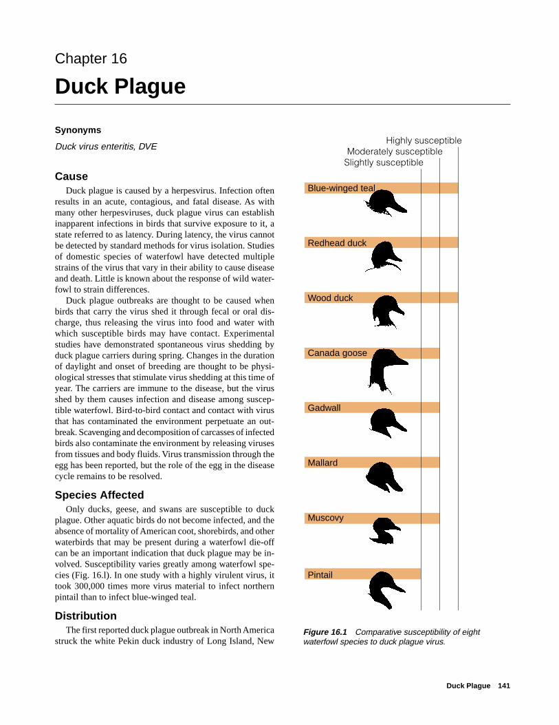
plague. Other aquatic birds do not become infected, and theabsence of mortality of American coot, shorebirds, and otherwaterbirds that may be present during a waterfowl die-offcan be an important indication that duck plague may be in-volved. Susceptibility varies greatly among waterfowl spe-cies (Fig. 16.l). In one study with a highly virulent virus, ittook 300,000 times more virus material to infect northernpintail than to infect blue-winged teal.
DistributionThe first reported duck plague outbreak in North America
struck the white Pekin duck industry of Long Island, NewFigure 16.1 Comparative susceptibility of eightwaterfowl species to duck plague virus.
Blue-winged teal
Redhead duck
Wood duck
Canada goose
Gadwall
Mallard
Muscovy
Pintail
Highly susceptibleModerately susceptible
Slightly susceptible
142 Field Manual of Wildlife Diseases: Birds
Figure 16.2 Frequency of duck plague since year of first outbreak (1967–1996).
Figure 16.3 Reported North American distribution of duck plague by period of first occurrence.
EXPLANATIONDuck plague activity index*
1.0 and over
0.50–0.99
0.10–0.49
0.01–0.09
No events recorded
Not calculated. First year of occurrence is current year
Year of initial outbreak
Suspected diagnosis, not confirmed by virus isolation
1980 *The activity index is an expression of the frequency of duck plague outbreaks in relation to time.
Total number of outbreaks x number of years with outbreaks
Current year – year of first outbreak within that state (x10)Index =
1988
1973
1972
1992
1980 1975
1988
1973
1973
1973
1981
1980
19861982
1986
1968
1967
1990
1968
(D.C.) 19751973
19931999
EXPLANATIONYear of first reported occurrence of duck plague
1967–69
1970–74
1975–79
1980–84
1985–89
1990–94
1995–99
No duck plague reported
Duck Plague 143
York in 1967. Since then, duck plague has broken out fromcoast to coast and from Canada to Texas. The frequency ofduck plague outbreaks has varied considerably geographi-cally. The greatest frequency of duck plague activity has beenreported in Maryland, followed by California, Virginia, andNew York (Fig. 16.2). The disease has also been reported inseveral Canadian Provinces since it first was observed in theUnited States (Fig. 16.3). First reported in the Netherlandsin 1923, duck plague has also been reported in several othercountries in Europe and in Asia since 1958. The frequencyof duck plague varies within different types of waterfowl,and failure to respond to these differences complicates dis-ease prevention and control efforts. The different types ofwaterfowl aggregations involved and the relative frequencyof duck plague activity within these different populationsare highlighted in Tables 16.1 and 16.2.
Despite the cumulative widespread geographic distribu-tion and frequent occurrence of duck plague in captive and
feral waterfowl in North America, wild waterfowl have beenaffected only infrequently. The only major outbreaks in mi-gratory waterfowl have happened in South Dakota and NewYork. In January 1973, more than 40,000 of 100,000 mal-lards and a smaller number of Canada geese and other spe-cies died at Lake Andes National Wildlife Refuge in SouthDakota while they were wintering there (Fig. 16.4). The onlyother duck plague event that caused substantial loss of wildwaterfowl occurred during February 1994 in the Finger Lakesregion of western New York State. Approximately 1,200 car-casses were recovered, primarily American black duck andmallard, with nearly three times as many black duck as mal-lard carcasses. The carcasses that were recovered were ap-proximately 24 percent of the black duck and 3 percent ofthe mallard populations present at the outbreak location.During the initial 1967 outbreak in white Pekin ducks onLong Island, several hundred wild waterfowl carcasses (pri-marily mallard and American black duck) were recovered
Table 16.2 Relative frequency of duck plague in different types of waterfowl within the United States.
Feral Common Increasing outbreaks, andcurrently prime virus source
Nonmigratory Occasional None; sporadic outbreaks
Migratory Rare None; rare
Table 16.1 Types of waterfowl involved in outbreaks of duck plague in the United States.
Waterfowl classification Population composition
Commercial Birds raised for consumptive markets; for example, white Pekin ducks.
Captive collections Zoological and other collections of birds for display and research.
Game farm Birds raised for release for sporting programs; for example, mallard ducks.
Feral Nonmigratory, nonconfined waterfowl of various species.
Nonmigratory Resident populations of native wild species; for example, mallard ducks andCanada geese.
Migratory North American waterfowl that breed in one geographic area and winter inanother before returning to their Northern breeding grounds.
144 Field Manual of Wildlife Diseases: Birds
Figure 16.4 During the 1973 outbreakof duck plague at Lake Andes NationalWildlife Refuge in South Dakota, morethan 40,000 mallards died.
Pho
to b
y M
ilton
Frie
nd
from adjacent Flanders Bay, apparently as a result of diseasetransmission from white Pekin ducks. Those carcasses rep-resented approximately 5 percent of the wild mallard andblack duck populations on Flanders Bay during the duckplague outbreak. Mortality in the white Pekin duck flockswas much greater, averaging 45 percent in mature ducks(2-year olds) and 17 percent in immature ducks (youngerthan 5 months of age). Equally important was the 25–40 per-cent decrease in egg production by mature breeder ducksthat were present during the outbreak. With the exception ofthe Lake Andes, Finger Lakes, and Flanders Bay outbreaks,duck plague in migratory waterfowl has been limited to asmall number of birds. All confirmed outbreaks have alsoinvolved commercial, avicultural, captive-raised, or feralwaterfowl.
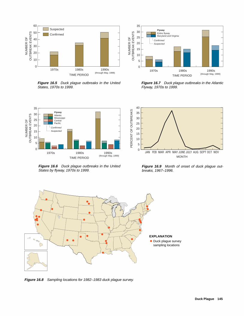
The pattern of duck plague within North America is thatof an emerging disease. The number of outbreaks being di-agnosed is increasing each decade (Fig. 16.5). The greatmajority of outbreaks occur within the Atlantic Flyway (Fig.16.6) and nearly all of those events are within Maryland andVirginia (Fig. 16.7). The factors responsible for the contin-ued emergence and geographic spread of duck plague withinNorth America are unknown, as is the distribution of duckplague among free-living North American waterfowl popu-lations.
Some individuals believe that a large number of surviv-ing wild waterfowl exposed to this disease at Lake Andesbecame disease carriers, that these disease carriers have per-petuated infections in other wild waterfowl, and that duckplague is now widespread among migratory waterfowl. How-ever, surveys of wild waterfowl conducted by the U.S.
Department of Agriculture in 1967 and by the National Wild-life Health Center (NWHC) from 1978 to 1986 and in 1982–1983 did not detect any evidence of duck plague carriers. Inthe latter NWHC survey, more than 4,500 waterfowl acrossthe United States were sampled (Fig. 16.8). Sampling sitesincluded major waterfowl concentration areas and areaswhere duck plague has been a recurrent disease problem incaptive and feral waterfowl. Although none of the birdssampled during either NWHC survey were shedding detect-able duck plague virus, the previously described problem ofinapparent carriers complicates interpretation of these results.New technology that was not yet developed at the time ofthat survey provides increased ability to detect duck plaguecarriers and resolve the question of sources for infection.
The absence of duck plague as a cause of mortality in thethousands of wild waterfowl necropsied by the NWHC pro-vides additional evidence that duck plague is not an estab-lished disease in wild North American waterfowl. These ex-aminations, performed since 1975, were of waterfowl founddead on National Wildlife Refuges and other major water-fowl concentration areas.
SeasonalityDuck plague outbreaks have been reported during every
month except August and September. Approximately 86 per-cent of these outbreaks occurred from March through June(Fig. 16.9). This pattern of spring outbreaks has also beenreported for captive waterfowl collections in England, and itmay be associated with the physiological changes referredto above.
Duck Plague 145
0
5
10
15
20
25
30
1970s 1980s 1990s
TIME PERIOD
NU
MB
ER
OF
OU
TB
RE
AK
EV
EN
TS
Confirmed
Suspected
AtlanticMississippiCentralPacific
(through May, 1999)
Flyway
35
0
5
10
15
20
25
30
1970s 1980s 1990s
TIME PERIOD
NU
MB
ER
OF
OU
TB
RE
AK
EV
EN
TS
Confirmed
Suspected
Entire flywayMaryland and Virginia
Flyway
35
(through May, 1999)
Figure 16.5 Duck plague outbreaks in the UnitedStates, 1970s to 1999.
Figure 16.6 Duck plague outbreaks in the UnitedStates by flyway, 1970s to 1999.
Figure 16.7 Duck plague outbreaks in the AtlanticFlyway, 1970s to 1999.
�
EXPLANATION
Duck plague survey sampling locations
�
��
� �
���
�
��
�
�
� �
� �
�
�
�
��
�
�
�
���
�
Figure 16.8 Sampling locations for 1982–1983 duck plague survey.
Figure 16.9 Month of onset of duck plague out-breaks, 1967–1996.
0
10
20
30
40
50
1970s 1980s 1990s
TIME PERIOD
NU
MB
ER
OF
O
UT
BR
EA
K E
VE
NT
SSuspected
Confirmed
60
(through May, 1999)
0
5
10
15
20
25
30
35
40
JAN FEB MAR APR MAY JUNE JULY AUG SEPT OCT NOV MONTH
PE
RC
EN
T O
F O
UT
BR
EA
KS
146 Field Manual of Wildlife Diseases: Birds
Field SignsThere is no prolonged illness associated with duck plague;
therefore, sick birds are seldom seen in the field, and birdsthat are healthy one day may be found dead the next. Theincubation period between virus exposure and death is gen-erally 3–7 days in domestic ducks, and experimental studieshave found that it is as long as 14 days in wild waterfowl.Wing-clipped mallards released to monitor the Lake Andesduck plague outbreak died 4–11 days after their release.
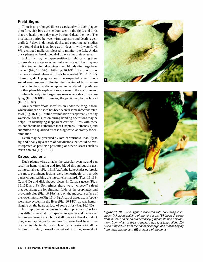
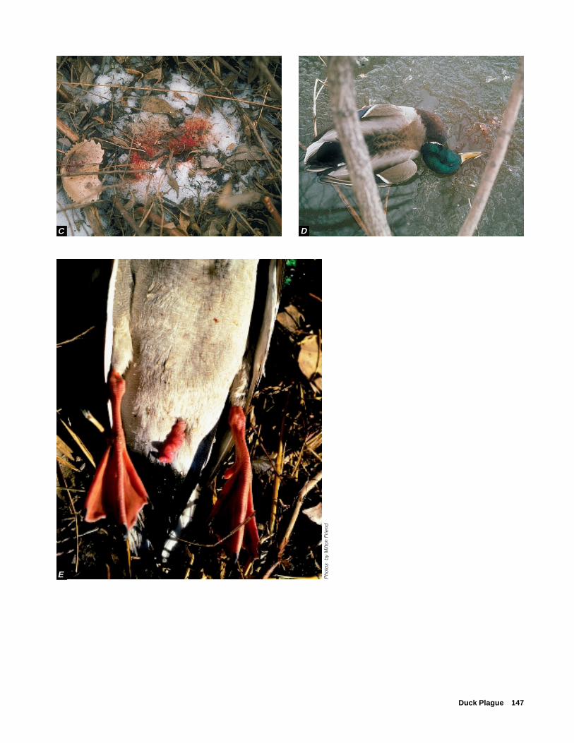
Sick birds may be hypersensitive to light, causing themto seek dense cover or other darkened areas. They may ex-hibit extreme thirst, droopiness, and bloody discharge fromthe vent (Fig. 16.10A) or bill (Fig. 16.10B). The ground maybe blood-stained where sick birds have rested (Fig. 16.10C).Therefore, duck plague should be suspected when blood-soiled areas are seen following the flushing of birds, whereblood splotches that do not appear to be related to predationor other plausible explanations are seen in the environment,or where bloody discharges are seen where dead birds arelying (Fig. 16.10D). In males, the penis may be prolapsed(Fig. 16.10E).
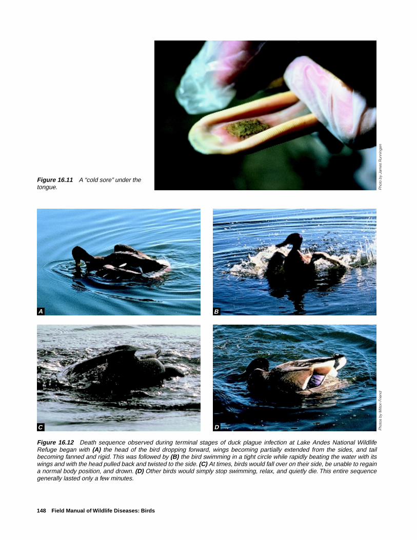
An ulcerative “cold sore” lesion under the tongue fromwhich virus can be shed has been seen in some infected water-fowl (Fig. 16.11). Routine examination of apparently healthywaterfowl for this lesion during banding operations may behelpful in identifying inapparent carriers. Birds with theselesions should be euthanized (see Chapter 5, Euthanasia) andsubmitted to a qualified disease diagnostic laboratory for ex-amination.
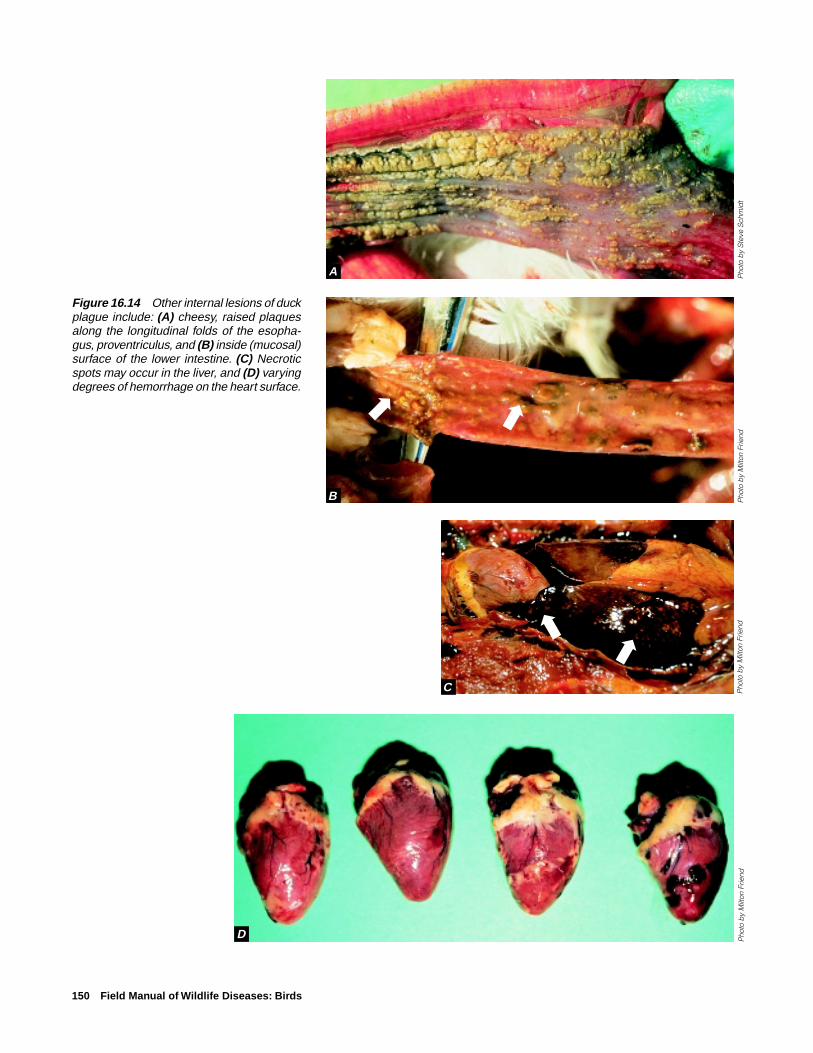
Death may be preceded by loss of wariness, inability tofly, and finally by a series of convulsions that could be mis-interpreted as pesticide poisoning or other diseases such asavian cholera (Fig. 16.12).
Gross LesionsDuck plague virus attacks the vascular system, and can
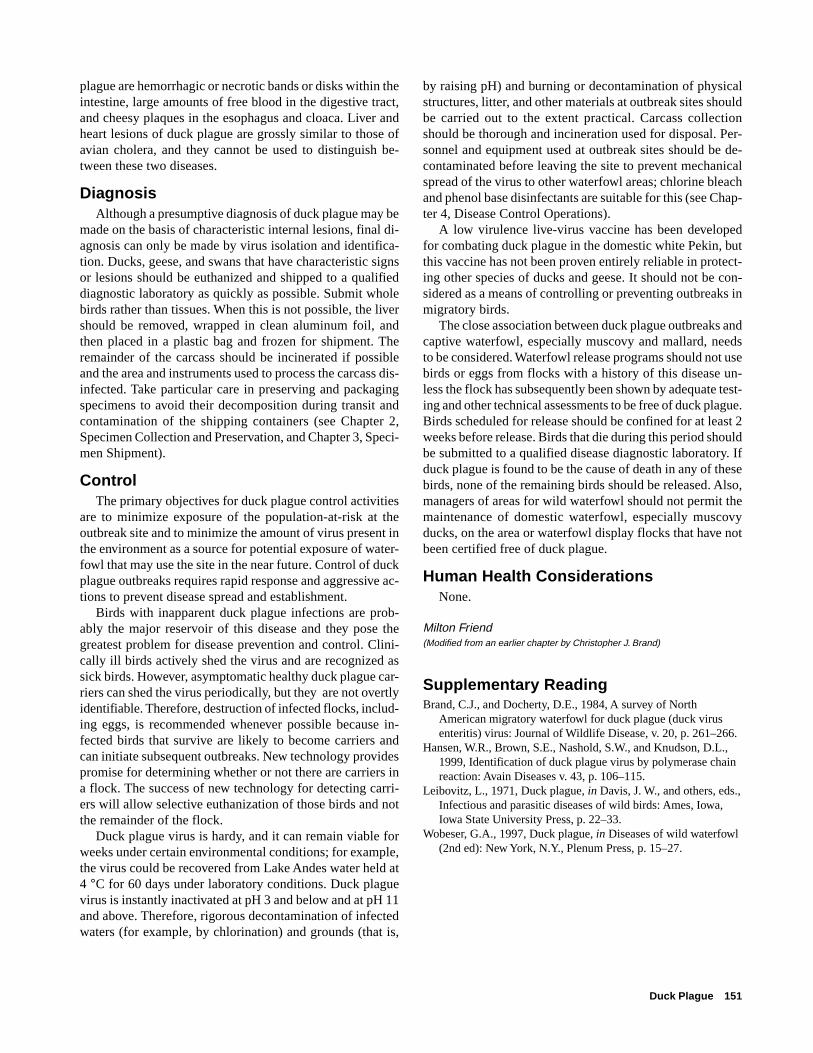
result in hemorrhaging and free blood throughout the gas-trointestinal tract (Fig. 16.13A). At the Lake Andes outbreak,the most prominent lesions were hemorrhagic or necroticbands circumscribing the intestine in mallards (Figs. 16.13B,C, and D) and disk-shaped ulcers in Canada geese (Figs.16.13E and F). Sometimes there were “cheesy,” raisedplaques along the longitudinal folds of the esophagus andproventriculus (Fig. 16.14A) and on the mucosal surface ofthe lower intestine (Fig. 16.14B). Areas of tissue death (spots)were also evident in the liver (Fig. 16.14C), as was hemor-rhaging on the heart surface of some birds (Fig. 16.14D).
It is important to recognize that the appearance of lesionsmay differ somewhat from species to species and that not alllesions are present in all birds at all times. Outbreaks of duckplague in captive and nonmigratory waterfowl have oftenresulted in infected birds with less distinct lesions. Of all thelesions illustrated, those of greatest value in diagnosing duck
Figure 16.10 Field signs associated with duck plague in-clude: (A) blood staining of the vent area; (B) blood drippingfrom the bill or a blood-stained bill; (C) blood-stained environ-ment from which a resting mallard has just taken flight; (D)blood-stained ice from the nasal discharge of a mallard dyingfrom duck plague; and (E) prolapse of the penis.
A
B
Duck Plague 147
Pho
tos
by
Milt
on F
riend
C D
E
148 Field Manual of Wildlife Diseases: Birds
Figure 16.11 A “cold sore” under thetongue. P
hoto
by
Jam
es R
unni
ngen
Figure 16.12 Death sequence observed during terminal stages of duck plague infection at Lake Andes National WildlifeRefuge began with (A) the head of the bird dropping forward, wings becoming partially extended from the sides, and tailbecoming fanned and rigid. This was followed by (B) the bird swimming in a tight circle while rapidly beating the water with itswings and with the head pulled back and twisted to the side. (C) At times, birds would fall over on their side, be unable to regaina normal body position, and drown. (D) Other birds would simply stop swimming, relax, and quietly die. This entire sequencegenerally lasted only a few minutes.
Pho
tos
by
Milt
on F
riend
A B
C D
Duck Plague 149
Figure 16.13 Appearance of major lesions of duck plague;(A) hemorrhage and free blood in the lumen of the gastrointes-tinal tract; (B and C) external appearance of hemorrhagicbands in mallard intestine; and (D) appearance of bands whenintestine is opened; (E) external appearance of similar lesionsin intestine of a Canada goose; and (F) buttonlike rather thanbandlike appearance of lesions when intestine is opened.
Pho
to b
y Ja
mes
Run
ning
en
A
B C
D
F
➡
➡ ➡
➡➡
➡
➡
➡
Pho
tos
by
Milt
on F
riend
(ex
cep
t pho
to D
)
E
150 Field Manual of Wildlife Diseases: Birds
Figure 16.14 Other internal lesions of duckplague include: (A) cheesy, raised plaquesalong the longitudinal folds of the esopha-gus, proventriculus, and (B) inside (mucosal)surface of the lower intestine. (C) Necroticspots may occur in the liver, and (D) varyingdegrees of hemorrhage on the heart surface.
Pho
to b
y M
ilton
Frie
nd
A
C
D
➡➡
➡
➡
B
Pho
to b
y S
teve
Sch
mid
tP
hoto
by
Milt
on F
riend
Pho
to b
y M
ilton
Frie
nd
Duck Plague 151
plague are hemorrhagic or necrotic bands or disks within theintestine, large amounts of free blood in the digestive tract,and cheesy plaques in the esophagus and cloaca. Liver andheart lesions of duck plague are grossly similar to those ofavian cholera, and they cannot be used to distinguish be-tween these two diseases.
DiagnosisAlthough a presumptive diagnosis of duck plague may be
made on the basis of characteristic internal lesions, final di-agnosis can only be made by virus isolation and identifica-tion. Ducks, geese, and swans that have characteristic signsor lesions should be euthanized and shipped to a qualifieddiagnostic laboratory as quickly as possible. Submit wholebirds rather than tissues. When this is not possible, the livershould be removed, wrapped in clean aluminum foil, andthen placed in a plastic bag and frozen for shipment. Theremainder of the carcass should be incinerated if possibleand the area and instruments used to process the carcass dis-infected. Take particular care in preserving and packagingspecimens to avoid their decomposition during transit andcontamination of the shipping containers (see Chapter 2,Specimen Collection and Preservation, and Chapter 3, Speci-men Shipment).
ControlThe primary objectives for duck plague control activities
are to minimize exposure of the population-at-risk at theoutbreak site and to minimize the amount of virus present inthe environment as a source for potential exposure of water-fowl that may use the site in the near future. Control of duckplague outbreaks requires rapid response and aggressive ac-tions to prevent disease spread and establishment.
Birds with inapparent duck plague infections are prob-ably the major reservoir of this disease and they pose thegreatest problem for disease prevention and control. Clini-cally ill birds actively shed the virus and are recognized assick birds. However, asymptomatic healthy duck plague car-riers can shed the virus periodically, but they are not overtlyidentifiable. Therefore, destruction of infected flocks, includ-ing eggs, is recommended whenever possible because in-fected birds that survive are likely to become carriers andcan initiate subsequent outbreaks. New technology providespromise for determining whether or not there are carriers ina flock. The success of new technology for detecting carri-ers will allow selective euthanization of those birds and notthe remainder of the flock.
Duck plague virus is hardy, and it can remain viable forweeks under certain environmental conditions; for example,the virus could be recovered from Lake Andes water held at4 °C for 60 days under laboratory conditions. Duck plaguevirus is instantly inactivated at pH 3 and below and at pH 11and above. Therefore, rigorous decontamination of infectedwaters (for example, by chlorination) and grounds (that is,
by raising pH) and burning or decontamination of physicalstructures, litter, and other materials at outbreak sites shouldbe carried out to the extent practical. Carcass collectionshould be thorough and incineration used for disposal. Per-sonnel and equipment used at outbreak sites should be de-contaminated before leaving the site to prevent mechanicalspread of the virus to other waterfowl areas; chlorine bleachand phenol base disinfectants are suitable for this (see Chap-ter 4, Disease Control Operations).
A low virulence live-virus vaccine has been developedfor combating duck plague in the domestic white Pekin, butthis vaccine has not been proven entirely reliable in protect-ing other species of ducks and geese. It should not be con-sidered as a means of controlling or preventing outbreaks inmigratory birds.
The close association between duck plague outbreaks andcaptive waterfowl, especially muscovy and mallard, needsto be considered. Waterfowl release programs should not usebirds or eggs from flocks with a history of this disease un-less the flock has subsequently been shown by adequate test-ing and other technical assessments to be free of duck plague.Birds scheduled for release should be confined for at least 2weeks before release. Birds that die during this period shouldbe submitted to a qualified disease diagnostic laboratory. Ifduck plague is found to be the cause of death in any of thesebirds, none of the remaining birds should be released. Also,managers of areas for wild waterfowl should not permit themaintenance of domestic waterfowl, especially muscovyducks, on the area or waterfowl display flocks that have notbeen certified free of duck plague.
Human Health ConsiderationsNone.
Milton Friend(Modified from an earlier chapter by Christopher J. Brand)
Supplementary ReadingBrand, C.J., and Docherty, D.E., 1984, A survey of North
American migratory waterfowl for duck plague (duck virusenteritis) virus: Journal of Wildlife Disease, v. 20, p. 261–266.
Hansen, W.R., Brown, S.E., Nashold, S.W., and Knudson, D.L.,1999, Identification of duck plague virus by polymerase chainreaction: Avain Diseases v. 43, p. 106–115.
Leibovitz, L., 1971, Duck plague, in Davis, J. W., and others, eds.,Infectious and parasitic diseases of wild birds: Ames, Iowa,Iowa State University Press, p. 22–33.
Wobeser, G.A., 1997, Duck plague, in Diseases of wild waterfowl(2nd ed): New York, N.Y., Plenum Press, p. 15–27.