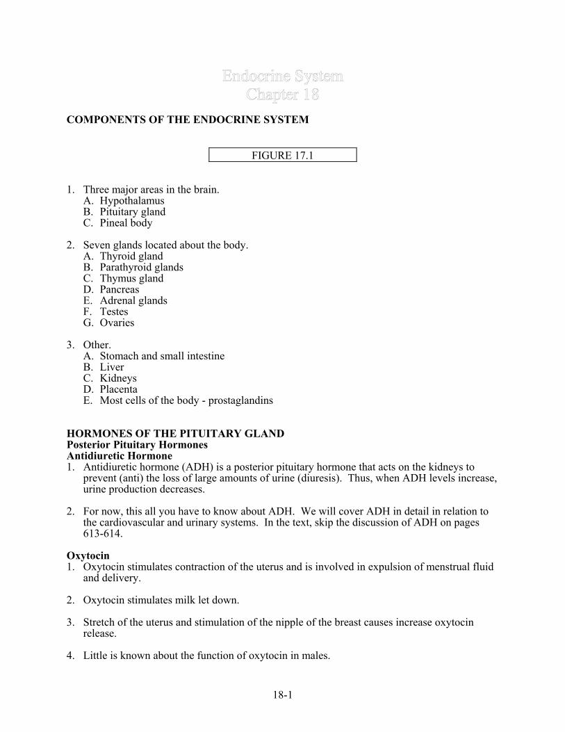
18-1 COMPONENTS OF THE ENDOCRINE SYSTEM FIGURE 17.1 1. Three major areas in the brain. A. Hypothalamus B. Pituitary gland C. Pineal body 2. Seven glands located about the body. A. Thyroid gland B. Parathyroid glands C. Thymus gland D. Pancreas E. Adrenal glands F. Testes G. Ovaries 3. Other. A. Stomach and small intestine B. Liver C. Kidneys D. Placenta E. Most cells of the body - prostaglandins HORMONES OF THE PITUITARY GLAND Posterior Pituitary Hormones Antidiuretic Hormone 1. Antidiuretic hormone (ADH) is a posterior pituitary hormone that acts on the kidneys to prevent (anti) the loss of large amounts of urine (diuresis). Thus, when ADH levels increase, urine production decreases. 2. For now, this all you have to know about ADH. We will cover ADH in detail in relation to the cardiovascular and urinary systems. In the text, skip the discussion of ADH on pages 613-614. Oxytocin 1. Oxytocin stimulates contraction of the uterus and is involved in expulsion of menstrual fluid and delivery. 2. Oxytocin stimulates milk let down. 3. Stretch of the uterus and stimulation of the nipple of the breast causes increase oxytocin release. 4. Little is known about the function of oxytocin in males.
Transcript
18-1
COMPONENTS OF THE ENDOCRINE SYSTEM FIGURE 17.1 1. Three major areas in the brain.
A. Hypothalamus B. Pituitary gland C. Pineal body
2. Seven glands located about the body. A. Thyroid gland B. Parathyroid glands C. Thymus gland D. Pancreas E. Adrenal glands F. Testes G. Ovaries
3. Other. A. Stomach and small intestine B. Liver C. Kidneys D. Placenta E. Most cells of the body - prostaglandins
HORMONES OF THE PITUITARY GLAND Posterior Pituitary Hormones Antidiuretic Hormone 1. Antidiuretic hormone (ADH) is a posterior pituitary hormone that acts on the kidneys to
prevent (anti) the loss of large amounts of urine (diuresis). Thus, when ADH levels increase, urine production decreases.
2. For now, this all you have to know about ADH. We will cover ADH in detail in relation to
the cardiovascular and urinary systems. In the text, skip the discussion of ADH on pages 613-614.
Oxytocin 1. Oxytocin stimulates contraction of the uterus and is involved in expulsion of menstrual fluid
and delivery. 2. Oxytocin stimulates milk let down. 3. Stretch of the uterus and stimulation of the nipple of the breast causes increase oxytocin
release. 4. Little is known about the function of oxytocin in males.
18-2
ANTERIOR Pituitary Hormones 1. Growth hormone (GH), also called somatotropin.
A. Effects. 1) Stimulates growth in most tissues.
a. Promotes protein synthesis. b. Promotes the growth of bone (especially at the epiphyseal plate). c. Stimulates the liver to produce hormones called somatomedins, which in turn
stimulate bone and cartilage growth. The best know somatomedins are insulin-like growth factor I and II.
2) Energy metabolism.
a. Increases the breakdown of lipids in adipose tissue and the release of fatty acids into the blood. The fatty acids are used by other tissues as a source of energy.
b. The use of fatty acids spares the use of blood glucose. Therefore blood glucose
levels are maintained or increase.
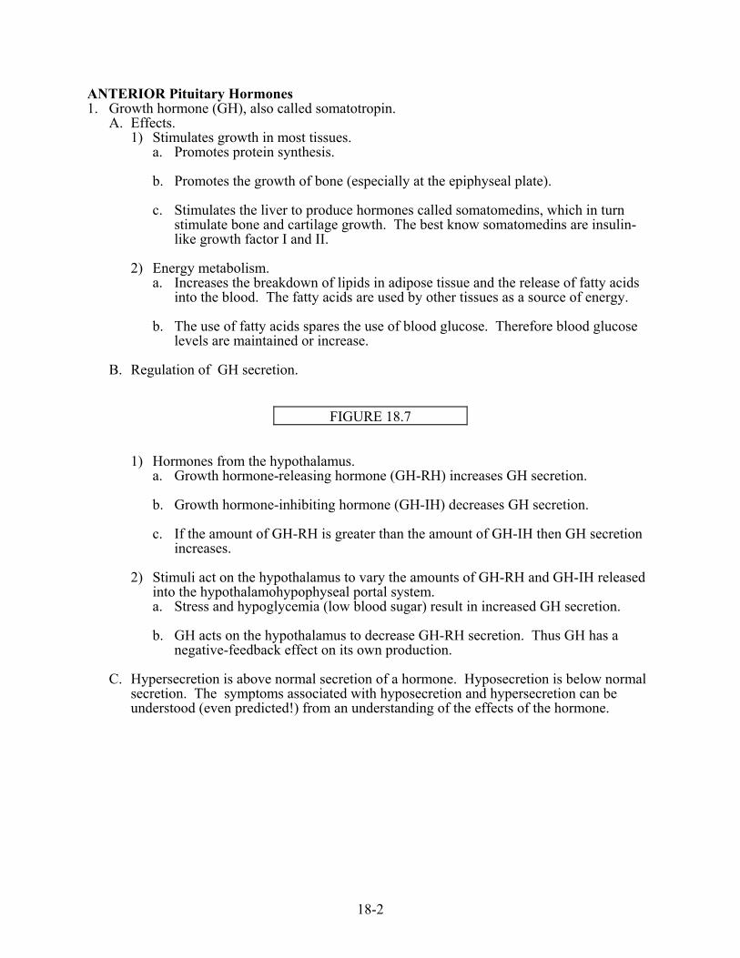
B. Regulation of GH secretion. FIGURE 18.7
1) Hormones from the hypothalamus. a. Growth hormone-releasing hormone (GH-RH) increases GH secretion. b. Growth hormone-inhibiting hormone (GH-IH) decreases GH secretion. c. If the amount of GH-RH is greater than the amount of GH-IH then GH secretion
increases.
2) Stimuli act on the hypothalamus to vary the amounts of GH-RH and GH-IH released into the hypothalamohypophyseal portal system. a. Stress and hypoglycemia (low blood sugar) result in increased GH secretion. b. GH acts on the hypothalamus to decrease GH-RH secretion. Thus GH has a
negative-feedback effect on its own production.
C. Hypersecretion is above normal secretion of a hormone. Hyposecretion is below normal secretion. The symptoms associated with hyposecretion and hypersecretion can be understood (even predicted!) from an understanding of the effects of the hormone.
18-3
Dwarfism results from hyposecretion of GH in children. How does decreased GH result in a person who is shorter than they would have otherwise been? What would be the result of hypersecretion of GH in children?
Would you expect a child with hyposecretion of GH to be thin or to be mildly obese?
Explain.
Acromegaly results from hypersecretion of GH in adults. Why does hypersecretion of
GH in an adult not change their height? Would a person with acromegaly have prominent eyebrow ridges and thick fingers, or would they appear normal? Explain.
How does hypersecretion of GH result in chronic hyperglycemia (elevated blood sugar),
which can lead to diabetes mellitus?
18-4
2. Thyroid stimulating hormone (TSH) or thyrotropin stimulates the release of thyroid hormones from the thyroid gland. Thyroid hormones are involved with metabolism. More later.
3. Adrenocorticotropic hormone (ACTH) stimulates the release of cortisol from the adrenal
gland. Cortisol is involved with metabolism. More later. 4. Melanocyte-stimulating hormone (MSH) stimulates melanocytes to increase melanin
production, causing skin to become darker. MSH is not a well understood hormone in humans.
5. Luteinizing hormone (LH) and follicle-stimulating hormone (FSH) are gonadotropins.
Gonadotropins stimulate the growth of the gonads (ovaries or testes) and the production of sex hormones (estrogen and progesterone in the female, testosterone in the male).
6. Prolactin stimulates the production of milk in the mammary glands. No clear role for
prolactin has been established in males.
18-5
Thyroid Gland FIGURE 18.8 1. The thyroid gland is located inferior to the larynx. 2. The thyroid gland is divided into two lobes connected by the isthmus. Histology 1. The thyroid gland contains many hollow balls of cells, called follicles. The follicles are
filled with a protein called thyroglobulin. Thyroid hormones are attached to the thyroglobulin, which functions as a storage molecule.
2. Parafollicular cells, scattered between the follicles, produce the hormone calcitonin. Thyroid Hormone Synthesis FIGURE 18.9 1. Dietary iodine (I) is ingested, converted to iodide (I-), absorbed into the blood, and
transported to the follicle. A. Iodide enters the follicle by active transport. This concentrates the iodide. B. Iodide transport requires TSH from the anterior pituitary.
2. Thyroglobulin is synthesized within the cells of the follicle. 3. Iodide is converted to iodine that binds to tyrosine (an amino acid) in thyroglobulin, which is
secreted into the lumen of the follicle. 4. The tyrosine and iodine molecules form the thyroid hormones: triiodothyronine (T3) and
tetraiodothyronine (T4) or thyroxine. T3 has three iodines and T4 has four iodines. Approximately a two week supply of thyroid hormones is stored within the follicles.
5. Thyroglobulin is removed from the lumen of the follicle by the follicular cells. 6. Thyroid hormones are separated from thyroglobulin and secreted into the blood. Thyroxine
(T4) is the major (90%) hormone secreted. The remaining thyroglobulin amino acids are reused to synthesize new thyroglobulin.
7. Important points: iodine and TSH are required for thyroid hormone synthesis. Transport in the Blood 1. Most thyroid hormones are transported bound to a plasma protein called thyroxine-binding
globulin (TBG). 2. TBG increases the half-life of thyroid hormones. Thyroid hormone levels do not fluctuate
very much.
18-6
3. Much [33% - 40%] of the T4 (thyroxine) is converted to T3, which is the major thyroid hormone that interacts with target tissues. [T3 is four times as potent as T4]
Mechanism of Action of Thyroid Hormones 1. The response of target cells is mediated through intracellular receptors in the nuclei. Thus
thyroid hormones stimulate the production of new proteins. 2. Thyroid hormones increase ATP and heat production in mitochondria. 3. The response time to thyroid hormones is about 1 week, so thyroid hormones are not
involved with minute to minute regulation. Effects of Thyroid Hormones 1. Thyroid hormones affect most tissues, but not all tissues respond in the same fashion. 2. Thyroid hormones are necessary for normal growth and development. 3. Metabolism.
A. Thyroid hormones increase the breakdown of glucose and lipids to make ATP, which is used for energy.
B Thyroid hormones increase protein synthesis for growth and development. C. Thyroid hormones increase metabolic rate and body temperature.
Effects of Hyposecretion and Hypersecretion of Thyroid Hormones Hypothyroidism Hyperthyroidism Decreased metabolic rate, Increased metabolic rate, low body temperature, high body temperature, intolerant to cold intolerant to heat Gain in body weight, Loss of body weight, reduced appetite increased appetite Reduced activity of sweat Copious sweating, skin and sebaceous glands, warm and flushed skin dry and cold Reduced heart rate Rapid heart rate Reflexes slowed Reflexes quicker and exaggerated, may exhibit tremors Myxedema (swelling of the face and body) Exophthalmos (protruding of the eyes) Iodide uptake decreased Iodide uptake increased Goiter (enlargement of the Goiter almost always develops thyroid gland) is possible
18-7
Why would you expect iodide use to decrease in hypothyroidism and increase in hyperthyroidism?
Regulation of Thyroid Hormone Secretion FIGURE 18.10 1. Thyrotropin-releasing hormone (TRH) is released from the hypothalamus.
A. Exposure to cold and stress increase TRH secretion, whereas food deprivation decreases TRH secretion.
B. TRH stimulates the release of TSH.
2. Thyroid stimulating hormone (TSH) is released from the anterior pituitary. A. TSH is the most important regulator of thyroid hormones. It is necessary for the uptake
of iodide (I-) and it stimulates increased thyroid hormone secretion. B. TSH also causes cells of the thyroid gland to increase in size and in number.
3. Negative-feedback control of thyroid hormone secretion. A. Thyroid hormones inhibit TSH secretion. B. Thyroid hormones inhibit TRH secretion.
Some Common Thyroid Disorders 1. Grave's disease is the second most common endocrine disorder (diabetes mellitus is the most
common). A. Grave's disease is hyperthyroidism characterized by goiter and exophthalmos. B. Grave's disease appears to be an autoimmune disease. Most patients have thyroid
stimulating immunoglobulin (TSI), a TSH-like immune globulin, in their plasma.
18-8
Would you expect TSH levels to be high or low? Why does this disorder produce goiter?
C. Treatment for Grave's disease includes drugs that prevent the synthesis of thyroid
hormones, surgical removal of part of the thyroid gland, or radioactive iodine that destroys part of the thyroid gland.
D. Thyroid storm results from large amounts of thyroid hormones. It usually occurs in
untreated, or poorly treated, Grave's disease. In severe cases, shock, coma, and death can result.
Which of the following symptoms would you expect to observe during thyroid storm?
High fever Dehydration (resulting from sweating) Tachycardia (above normal heart rate)
2. Iodine deficiency in the diet is not usually a problem in the USA because table salt is fortified with iodine.
What effect would iodine deficiency have on blood TSH levels? Could this cause goiter?
Explain.
3. Cretinism is hypothyroidism in the infant.
A. The fetus does not make its own thyroid hormones and cretinism can be caused by maternal iodine deficiency. Congenital (present at birth) errors in thyroid hormone synthesis can result in cretinism in infants.
B. Cretinism results in mental retardation and a short, grotesque appearance. C. If detected early, cretinism can be treated with thyroid hormones. All babies in the U.S.
are screened for low thyroid hormone two to five days after birth.
18-9
Calcitonin 1. Calcitonin is secreted by the parafollicular cells of the thyroid gland. 2. Calcitonin affects osteoclasts .
calcitonin , osteoclast activity , Ca2+ movement , Ca2+ in blood (bone breakdown) from blood into bone
3. Regulation. Blood calcium levels directly affect the parafollicular cells to produce a
negative-feedback loop.
Ca2+ in blood , calcitonin , Ca2+ in blood 4. The role of calcitonin in humans is unclear.
A. Other hormones (e.g., parathyroid hormone and vitamin D) are more important than calcitonin in regulating calcium levels, and probably mask the effects of calcitonin.
B. Humans are relatively insensitive to the actions of calcitonin.
1) Large doses of calcitonin are required to produce hypocalcemia (low blood calcium levels).
2) Calcitonin deficiency produces no known pathology.
C. Calcitonin is being used to treat osteoporosis because it depresses osteoclast activity.
PARATHYROID GLANDS FIGURE 18.11 1. The parathyroid glands are located on the posterior surface of the thyroid gland. There are
usually four masses of parathyroid glands, consisting of densely packed cells. 2. The parathyroid glands produce parathyroid hormone (PTH). The overall effect of
parathyroid hormone is to increase blood calcium levels and decrease blood phosphate levels.
FIGURE 18.12
A. PTH increases the release of calcium from bone into the blood.
PTH , osteoclast activity , Ca2+ in blood
18-10
B. When urine is formed, calcium moves from the blood into the urine. To prevent calcium loss, the calcium is later reabsorbed from the urine.
PTH , Ca2+ reabsorption from urine , Ca2+ in blood
C. Calcium absorption from the small intestine depends upon active vitamin D. Activation
of vitamin D occurs in the kidney under the influence of PTH.
PTH , active vitamin D in kidney , Ca2+ absorption , Ca2+ in blood in small intestine
3. Regulation of PTH. Blood calcium levels affect the parathyroid glands directly to produce a negative-feedback effect, which maintains calcium homeostasis. Blood Ca2+ , PTH , which results in blood Ca2+ or Blood Ca2+ , PTH , which results in blood Ca2+ If person did not have enough calcium in their diet, what would happen to:
Levels of PTH Increase Decrease Release of calcium from bones Increase Decrease Calcium reabsorption from the kidneys Increase Decrease Calcium absorption from the Increase small intestine Decrease
4. Hypoparathyroidism is a lower than normal level of PTH. A. Hypoparathyroidism is caused by removal of the parathyroid glands with the thyroid
gland or is of unknown cause. B. Hypoparathyroidism results in hypocalcemia, which is lower than normal blood calcium
levels.
18-11
Hypoparathyroidism can result in increased neuromuscular excitability, tetany, laryngospasm, and death from asphyxiation can result. Explain.
5. Hyperparathyroidism is a higher than normal level of PTH.
A. Primary hyperparathyroidism results from abnormal parathyroid function, such as a tumor. Primary hyperparathyroidism can result in hypercalcemia, which is higher than normal blood calcium levels. Calcium carbonate salts may be deposited throughout the body, especially in the renal tubules (kidney stones), lungs, blood vessels, and gastric mucosa.
B. Secondary hyperparathyroidism is caused by conditions that reduce blood calcium
levels, which stimulates PTH secretion. Examples are inadequate calcium in the diet, inadequate vitamin D, pregnancy, or lactation.
A man is admitted to the emergency room with a broken femur. X-rays reveal than not
only is the bone broken, but that it probably broke because it was weakened by increased bone resorption. Upon questioning, it was learned that the man has very poor dietary habits, including little intake of dairy products. However, blood test revealed that his blood calcium levels are normal. Explain these observations.
ADRENAL GLANDS FIGURE 18.13 1. There are two adrenal glands. 2. Each adrenal gland is located on the superior aspect of the kidney. The adrenal glands are
sometimes called the suprarenal glands because of their location.
18-12
Histology 1. The medulla is the inner layer. 2. The cortex is the outer layer. The cortex is subdivided into three indistinct parts.
A. The zona glomerulosa is the outermost part of the cortex. B. The zona fasciculata is the middle part of the cortex. C. The zona reticularis is the innermost part of the cortex.
Hormones of the Adrenal Medulla 1. The adrenal medulla secretes epinephrine (80%) and norepinephrine (20%). These are
structurally similar chemicals that produce similar responses in the body. Tyrosine -----> Dopa -----> Dopamine -----> Norepinephrine -----> Epinephrine (Noradrenalin) (Adrenalin)
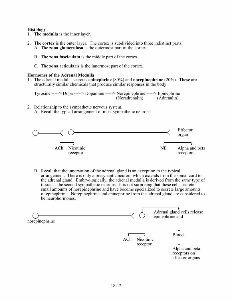
2. Relationship to the sympathetic nervous system. A. Recall the typical arrangement of most sympathetic neurons.
Effector organ ACh Nicotinic NE Alpha and beta receptor receptors
B. Recall that the innervation of the adrenal gland is an exception to the typical arrangement. There is only a presynaptic neuron, which extends from the spinal cord to the adrenal gland. Embryologically, the adrenal medulla is derived from the same type of tissue as the second sympathetic neurons. It is not surprising that these cells secrete small amounts of norepinephrine and have become specialized to secrete large amounts of epinephrine. Norepinephrine and epinephrine from the adrenal gland are considered to be neurohormones.
Adrenal gland cells release epinephrine and norepinephrine Blood ACh Nicotinic receptor Alpha and beta receptors on effector organs
18-13
3. Effects of epinephrine and norepinephrine. FIGURE 18.14
A. Epinephrine and norepinephrine are sympathomimetic (mimic the effects of the sympathetic division of the ANS). Secretion of these hormones is part of the "fight or flight" response.
B. Increase energy sources.
1) Stimulate the breakdown of glycogen to glucose in the liver. The glucose is released from the liver and causes an increase in blood sugar levels. The glucose is used as a source of energy by other tissues.
Name another hormone we have studies that causes an increase in blood sugar levels.
2) Stimulate the breakdown of glycogen to glucose in skeletal muscle. The glucose is
used as a source of energy by the skeletal muscle, which can't release glucose. 3) Increase the breakdown of fats in adipose tissue. Fatty acids are released into the
blood and used by other tissues, especially skeletal muscle, as a source of energy.
C. Cardiovascular effects. 1) Increase heart rate and force of contraction of the heart result in increased blood
pressure and increased delivery of blood. 2) Reroute blood flow.
a) Vasodilation of blood vessels in cardiac muscle and skeletal muscle reroutes blood to these organs, which are essential for activity.
b) Vasoconstriction of blood vessels in the skin, kidneys, and gastrointestinal tract
reroutes blood away from organs not essential for activity.
4. Regulation of adrenal medulla hormones. A. Epinephrine and norepinephrine are released as a result of sympathetic stimulation. B. Secretions increase in response to low blood sugar, emotional excitement, stress,
exercise, low blood pressure, or injury.
5. Disorders of the adrenal medulla. A. Hypersecretion can result from pheochromocytoma, a benign adrenal tumor, or
neuroblastoma, a malignant adrenal tumor.
18-14
Which of the following symptoms would you expect to observe as a result of hypersecretion of the adrenal medulla?
Bradycardia (heart rate slower than normal) Pallor (skin paler than normal) Sweating Hypertension (blood pressure higher than normal)
B. Hyposecretion produces no known clinical conditions.
Hormones of the Adrenal Cortex 1. The adrenal cortex secretes corticosteroids (cortico = cortex; steroid = a type of lipid). 2. There are three general groups of corticosteroids.
A. Mineralocorticoids (aldosterone). Secreted by the zona glomerulosa, these hormones affect the "minerals" sodium, potassium, and hydrogen.
B. Glucocorticoids (cortisol). Secreted by the zona fasciculata, these hormones affect
glucose metabolism. C. Androgens. Secreted by the zona reticularis, these are "male sex hormones."
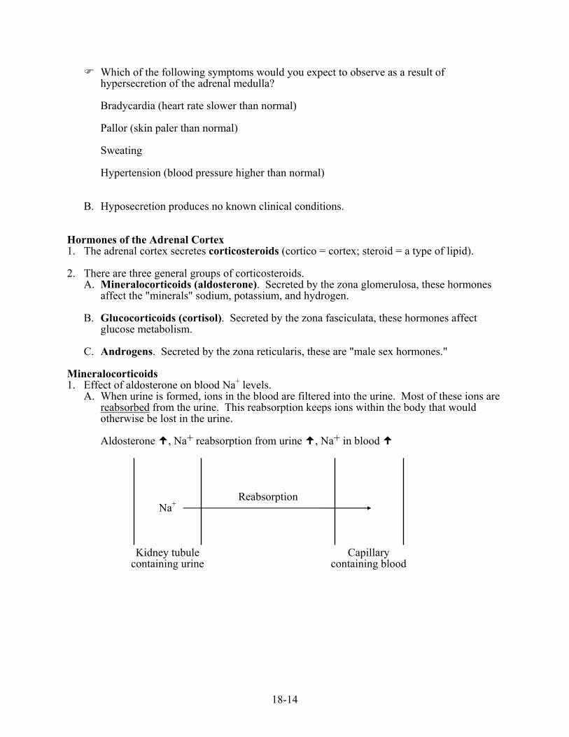
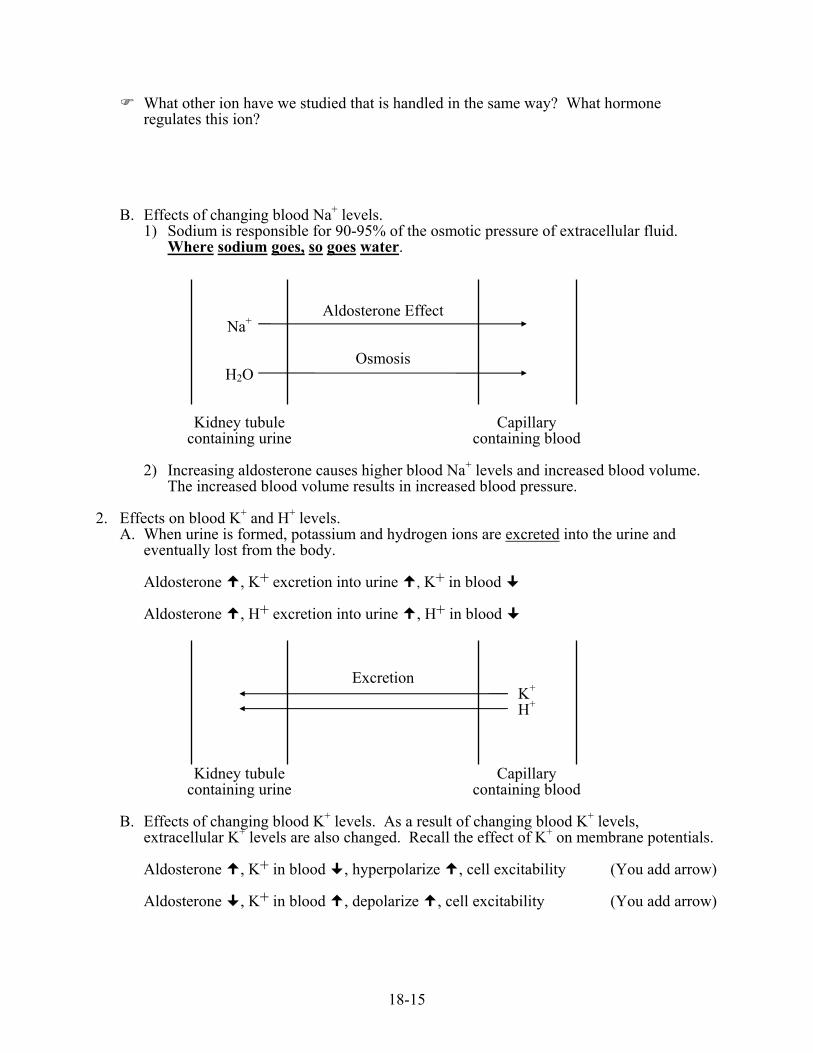
Mineralocorticoids 1. Effect of aldosterone on blood Na+ levels.
A. When urine is formed, ions in the blood are filtered into the urine. Most of these ions are reabsorbed from the urine. This reabsorption keeps ions within the body that would otherwise be lost in the urine. Aldosterone , Na+ reabsorption from urine , Na+ in blood
B. Effects of changing blood K+ levels. As a result of changing blood K+ levels, extracellular K+ levels are also changed. Recall the effect of K+ on membrane potentials. Aldosterone , K+ in blood , hyperpolarize , cell excitability (You add arrow) Aldosterone , K+ in blood , depolarize , cell excitability (You add arrow)
18-16
C. Effects of changing blood H+ levels. As a result of changing blood H+ levels, extracellular H+ levels are also changed. By definition, acidosis is below normal blood pH, and alkalosis is above normal blood pH. Aldosterone , H+ in blood , pH (You add arrow) The condition that results from this pH change is called _________________________, and it can cause convulsions. Aldosterone , H+ in blood , pH (You add arrow) The condition that results from this pH change is called _________________________, and it can cause coma.
3. Regulation of aldosterone will be considered with the cardiovascular and urinary systems. 4. Addison's disease results from low levels of aldosterone and cortisol. It can be caused by
autoimmune disease or infections, such as tuberculosis. Which of the following symptoms are produced by low levels of aldosterone?
Decreased blood Na+ Hyponatremia Yes No Elevated blood K+ Hyperkalemia Yes No Elevated blood H+ Acidosis Yes No
In Addison’s disease would you expect lower than normal or higher than normal blood
pressure?
18-17
In Addison’s disease, would you expect nonresponsive, weak skeletal muscles or skeletal muscles that exhibit tremors and tetany? Explain.
5. Aldosteronism results from excess production of aldosterone. It can result from an adrenal tumor (primary aldosteronism) or from stimulation of aldosterone production by an extraneous factor (secondary aldosteronism, e.g. renin secretion by the kidneys). Which of the following would you expect in aldosteronism?
Increased blood Na+ Hypernatremia Yes No Decreased blood K+ Hypokalemia Yes No Decreased blood H+ Alkalosis Yes No
Glucocorticoids 1. Effects.
A. Metabolic effects. Cortisol increases blood sugar and the conversion of glucose to glycogen. 1) Cortisol increases fat and protein metabolism.
a) Fats are broken down and are used as sources of energy, which spares glucose use.
b) There is decreased use of glucose and amino acids by skeletal muscle, which
spares glucose use. The muscles then rely more on fats. c) Increased protein breakdown, especially in skeletal muscle, makes amino acids
available. The liver uses the amino acids to synthesize glucose, which can be released from the liver causing a rise in blood sugar levels.
2) The liver stores excess glucose as glycogen.
18-18
Name two other hormone that we have studied that increase blood sugar levels?
B. Developmental effects. Glucocorticoids are necessary for the maturation of fetal tissues. C. Antiinflammatory effects.
1) Glucocorticoids decrease the inflammatory response by decreasing the number of white blood cells and the secretion of inflammatory chemicals from tissues.
2) Glucocorticoids function to prevent over reaction of the body to stress, injury, and
disease. 2. Cushing's syndrome results from hypersecretion of cortisol and androgens. Most cases
result from over production of ACTH caused by an anterior pituitary tumor. Nonpituitary tumors of the lung, thymus, pancreas, and kidney can also produce ACTH.
Would you predict hypoglycemia or hyperglycemia in Cushing's syndrome? Explain.
Explain how muscular atrophy (decrease in the amount of muscle tissue), muscular
weakness, and osteoporosis (hint: a component of bone is collagen) could develop in Cushing's syndrome.
18-19
Would a person with Cushing's syndrome be more or less susceptible to infections? Explain.
3. Regulation of cortisol. FIGURE 18.15
A. Stress or hypoglycemia (low blood sugar levels) increases the release of corticotropin-releasing hormone (CRH) from the hypothalamus.
B. CRH stimulates the release of adrenocorticotropic hormone (ACTH) from the anterior
pituitary. C. ACTH stimulates the secretion of cortisol from the adrenal cortex. Without ACTH the
adrenal cortex atrophies. D. Cortisol has a negative-feedback effect on the hypothalamus and the anterior pituitary,
which decreases the secretion of CRH and ACTH. Can you explain why stress might result in getting sick?
18-20
A drug similar to cortisol, called cortisone, is sometimes given to people with severe allergies. How would this be useful?
Taking cortisone chronically can damage the adrenal cortex. Eventually the adrenal cortex
may be unable to produce cortisol. Explain how that might occur.
Addison's disease results from low levels of aldosterone and cortisol. ACTH is similar in
structure to melanocyte stimulating hormone (MSH) and can also cause an increase in melanin production. Would you expect someone with Addison's disease to have increased or decreased skin pigmentation. Explain.
Adrenal Androgens 1. The adrenal cortex produces weak androgens (male sex hormones). These are converted by
peripheral tissues (especially the liver) into the more powerful androgen testosterone. 2. ACTH is known to increase adrenal androgen secretion, but is not believed to be the main
regulator of secretion because adrenal androgen secretion changes independently of ACTH. A yet to be identified pituitary hormone may be responsible.
3. In adult males, adrenal androgens have little effect because so much more testosterone is
produced in the testes. In adult females, adrenal androgens stimulate pubic and axillary hair growth and sex drive.
4. Adrenogenital syndrome results from hypersecretion of androgens.
A. In male children this produces early sexual development and short stature. B. In females, if hypersecretion occurs during fetal development, masculinization of the
genitalia may occur. In adult females, development of facial hair, a deeper voice, and increased sex drive can occur.
18-21
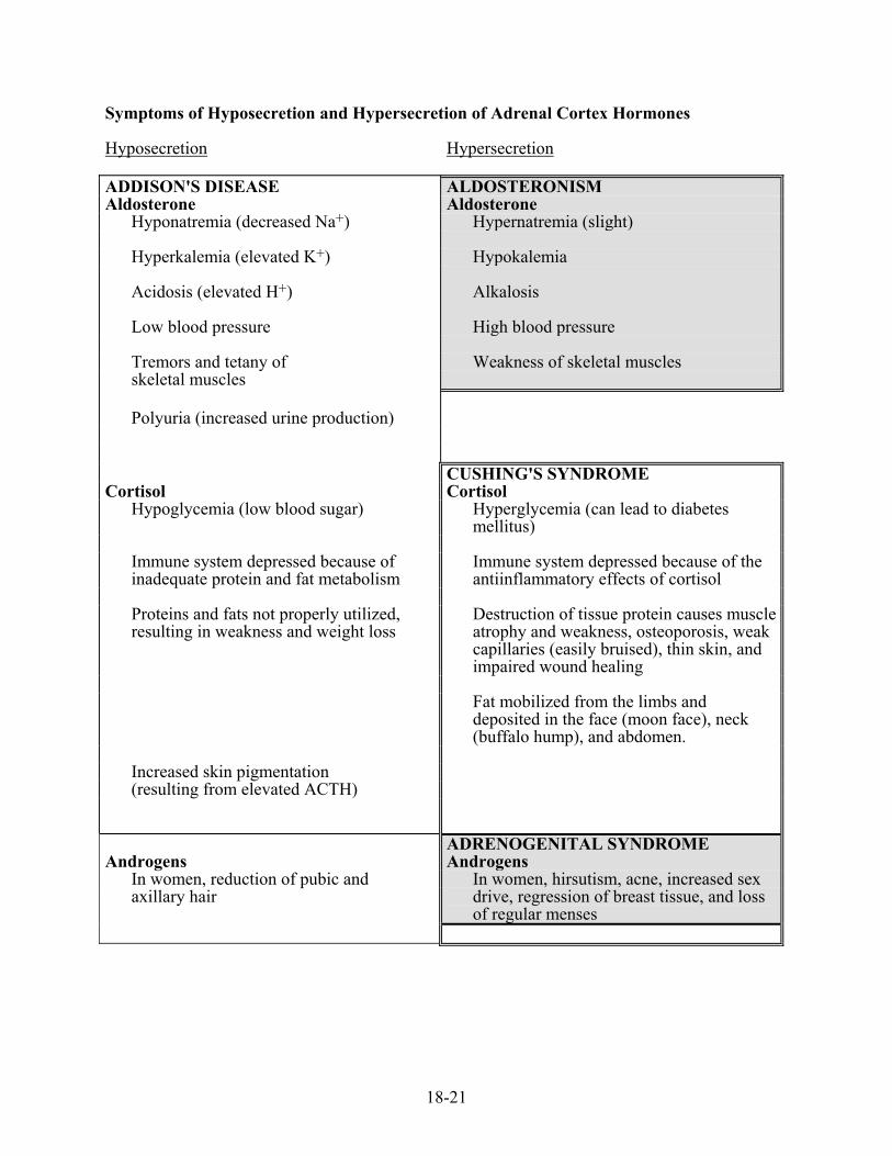
Symptoms of Hyposecretion and Hypersecretion of Adrenal Cortex Hormones Hyposecretion Hypersecretion ADDISON'S DISEASE ALDOSTERONISM Aldosterone Aldosterone
Hyponatremia (decreased Na+) Hypernatremia (slight) Hyperkalemia (elevated K+) Hypokalemia Acidosis (elevated H+) Alkalosis Low blood pressure High blood pressure Tremors and tetany of Weakness of skeletal muscles skeletal muscles Polyuria (increased urine production) CUSHING'S SYNDROME
Cortisol Cortisol Hypoglycemia (low blood sugar) Hyperglycemia (can lead to diabetes
mellitus) Immune system depressed because of inadequate protein and fat metabolism
Immune system depressed because of the antiinflammatory effects of cortisol
Proteins and fats not properly utilized, resulting in weakness and weight loss
Destruction of tissue protein causes muscle atrophy and weakness, osteoporosis, weak capillaries (easily bruised), thin skin, and impaired wound healing
Fat mobilized from the limbs and
deposited in the face (moon face), neck (buffalo hump), and abdomen.
Increased skin pigmentation (resulting from elevated ACTH) ADRENOGENITAL SYNDROME
Androgens Androgens In women, reduction of pubic and In women, hirsutism, acne, increased sex axillary hair drive, regression of breast tissue, and loss
of regular menses
18-22
PANCREAS FIGURE 18.16 The pancreas is located along the superior part of the small intestine (deep to the stomach). Histology 1. Exocrine gland.
A. Acini (singular acinus) are sac-like structures connected to ducts. The acini secrete enzymes that move down a duct system into the small intestine.
B. The enzymes breakdown foods in the small intestine.
2. Endocrine gland. A. The pancreatic islets (islets of Langerhans) secrete hormones. B. Within the islets, alpha cells secrete glucagon and beta cells secrete insulin.
Effects of Increased Insulin on Its Target Tissues 1. Most tissues of the body (except as noted below).
A. Insulin increases glucose (blood sugar) movement into most cells by facilitated diffusion by stimulating an increase in the number of carrier molecules within plasma membranes. As a result, blood sugar levels decrease.
B. Insulin increases the active transport of many amino acids into most cells. The amino
acids are used for energy, converted into glucose, or are used to synthesize proteins.
2. Skeletal muscle. A. Insulin promotes the uptake of glucose by skeletal muscle. B. Excess glucose is converted into glycogen, which functions as an energy storage
molecule.
3. Liver. A. Glucose moves into or out of liver cells by facilitated diffusion. However, insulin does
not change the number of carrier molecules in the membrane of liver cells, and glucose transport by liver cells does not respond to insulin.
B. Excess glucose is converted into glycogen.
4. Adipose tissue. A. Insulin increases the transport of glucose into adipose cells by facilitated diffusion. B. Insulin activates an enzyme that promotes the uptake of fatty acids. C. The glucose is convert into glycerol, which combines with the fatty acids to form
triglycerides (stored lipids).
18-23
5. Nervous tissue. A. Glucose is the main source of energy for nervous tissue. B. Glucose transport into most nervous tissues does not depend upon insulin. However,
because insulin regulates blood sugar levels, insulin is very important for the proper functioning of nervous tissue.
C. Exceptions: glucose movement into the satiety (hunger) center and certain sympathetic
centers in the hypothalamus depends upon insulin.
6. Overview: What effect does increased insulin have on Uptake of glucose Increase or decrease Glycogen synthesis Increase or decrease Fat synthesis Increase or decrease Blood sugar levels Increase or decrease
Effects of Decreased Insulin on Its Target Tissues 1. Decreased insulin decreases the uptake of glucose by most tissues and increases the release
of glucose from the liver. As a result, blood sugar levels are maintained or increase. 2. Skeletal muscle uses stored glycogen as an energy source. Skeletal muscle does not release
glucose. However, proteins can be broken down, resulting in the release of amino acids into the blood.
3. Liver.
A Decreased insulin promotes the breakdown of glycogen and the release of glucose from the liver. The glucose is an energy source for nervous tissue between meals or during exercise.
B. Amino acids are used to synthesize glucose.
4. Adipose tissue. A. Decreased insulin promotes the breakdown of fats and the release of fatty acids into the
blood. The fatty acids are used by tissues as an energy source, which spares glucose use. B. Decreased insulin can stimulate the formation of ketones from fatty acids.
1) Ketones are released from the liver into the blood, and they can be utilized by other tissues as a source of energy.
2) Ketones are acidic substances. Excessive amounts of ketones can lower blood pH.
18-24
Effects of Increased Glucagon on Its Target Tissues 1. Glucagon affects primarily the liver. 2. Effects.
A. Glucagon increases blood sugar by stimulating the release of glucose from the liver. 1) Glucagon stimulates the breakdown of glycogen to glucose. 2) Glucagon stimulates the formation of glucose from amino acids and fats.
B. Glucagon stimulates the breakdown of fats and the formation of ketones.
3. Note that insulin and glucagon have opposite effects. A. Insulin causes the movement of energy molecules into tissues where they are used for a
source of energy or they are stored. Consequently, insulin causes blood sugar levels to decrease.
Think of insulin as the “after the meal” hormone. B. Glucagon causes the production and release of energy molecules from the liver into the
blood. These molecules can be removed from the blood and used by other tissues as sources of energy. Glucagon causes blood sugar levels to increase.
Think of glucagon as the “before the next meal” hormone.
Regulation of Pancreatic Hormone Secretion FIGURE 18.17 1. Regulation of insulin secretion.
A. Blood glucose levels are the most important factor. Certain amino acids can also cause an increase in insulin secretion. blood glucose , amino acids , insulin (following a meal) blood glucose , amino acids , insulin (exercise, fasting)
B. Hormones released from the GI (gastrointestinal) tract increase insulin secretion. The hormones (e.g., gastrin, secretin, cholecystokinin) are released when food enters the GI tract.
18-25
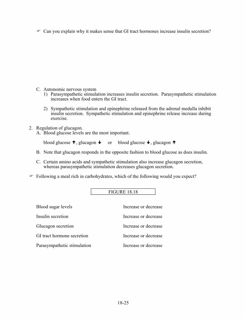
Can you explain why it makes sense that GI tract hormones increase insulin secretion?
C. Autonomic nervous system
1) Parasympathetic stimulation increases insulin secretion. Parasympathetic stimulation increases when food enters the GI tract.
2) Sympathetic stimulation and epinephrine released from the adrenal medulla inhibit
insulin secretion. Sympathetic stimulation and epinephrine release increase during exercise.
2. Regulation of glucagon.
A. Blood glucose levels are the most important. blood glucose , glucagon or blood glucose , glucagon
B. Note that glucagon responds in the opposite fashion to blood glucose as does insulin. C. Certain amino acids and sympathetic stimulation also increase glucagon secretion,
whereas parasympathetic stimulation decreases glucagon secretion. Following a meal rich in carbohydrates, which of the following would you expect?
FIGURE 18.18
Blood sugar levels Increase or decrease Insulin secretion Increase or decrease Glucagon secretion Increase or decrease GI tract hormone secretion Increase or decrease Parasympathetic stimulation Increase or decrease
18-26
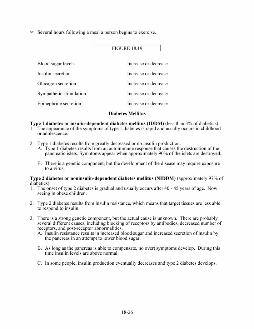
Several hours following a meal a person begins to exercise. FIGURE 18.19
Blood sugar levels Increase or decrease Insulin secretion Increase or decrease Glucagon secretion Increase or decrease Sympathetic stimulation Increase or decrease Epinephrine secretion Increase or decrease
Diabetes Mellitus Type 1 diabetes or insulin-dependent diabetes mellitus (IDDM) (less than 3% of diabetics) 1. The appearance of the symptoms of type 1 diabetes is rapid and usually occurs in childhood
or adolescence. 2. Type 1 diabetes results from greatly decreased or no insulin production.
A. Type 1 diabetes results from an autoimmune response that causes the destruction of the pancreatic islets. Symptoms appear when approximately 90% of the islets are destroyed.
B. There is a genetic component, but the development of the disease may require exposure
to a virus.
Type 2 diabetes or noninsulin-dependent diabetes mellitus (NIDDM) (approximately 97% of diabetics) 1. The onset of type 2 diabetes is gradual and usually occurs after 40 - 45 years of age. Now
seeing in obese children. 2. Type 2 diabetes results from insulin resistance, which means that target tissues are less able
to respond to insulin. 3. There is a strong genetic component, but the actual cause is unknown. There are probably
several different causes, including blocking of receptors by antibodies, decreased number of receptors, and post-receptor abnormalities. A. Insulin resistance results in increased blood sugar and increased secretion of insulin by
the pancreas in an attempt to lower blood sugar. B. As long as the pancreas is able to compensate, no overt symptoms develop. During this
time insulin levels are above normal. C. In some people, insulin production eventually decreases and type 2 diabetes develops.
18-27
3. Treatment. A. Diet and exercise are important for both types of diabetes. Some people with type 2
diabetes can control their diabetes through diet and exercise. B. Injected insulin is required for type 1 diabetes. Injected insulin is used by about 25-30%
of type 2 diabetics and about 50% of type 2 diabetics use oral drugs that stimulate the pancreas to produce more insulin, improve the body’s response to insulin, or slow carbohydrate digestion (resulting in a slower rise in blood sugar).
Symptoms of Untreated Diabetes Mellitus 1. Hyperglycemia - elevated blood glucose. Why do blood glucose levels become elevated in type 1 diabetes?
Why do blood glucose levels become elevated in type 2 diabetes?
2. Glucosuria - excess glucose in the urine.
A. When urine is first formed, normally all the glucose in blood enters the urine. Later in urine production, all the glucose is reabsorbed from the urine into the blood. When blood glucose levels become very high, more glucose enters the urine than can be reabsorbed . Consequently, the glucose "spills over" into the urine.
B. A simple test of the urine can detect glucose. A positive test for glucose is very strong
evidence for diabetes mellitus.
18-28
3. The three "P's" of untreated diabetes mellitus: polyuria (copious urine production), polydipsia (thirst), and polyphagia (excess eating).
Explain the basis for each of these symptoms.
Polyuria
Polydipsia
Polyphagia (hint: what is the effect of insulin on the hunger center in the brain?)
4. Ketosis/acidosis in type 1 diabetes.
A. Ketosis is elevated ketones (acetoacidic acid, beta-hydroxybutyric acid, and acetone) in the blood. 1) Recall that decreased insulin results in the formation of ketones. 2) Ketosis does not usually develop in type 2 diabetes because there is enough insulin
present.
B. Ketonurea is enhanced excretion of ketones in the urine. A simple test of the urine can detect ketones.
18-29
C. Acetone breath results from ketones excreted in the breath. D. Acidosis results from ketone formation by the liver and the release of fatty acids from
adipose tissue. Recall that acidosis can depress neuron activity. Untreated acidosis can become severe enough to cause coma.
E. Deep, rapid respirations are one method of compensating for acidosis (more later when
we get to the respiratory system). 5. Hyperosmolarity in type 2 diabetes.
A. Hyperosmolarity results from very high levels of glucose in the blood. B. In type 2 diabetes, the amount of insulin present is not enough for normal glucose
transport because of insulin resistance. The result is an increase in blood sugar. The amount of insulin present is enough to prevent ketosis/acidosis.
C. As the concentration of glucose in the blood increases, water moves out of neurons by
osmosis. The resulting dehydration results in drowsiness, depressed reflexes, and even coma.
D. Although blood sugar levels increase in type 1 diabetes, they do not increase to the high
levels seen in type 2 diabetes, because in type 1 diabetes ketosis/acidosis occurs first.
6. Long term effects. A. The cause or causes of the many devastating effects of diabetes are not completely
known. 1) Chronic, higher than normal levels of glucose are the number one suspect. 2) Careful monitoring of blood glucose followed by the appropriate insulin dose helps to
keep glucose levels closer to normal. Data indicates this can dramatically reduce the long term complications of diabetes.
B. Complications.
1) Retinopathy results in impaired vision and blindness. 2) Nephropathy results in protein excretion by the kidneys and eventual kidney failure. 3) Neuropathy results in sensations of pain, coldness, tingling, or burning; muscle
weakness and paralysis; loss of bladder and bowel control; and impotence. 4) Cardiovascular disease such as atherosclerosis and reduced blood delivery to tissues,
which can result in amputation. 5) Increase risk of infections and poor wound healing.
18-30
Insulin Shock Description 1. Too much insulin is taken relative to the amount of glucose ingested. For example, a
diabetic skips a meal. 2. Increased physical activity decreases blood glucose as the glucose is utilized, causing a
relative excess of insulin. Symptoms 1. The nervous system has an inadequate supply of glucose. Symptoms include headache,
drowsiness, fatigue, coma, convulsive seizures, and death. 2. In response to the low blood glucose, the sympathetic division is activated and the adrenal
gland is stimulated to release epinephrine. What is the advantage of stimulating epinephrine release?
3. Symptoms resulting from epinephrine and activation of the sympathetic division include pale
skin, tachycardia (increased heart rate), increased nervous system excitability, and tremors. Activation of the sympathetic division caused by low blood glucose can also cause sweating (remember that sweat glands have cholinergic receptors, which do not respond to epinephrine).
18-31
Practice problem: A patient arrives in an unconscious condition. A medical emergency bracelet reveals that he is a diabetic. The patient may be in diabetic coma, hyperosmolar coma, or insulin shock. How could you tell which, and what treatment would you recommend for each condition?
Diabetic coma: caused by acidosis in type 1 diabetes. Hyperosmolar coma: caused by dehydration in type 2 diabetes; elevated blood glucose
levels result in the movement of water from tissues into blood, causing tissue dehydration and dysfunction.
Insulin shock: caused by decreased blood glucose resulting from too much insulin. Diabetic Hyperosmolar Insulin Coma Coma Shock (Type 1) (Type 2) Insulin levels (low, normal, high) Blood glucose levels (low, high, very high) Glucose in urine (yes, no) Sweating (yes, no) Pale skin (yes, no) Blood pH (normal, acidosis) Ketones in blood, urine, and breath (yes, no) Deep respirations (yes, no) Dehydrated (yes, no) Treatment 1. Insulin injection 2. Glucagon injection 3. Glucose injection [Standard treatment for EMT for an unconscious person with a diabetic ID
is a glucose drip. Rationale: if insulin shock, glucose will fix. If diabetic coma or hyperosmolar coma, will not make matters much worse. Can fix later with insulin.
18-32
HORMONES OF THE PINEAL BODY, THYMUS GLAND, AND OTHERS 1. Pineal Body. [All mammals, except the anteater, armadillo, sloth, and manatee have a pineal
body. Some lizards, frogs, and lampreys have a pineal eye (“third eye”) that innervates the pineal body.]
FIGURE 18.20
A. The pineal body is located in the brain (epithalamus) and secretes the hormone melatonin in response to the amount of light the organism is exposed to.
B. An increase in light exposure (e.g., increased day length) results in a decrease in
melatonin. A decrease in light (e.g., decreased day length) causes an increase in melatonin secretion.
C. In some animals, melatonin inhibits the release of gonadotropin-releasing hormone
(GnRH) from the hypothalamus. This inhibits the release of follicle-stimulating hormone (FSH) and luteinizing hormone (LH) from the anterior pituitary. Without these hormones, reproductive function is inhibited.
D. In many animals melatonin is used to time seasonal changes, such as breeding cycles,
migration, color coat changes, loss of antlers, etc. 1) In fall and winter as day length shortens, light exposure decreases and melatonin
secretion increases. The effect is to inhibit reproductive organs. 2) In spring and summer day length increases, light exposure increases, melatonin
production decreases, and reproductive organs and behaviors develop.
E. In humans the role of melatonin is unclear. 1) Melatonin may be involved with the onset of puberty.
a. Destruction of the pineal body leads to early sexual development. b. Tumors that increase melatonin secretion result in retarded development of the
reproductive system.
2) Melatonin may be involved with fertility, sleep induction, mood, mental illness, and circadian rhythms (24 hour biological clocks).
2. Thymus Gland
A. The thymus gland is located superior to the heart in the thorax and neck. B. The thymus gland produces the hormone thymosin, which is involved with the
maturation of immune system cells (T cells, a special type of white blood cell).
3. Gastrointestinal Tract
18-33
HORMONELIKE SUBSTANCES 1. Prostaglandins.
A. Prostaglandins are fatty acids produced by most, if not all, tissues. They are secreted in small amounts and effect cells in the immediate area. B. Prostaglandins produce a variety of responses throughout the body.
1) Prostaglandins stimulate uterine contractions, for example, during delivery and menstruation. a. Inhibit prostaglandin production to prevent premature labor and relieve menstrual
cramps. b. Stimulate prostaglandin production to induce labor or abortion. The IUD
prevents embryo implantation by stimulating uterine contractions.
2) Prostaglandins stimulate the inflammatory response, fever, and symptoms of pain. Prostaglandin inhibitors reduce inflammation, fever, and pain. For example, aspirin is a prostaglandin inhibitor.
3) Prostaglandins inhibit gastric secretions in the stomach. A side effect of taking
prostaglandin inhibitors is over production of gastric secretions with accompanying irritation to the GI tract.
4) For the future: need to find drugs that activate or inhibit prostaglandins in one
system. At present, prostaglandin drugs have wide-spread effects, producing unwanted side effects.
2. Endorphins and enkephalins.
A. Endorphins and enkephalins are produced by many areas of the brain (especially the limbic system and hypothalamus), the dorsal horns of the spinal cord, the placenta, and the GI tract.
B. Endorphins and enkephalins act to inhibit neuron transmission. They bind to the same
receptors as morphine, and are involved with decreasing sensitivity to painful stimuli.