72 Figures 72 Figures 72 Figure 7A Figv 3S 75 - 84 Phyllotheca cf. indica x 2 - 73 The free leaflets of success" odes are longer than the internodes, but the leal ippe"r to have been positioned in such a way as to prevent most of the direct overlap between whorls. 72 - N-Bgv 131 73 - N-Bgv 53-'. A succession of nodes with short internodes. N-Bgv 503 - 81 These specimens are all of dorsiventra1ly flattened discs typical of the species. 75 - N-Bgv 545 78 - N-Bgv 541 81 - N-Bgv 244 76 - N-Bgv 511 79 - N-Bgv 515 77 - N-Bgv 523 80 - N-Bgv 80 (figure 78 see also figure 62 c). Figures 82 - 84 Specimens that appear less robust than usual. 82 - N-Bgv 522 83 - N-Bgv 246 84 - N-Bgv 285.
Transcript
72
Figures 72
Figures 72
Figure 7A
Figv 3S 75
- 84 Phyllotheca cf. indica x 2
- 73 The free leaflets of success" odes are longer thanthe internodes, but the leal ippe"r to have beenpositioned in such a way as to prevent most of thedirect overlap between whorls. 72 - N-Bgv 131 73 - N-Bgv 53-'.
A succession of nodes with short internodes. N-Bgv 503
- 81 These specimens are all of dorsiventra1ly flatteneddiscs typical of the species.75 - N-Bgv 545 78 - N-Bgv 541 81 - N-Bgv 24476 - N-Bgv 511 79 - N-Bgv 51577 - N-Bgv 523 80 - N-Bgv 80 (figure 78 see also figure 62 c).
Figures 82 - 84 Specimens that appear less robust than usual.82 - N-Bgv 522 83 - N-Bgv 246 84 - N-Bgv 285.
73
74
The specimens in figures 82 - 87 all show the typical strap-like leaflets with the marginal strand, but are slightly less robust or rigid than the usual specimens, in other features they resemble them closely. A further group of long leaved specimens is seen in figures 88 - 96, 96a. The free leaflets tend to radiate from the disc in an ordered but lax manner, they too are less robust looking than usual.
3.2.3.1.1 Phyllotheca with long tapering leaflets.
Figures 97 and 98 both have dorsiventrally preserved whorls with relatively long leaves (15 millimetres) that are tapering over their whole length. There are very clear longitudinal strands with lateral flanges at the margin of the free leaflets which persist the whole length. At the base of the free leaflet where it unites to become the disc, there is a very slight widening of the leaf, with a blunt V-shaped notch between adjacent leaflets. The united disc area is not particularly well developed ( 2 - 4 millimetres wide). The midrib is hardly visible. The lateral longitudinal lines end abruptly at the base of the leaflet, commissural lines very faint in the disc.
Figure 99 similarly has tapering leaflets, but only 7,9 millimetres long, the disc being well developed (6,1 millimetres wide). Heavy longitudinal lines on the free leaflet margins are present, ending abruptly at the disc margin. Midribs and commissural lines are clearly visible on the disc. None of these specimens show any signs of recurved or reflexed leaflets. The specimen in figure 100 is preserved as an inverted cup. The united disc-like area has not spread horizontally, which may be a developmental feature. The leaflets are long and narrow, tapering to a fine point, with longitudinal strands and commissural lines clear, but the midribs art poorly defined.
A few specimens show very fine free leaflets, e.g. in figures 101 - 103. The first two have the leaflets fairly lax, spreading in relatively disordered manner, while the last one hao the leaflets placed very regularly, the thin strap-like leaflet expanding at the base to ftrm the united disc. In all the lateral longitudinal strands are visible while the midrib is only faintly indicated.
3.2.3.1.2 Phyl lotheca with long, thin leaflets. Iftfc-"'.
Several specimens have free leaflets that are long and thin in relation to the united disc area. Due to the length and width of the leaflets,
Figures 85 -
Figures 85 -
75
87 Phyllotheca cf. indica x 2
87 Further examples of leaves that are slightly atypical. The free leaflets appear less rigid than usual. In other aspects they agree closely with normal specimens. 85 - N-Bgv 87 86 - N-Bgv 113 87 - N-Bgv 59 pto.
7£
77
Figures 88 - 96a Phyllotheca cf. indica x 2
Figures 88 - 96a A further group of slightly atypical specimens.The long free leaflets are less robust looking and some leaflets appear to be more lax than usual.88 - N-Bgv 100 91 - N-Bgv 221 94 - N-Bgv 28989 - N-Bgv 525 92 - N-Bgv 85 95 - N-Bgv 7590 - N-Bgv 243 93 - N-Bgv 79 96 - N-Bgv 117
96a - N-Bgv 308
18
79
Figures 97
Figures 97
Figure 99
Figure 100
Figure 101
Phyllotheca with long tapering leaves probably P.cf.indica x 2
and 98 Dorsiventrally preserved whorls with long leaflets that taper over their whole length.97 - N-MN 931b 98 - N-MN 933a
A well developed united disc subtends tapering leaflets.N - Blw 86a (see also figure 105).
The united disc has been preserved as an inverted cup, not spread horizontally. This may be a developmental feature. N~Blw 85a (see also figure 104).
- 103 Specimens with fine free leaflets. The laterallongitudinal strands are present on all the specimens, while the midribs are hard.y discernible.101 - N-Bgv 242*249 102 - N-Bgv 247a 103 - N-Bgv 538
to
Figures 104 - 105 Phyllotheca cf. indica Scale is 1 cm
Figure 104 A small specimen that has an unexpanded united discpreserved as an inverted cup-like projection. The lateral strands are clearly preserved, midribs obscure.N-Blw 85a (see also figure 100).
Figure 105 A specimen with well developed disc. The leaflets tend to shoulder out more than usual. The commissural lines are very clear. N-Blw 86a (see also figure 99).
82
105
I
83
they are not as obviously tapering as the "Phyllotheca with long tapering leaves" group.
The longitudinal strand is visible, while the midrib is faint. The leaflets appear to have been rigid as they are not bent or curved in their fossil state. The specimens in figures 106 - 109 have the leaflets undistorted as they reach the united area, while in figures 109 -111 the specimens each have the leaflets widening at that point.Figure 109 shows portion of the closely adhering sheath.
3.2. 3.1.3 Ragged Phy 1 lotheca . HI - MS.
A few specimens show whorls that have the free leaflets disarranged and bent. The leaflets are relatively short and slightly tapered.The tips are not preserved . The leaflets expandat their base, to have wide "shoulders" at the margin of the united disc. The commissural lines are visible, while the midrib is not clear, the lateral longitudinal strand is promineii*". Figures 112 - 114 and 117 show fairly large whorls, while figure 115 is a smaller whorl that has the same characjlter. Figure 116 shows intermediate characters between the usual P.cf.indica a:id this group in that the leaflets appear fairly rigid (P. cf.i ndica) but at their base they definitely "shoulder" out.The specimen in figure 118 similarly has well defined "shjjlders" but the free leaflets are narrower than in the previous specimen.Commi ss'tral lines are clearly defined and the midribs are faint.
3.2.3. 1.4 Unusually large Phyl lotheca remains . 11̂ -121.-----------------------------------
By the large nature of these specimens they are incomplete. The most complete specimen (fig. 119) is preserved with three successive whorls. The united discs are preserved as fans off the node with a width of ahoi't 14 millimetres. In the topmost whorl the free leaflets are indicated, the bases shoulder out, the notchrs between the leaflets are u - or bluntly v - shaped. The midribs are faintly visible. Commissural lines are present/by indication of a depressed arearather than by a distinct line. Figure 120 shows fragments of five successive nodes. The united disc is not 11 developed, but islarge, some free leaflets are preserved, figure 121 has some short leaflets. Figures 122 - 124 show large discs with relatively straplike leaf bases preserved, while figures 125 - 128 appear to have short robust tapering leaves on a robust stem, unlike the delicate leaves of P.cf.austra1i s■ The specimen in figure 129 has the remains of two
84
Figures 106 - 111 Phyllotheca with long, thin leafletsProbably Phyllotheca cf.indica x 2
Figures 106 - 108 The united disc area is relatively small in relationto the length of the free leaflsts, which appear to have been rigid during life. The leaflets continue undistorted into the united disc area 106 - N-Bgv 69 107 - N-Bgv 70 108 - N-Bgv 70
Figures 109 - 111 The leaflets widen slightly as they reach the marginof the united disc area.109 - N-Bgv 62 110 - N-Bgv 546 111 - N-Bgv 91
86
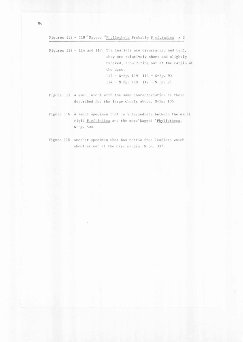
Figures 112 - 118 "Ragged "Phy1lotheca Probably P.cf.indica x 2
Figures 112 - 114 and 117. The leaflets are disarranged and bent,they are relatively short and slightly tapered, shou1 ̂ .jring out at the margin of the disc.112 - N-Bgv 119 113 - N-Bgv 90 114 - N-Bgv 110 117 - N-Bgv 71
Figure 115 A small whorl with the same characteristics as those described for the large whorls above. N-Bgv 105.
Vigure 116 A small specimen that is intermediate between the usual rigid P.cf.ind ica and the more’Ragged "Phyllotheca.N-Bgv 500.
Figure 118 Another specimen that has narrow free leaflets wnich shoulder out at the disc margin. N-Bgv 337.
V
88
Figures 119 - 123 Unusually large Phyllotheca remains. ProbablyP.cl.indica x 2
Figure 119 Three nodes with their united disc are preserved. The margin of the upper disc clearly shows the continuation of the free leaflets. N-Bgv 109.
Figures 120 - 123 Fragments of large specimens with some free leafletsattached. 120 - N-Bgv 509 121 - N-Bgv 507 122 - N-Bgv 506 123 - N-Bgv 294.
90
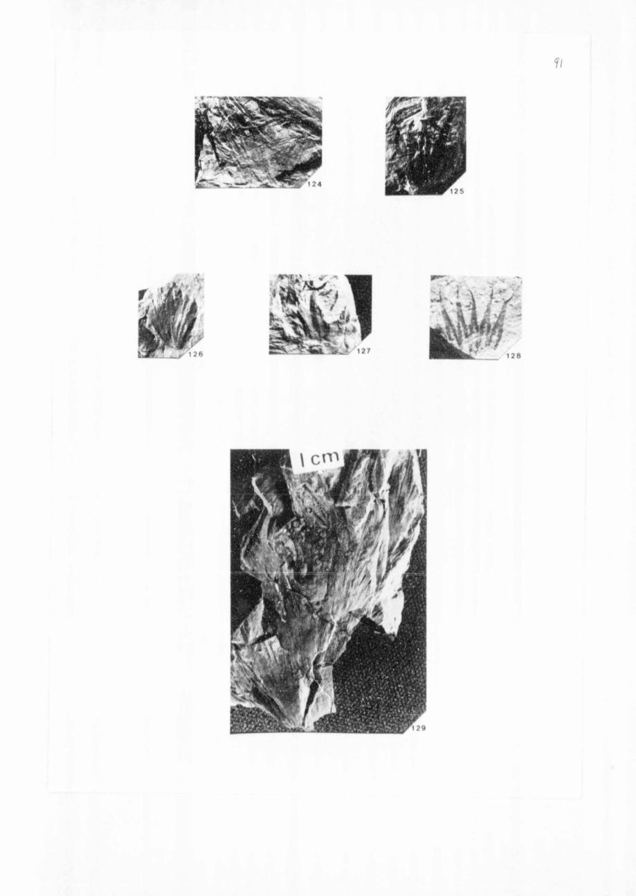
Figures 124 - 129 Unusually large Phyllotheca remains.Probably P.cf.indica x 2
Figure 124 The large united disc area continues into stiap-like leaflets. N-Bgv 104.
Figures 125 - 128 Fragments of united discs with robust, apparentlytapering free leaflets.125 - N-Bgv 288 126 - N-Bgv 106 127 - N-Bgv 111 128 - N-Bgv 102
Figure 129 The remains of two whorls with unusually long leaves that taper slightly. N-Bgv 303.
92
whorls with unusually long leaves, that taper slightly. Midribs and commissural lines are visible.
3.2.3.1.5 Fine Phy 1 lotheca . t1ol 1 1 4.
A few regains of a small delicate Phy1lotheca are preserved on the specimen figured in figure 130. Example 1 shows eight whorls in succession, most preserved laterally with one preserved dorsiventrally. The leaflets are between 0,1 - 0,2 millimetre wide at their base.The united disc is one millimetre wide and the largest preserved leaflet about 10 millimetres long. Example 2 shows a growing tip of the same species, with a thin-leaved tuft at the apex. Example 3 shows three poorly preserved whorls, which have between 14 - 16 leaflets per whorl. The midribs are clear, commissural lines on the disc are faint, and longitudinal striae are present on the leaf margin. A fairly long sheath appears to be present covering most of the internode.Figure 134 (2) shows another fragment of this type of Phy11otlu t, probably from the growing tip.
3.2.3.1.6 Very small whorls of Phy 1 lotheca . US.
A few specimens of small whorls that do not clearly belong to any one the above groups are shown in figures 131 - 135. In figures 133 (1)134 (1) the whorls are stellar, with tapering leaflets, the midribs
- not clear and the commissural lines are not visible. Figures 133 (2)135 (1) and (2) show leaflets that are more strap-like and somewhat
reflexed and bent. The midrib is faintly visible and the longitudinal strands are visible on some of the leaflets. Figure 131 shows three whorls which were probably consecutive. The lowest one is preserved horizontally the other two laterally; the leaflets are tapering and shoulder out to form the united disc. The leaflets appear to be slightly curved towards the stem when seen in side view. Figure 132has one isolated minuscule whorl, of which the united disc area is poorly developed. The leaves are strap-like with the midrib visible, and there are faint longitudinal strar.Js at the margin of the leaflets, a fragmentary whorl lies next to it.
3.2.3.1.7 Detached sheath of main stem of P.cf . indica
One specimen (fig. 136) having seventeen clear grooves and eighteen flattened ridges over 19 millimetres width has the 1 idges terminating
93
Figure 130 Fine Phy1lotheca Doubtfully P .cf.indiea x 2
1) (Lower left hand quarter). A shoot with eight whorls in succession, showing the delicate nature of these specimens.
2) (Lower right hand quarter). A growing tip of the same species, poorly preserved.
3) (Upper right hand quarter). Three whorls, midribs and longitudinal strands visible on specimens under magnification, not so clear on photographed specimens.N-Bgv 131
Figure 134(2) (Lower whorl) Another specimen of a Fine Phyllotheca, probably from near the growing tip. N-Bgv 76 x 2.
Figures 131 - 135 Very small whorls of Phyllotheca. ProbablyP.cf.indica x 2
Figures 130 - 135
Figure 131 Three consecutive ? whorls. The free leaflets shoulder out at the united disc margin. N-bgv 545.
Figure 132 A very small whorl with small disc area, and strap-like leaves. N-MN 930a.
Figure 133 1) (Upper whorl) Small stellar whorl, with slightly tapering leaf lets.
2) Lower whorl. R( nt, strap-like free leaflets on a small whor1.N-Bgv 81
Figure 134 1) Upper. A stellar whorl. N-Bgv 76.2) See above.
Figure 135 1) and 2) Small whorls with strap-like leaves. N-Bgv 84
95
in free leaflets, that continue strap-like for a short distance. The longest leaflet is 9 millimetres long by 1 millimetre wide. There is an indication of a midrib, but the longitudinal strands are either not present or indistinct. The united part of the sheath is 20 millimetres long, terminating at a node with some smooth stem showing below it.
Whether this is really a sheath off a main stem, that has relatively short free leaflets adhering close to the stem is not known. The structure is suggestive of such a function, although the "leaflets" may be partially rotted ridges of a stem that has decomposed. The uniform limit of the joined sheath giving way to the free leaflets Joes not support a partially rotted stem suggestion. For the present it is regarded as a sheath with short free leaflets belonging to the main stem, probably responsible for photosynthesis prior to the foliar branches taking over that function.
3.2.3.1.8 Ph\1 lotheca shoot tips from Bergville . ijc- isi
There are many examples of shoot tips from Borgville, but in most thr finer details are obscured by the tightly overlapping leaflets.In tigure 140 the apical tuft is closely pressed together with the outer leaflets, about 8 millimetres long, covering all the inner details. The specimen in figure 141 has the aj^ial tuft preserved in i;wo layers of whorls arising from a substantial stem of about3,5 millimetres diameter. Figure 142 has a slightly expanded shoot tip, the apical tuft being distinguishable from two lower whorls. The tuft leaflets in this specimen are 8,4 millimetres long and 0,4 millimetre wide. The disc area is clearly developed, and about 2,3 millimetres wide, the sheath being about 4,2 millimetres long. Vhe stem diameter in roughly 1,7 millimetres. The middle whorl has free leaflets of about 8 millimetres long by 0,4 millimetre wide. The disc is 1,0 millimetre wide and the sheath 2,8 millimetres long. The leaflets of the lowest whorl reach the whole length up to the apical tuft tip, being 17,7 millimetres long and 0,5 millimetre wide. The width of the disc is 2,3 millimetres and the length of the sheath 3,0 millimetres with a sheath width of 3,3 millimetres. Within the apical tuft there is an area that appears to be a growing point, indicated by tightly overlapping leaflets. In many leaflets the midribs are already differentiated. The leaflets taper to a fine point. Figure 143 shows a similarly arranged apical area, that is slightly mote expanded and only having two whorls.
96
The specimen in figure 144 is probably the apical portion slightly expanded, as the tuft is not tightly drawn together. Figure 146 has the apical tuft arising out of an expanded disc. The two whorls are fairly close together thus obscuring details, but it would appear that the sheath is not developed. The united disc of the lower whorl is 6,7 mi’li- metres wide, the free leaflets 11,6 millimetres long by 0,6 millimetre wide and the stem diameter is about ',5 millimetres. The leaflets at the very tip probably belong to a third, partially hidden whorl. Thus the sheath must row with the expanding internode, but there is no external sign of a meristematic region. In figure 145 the apex similarly arises out of an expanded whorl with free leaflets about 3,5 millimetres long, but the disc and sheath are not clear. The apical tuft is 3,3 millimetres long. Specimens N-Bgv 38 and 515 (not figured) are typical tips but rather poorly preserved.
A delightful specimen (fig. 151) shows a main stem more than 15,3 millimetres wide, subtending a branch at the node. The base of the branch is4,4 millimetres wide, and the whole branch, inclusive of the apical tuft is 11,3 millimetres long. There are about eighteen leaflets per whorl o£ the branch. The free leaflets are 2,3 millimetres long and 0,3 millimetre wide. The united disc is 2,1 millimetres wide. Further detail is obscured by preservationaI defects.
Several other specimens (figs. 147 - 150) are all thought to represent stem tips. These have tightly overlapping leaflets adhering to the stem, with definite indications of nodes only. Figure 149 shows a possible associatior with longer leaves. The specimen in figure 148 is 22 millimetres long, and divisible into four nodes, while figure 147 has a specimen which is 27,4 millimetres long with between four to five nodes preserved. There are no expanded discs in this latter specimen and the leaflets length whorl: - (a) 2,0 mm, (b) 2,0 mm, (c) 3,8 mm. Width leaflets, whorls: - (a) 0,1 mm, (b) 0,3 mm, (c) 0,4 mm.
A final specimen (fig.150) shows a stem 30 millimetres long and about eight millimetres wide that has up to ten nodes indicated. The internodes are sharply ridged but no further detail is preserved.
These stem tips are all more robust than the shoot tips. No evident £cj- branch buds wai < lel-fi lrr(.
97Figures 136 - 151
Figure 136 Detached sheath of main stem of P.cf.indica.Notice the uniform limit of the free leaflets x /. N-Bgv 21.
Figures 137 - 139 Phy1lotheca from Antarctica
Figure 137 The central stem with three linear leaflets is visible. This specimen is only doubtfully assigned to the Sphenopsids. x 1 F-Ant 35.
Figures 138 and 139 Two fragmentary whorls X 2 138 F-Ant 19139 - F-Ant 31.
Figures 140 - 151 P.cf.indica shoot and stem tips x 2
Figure 140 A shoot tip apical tuft tightly pressed together and obscured by the outer leaflets. N-Bgv 61.
Figure 141 The shoot apical tuft is surrounded by another lower whorl of leaflets. N-Bgv 71.
Figure 142 A shoot tip with the apical tuft clearly distinguishable from the lower whorls. The lengthening of the internodes is clearly observable. N-Bgv 248.
Figures 143 ~ 146 Apical tufts arising out of more or less expandedwhorls. 143 - N-Bgv 132 144 - N-Bgv 525 145 - N-Bgv 531 146 - N-Bgv 95.
Figure 147 - 150 These specimens are thought to represent main stemtips with tightly overlapping leaflets adhering to the stem and short internodes. 149 appears to be associated with longer leaflets but does not have the topical apical tuft of the shoot tins.<147 -N-Bgv 134 148 - v 483 149 -- N-Bgv 54 150 - N-Bgv 286
Figure 151 A main stem subtending a branch at the node.The branch is little more than an apical tuft with an apparently expanded lower interncde.N-Bgv 151.
Author Benecke Anna Katherina
Name of thesis The Sphenopsida of the South African Upper Permian. 1977
PUBLISHER: University of the Witwatersrand, Johannesburg
Copyright Notice: All materials on the Un i ve r s i t y o f the Wi twa te r s rand , Johannesbu rg L ib ra ry website are protected by South African copyright law and may not be distributed, transmitted, displayed, or otherwise published in any format, without the prior written permission of the copyright owner.
Disclaimer and Terms of Use: Provided that you maintain all copyright and other notices contained therein, you may download material (one machine readable copy and one print copy per page) for your personal and/or educational non-commercial use only.
The University of the Witwatersrand, Johannesburg, is not responsible for any errors or omissions and excludes any and all liability for any errors in or omissions from the information on the Library website.