Final Progress Report for Research Projects Funded by Health Research Grants Instructions: Please complete all of the items as instructed. Do not delete instructions. Do not leave any items blank; responses must be provided for all items. If your response to an item is “None”, please specify “None” as your response. “Not applicable” is not an acceptable response for any of the items. There is no limit to the length of your response to any question. Responses should be single-spaced, no smaller than 12-point type. The report must be completed using MS Word. Submitted reports must be Word documents; they should not be converted to pdf format. Questions? Contact Health Research Program staff at 717-783-2548. 1. Grantee Institution: Children’s Hospital of Pittsburgh of the UPMC Health System 2. Reporting Period (start and end date of grant award period): 1/1/2012 – 12/31/2013 3. Grant Contact Person (First Name, M.I., Last Name, Degrees): David H. Perlmutter, MD 4. Grant Contact Person’s Telephone Number: 412-692-6081 5. Grant SAP Number: 4100057656 6. Project Number and Title of Research Project: Project #1 Regulatory T cells and Tolerance after Blood and Marrow Transplantation 7. Start and End Date of Research Project: 1/1/2012 – 12/31/2013 8. Name of Principal Investigator for the Research Project: Paul Szabolcs, MD 9. Research Project Expenses. 9(A) Please provide the total amount of health research grant funds spent on this project for the entire duration of the grant, including indirect costs and any interest earned that was spent: $227,201.68 9(B) Provide the last names (include first initial if multiple individuals with the same last name are listed) of all persons who worked on this research project and were supported with health research funds. Include position titles (Principal Investigator, Graduate Assistant, Post-doctoral Fellow, etc.), percent of effort on project and total health research funds expended for the position. For multiple year projects, if percent of effort varied from year to year, report in the % of Effort column the effort by year 1, 2, 3, etc. of the project (x% Yr 1; z% Yr 2-3).
Transcript
Final Progress Report for Research Projects Funded by
Health Research Grants
Instructions: Please complete all of the items as instructed. Do not delete instructions. Do not
leave any items blank; responses must be provided for all items. If your response to an item is
“None”, please specify “None” as your response. “Not applicable” is not an acceptable response
for any of the items. There is no limit to the length of your response to any question. Responses
should be single-spaced, no smaller than 12-point type. The report must be completed using
MS Word. Submitted reports must be Word documents; they should not be converted to pdf
format. Questions? Contact Health Research Program staff at 717-783-2548.
1. Grantee Institution: Children’s Hospital of Pittsburgh of the UPMC Health System
2. Reporting Period (start and end date of grant award period): 1/1/2012 – 12/31/2013
3. Grant Contact Person (First Name, M.I., Last Name, Degrees): David H. Perlmutter, MD
4. Grant Contact Person’s Telephone Number: 412-692-6081
5. Grant SAP Number: 4100057656
6. Project Number and Title of Research Project: Project #1 Regulatory T cells and
Tolerance after Blood and Marrow Transplantation
7. Start and End Date of Research Project: 1/1/2012 – 12/31/2013
8. Name of Principal Investigator for the Research Project: Paul Szabolcs, MD
9. Research Project Expenses.
9(A) Please provide the total amount of health research grant funds spent on this project for
the entire duration of the grant, including indirect costs and any interest earned that was
spent:
$227,201.68
9(B) Provide the last names (include first initial if multiple individuals with the same last
name are listed) of all persons who worked on this research project and were supported with
health research funds. Include position titles (Principal Investigator, Graduate Assistant,
Post-doctoral Fellow, etc.), percent of effort on project and total health research funds
expended for the position. For multiple year projects, if percent of effort varied from year to
year, report in the % of Effort column the effort by year 1, 2, 3, etc. of the project (x% Yr 1;
z% Yr 2-3).
2
Last Name, First Name Position Title % of Effort on
Project
Cost
Chen, Xiaohua Research Assistant Prof. 40% $53,458.29
Szabolcs, Paul Principal Investigator 10% $40,037.29
9(C) Provide the names of all persons who worked on this research project, but who were not
supported with health research funds. Include position titles (Research Assistant,
Administrative Assistant, etc.) and percent of effort on project. For multiple year projects, if
percent of effort varied from year to year, report in the % of Effort column the effort by year
1, 2, 3, etc. of the project (x% Yr 1; z% Yr 2-3).
Last Name, First Name Position Title % of Effort on Project
9(D) Provide a list of all scientific equipment purchased as part of this research grant, a short
description of the value (benefit) derived by the institution from this equipment, and the cost
of the equipment.
Type of Scientific Equipment Value Derived Cost
Electronic multichannel
pipettes 5-100ul
Eppendorf $1333.80
Electronic multichannel
pipettes 50-1200ul
Eppendorf $1526.00
Electronic pipettes 50-1000ul Eppendorf $630.59
10. Co-funding of Research Project during Health Research Grant Award Period. Did this
research project receive funding from any other source during the project period when it was
supported by the health research grant?
Yes_________ No_____X_____
If yes, please indicate the source and amount of other funds:
3
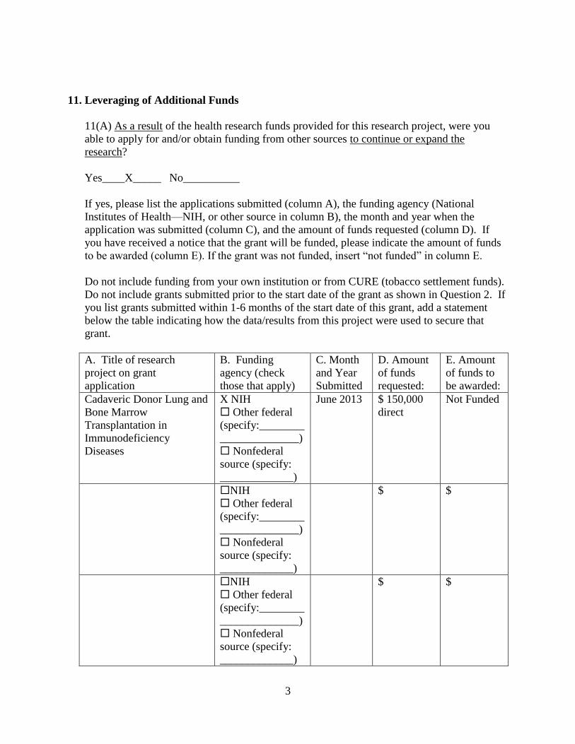
11. Leveraging of Additional Funds
11(A) As a result of the health research funds provided for this research project, were you
able to apply for and/or obtain funding from other sources to continue or expand the
research?
Yes____X_____ No__________
If yes, please list the applications submitted (column A), the funding agency (National
Institutes of Health—NIH, or other source in column B), the month and year when the
application was submitted (column C), and the amount of funds requested (column D). If
you have received a notice that the grant will be funded, please indicate the amount of funds
to be awarded (column E). If the grant was not funded, insert “not funded” in column E.
Do not include funding from your own institution or from CURE (tobacco settlement funds).
Do not include grants submitted prior to the start date of the grant as shown in Question 2. If
you list grants submitted within 1-6 months of the start date of this grant, add a statement
below the table indicating how the data/results from this project were used to secure that
grant.
A. Title of research
project on grant
application
B. Funding
agency (check
those that apply)
C. Month
and Year
Submitted
D. Amount
of funds
requested:
E. Amount
of funds to
be awarded:
Cadaveric Donor Lung and
Bone Marrow
Transplantation in
Immunodeficiency
Diseases
X NIH
Other federal
(specify:________
______________)
Nonfederal
source (specify:
_____________)
June 2013 $ 150,000
direct
Not Funded
NIH
Other federal
(specify:________
______________)
Nonfederal
source (specify:
_____________)
$ $
NIH
Other federal
(specify:________
______________)
Nonfederal
source (specify:
_____________)
$ $
4
11(B) Are you planning to apply for additional funding in the future to continue or expand
the research?
Yes____X_____ No__________
If yes, please describe your plans:
We hope to extend our studies to patients and we would seek RO1 support from NIH to study
Treg immune function in mixed chimerism and GVHD
12. Future of Research Project. What are the future plans for this research project?
1. We plan to perform experiments on newly accrued patients to extend and confirm our
findings of Foxp3/Helios co-expressing Treg deficiency in GvHD by testing on more
subjects.
2. Further characterize Treg subsets (cytokine responsiveness, functional pathways).
3. Refine our single cell Treg cloning strategy to develop therapeutic cell products in the
future.
13. New Investigator Training and Development. Did students participate in project
supported internships or graduate or post-graduate training for at least one semester or one
summer?
Yes_________ No__X____
If yes, how many students? Please specify in the tables below:
Undergraduate Masters Pre-doc Post-doc
Male
Female
Unknown
Total
Undergraduate Masters Pre-doc Post-doc
Hispanic
Non-Hispanic
Unknown
Total
5
Undergraduate Masters Pre-doc Post-doc
White
Black
Asian
Other
Unknown
Total
14. Recruitment of Out-of–State Researchers. Did you bring researchers into Pennsylvania to
carry out this research project?
Yes_________ No____X___
If yes, please list the name and degree of each researcher and his/her previous affiliation:
15. Impact on Research Capacity and Quality. Did the health research project enhance the
quality and/or capacity of research at your institution?
Yes_________ No____X____
If yes, describe how improvements in infrastructure, the addition of new investigators, and
other resources have led to more and better research.
16. Collaboration, business and community involvement.
16(A) Did the health research funds lead to collaboration with research partners outside of
your institution (e.g., entire university, entire hospital system)?
Yes__X___ No__________
If yes, please describe the collaborations:
We have entered into discussions how we may perform tolerance studies and Treg analysis
on upcoming studies supported by the PIDTC consortium.
http://rarediseasesnetwork.epi.usf.edu/PIDTC/
16(B) Did the research project result in commercial development of any research products?
If yes, please describe commercial development activities that resulted from the research
project:
16(C) Did the research lead to new involvement with the community?
Yes_________ No____X____
If yes, please describe involvement with community groups that resulted from the
research project:
17. Progress in Achieving Research Goals, Objectives and Aims. List the project goals, objectives and specific aims (as contained in the grant agreement).
Summarize the progress made in achieving these goals, objectives and aims for the period
that the project was funded (i.e., from project start date through end date). Indicate whether
or not each goal/objective/aim was achieved; if something was not achieved, note the reasons
why. Describe the methods used. If changes were made to the research
goals/objectives/aims, methods, design or timeline since the original grant application was
submitted, please describe the changes. Provide detailed results of the project. Include
evidence of the data that was generated and analyzed, and provide tables, graphs, and figures
of the data. List published abstracts, poster presentations and scientific meeting presentations
at the end of the summary of progress; peer-reviewed publications should be listed under
item 20.
This response should be a DETAILED report of the methods and findings. It is not sufficient
to state that the work was completed. Insufficient information may result in an unfavorable
performance review, which may jeopardize future funding. If research findings are pending
publication you must still include enough detail for the expert peer reviewers to evaluate the
progress during the course of the project.
Health research grants funded under the Tobacco Settlement Act will be evaluated via a
performance review by an expert panel of researchers and clinicians who will assess project
work using this Final Progress Report, all project Annual Reports and the project’s strategic
plan. After the final performance review of each project is complete, approximately 12-16
months after the end of the grant, this Final Progress Report, as well as the Final Performance
Review Report containing the comments of the expert review panel, and the grantee’s written
response to the Final Performance Review Report, will be posted on the CURE Web site.
There is no limit to the length of your response. Responses must be single-spaced below,
no smaller than 12-point type. If you cut and paste text from a publication, be sure
symbols print properly, e.g., the Greek symbol for alpha () and beta (ß) should not
print as boxes () and include the appropriate citation(s). DO NOT DELETE THESE
INSTRUCTIONS.
7
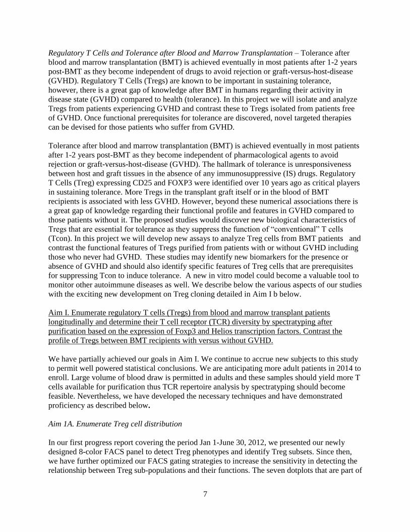
Regulatory T Cells and Tolerance after Blood and Marrow Transplantation – Tolerance after
blood and marrow transplantation (BMT) is achieved eventually in most patients after 1-2 years
post-BMT as they become independent of drugs to avoid rejection or graft-versus-host-disease
(GVHD). Regulatory T Cells (Tregs) are known to be important in sustaining tolerance,
however, there is a great gap of knowledge after BMT in humans regarding their activity in
disease state (GVHD) compared to health (tolerance). In this project we will isolate and analyze
Tregs from patients experiencing GVHD and contrast these to Tregs isolated from patients free
of GVHD. Once functional prerequisites for tolerance are discovered, novel targeted therapies
can be devised for those patients who suffer from GVHD.
Tolerance after blood and marrow transplantation (BMT) is achieved eventually in most patients
after 1-2 years post-BMT as they become independent of pharmacological agents to avoid
rejection or graft-versus-host-disease (GVHD). The hallmark of tolerance is unresponsiveness
between host and graft tissues in the absence of any immunosuppressive (IS) drugs. Regulatory
T Cells (Treg) expressing CD25 and FOXP3 were identified over 10 years ago as critical players
in sustaining tolerance. More Tregs in the transplant graft itself or in the blood of BMT
recipients is associated with less GVHD. However, beyond these numerical associations there is
a great gap of knowledge regarding their functional profile and features in GVHD compared to
those patients without it. The proposed studies would discover new biological characteristics of
Tregs that are essential for tolerance as they suppress the function of “conventional” T cells
(Tcon). In this project we will develop new assays to analyze Treg cells from BMT patients and
contrast the functional features of Tregs purified from patients with or without GVHD including
those who never had GVHD. These studies may identify new biomarkers for the presence or
absence of GVHD and should also identify specific features of Treg cells that are prerequisites
for suppressing Tcon to induce tolerance. A new in vitro model could become a valuable tool to
monitor other autoimmune diseases as well. We describe below the various aspects of our studies
with the exciting new development on Treg cloning detailed in Aim I b below.
Aim I. Enumerate regulatory T cells (Tregs) from blood and marrow transplant patients
longitudinally and determine their T cell receptor (TCR) diversity by spectratyping after
purification based on the expression of Foxp3 and Helios transcription factors. Contrast the
profile of Tregs between BMT recipients with versus without GVHD.
We have partially achieved our goals in Aim I. We continue to accrue new subjects to this study
to permit well powered statistical conclusions. We are anticipating more adult patients in 2014 to
enroll. Large volume of blood draw is permitted in adults and these samples should yield more T
cells available for purification thus TCR repertoire analysis by spectratyping should become
feasible. Nevertheless, we have developed the necessary techniques and have demonstrated
proficiency as described below.
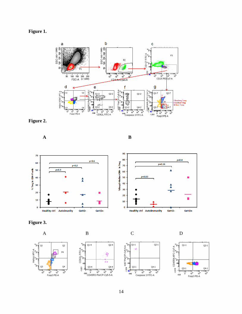
Aim 1A. Enumerate Treg cell distribution
In our first progress report covering the period Jan 1-June 30, 2012, we presented our newly
designed 8-color FACS panel to detect Treg phenotypes and identify Treg subsets. Since then,
we have further optimized our FACS gating strategies to increase the sensitivity in detecting the
relationship between Treg sub-populations and their functions. The seven dotplots that are part of
8
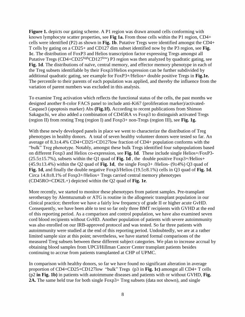
Figure 1. depicts our gating scheme. A P1 region was drawn around cells conforming with
known lymphocyte scatter properties, see Fig 1a. From those cells within the P1 region, CD4+
cells were identified (P2) as shown in Fig. 1b. Putative Tregs were identified amongst the CD4+
T cells by gating on a CD25+ and CD127 dim subset identified now by the P3 region, see Fig.
1c. The distribution of FoxP3 and Helios transcription factor expressing Tregs amongst all
Putative Tregs (CD4+CD25highCD127low) P3 region was then analyzed by quadratic gating, see
Fig. 1d. The distributions of naïve, central memory, and effector memory phenotype in each of
the Treg subsets identifiable by their Foxp3/Helios expression can be further subdivided by
additional quadratic gating, see example for FoxP3+/Helios+ double positive Tregs in Fig.1e.
The percentile to their parents of each population was applied, and thereby the influence from the
variation of parent numbers was excluded in this analysis.
To examine Treg activation which reflects the functional status of the cells, the past months we
designed another 8-color FACS panel to include anti-Ki67 (proliferation marker)/activated-
Caspase3 (apoptosis marker) Abs (Fig.1f). According to recent publications from Shimon
Sakaguchi, we also added a combination of CD45RA vs Foxp3 to distinguish activated Tregs
(region II) from resting Treg (region I) and Foxp3+ non-Tregs (region III), see Fig. 1g.
With these newly developed panels in place we went to characterize the distribution of Treg
phenotypes in healthy donors. A total of seven healthy volunteer donors were tested so far. An
average of 8.3±4.4% CD4+CD25+CD127low fraction of CD4+ population conforms with the
“bulk” Treg phenotype. Notably, amongst these bulk Tregs identified four subpopulations based
on different Foxp3 and Helios co-expression, see Fig. 1d. These include single Helios+/FoxP3-
(25.5±15.7%), subsets within the Q1 quad of Fig. 1d , the double positive Foxp3+/Helios+
(45.9±13.4%) within the Q2 quad of Fig. 1d, the single Foxp3+ /Helios- (9±4%) Q3 quad of
Fig. 1d, and finally the double negative Foxp3/Helios (19.5±8.1%) cells in Q3 quad of Fig. 1d.
Circa 14.8±8.1% of Foxp3+/Helios+ Tregs carried central memory phenotypes
(CD45RO+CD62L+) depicted within the Q2 quad of Fig. 1e.
More recently, we started to monitor these phenotypes from patient samples. Pre-transplant
serotherapy by Alemtuzumab or ATG is routine in the allogeneic transplant population in our
clinical practice; therefore we have a fairly low frequency of grade II or higher acute GvHD.
Consequently, we have been able to test so far only three BMT recipients with GVHD at the end
of this reporting period. As a comparison and control population, we have also examined seven
cord blood recipients without GvHD. Another population of patients with severe autoimmunity
was also enrolled on our IRB-approved protocol and was tested. So far three patients with
autoimmunity were studied at the end of this reporting period. Undoubtedly, we are at a rather
limited sample size at this point; nevertheless, we have started formal comparisons of the
measured Treg subsets between these different subject categories. We plan to increase accrual by
obtaining blood samples from UPCI/Hillman Cancer Center transplant patients besides
continuing to accrue from patients transplanted at CHP of UPMC.
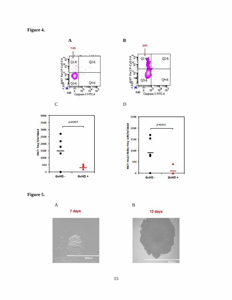
In comparison with healthy donors, so far we have found no significant alteration in average
proportion of CD4+CD25+CD127low “bulk” Tregs (p3 in Fig. 1c) amongst all CD4+ T cells
(p2 in Fig. 1b) in patients with autoimmune diseases and patients with or without GVHD, Fig.
2A. The same held true for both single Foxp3+ Treg subsets (data not shown), and single
9
Helios+ Tregs (data not shown). However, unexpectedly, compared to healthy controls, we
found, a significant decrease in the Foxp3+/Helios+ (double positive) Treg fraction and their
central memory compartment in patients with autoimmune diseases, while this population was
comparable to healthy volunteers in cord blood transplant patients with or without GvHD (Fig.
2B). These results suggest that Foxp3 and Helios co-expressing “double positive” central
memory Tregs, rather than single Foxp3+ or Helios+ Treg, may play an important role in the
maintenance of immune tolerance.
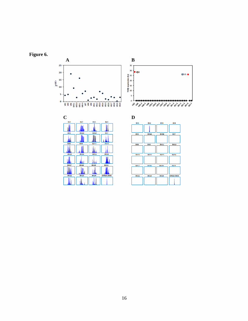
When we restricted the analysis on the brightest subpopulation within the Foxp3+Helios+ central
memory Tregs (Fig.3A) displaying the highest fluorescence intensity for these transcription
factors (depicted P4 in Q2) and analyzed their distribution within the “Sakaguchi regions” (as
defined by CD45RA/Foxp3 expression amongst CD4+/CD25 bright cells), we found no resting
CD45RA+ Tregs (Fig. 3 D), however, the majority of them belong to “Sakaguchi region II”-aka
“activated Tregs” which are CD45RA-/Foxp3high (Q4-1 in Fig. 3D). These cells have undergone
significant proliferation, identifiable by the strong intranuclear expression of Ki67 (see events in
Q1-1 in Fig. 3C).
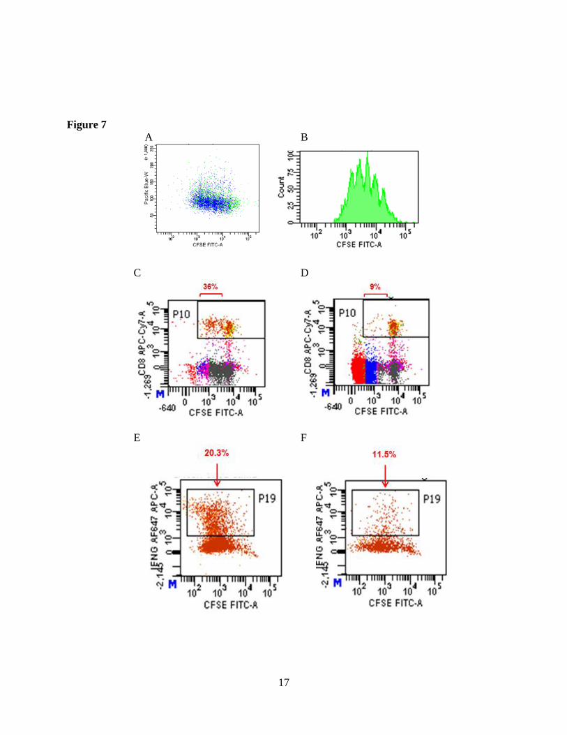
When we examined the Treg profile of those subjects with recent acute GvHD, we found little if
any proliferation (low expression of intracellular (ic) Ki67 in Q1-6 Fig. 4A) amongst CD4+Treg
and Foxp3+/Helios+ Treg carrying central memory phenotype, in comparison with that in a cord
blood patient in good clinical condition, free of any GVHD, (Q1-6 Fig. 4B) . The analysis was
performed at the same time-point, ~ 100 days post-transplantation. There was statistically
significant reduction in proliferating “bulk” Tregs (Fig. 4C) in those with GVHD impacting just
as well the Foxp3+Helios+ co-expressing Treg subset that displays (CD45RA-
/CD45RO+/CD62L+) central memory phenotype (Fig. 4D). It is plausible that reduced
proliferative capacity and possible exhaustion of Foxp3+Helios+ Tregs is a critical event in the
development of acute GvHD. We hypothesize that the subjects’ immune environment, e.g.
cytokine milieu that influence Treg function may also play an important role on Treg survival
and proliferative capacity, which in turn may feed back into the pathogenesis of GVHD. Recent
publications (Shamim et al Human Immunology, 2013;10:1111; Kim et al Transplantation
2012;94:1250) have suggested that single nucleotide polymorphism (SNP) in several cytokines
are associated with occurrence of acute GvHD. Targeted modulation of the immune
environment to support Treg function may become a new way to favorably modulate and treat
acute GvHD. Methods that would favorably impact Treg kinetics and function may be as
effective or more than infusion of bulk Tregs expanded ex vivo from allogeneic healthy donors.
Importantly, these new findings were detected directly from unmanipulated patient samples to
reflect human disease state without experimental influence leading to accidental bias. If the
findings were confirmed in a larger cohort, future Treg expansion strategies should focus on
generating Tregs with stable and functionally and phenotypically defined subsets. This approach
may result in superior therapeutic responses for GvHD and possibly in autoimmune disease as
well where autologous Tregs would be expanded without fears of GVHD. In the immediate
future we plan to confirm on larger patient populations these findings on functionally and
phenotypically distinct Treg subsets, we could 1) devise more powerful biomarker assays to
predict the presence or absence of overall tolerance versus exaggerated/pathological immune
responses ( i.e. GVHD) post-HSCT, and 2) design new therapeutical interventions.
10
In summary, these studies have yielded some exciting findings sufficient for generating new
hypotheses. We plan to submit these for new funding agencies to extend and validate the above
preliminary findings in a large patient cohort. Towards these goals, we have successfully reached
out to colleagues at the Hillman Cancer Center of the University of Pittsburgh Cancer Institute
(UPCI) to enroll their patients on these IRB approved studies.
Aim 1B. Treg single cell cloning
Because Foxp3 and Helios genes are transcription factors it is not feasible to purify viable
primary human Treg sub-populations by gating on these intracellular markers. Therefore, we
have developed a novel single cell cloning technique with the goal to characterize Treg sub-
populations. After successful cell expansion, we may sacrifice a fraction of the clone for
characterization while many more are available for functional studies. Conventional method of
T-cell cloning requires feeder cells, in particular using PBMC to support single cell growth, may
cause contamination with unwanted cells or more likely it may lead to additional cell contacts
and soluble signals that could result in functional bias of Tregs expanding from single cells in an
uncontrolled milieu. After months of pilot experiments, we have made progress towards a new
protocol for single Treg cloning (Fig. 5 and Fig. 6), with a remarkable ~ 20% cloning efficiency
for single Treg clones, (manuscript in preparation). To verify single cell clonality, we established
TCR qPCR and TCR immunoscope for fast screening single cell cultures, another technical
advance in this field, (manuscript in preparation). Figure 6 shows TCR qPCR profiles tested
from the PBMC of a healthy donor (Fig. 6A) or from the single cell cloning (Fig. 6B); and TCR
immunoscope profiles representing the PBMC of a healthy donor (Fig. 6C), or the clonal
progeny of a single cell (Fig. 6D).
Aim IB TCR repertoire analysis
Our original hypothesis has been that the TCR repertoire of circulating Tregs from patients with
GvHD will differ from that seen in patients not having experienced GVHD. The main obstacle to
test this hypothesis has been the low cell numbers of Tregs we have isolated, reflecting both
CD4+ lymphopenia post transplant and the overall small volume of blood samples obtained from
young children (4-8 ml range). We have nevertheless titrated the assay to the lowest cell number
sufficient for efficient RNA purification. By adding carrier during the purification procedure, we
can purify RNA from 1000 cells (data not shown). However, we have not yet obtained
reproducibly clear signals on TCR immunoscope with RNA purified from 1000 cell. We are
pursuing experiments with pre-PCR amplification (Life Technologies) to enhance PCR signals.
Aim II. Design and implement functional assays to characterize and to quantify the biological
profile and suppressive capacity of purified Treg in vitro as they are mixed with conventional T-
cells from the same BMT recipient. Following non-specific and transplant recipient specific
activation of Tregs and Tcon in the same co-cultures to model the in vivo scenario, we will
analyze Tregs by qPCR for cytokine and homing receptors, co-stimulator, and other critical
suppressor molecules while Tcon will be tested in parallel for evidence of receiving suppressive
signals.
11
We have partially achieved our goals as described in Aim II, We have developed and validated a
sensitive functional assay as described below. We are anticipating more adult patients to enroll
between the Summer of 2014 and Spring of 2015 . These new datasets will permit correlative
analysis of Treg function and clinical correlates, most importantly the presence or absence of
GVHD. In adult subjects larger blood draw is permitted and these newly enrolled adult subject
can provide more T cells for purification and for analysis. Nevertheless, we have developed the
necessary techniques and have demonstrated proficiency as described below.
After initially setting up a “standard” CFSE proliferation assay developed by others years ago,
we have further optimized the conditions to obtain differentiation peaks to give much distinct
proliferation features. Figure 7A shows differentiation peaks generated at day 4 of a Tcon
culture. We are currently titrating the dose of antigen presenting cells (APC), employing both
live cells and artificial CD3/CD28 T cell stimulatory beads to determine the best conditions for
in vitro Tcon activation to sufficiently quantify the suppressive features from Treg. We have had
surprising proliferative responses from Tregs in the presence of unnecessarily potent stimulatory
signals.
We are also in the midst of developing other functional assays that may directly reflect other
Treg cell functions rather than inhibit of cell proliferation in response to non-antigen-specific
signals.
i. We have started monitoring Treg suppression of cytokine secretion by Tcons. IFN is
one of main cytokine secreted by functional Th1 type Tcons. Reduced IFN production
by Tcons reflects an inhibition by Tregs in the co-culture. Importantly, Tregs usually
secrete very low levels of IFN. Therefore, any alteration in IFN secretion in Tcon/Treg
co-culture medium will indicate changes of Tcon function. We are performing
experiments to establish and validate cytokine suppression assay(s). After 3-5 days co-
culture of Tcon and Treg, the supernatant of the cultures are collected for cytokine
analysis by Bioplex protein assay.
ii. While Bioplex assay is well suited to test batched Treg/Tcon culture supernatants, we
will also establish real-time PCR which is very specific, quantitative, and sensitive. It
can be applied to test individual samples.
iii. Multicolor FACS panel will be broadened in scope by examining the CFSE labelled
Tcons for de novo expression of surface CD69, CD154 activation markers in parallel
with intracellular IL-2 and IFN in response to APC. The putative Treg populations
would inhibit these functional events and their percent suppression can be measured
with titrated dose of Treg.
In summary, by using our newly established multi-color FACS panels and gating strategy, we
found preliminary evidence for reduced proliferation of Foxp3+Helios+ central memory Tregs in
the tested patients with acute GVHD (allogeneic responses). In the case of poorly controlled and
clinically active autoimmune disease, we found a significant decrease in the Foxp3+Helios+
central memory Treg population. These findings suggest a positive correlation between the
presence of Foxp3+Helios+ central memory Tregs above a threshold limit in achieving and
sustaining immune tolerance. If these findings are confirmed in sufficiently large patient groups,
these new findings could have significant clinical impact in the future. We may be able to
12
modulate the environment to promote survival and function of specific Treg subsets while in
parallel we are developing methods to single cell clone and expand Tregs with favorable
phenotype and suppressive function. Importantly, the preliminary dataset supported by CURE
was obtained directly from non-manipulated patient samples to accurately reflect the actual state
of human disease such as GVHD and/or autoimmunity.
Materials and Methods
Flow cytometric analysis of Treg phenotypes-for Aim I.
Peripheral blood mononuclear cells (PBMCs) were collected from the heparinized blood by
standard Ficoll-Hypaque density gradient centrifugation. The immune phenotype of cells was
tested by analysis on an 8 color FACS Canto II flow cytometer (BD Bioscience). The
monoclonal antibodies used were anti-CD4-BV510, CD8-APC-H7, CD25-PE-Cy7, CD127-
PerCP-Cy5.5, Caspase 3-FITC from BD Bioscience. The lymphocytes were first gated in
SSC/FCS, followed by gating on CD4 T cells, and the CD25+CD127low Treg was then selected.
The central memory Treg was determined by quadratic gating on CD45RO vs CD62L on the
Tregs. The proliferation vs apoptotic cells were distinguished by quadratic gating of Ki67 vs
Caspase 3 on a giving population.
Treg isolation and culture -for Aim II.
The T cells were enriched from PBMNC by using EasySep® human T cell enrichment kit (Stem
Cell Technologies). The CD4+/CD25+/CD127low/CD49d lowTregs were then isolated with
EasySep® human CD4+CD25+CD127lowCD49d- enrichment kit (Stem Cell Technologies).
Isolated Tregs were then cultured in XIVIVO 15 medium (Life Sciences) with 10% human
serum.
TCR immunoscope -for Aim I.
The RNA was purified from cells with RNeasy Mini Kit (QIAGEN). Complementary DNA
(cDNA) was synthesized by using Superscript II reverse transcripts and random hexamers rimer
(Life Technologies) according to the manufacturer’s instructions. PCR was performed by using
Hotstar PCR Kit (QIAGEN). The PCR products were then run on an ABI Prism 3130 Genetic
Analyzer (Life Technologies).
CFSE-labeled T cell proliferation assay - for Aim II.
T cells were labeled with CellTrace CFSE cell proliferation kit (Life Technologies) and cultured
in XVIVO 15 with 5% human serum. On day 4, the cells were harvested and the division peaks
of CFSE fluoresce were detected on FACSCanto II.
Treg suppression assay - for Aim II.
PBMNC were labeled with CFSE CellTrace kit (Life Technologies) and were cultured with Treg
at ratio 1:1. On the day 4th, both PBMNC and Treg were collected to determine inhibition of
division peaks with FACSCanto II.
13
Figure Legends
Figure 1. FACS gating scheme for Treg phenotypes. Lymphocytes were identified in dot
plots (a) by FSC vs SSC. Thereafter, CD4+ cells were gated (b). CD4+ T cells with CD25
positivity and CD127 low expression were gated next (c). The distribution of Foxp3+ and
Helios+ cells in CD4+CD25highCD127low Tregs were analyzed by quadratic gating (d). The
distribution of CD45RO+/CD62L+ (central memory) versus other phenotypes in each
Foxp3/Helios population were analyzed by additional quadratic gating (e); intracellular Ki67 and
Activated Caspase 3 expression (f) and CD45RA/Foxp3 (g) were also tested.
Figure 2. The comparison of the distribution for CD4+CD25+CD127low population in CD4+
cells (A) or for Foxp3+Helios+ central memory Treg in CD4+CD25+CD127low cells (B) in
healthy donors, in patients with active autoimmunity or transplant recipients with or without
GVHD post-HSCT.
Figure 3. Characterization of Foxp3+Helios+ Tregs with the highest expression of these
transcription factors (A). Gating on the brightest dual positive Tregs to analyze expression of
CD45RO+CD62L+ (B), and Ki67 (C); into “Sakaguchi region” II: CD45RA-Foxp3high active
Treg (D).
Figure 4. Comparison of intracellular (A) Ki67 expression in Treg cells of a patient with
acute GVHD or (B) of a patient without GVHD. Absolute numbers of Ki67+ Treg/ml blood in
(C) 5 patients without aGVHD vs 3 patients with a GVHD. (D) Contrasting values of absolute
numbers of Ki67+ Foxp3+Helios+Treg/ml within the central memory phenotype in the same
patient groups as in (C).
Figure 5. Single Treg clone expansion as reproduced by phase contrast inverted microscope at
20x after 7 days of culture (A) or at 12 days of culture (B).
Figure 6. Identification of single cell clonality by TCR qPCR in single cell cloning (B) in
comparison with bulk unselected PBMNC in a healthy donor (A); or by using TCR
immunoscope in single cell cloning (D), in comparison with PBMNC obtained from a healthy
volunteer donor (C).
Figure 7. Treg suppression assay based on CFSE-labeled Tcon proliferation as detected by
dilution of CFSE in dotplot (A) or division peaks in histogram represent successive generations
of Tcon (B). Compared to Tcon alone (C and E) when Treg is added at 1:1 ratio, proliferation
(D) and IFN secretion (F) are both depressed in autologous Tcon cells.
14
Figure 1.
Figure 2.
A B
Figure 3.
A B C D
15
Figure 4.
A B
C D
Figure 5.
A B
16
Figure 6.
A B
C D
17
Figure 7
A B
C D
E F
18
18. Extent of Clinical Activities Initiated and Completed. Items 18(A) and 18(B) should be
completed for all research projects. If the project was restricted to secondary analysis of
clinical data or data analysis of clinical research, then responses to 18(A) and 18(B) should
be “No.”
18(A) Did you initiate a study that involved the testing of treatment, prevention or
diagnostic procedures on human subjects?
______Yes
X__No
18(B) Did you complete a study that involved the testing of treatment, prevention or
diagnostic procedures on human subjects?
______Yes
X____No
If “Yes” to either 18(A) or 18(B), items 18(C) – (F) must also be completed. (Do NOT
complete 18(C-F) if 18(A) and 18(B) are both “No.”)
18(C) How many hospital and health care professionals were involved in the research
project?
______Number of hospital and health care professionals involved in the research
project
18(D) How many subjects were included in the study compared to targeted goals?
______Number of subjects originally targeted to be included in the study
______Number of subjects enrolled in the study
Note: Studies that fall dramatically short on recruitment are encouraged to
provide the details of their recruitment efforts in Item 17, Progress in Achieving
Research Goals, Objectives and Aims. For example, the number of eligible
subjects approached, the number that refused to participate and the reasons for
refusal. Without this information it is difficult to discern whether eligibility
criteria were too restrictive or the study simply did not appeal to subjects.
18(E) How many subjects were enrolled in the study by gender, ethnicity and race?
Gender:
______Males
______Females
______Unknown
Ethnicity:
______Latinos or Hispanics
______Not Latinos or Hispanics
______Unknown
19
Race:
______American Indian or Alaska Native
______Asian
______Blacks or African American
______Native Hawaiian or Other Pacific Islander
______White
______Other, specify:
______Unknown
18(F) Where was the research study conducted? (List the county where the research
study was conducted. If the treatment, prevention and diagnostic tests were offered in
more than one county, list all of the counties where the research study was
conducted.)
19. Human Embryonic Stem Cell Research. Item 19(A) should be completed for all research
projects. If the research project involved human embryonic stem cells, items 19(B) and
19(C) must also be completed.
19(A) Did this project involve, in any capacity, human embryonic stem cells?
______Yes
___X_ No
19(B) Were these stem cell lines NIH-approved lines that were derived outside of
Pennsylvania?
______Yes
______ No
19(C) Please describe how this project involved human embryonic stem cells:
20. Articles Submitted to Peer-Reviewed Publications.
20(A) Identify all publications that resulted from the research performed during the funding
period and that have been submitted to peer-reviewed publications. Do not list journal
abstracts or presentations at professional meetings; abstract and meeting presentations should
be listed at the end of item 17. Include only those publications that acknowledge the
Pennsylvania Department of Health as a funding source (as required in the grant
agreement). List the title of the journal article, the authors, the name of the peer-reviewed
publication, the month and year when it was submitted, and the status of publication
(submitted for publication, accepted for publication or published.). Submit an electronic
copy of each publication or paper submitted for publication, listed in the table, in a PDF
version 5.0.5 (or greater) format, 1,200 dpi. Filenames for each publication should include
the number of the research project, the last name of the PI, and an abbreviated title of the
publication. For example, if you submit two publications for Smith (PI for Project 01), one
20
publication for Zhang (PI for Project 03), and one publication for Bates (PI for Project 04),
the filenames would be:
Project 01 – Smith – Three cases of isolated
Project 01 – Smith – Investigation of NEB1 deletions
Project 03 – Zhang – Molecular profiling of aromatase
Project 04 – Bates – Neonatal intensive care
If the publication is not available electronically, provide 5 paper copies of the publication.
Note: The grant agreement requires that recipients acknowledge the Pennsylvania
Department of Health funding in all publications. Please ensure that all publications listed
acknowledge the Department of Health funding. If a publication does not acknowledge the
funding from the Commonwealth, do not list the publication.
Title of Journal
Article:
Authors: Name of Peer-
reviewed
Publication:
Month and
Year
Submitted:
Publication
Status (check
appropriate box
below):
1.
Submitted
Accepted
Published
2.
Submitted
Accepted
Published
3.
Submitted
Accepted
Published
20(B) Based on this project, are you planning to submit articles to peer-reviewed publications
in the future?
Yes____X____ No__________
If yes, please describe your plans:
The following manuscripts will be published if they are confirmed with large data sets:
1. Deficiency of Foxp3/Helios co-expressing regulatory T-cells correlates with activity in
autoimmunity. (an abstract was submitted to annual AAI meeting)
2. Inefficiency in proliferation of Foxp3/Helios co-expressing regulatory T-cells in those
with GvHD post-blood and marrow transplantation.
3. Heterogeneity of regulatory T-cell single clones in healthy donors.
4. Characterization of regulatory T-cell clones in the patients with acute GvHD.
21. Changes in Outcome, Impact and Effectiveness Attributable to the Research Project.
Describe the outcome, impact, and effectiveness of the research project by summarizing its
impact on the incidence of disease, death from disease, stage of disease at time of diagnosis,
21
or other relevant measures of outcome, impact or effectiveness of the research project. If
there were no changes, insert “None”; do not use “Not applicable.” Responses must be
single-spaced below, and no smaller than 12-point type. DO NOT DELETE THESE
INSTRUCTIONS. There is no limit to the length of your response.
None
22. Major Discoveries, New Drugs, and New Approaches for Prevention Diagnosis and
Treatment. Describe major discoveries, new drugs, and new approaches for prevention,
diagnosis and treatment that are attributable to the completed research project. If there were
no major discoveries, drugs or approaches, insert “None”; do not use “Not applicable.”
Responses must be single-spaced below, and no smaller than 12-point type. DO NOT
DELETE THESE INSTRUCTIONS. There is no limit to the length of your response.
Our major findings so far are the lack of proliferative capacity for foxp3/helios co-expressing
Treg in the patients with acute GVHD, and overall decrease of those Tregs in autoimmune
diseases. If these very preliminary findings were to be confirmed, it may guide the treatment
of GvHD by focusing on restoring the proliferative potential of Foxp3/Helios co-expressing
Tregs as this subset may become a potential biomarker for onset of and activity
autoimmunity.
23. Inventions, Patents and Commercial Development Opportunities.
23(A) Were any inventions, which may be patentable or otherwise protectable under Title 35
of the United States Code, conceived or first actually reduced to practice in the performance
of work under this health research grant? Yes X No
If “Yes” to 23(A), complete items a – g below for each invention. (Do NOT complete items
a - g if 23(A) is “No.”)
a. Title of Invention:
Generation of regulatory T cell clones with superior function
b. Name of Inventor(s):
Paul Szabolcs MD
Xiaohua Chen PhD
c. Technical Description of Invention (describe nature, purpose, operation and physical,
chemical, biological or electrical characteristics of the invention):
A new strategy in single regulatory T cell cloning from bulk Treg population or
special sub-population, which also includes identifying single clones by TCRB
immunoscope and TCRB real-time PCR.
22
d. Was a patent filed for the invention conceived or first actually reduced to practice in
the performance of work under this health research grant?
e. Yes No X
If yes, indicate date patent was filed:
f. Was a patent issued for the invention conceived or first actually reduced to practice in
the performance of work under this health research grant?
g. Yes No X
If yes, indicate number of patent, title and date issued:
Patent number:
Title of patent:
Date issued:
h. Were any licenses granted for the patent obtained as a result of work performed under
this health research grant? Yes No X
If yes, how many licenses were granted?
i. Were any commercial development activities taken to develop the invention into a
commercial product or service for manufacture or sale? Yes No X
If yes, describe the commercial development activities:
23(B) Based on the results of this project, are you planning to file for any licenses or patents,
or undertake any commercial development opportunities in the future?
Yes___X_____ No__________
If yes, please describe your plans:
Single cell cloning of regulatory T-cells
24. Key Investigator Qualifications. Briefly describe the education, research interests and
experience and professional commitments of the Principal Investigator and all other key
investigators. In place of narrative you may insert the NIH biosketch form here; however,
please limit each biosketch to 1-2 pages. For Nonformula grants only – include information
for only those key investigators whose biosketches were not included in the original grant
application.
23
BIOGRAPHICAL SKETCH Provide the following information for the Senior/key personnel and other significant contributors in the order listed on Form Page 2.
Follow this format for each person. DO NOT EXCEED FOUR PAGES.
NAME
Paul Szabolcs POSITION TITLE
Professor of Pediatrics and Immunology, University of Pittsburgh School of Medicine
eRA COMMONS USER NAME (credential, e.g., agency login)
szabo001
EDUCATION/TRAINING (Begin with baccalaureate or other initial professional education, such as nursing, include postdoctoral training and residency training if applicable.)
INSTITUTION AND LOCATION DEGREE
(if applicable) MM/YY FIELD OF STUDY
Pecs University School of Medicine, Pecs, Hungary Transferred 1983 Medicine
Semmelweis University School of Medicine, Budapest, Hungary
Bellevue Hospital, New York
Cornell University Medical College, New York Hospital, Memorial Sloan Kettering Cancer Center, New York, NY
Sloan-Kettering Cancer Center, New York, NY
New York Hospital, Memorial Sloan-Kettering Cancer Center, New York, NY
Memorial Sloan-Kettering Cancer Center, New York, NY
M.D. Summa
cum laude
Residency
Fellowship
Postdoctoral Fellowship
Chief Fellow
Special Fellow
1985
1987-90
1990-93
1991-93
1992-93
1993-94
Medicine
Pediatrics
Pediatric Hematology/Oncology
Molecular Biology
Pediatric Hematology/Oncology
Department of Pediatrics
1987 - 90 Intern, Resident, Department of Pediatrics, New York University-Bellevue Hospital Center, NY, NY 1989 - 90 Teaching Assistant, New York University Medical Center-Bellevue Hospital Center, NY,NY 1990 –93 Clinical Fellow, Pediatric Hematology-Oncology, Cornell University Medical College,(CUMC) New York
Hospital/Memorial Sloan-Kettering Cancer Center (MSKCC), NY, NY 1992 –94 Chief Fellow, Pediatric Hematology-Oncology, MSKCC/CUMC 1994 –98 Instructor, Bone Marrow Transplant Service, Department of Pediatrics, MSKCC, NY 08/98-06/06 Assistant Professor Pediatrics, Stem Cell Transplant Program, Duke University Medical Center,
Durham, NC
2001-2011 2006-2011 2011-2014
Assistant Professor of Immunology, Duke University Medical Center, Durham, NC Associate Professor Pediatrics, Blood and Marrow Transplant Program, Duke University Medical Center, Durham, NC Visiting Professor of Pediatrics, University of Pittsburgh School of Medicine Chief, Division of Blood and Marrow Transplantation and Cellular Therapies, Children’s Hospital of Pittsburgh of UPMC
2014-present
Professor of Pediatrics and Immunology, University of Pittsburgh School of Medicine
Other Experience and Professional Memberships 1986 – 87 Research Assistant, The Hospital of the University of Pennsylvania Cancer Center,
Philadelphia, PA 8/91 - 2/93 Postdoctoral Fellow, Department of Molecular Biology Sloan-Kettering Institute,
NY, NY 3/93 – 5/94 Guest Investigator, Laboratory of Cellular Physiology and Immunology, The
Rockefeller University, NY 1994 – 97
Research Associate, Laboratory of Cellular Physiology and Immunology, The Rockefeller University, NY
American Society of Hematology (ASH) American Society for Blood and Marrow Transplantation (ASBMT) American Association of Immunologists (AAI) Fellow, American Academy of Pediatrics Center for International Blood and Marrow Transplant Research (CIBMTR): Immune Deficiencies/Inborn Errors Working Committee Center for International Blood and Marrow Transplant Research (CIBMTR): Graft-vs-Host Disease Working Committee International Society for Cellular Therapy (ISCT) Society for Pediatric Research (SPR) Member Center for International Blood and Marrow Transplant Research (CIBMTR): Infection and Immune Reconstitution Working Committee – Co Chair Clinical Immunology Society University of Pittsburgh Cancer Institute American Pediatric Society
Honors and Awards: 1980 1st Prize for Critical Thesis, Human globin genes, Pecs University of Medical
Sciences, Pecs, Hungary 1990 Elected “Best Student of the Year”, Pecs University of Medical Sciences, Pecs,
Hungary 2000 Lisa Stafford Memorial Research Prize, Duke University Comprehensive Cancer
Center, Durham, NC
C. Selected Peer-reviewed Publications
1. Iwata Y, Matsushita T, Horikawa M, Dilillo DJ, Yanaba K, Venturi GM, Szabolcs P, Bernstein SH, Magro CM, Williams AD, Hall RP, St Clair EW, Tedder TF. Characterization of a rare IL-10-competent B cell subset in humans that parallels mouse regulatory B10 cells. Blood, 2011, Jan 13;117:530-41 PMCID: 3031478
2. Szabolcs P. T-lymphocyte recovery and function after cord blood transplantation. Immunol Res.
2011 April, 49; 56-69. PMID: 21128006
3. Kamani, N. R., Kumar, S., Hassebroek, A., Eapen, M., Lerademacher, J., Casper, J., Cowan, M., Sanchez de Toledo, J., Ferster, A., Szabolcs, P., Wingard, J. R., Horwitz, E., Filipovich, A. H. Malignancies after Hematopoietic Cell Transplantation for Primary Immune Deficiencies: A Report from the Center for International Blood and Marrow Transplant Research. Biol. Blood and Marrow Transplantation, 2011 epub May 20, PMID: 21658461.
4. Parikh S, Szabolcs P. Reduced-intensity conditioning (RIC) in children with nonmalignant disorders (NMD) undergoing unrelated donor umbilical cord transplantation (UCBT). Biol Blood Marrow Transplant. 2012 Jan; 18(1 Suppl):S53-5. PMID: 22226113
5. Szabolcs P, Burlingham WJ, Thomson AW. Tolerance after solid organ and hematopoietic cell transplantation. Biol Blood Marrow Transplant. 2012 Jan; 18(1 Suppl):S193-200. PMID: 22226107
6. Bunin N, Small T, Szabolcs P, Baker KS, Pulsipher MA, Torgerson T. NCI, NHLBI/PBMTC first international conference on late effects after pediatric hematopoietic cell transplantation: persistent immune deficiency in pediatric transplant survivors. Biol Blood Marrow Transplant. 2012 Jan; 18(1):6-15 PMID: 22100979
7. Eapen M, Ahn KW, Orchard PJ, Cowan MJ, Davies SM, Fasth A, Hassebroek A, Ayas M, Bonfim C, O'Brien TA, Gross TG, Horwitz M, Horwitz E, Kapoor N, Kurtzberg J, Majhail N, Ringden O, Szabolcs P, Veys P, Baker KS. Long-term survival and late deaths after hematopoietic cell transplantation for primary immunodeficiency diseases and inborn errors of metabolism. Biol Blood Marrow Transplant. 2012 Sep; 18(9):1438-45. PMID: 22430083
8. Kanda, J, Chiou LW, Szabolcs P, Sempowski GD, Rizzieri DA, Long GD, Sullivan KM, Gasparetto C, Chute JP, Morris A, McPherson J, Hale J, Livingston JA, Broadwater, G, Niedzwiecki D, Chao NJ, Horwitz ME. Immune recovery in adult patients after myeloablative dual umbilical cord blood,
matched sibling, and matched unrelated donor hematopoietic cell transplantation. Biol Blood Marrow Transplant. 2012 Nov 18. PMID: 22698485
9. Parikh SH, Mendizabal A, Benjamin CL, Komanduri KV, Antony J, Petrovic A, Hale G, Driscoll TA, Martin PL, Page KM, Flickinger K, Moffet J, Niedzwiecki, D, Kurtzberg, J, Szabolcs P. A Novel Reduced-Intensity Conditioning Regimen for Unrelated Umbilical Cord Blood Transplantation in Children with Nonmalignant Diseases. Biol Blood Marrow Transplant. 2013 Dec 1. pii: S1083-8791(13)00558-2. doi: 10.1016/j.bbmt.2013.11.021 [Epub ahead of print]. PMID: 24296492 [PubMed – as supplied by publisher]