Journal of Clinical Investigation Vol. 41, No. 5, 1962 EQUILIBRIUM DIALYSIS STUDIES OF THE BINDING OF THYROXINE BY HUMAN SERUM ALBUMIN * By KENNETH STERLING, PHILIP ROSEN t AND MILTON TABACHNICK t (From the Department of Internal Medicine, New York State Psychiatric Institute, and the Department of Psychiatry, Columbia University College of Physicians and Surgeons, New York, N. Y.) (Submitted for publication July 20, 1961; accepted December 28, 1961) In previous work from this laboratory (1) stud- ies on equilibrium dialysis were carried out to determine the binding constants for the inter- action of human serum albumin with thyroxine and its analogs. The classical technique of equi- librium dialysis as described by Scatchard and others (2-6) was employed. At present it seems clear that thyroxine in hu- man serum migrates to at least three different sites during electrophoresis at pH 8.6 (7-10). The three sites most frequently reported have been the thyroxine-binding a-globulin (TBG), albumin, and the "prealbumin" region. The thyroxine- binding a-globulin has generally been considered of major physiological importance, the others be- ing regarded as secondary carriers. Albumin is the only one of the thyroxine carriers of serum readily available in pure form and with a known molecular weight. The findings of the previous work (1) ap- peared compatible with the hypothesis that the anionic phenolate groups of thyroxine and triiodo- thyronine interact with free cationic groups on the protein molecule. In the present report the ob- servations have been extended to studies with variations in temperature and pH, as well as to experiments with mercaptalbumin, isooctane-ex- tracted albumin, and acetylated albumin prepara- tions. The present findings provide further sup- port for the concept of an interaction of the anionic phenolate groups of thyroxine and four cationic groups on the protein molecule. More- * Work done under U. S. Atomic Energy Commission contract AT (30-1) -2361, and supported by grants from the American Cancer Society and the National Institutes of Health (A-2770 Endo). t Present address: Department of Chemistry, New York University, Washington Square 3, New York, N. Y. t Present address: Department of Biochemistry, New York Medical College, New York, N. Y. over, the evidence suggests that the four primary binding sites may be e-amino groups of lysine resi- dues. The binding constants for the albumin-thy- roxine interaction have been employed to calcu- late the theoretical free thyroxine concentration in human serum. MATERIALS AND METHODS Most of the albumin used was supplied by the Ameri- can Red Cross; these amorphous albumin solutions had been stabilized by the addition of 0.02 M sodium caprylate and 0.02 M sodium acetyltryptophanate. Consequently, all Red Cross albumin preparations were dialyzed against several changes of isotonic saline solution and then de- ionized by passage through Amberlite MB-1 resin, ac- cording to the method of Dintzis (11). Such treatment has produced isoionic albumin, freed of added stabilizers (11). Bound fatty acids (12) may be removed to an extent of approximately 1 mole of fatty acid remaining per mole of albumin (6, 13) after passage through the mixed-bed resin. Upon electrophoretic analysis the deionized amorphous albumin preparations were found to be 98 per cent pure. Addition of minute tracer amounts of IJ1-labeled thyrox- ine, followed by electrophoresis at pH 8.6 in Tris-maleate buffer, demonstrated that the albumin solutions were free of detectable thyroxine-binding a-globulin or prealbumin. Crystalline human mercaptalbumin, which had been passed through the mixed-bed resin, Amberlite MB-1, was supplied as a dry powder by the Protein Foundation, Jamaica Plain, Mass., though the courtesy of Drs. J. L. Oncley and R. B. Pennell. For studies of albumin free of bound fatty acid, the amorphous Red Cross albumin, which had been deionized, was subjected to isooctane extraction by the method of Goodman (13). For studies of the mechanism of binding, acetylated human serum albumin preparations were made. Acetyla- tion was carried out after the procedure of Fraenkel- Conrat, Bean and Lineweaver (14). The acetic anhy- dride was added gradually over a 60-minute period to a 5 per cent protein solution in half-saturated sodium ace- tate, the reaction mixture being held in an ice bath. The acetylated protein was precipitated at pH 4.0 and col- lected by centrifugation (15), then dialyzed against three 1021
Transcript
Journal of Clinical InvestigationVol. 41, No. 5, 1962
EQUILIBRIUM DIALYSIS STUDIES OF THE BINDING OF THYROXINEBY HUMAN SERUM ALBUMIN *
By KENNETH STERLING, PHILIP ROSEN t AND MILTON TABACHNICK t(From the Department of Internal Medicine, New York State Psychiatric Institute, and the
Department of Psychiatry, Columbia University College of Physicians andSurgeons, New York, N. Y.)
(Submitted for publication July 20, 1961; accepted December 28, 1961)
In previous work from this laboratory (1) stud-ies on equilibrium dialysis were carried out todetermine the binding constants for the inter-action of human serum albumin with thyroxineand its analogs. The classical technique of equi-librium dialysis as described by Scatchard andothers (2-6) was employed.At present it seems clear that thyroxine in hu-
man serum migrates to at least three different sitesduring electrophoresis at pH 8.6 (7-10). Thethree sites most frequently reported have been thethyroxine-binding a-globulin (TBG), albumin,and the "prealbumin" region. The thyroxine-binding a-globulin has generally been consideredof major physiological importance, the others be-ing regarded as secondary carriers. Albumin isthe only one of the thyroxine carriers of serumreadily available in pure form and with a knownmolecular weight.The findings of the previous work (1) ap-
peared compatible with the hypothesis that theanionic phenolate groups of thyroxine and triiodo-thyronine interact with free cationic groups on theprotein molecule. In the present report the ob-servations have been extended to studies withvariations in temperature and pH, as well as toexperiments with mercaptalbumin, isooctane-ex-tracted albumin, and acetylated albumin prepara-tions. The present findings provide further sup-port for the concept of an interaction of theanionic phenolate groups of thyroxine and fourcationic groups on the protein molecule. More-
* Work done under U. S. Atomic Energy Commissioncontract AT (30-1) -2361, and supported by grants fromthe American Cancer Society and the National Institutesof Health (A-2770 Endo).
t Present address: Department of Chemistry, NewYork University, Washington Square 3, New York, N. Y.t Present address: Department of Biochemistry, New
York Medical College, New York, N. Y.
over, the evidence suggests that the four primarybinding sites may be e-amino groups of lysine resi-dues. The binding constants for the albumin-thy-roxine interaction have been employed to calcu-late the theoretical free thyroxine concentration inhuman serum.
MATERIALS AND METHODS
Most of the albumin used was supplied by the Ameri-can Red Cross; these amorphous albumin solutions hadbeen stabilized by the addition of 0.02 M sodium caprylateand 0.02 M sodium acetyltryptophanate. Consequently,all Red Cross albumin preparations were dialyzed againstseveral changes of isotonic saline solution and then de-ionized by passage through Amberlite MB-1 resin, ac-cording to the method of Dintzis (11). Such treatmenthas produced isoionic albumin, freed of added stabilizers(11). Bound fatty acids (12) may be removed to anextent of approximately 1 mole of fatty acid remainingper mole of albumin (6, 13) after passage through themixed-bed resin.Upon electrophoretic analysis the deionized amorphous
albumin preparations were found to be 98 per cent pure.Addition of minute tracer amounts of IJ1-labeled thyrox-ine, followed by electrophoresis at pH 8.6 in Tris-maleatebuffer, demonstrated that the albumin solutions were freeof detectable thyroxine-binding a-globulin or prealbumin.
Crystalline human mercaptalbumin, which had beenpassed through the mixed-bed resin, Amberlite MB-1, wassupplied as a dry powder by the Protein Foundation,Jamaica Plain, Mass., though the courtesy of Drs. J. L.Oncley and R. B. Pennell.For studies of albumin free of bound fatty acid, the
amorphous Red Cross albumin, which had been deionized,was subjected to isooctane extraction by the method ofGoodman (13).For studies of the mechanism of binding, acetylated
human serum albumin preparations were made. Acetyla-tion was carried out after the procedure of Fraenkel-Conrat, Bean and Lineweaver (14). The acetic anhy-dride was added gradually over a 60-minute period to a5 per cent protein solution in half-saturated sodium ace-tate, the reaction mixture being held in an ice bath. Theacetylated protein was precipitated at pH 4.0 and col-lected by centrifugation (15), then dialyzed against three
1021
K. STERLING, P. ROSEN AND M. TABACHNICK
successive daily changes of deionized water at 4VC. Thedegree of acetylation was controlled by the amount ofacetic anhydride added to each protein solution. Theextent of acetylation was subsequently measured by theninhydrin reaction.
All protein solutions were stored frozen. Repeatedfreezing and thawing had no detectable effect on the thy-roxine-albumin interaction. The protein concentrationswere measured by the biuret reaction as described byGornall, Bardawill and David (16), employing as stand-ards protein solutions prepared from weighed amorphousproteins, or checked by micro-Kjeldahl determinations,or both. Protein concentrations were also determinedspectrophotometrically at 280 m/A, with an extinction co-efficient of 5.3 for a 1 per cent solution of human se-rum albumin (17).
I3"-labeled L-thyroxine was obtained from E. R. Squibband Co. or from Abbott Laboratories. NonradioactiveL-thyroxine was purchased from Mann Research Co., NewYork, N. Y. The radioactive tracer was added to solu-tions of carrier thyroxine which had been diluted from astock solution prepared by dissolving a weighed amountof the material. The tracer, in the presence of adequateamounts of carrier, was stable at 4°C for 3 weeks ormore upon chromatographic analysis, whereas in dilutesolution the carrier-free tracer underwent decomposition.Each radioactive shipment was checked for purity by
paper chromatography on duplicate 56-cm strips of What-man 3MM paper, previously prepared with applicationof nonradioactive carriers at the origin. Descendingpaper chromatography was carried out with a butanol:dioxane: ammonia system (4: 1: 5, vol/vol) for 16 hours;the dried paper strips were then scanned with a Nuclear-Chicago Actigraph IIB, model C-100 B. After scanning,the position of carriers was obtained by color develop-ment upon spraying with palladium chloride for iodideand 4-aminoantipyrene for thyroxine and analogs (18).In the studies illustrated, no radioactive peaks other thanthyroxine were observed, except for small traces of in-organic iodide (less than 2 per cent). In a few instancesshipments with iodide contamination in excess of 2 percent were employed, with appropriate corrections, basedon the observation that binding of iodide by albumin wassmall in comparison with thyroxine binding. Paperchromatography repeated after dialysis revealed no al-teration of thyroxine under the experimental conditions.The following buffers were employed for dialysis:
potassium phosphate, pH 7.0 and pH 7.4, 0.15 ionicstrength; Tris-sodium chloride, pH 7.4 and pH 8.6 (0.05M Tris-0.1 M NaCl); sodium borate, pH 8.6, 0.15 ionicstrength; sodium glycinate, pH 9.$, 0.2 M glycinate; po-tassium phosphate, pH 10.5, 10.9, and 11.2 with 0.15ionic strength.The phosphate buffers were isotonic (ionic strength.
0.15) and the Tris-sodium chloride buffers were veryclose to this ionic strength. The sodium glycinate buf-fer was somewhat hypertonic.
Equilibrium dialysis procedure. Dialysis bags weremade from Visking sausage casing (30/32 inch). The
dialysis membranes were specially cleaned by soakingthem first in 0.1 M nitric acid for 16 to 24 hours, and thenfor 2 to 3 days in 0.01 M nitric acid, followed by storagein deionized water at 4°C (1). Prior to use the bagswere washed repeatedly with deionized or glass-distilledwater and dried for 6 to 16 hours at room temperature.To insure optimal mixing, a marble was placed withinthe bag before addition of protein solution. Five ml ofprotein solution with buffer was added inside the bag and5 ml of isotonic buffer solution was added to the outside.Dialysis was carried out in 50-ml plastic centrifuge tubes(28 X 102 mm); adsorption of thyroxine in dilute solu-tion by glassware was obviated by the use of plastic(cellulose nitrate) tubes. The tubes were placed in awater-bath shaker with temperature at 380 or 30°C andin a few instances at 5°C.Recovery of I13'-thyroxine ranged from 95 to 105 per
cent, even in control runs in the absence of protein, indi-cating no significant binding by the dialysis bags or by thewalls of the plastic tubes.Owing to the limited solubility of thyroxine at pH 7.4,
it was necessary to work with low thyroxine concentra-tions, and it was not feasible to vary the thyroxine con-centrations more than tenfold. Higher concentrationsresulted in visible precipitation. In most of the studies theconcentration of thyroxine ranged from 2.6 X 10' M to2.6 x 10-' M, expressed as the final concentration in the5-ml protein solution. As shown below, this tenfoldvariation of thyroxine concentration proved adequate forstudies of the primary binding sites. With the increasedsolubility of thyroxine in more alkaline solutions, a greaterrange of concentrations was possible. At pH 9.8 theconcentration of thyroxine was varied 90-fold withoutdifficulty. The albumin concentration usually employedwithin the dialysis bag was 0.1 g per 100 ml (1.45 x10' M). The use of higher concentrations of albumin,such as 0.2 g per 100 ml, restricted the possible ratios ofthyroxine to protein because of the limited thyroxinesolubility. Variations in protein concentration had noappreciable effect upon the results obtained. Thereforethe more dilute albumin solutions were used in themajority of the experiments, since they permitted alarger number of experimental points.EDTA at a final concentration of 3.4 X 10' M was also
added (1). Presumably the EDTA prevents thyroxinein dilute solution on the nonprotein side of the bag fromforming complexes with minute traces of metal ions (19,20). When dialysis studies were run without EDTA,proper equilibrium was not obtained in tubes to whichthe thyroxine was added outside the bag.To achieve equilibrium, a period of 4 hours was ade-
quate at 38°C, but overnight (16 hours) shaking in thewater bath was usually employed and was always usedat temperatures below 38°. Appreciable bacterial growthwas not observed. In control tubes with no protein in-side the bag, radioactivity was essentially the same inboth compartments after 4 or 16 hours of shaking at38°C. With the dilute protein solutions employed, nosignificant volume change was observed, although similar
1022
EQUILIBRIUM DIALYSIS STUDIES: THYROXINE BINDING BY HUMAN SERUM ALBUMIN 1023
Fraction Bound =cpm inside-cpm outside
total cpm
A
v moles T4 boundV mole albumin
As v-, 0,A-kn
. ma ~~As 57A--'oAs /AR0
% v n
A a concentration (activity) of free T4
maximum moles T4 boundn = mole albumin
km apparent association constant
A, =kn-kV
FIG. 1. EQUILIBRIUM DIALYSIS STUDY OF THYROXINE BINDING BY HUMAN SERUM ALBUMIN (SCATCHARD PLOT).
studies with undiluted serum, described separately (21),resulted in a volume change of 0.5 ml owing to osmosisunder the conditions described.
In all studies a series of graded increments of thyroxine(carrier thyroxine mixed with tracer) was employed withthe same protein solution. No change in pH occurredduring dialysis, as measured with the Beckman model GpH meter. After equilibration, aliquots were taken fromboth the inside and outside of the dialysis bag andcounted in a Packard Auto-Gamma well-type scintillationcounter. The fraction of thyroxine bound was obtainedfrom the quotient: (cpm inside - cpm outside) /total cpm.The Scatchard approach (2-4) was used to obtain the
binding constants for the interaction of thyroxine and al-bumin. Figure 1 illustrates the Scatchard equation anddefines the symbols. From the protein concentration em-ployed, the thyroxine concentration added, and the "frac-tion bound," the value for p was computed for each of thetubes of the study. The value for A, the free thyroxineconcentration, was obtained from counts per minute outsideby employing the known thyroxine concentration added.The values Qf P/A plotted against i give a straight linewhen all the binding sites are equivalent and independent(2-6). As illustrated in the idealized Scatchard plot inFigure 1, the dotted line extrapolations to the axes areemployed to obtain binding constants. The intercept onthe p axis is it, as P/A approaches zero as a limit, andthe intercept on the P/A axis is knt as P approaches zeroas a limit. The quotient kn/ln gives k. the apparent asso-ciation constant, an expression that is the reciprocal ofthe dissociation constant. The Scatchard equation may be
derived from the mass law (2-5). When binding takesplace at more than one set of sites with different associa-tion constants, deviations from linearity are observed.Curvature may also occur in the binding of di- or tri-valent ions by proteins, since the addition of successiveions to each of a class of essentially identical sites mayproduce differences in apparent association constants be-cause of appreciable alteration of the net charge of theprotein-ion complex (2-6).The binding of each concentration of thyroxine was
always determined by setting up tubes in duplicate. Inone tube the labeled compound was added to the proteinsolution inside the dialysis bag; in the other tube it wasadded to the protein-free solution outside. If both of theduplicate tubes reach equilibrium there should be the samefraction of IP3`-thyroxine bound. Therefore, p and Ashould be the same in both cases where the interaction isa true equilibrium. Figure 2 shows that reasonablecorrespondence between duplicates is actually obtained,and that a linear relationship exists between p/A and pfor the binding of thyroxine by crystalline human mer-captalbumin at pH 8.6 and 30'C. This experiment wastypical of all acceptable studies under the varying ex-perimental conditions; reasonable equilibration was indi-cated by the close correspondence of "inside" and "out-side" points. In the presence of EDTA the values ofpaired tubes usually agreed within 5 per cent.
Inispection of Figure 2 reveals that all the triangles areabove the line and all the circles below it. This was typi-cal of all studies except that occasionally some of the in-side and outside points corresponded so closely as to be
K. STERLING, P. ROSEN AND M. TABACHNICK
6
5
VA
X10-5
4
3
2
0
"A~~~~""\0~~~~~"
A n\
1 2 3 4 5v
FIG. 2. EQUILIBRIUM DIALYSIS STUDY OF L-TIBINDING BY CRYSTALLINE HUMAN MERCAPTALBUMI
pH 8.6, 0.05 M Tris-0.1 M NaCl; A\ = thyroxito protein solution inside bag; 0 = thyroxinenonprotein solution outside bag.
indistinguishable. The line was determinedrepresenting the mean values of the inside anpoints. Figures subsequent to Figure 2 illusmean values.
In the absence of EDTA the points obtaittubes to which thyroxine was added to the nside of the bag were quite far below a line drawlthe points obtained from inside additions, as ipreviously (1). Therefore, EDTA was nece.equilibration only where thyroxine was adderthe bag but was employed in all studies nevertluniformity of conditions.
RESULTS AND COMMENTS
1. The binding of thyroxine by crystalman mercaptalbumin. The findings in <equilibrium dialysis study are illustratedure 2. The binding data were analyzed bof the Scatchard equation as described ir1 and Methods. Satisfactory correspbetween the duplicates (triangles and circlfigure) is evident upon inspection; suchtory agreement was obtained only in theof EDTA. A linear relationship existsP/A and P. The extrapolations to the Pkn, and the i axis, n, are shown by the dottThe intercepts gave a value of 4.43 for nvalue obtained was 665,000. The quotient (4.43 equals 150,000, the value for k, the asiconstant. An association constant of thistude represents a somewhat firmer bindi
that of many small molecules known to be boundto albumin, such as methyl orange (5) and tryp-tophan (6), but much less firm binding than theinteraction of human serum albumin with the freefatty acids of serum (12).The results of albumin-thyroxine interactions at
pH 8.6 are quite similar to the findings at pH 7.4,as shown below; however, this is not the casewhen the small molecule is triiodothyronine, as issubsequently discussed. At pH 8.6 the same re-sults were obtained with Tris-sodium chloride andwith sodium borate buffers.
2. The binding of thyroxine by human serum al-6 7 bumin at pH 7.4 and 380C. The interaction of
L-thyroxine with amorphous preparations of se-HYROXINE rum albumin was studied at pH 7.4 in two bufferEN. 300C, systems: 1) potassium phosphate buffer, ionic
addedto strength 0.15; and 2) Tris-sodium chloride (0.05M Tris-0.1 M NaCl). The two buffer systemsgave similar results at the same pH. A typical
by points experiment with dialyzed and deionized amor-d outside phous albumin is illustrated in Figure 3. Thetrate the value for kn is 420,000, n is 4.2, and k is there-
fore 100,000. It will be observed that the two
oonprotein points near the ordinate are not close to the line.n through Such scattering, above or below the line, was fre-illustrated quent in most experiments for the points repre-ssary for senting the smallest amounts of thyroxine. It isd outside evident on mathematical grounds that less accuratehelerqq for
1line hu-a typicalin Fig-y means1 Figureondencees in thesatisfac-presencebetween/A axis,ted lines.; the kn565,000/sociations nmagni-ing than
6
vA
x10-5
4
3
2
0 1 2 3 4V
5 6 1
FIG. 3. EQUILIBRIUM DIALYSIS STUDY OF L-THYROXINEBINDING BY HUMAN SERUM ALBUMIN. 380C, pH 7.4,p1hosphate buffer, 0.15 ionic strength. The points repre-sent the mean values of "inside" and "outside" additionsfor each amount of thyroxine.
0
1024
EQUILIBRIUM DIALYSIS STUDIES: THYROXINE BINDING BY HUMAN SERUM ALBUMIN 1025
data can be obtained where the "fraction bound"is higk (in excess of 90 per cent), since the nu-merator represents the difference between a largeand a small number. The most accurate data wereinvariably obtained for points in the intermediateportion of the curve. At pH 7.4 the limited solu-bility of thyroxine made it impossible to obtainpoints for higher concentrations of thyroxine thanare illustrated. Inspection of the dotted line ex-trapolations to the axes reveal that the extrapola-tion to the abscissa is longer than that to the ordi-nate. Consequently the values for n may be sub-ject to an error of about 10 per cent and may besomewhat less accurate than the values for kn,owing to the shorter extrapolation required. Inten equilibrium dialysis experiments with amor-phous albumin in phosphate buffer at pH 7.4, themean value for n was 4.0 ± 0.3 (standard devia-tion), and the mean value for k was 107,000 +12,000. In eight experiments at pH 8.6 withTris-sodium chloride buffer, the mean value forn was 4.1 + 0.2 and the mean value for k was110,000 + 15,000 (cf Table I).
It is evident that the results at pH 7.4 and 8.6do not differ appreciably. The results selectedfor illustration showed the least scatter of experi-mental points and are considered the most ac-curate. Since the number of binding sites shouldbe an integer, it may be inferred from the data thatthe albumin molecule has a primary class of fourthyroxine binding sites with an apparent associa-tion constant of approximately 100,000. No im-mediate explanation is apparent for the differencein association constants of 150,000 for crystallinehuman mercaptalbumin and 100,000 for amor-phous human albumin, which differ in that eachof the mercaptalbumin molecules possesses a freesulfhydryl group. The amorphous albumin prep-arations from the American Red Cross may bepresumed to consist partly of mercaptalbumin, al-though the proportion was not determined in theindividual lots. These amorphous preparationshad been stabilized prior to acquisition by the ad-dition of 0.02 M sodium caprylate (octanoate)and 0.02 M sodium acetyltryptophanate. Thisconcentration corresponds to 5.5 molecules ofcaprylate and acetyltryptophanate per albuminmolecule in the original solution. The binding ofcaprylate to albumin is relatively weak (22), andmuch of it was probably removed by the dialysis
against isotonic saline prior to use. Moreover,passage through the mixed-bed resin removes thesepreservatives as well as much of the bound fattyacid (11-13). Since both the crystalline mercap-talbumin and the amorphous albumin preparationshad been passed through Amberlite MB-1, onecannot assume that differences in the amount offatty acids bound by the two proteins could ex-plain the discrepancy between the association con-stants. However, in the absence of data onbound fatty acids, this possibility remains to beexcluded (cf 6, below).
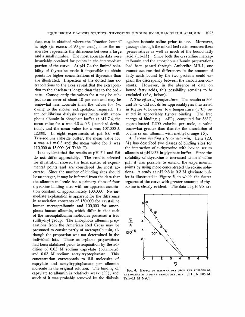
3. The effect of temperature. The results at 300and 380C did not differ appreciably; as illustratedin Figure 4, however, low temperature (50C) re-sulted in appreciably tighter binding. The freeenergy of binding (- AF), computed for 380C,approximated 7,200 calories per mole, a valuesomewhat greater than that for the association ofbovine serum albumin with methyl orange (5).
4. Second binding site on albumin. Lein (23,24) has described two classes of binding sites forthe interaction of L-thyroxine with bovine serumalbumin at pH 9.75 in glycinate buffer. Since thesolubility of thyroxine is increased at an alkalinepH, it was possible to extend the experimentalpoints by using more concentrated thyroxine solu-tions. A study at pH 9.8 in 0.2 M glycinate buf-fer is illustrated in Figure 5, in which the flattersegment of the curve with greater amounts of thy-roxine is clearly evident. The data at pH 9.8 are
6
51
V
A
x105
4
3
2
0 I 2 3 4
VFIG. 4. EFFECT OF TEMPERATURE UPON
THYROXINE BY HUMAN SERUM ALBUMIN.
Tris-0.1 M NaCl.
5 6
THE BINDING OF
pH 8.6, 0.05 M
5.
300
. . . ~~~~~~~~~~~'. v%
K. STERLING, P. ROSEN AND M. TABACHNICK
6
5
V
A
X10-5
4
3
2
I z 4
V
FIG. 5. SECOND BINDING SITE ON THE ALBUMIN MOLECULE.300C, pH 9.8, 0.2 M glycinate buffer.
best described by assuming the existence of twobinding sites: n1 = 3, k, = 143,000; n2 = approxi-mately 20, k2 = approximately 1,000. The valuesfor n2 and k2 represent estimates based upon ex-
trapolations of the flat segment. Theoreticalcurves constructed with these constants resultedin the closest approximation to the experimentalpoints. Owing to the extremely low concentrationof free thyroxine in serum, only the first bindingsite has physiological importance (cf Discussion).
5. The effect of pH. A series of amorphous al-bumin experiments with varying pH is presentedin Table I. It should be pointed out that at pH7.0 the limitation of thyroxine solubility permittedonly a few satisfactorily equilibrated points at thelowest values for i; therefore, lines could bedrawn with reasonable certainty in only two offour experiments attempted. The results inTable I represent the single most satisfactory ex-
periment of the four, and these values should beregarded as the best approximations obtainable at
pH 7.0. The appreciable diminution in binding atpH 10.9 is evident. At pH 11.2 with phosphatebuffer, the data yielded a low, essentially hori-zontal curve indicative of pronounced reduction inbinding. As discussed below, the reduction inbinding with strongly alkaline pH is compatiblewith removal of protons from free -NH3+ groupson the protein.
6. Isooctane-extracted albumin. Goodman (12)has reported that serum albumin has a primaryclass of two binding sites for fatty acids, with an
association constant of 6 x 10⁷ for palmitate. Toinvestigate the possibility that fatty acids mightinterfere with the binding of thyroxine, studieswere carried out with deionized amorphous albu-min preparations which had been subjected to iso-octane extraction to free them of bound fatty acids.The equilibrium dialysis study in Figure 6 re-
vealed no diminution in the number of primarybinding sites (n = 4.2) and, an increased associa-tion constant (k = 148,000). Similar findingswere obtained at pH 7.4. The mean values offour studies with isooctane-extracted albumin were
n = 4.2 and k = 163,000. The reason for thefirmer binding is not evident; possibly, removalof the long-chain fatty acid may free a nonpolarpart of the protein molecule important in the in-teraction. Addition of 2 moles of palmitate per
mole of protein to an isooctane-extracted albuminpreparation resulted in a curve indistinguishablefrom the original, as shown (Figure 6).
7. Nature of the principal binding sites of albu-min. Since Klotz and Walker (25) presented evi-dence in support of the interaction of the anionicdye, methyl orange, and the c-amino groups of ly-sine residues of serum albumin, it seemed reason-
able to suppose that these groups might also beinvolved in the binding of the anionic molecule,
TABLE IEffect of pH on the binding of L-thyroxine by human serum albumin *
No.of
pH Buffer expts. kint nl ki k2n2 n2 k2
7.0 Phosphate 1 370,000 3.5 106,0007.4 Phosphate 10 428,000 4.0 i 0.3 107,000 4 12,0008.6 Tris-NaCI 8 450,000 4.1 ± 0.2 110,000 i 15,0009.8 Glycinate 1 430,000 3.0 143,000 20,000 ca. 20 ca. 1,0009.8 Glycinate 7 420,000 3.3 i 0.4 127,000 i 42,000
10.9 Phosphate 1 150,000 2.0 75,000
* Mean values and standard deviations from the mean. For glycinate at pH 9.8, the single most accurate experi-ment is given in addition.
I I I
i
I
I If% 1 03 A r. a 7 Q
1026
0A 0 r
EQUILIBRIUM DIALYSIS STUDIES: THYROXINE BINDING BY HUMAN SE'RUM ALBUMIN 1027
4-A
x105 3! :x.2."
0 I 2 3 4 5 6 7
FIG. 6. EFFECT OF PALMITATE ON THE BINDING OFL-THYROXINE BY ISOOCTANE-EXTRACTED HUMAN SERUM
ALBUMIN. 30'C, pH 8.6, 0.05 M Tris-0.1 M NaCi.
6
5
V
A
x 10-5
4
3
21
0 1 2 3 4 5
V
6 7
FIG. 7. THE BINDING OF THYROXINE BY ACETYLATED
HUMAN SERUM ALBUMIN PREPARATIONS. 30'C, pH 8.6,0.05 M Tris-0.1 M NaCl.
thyroxine. A means of testing this hypothesiswas provided by studies in which the free aminogroups of the protein were blocked by acetylation(14). As illustrated in Figure 7, acetylation of 11per cent of the lysyl e-amino groups resulted inreduction of n from 4 to 3, and reduction of theassociation constant by one-third (from 100,000to 70,000). Heavier acetylation, which producedblocking of 45 to 75 per cent of the protein aminogroups, resulted in more pronounced reduction ofbinding.The behavior as a function of variation of pH
is consistent with an interaction of free -NH3+groups and the small anionic molecule, thyroxine.Thus, at very strongly alkaline reaction (pH 10.9and 11.2) in the presence of isotonic phosphatebuffer, the binding of L-thyroxine is markedly re-
duced, presumably because most of the - NH,+groups have lost a proton to become -NH2groups.
As previously reported (1), the free cationicgroups on the protein molecule are believed to in-teract with the anionic phenolate groups of thyrox-ine and triiodothyronine. At pH 7.4 triiodothyro-nine was bound only one-tenth as tightly as
L-thyroxine and D-thyroxine. The difference inbehavior was attributed to the lesser degree ofionization of the phenolic hydroxyl group of tri-iodothyronine, which at pH 7.4 is only one-tenthas highly ionized as that of thyroxine. At pH 8.6,with greater ionization to the phenolate form, tri-
iodothyronine was found to be more firmly boundby albumin. While other factors besides electro-static attraction may be involved in binding, thedata indicate an important role for the interac-tion of free cationic groups on the protein mole-cule and the anionic phenolate groups of thyroxineand triiodothyronine. The interaction suggestedin Figure 8 may be regarded as a tentative andsimplified hypothesis, consistent with the data athand but not definitely proven. An alternative ex-
planation may be advanced for the observations ofreduced binding at strongly alkaline reaction (pH10.9 and 11.2), as well as for the behavior of theacetylated proteins. In both circumstances theprotein molecule should have a greater net nega-
tive charge because of the loss of free cationic
NH'I
NH3I3(ALBUMIN MOLECULE)
NH 3
0-
I I-
THYROXINEFIG. 8. SCHEMATIC
NH 3
pH 7.4
PHENOLIC HYDROXYLGROUP 82% IONIZED
REPRESENTATION OF HYPOTHESIS OF
THYROXINE-ALBUMIN INTERACTION.
NATIVE ALBUMIN
1% ACETYLATED
45% ACETYLATED
75% ACETYLATED
I
K. STERLING, P. ROSEN AND M. TABACHNICK
(-NH3+) groups, whether by acetylation or byloss of a proton. Thus, the reduction of affinitybetween a more negatively charged protein mole-cule and the thyroxine anion does not require thespecific hypothesis suggested in Figure 8. More-over, configurational changes or other possible al-terations have not been assessed. Further studieswith modified proteins and with thyroxine analogsmay provide more evidence concerning the natureof the interaction.
DISCUSSION
The theoretical treatment of the interaction ofsmall molecules with proteins has been consideredextensively by Scatchard (2-4), Klotz (5), andmany other workers. Scatchard has simplifiedthe theoretical consideration of the problem bydiscussing it under the four questions listed below;the answers for the thyroxine-albumin interactionare provided by the present data.
1. "How many?" (How many small moleculesare bound by each protein molecule?) At physi-ological pH, in isotonic phosphate or Tris-NaClbuffer, n = 4, signifying that the human serum al-bumin molecule may bind four thyroxine mole-cules at its primary binding sites.
2. "How tightly?" (How tightly are the smallmolecules bound to the protein?) The apparentassociation constant of approximately 100,000 is ofan order of magnitude comparable with the bindingof many drugs and dyes. Albumin binds thyrox-ine somewhat more tightly than it binds the anionicdye, methyl orange (5), or tryptophan (6), butmuch less tightly than it binds the free fatty acidsof serum (12). In contrast, triiodothyronine isbound only one-tenth as tightly as thyroxine atpH 7.4, a difference attributed to the greater de-gree of dissociation of the phenolic hydroxyl groupof thyroxine.
3. "Where ?" (At what sites on the proteinmolecule does binding occur?) Since the nega-tively charged phenolic hydroxyl group appearsto be one site of attachment of the iodothyroninemolecule, it seems reasonable to suppose that freecationic groups on the protein are binding sites.The reduction of binding at strongly alkaline re-action (pH 10.9 and 11.2) is compatible withsuch a hypothesis. Experimental support is af-forded by the studies in which diminished binding
was observed after lysyl e-amino groups of theprotein had been blocked by acetylation. The evi-dence is not, however, a conclusive demonstrationthat -NH3+ groups are the specific binding sites.In strongly alkaline solutions where -NH3+ groupslose protons, or with these groups blocked, theprotein molecule would have a greater net nega-tive charge that could reduce its affinity for thethyroxine anion.
4. "Why ?" (What is the biological signifi-cance of binding, if any?) In normal serum, thealbumin molecules are present in vast excess.The ratio approximates 10,000 albumin moleculesto one of thyroxine. Under these circumstancesan even smaller minority than 1/10,000 of the al-bumin molecules would carry a bound thyroxinemolecule, since most of the serum thyroxine isbound by the thyroxine-binding a-globulin and pre-albumin fractions. Therefore, the tightness of al-bumin binding is not a limiting factor. Neverthe-less, it has biological relevance as a secondarycarrier.From the binding constants established for hu-
man serum albumin it is possible to estimate thefree thyroxine concentration of serum by employ-ing the observation that approximately 15 percent of serum thyroxine is albumin-bound uponelectrophoresis. With the addition of traceramounts of thyroxine, the mean value of 15 percent has been obtained both by Ingbar andFreinkel (7) and Robbins and Rall (26) with re-spective buffer systems of pH 8.6 and 8.4; theprecise value at pH 7.4 has not thus far been de-termined in a system that separates the prealbu-min carrier. However, the use of 15 per centseems reasonable as a first-order approximation,in view of the similarity of albumin binding of thy-roxine at pH 7.4 and pH 8.6. The 85 per centbound by the other carriers was ignored for thepresent calculation.The equation most conveniently applied is that
of Scatchard-i = nkA/(l + kA)-which repre-sents the form from which the equation v/A =kn - kP is derived algebraically. If an averagenormal butanol-extractable iodine of 5.1 ug per100 ml is assumed, this would correspond to aserum thyroxine concentration of 7.78 jug per 100ml, or 1 X 1J7 M. Since approximately 15 percent of the total serum thyroxine is albumin-
1028
EQUILIBRIUM DIALYSIS STUDIES: THYROXINE BINDING BY HUMAN SERUM ALBUMIN 1029
bound, the value for the thyroxine-albumin com-plex would be 1.5 x 10⁻⁸ M. The mean normalconcentration of albumin of 4.5 g per 100 ml cor-responds to a concentration of 6.5 x 10⁻⁴ M.Therefore v=(1.5 X 10⁻⁸ M)/(6.5 X 10-4 M) =2.3 x 10⁻⁵. Since the equilibrium dialysis stud-ies reported have given values of 4 for n, and 100,-000 for k, we may substitute in the equation andsolve for A, the free thyroxine concentration ofserum. The value of 0.6 x 10⁻¹⁰ M obtained cor-responds to the predictions of Robbins and Rall(8, 9). A value of 1.3 x 10¹⁰ M has been ob-tained in this laboratory by dialysis of whole se-rum, as reported separately (21). The discrep-ancy may be attributed to the extrapolation ofbinding data to undiluted serum with its manysmall molecules (27), the partial removal of fattyacids bound to albumin, and the use of data fromelectrophoresis at pH 8.6.
SUMMARY
1. Equilibrium dialysis has been employed toinvestigate the binding of thyroxine by human se-rum albumin. Various temperatures and bufferswere used, including 380C, pH 7.4, and 0.15 ionicstrength (isotonic).
2. Detailed studies of various albumin prepara-tions yielded results consistent with four primarybinding sites per molecule, with an apparent as-sociation constant of approximately 100,000.
3. Studies exploring variation in pH and be-havior of acetylated albumin preparations sug-gested that four cationic groups on the proteinmolecule interact with the anionic phenolate groupsof thyroxine and its analogs. The evidence is com-patible with the hypothesis that the four primarybinding sites are e-amino groups of lysine residuesof albumin, but the nature of the binding sites isnot considered proven.
4. With the observation that 15 per cent of thetotal serum thyroxine is bound to albumin, it waspossible to employ the binding constants obtainedto compute the theoretical free thyroxine concen-tration of serum; the value calculated was 0.6 x10⁻¹⁰ M.
ACNOWLEDGMENTS
The authors wish to express gratitude to Robert Dan-ziger, Alexander Hegedus and Thornton S. Walker forvaluable technical assistance.
REFERENCES
1. Sterling, K., and Tabachnick, M. Determination ofthe binding constants for the interaction of thy-roxine and its analogues with human serum al-bumin. J. biol. Chem. 1961, 236, 2241.
2. Scatchard, G. The attractions of proteins for smallmolecules and ions. Ann. N. Y. Acad. Sci. 1949,51, 660.
3. Scatchard, G., Hughes, W. L., Jr., Gurd, F. R. N.,and Wilcox, P. E. The interaction of proteinswith small molecules and ions in Chemical Speci-ficity in Biological Interactions, F. R. N. Gurd,Ed. New York, Academic Press, 1954, p. 193.
4. Scatchard, G., Coleman, J. S., and Shen, A. L.Physical chemistry of protein solutions. VII.The binding of some small anions to serum al-bumin. J. Amer. chem. Soc. 1957, 79, 12.
5. Klotz, I. M. Protein interactions in The Proteins;Chemistry, Biological Activity, and Methods, H.Neurath and K. Bailey, Eds. New York, Aca-demic Press, 1953, vol. 1, part B, p. 727.
6. McMenamy, R. H., and Oncley, J. L. The specificbinding of L-tryptophan to serum albumin. J. biol.Chem. 1958, 233, 1436.
7. Ingbar, S. H., and Freinkel, N. Regulation of theperipheral metabolism of the thyroid hormones.Recent Progr. Hormone Res. 1960, 10, 353.
8. Robbins, J., and Rall, J. E. The interaction of thy-roid hormones and protein in biological fluids.Recent Progr. Hormone Res. 1957, 13, 161.
9. Robbins, J., and Rall, J. E. Proteins associated withthe thyroid hormones. Physiol. Rev. 1960, 40, 415.
10. Sterling, K., and Tabachnick, M. Paper electropho-retic demonstration of thyroxine-binding prealbu-min fraction in serum. Endocrinology 1961, 68,1073.
11. Dintzis, H. M. Studies on the Dielectric Propertiesof Human Serum Mercaptalbumin Solutions.Thesis, Harvard University, 1952.
12. Goodman, D. S. The interaction of human serumalbumin with long-chain fatty acid anions. J. Amer.chem. Soc. 1958, 80, 3892.
13. Goodman, D. S. Preparation of human serum albu-min free of long-chain fatty acids. Science 1957,125, 1296.
14. Fraenkel-Conrat, H., Bean, R. S., and Lineweaver,H. Essential groups for the interaction of ovomu-coid (egg white trypsin inhibitor) and trypsin, andfor tryptic activity. J. biol. Chem. 1949, 177, 385.
15. Tabachnick, M., and Sobotka, H. Azoproteins. II.A spectrophotometric study of the coupling ofdiazotized arsanilic acid with proteins. J. biol.Chem. 1960, 235, 1051.
16. Gornall, A. G., Bardawill, C. J., and David, M. M.Determination of serum proteins by means of thebiuret reaction. J. biol. Chem. 1949, 177, 751.
17. Cohn, E. J., Hughes, W. L., Jr., and Weare, J. H.Preparation and properties of serum and plasma
K. STERLING, P. ROSEN AND M. TABACHNICK
proteins. XIII. Crystallization of serum albuminsfrom ethanol-water mixtures. J. Amer. chem. Soc.1947, 69, 1753.
18. Albright, E. C., and Larson, F. C. Metabolism ofL-thyroxine by human tissue slices. J. clin. Invest.1959, 38, 1899.
19. Gemmill, C. L., and Plunkett, R. L. The kinetics ofthe inhibition by thyroxine of the cupric chloridecatalyzed oxidation of ascorbic acid. Arch. Bio-chem. 1952, 36, 434.
20. Lardy, H. Effect of thyroid hormones on enzyme sys-
tems in The Thyroid, A. Edelmann, H. J. Curtisand M. E. Koshland, Eds. Upton, N. Y., Brook-haven Nat. Lab., 1954, p. 90.
21. Sterling, K., and Hegedus, A. Measurement of freethyroxine concentration in human serum. J. clin.Invest. 1962, 41, 1031.
22. Teresi, J. D., and Luck, J. M. The combination oforganic anions with serum albumin. VIII. Fattyacid salts. J. biol. Chem. 1952, 194, 823.
23. Lein, A. Thyroxine binding by bovine serum albu-min. Fed. Proc. 1952, 11, 91.
24. Lein, A. The binding of thyroxine, diiodotyrosine,and triiodothyronine by bovine serum proteins.Abstracts of Papers of 123rd Meeting of Amer.Chem. Soc., Div. of Biol. Chem., Section 42,March, 1953, p. 16C.
25. Klotz, I. M., and Walker, F. M. The binding of or-
ganic ions by proteins. Charge and pH effects. J.Amer. chem. Soc. 1947, 69, 1609.
26. Rall, J. E. Personal communication.27. McMenamy, R. H., Lund, C. C., Van Marcke, J., and
Oncley, J. L. The binding of L-tryptophan in hu-man plasma at 370 C. Arch. Biochem. 1961, 93,135.