First Crystal Structure of a Fungal High-redox PotentialDye-decolorizing PeroxidaseSUBSTRATE INTERACTION SITES AND LONG-RANGE ELECTRON TRANSFER*
Received for publication, July 12, 2012, and in revised form, December 11, 2012 Published, JBC Papers in Press, December 12, 2012, DOI 10.1074/jbc.M112.400176
Eric Strittmatter‡1, Christiane Liers§, Rene Ullrich§, Sabrina Wachter‡, Martin Hofrichter§, Dietmar A. Plattner‡,and Klaus Piontek‡1,2
From the ‡Institute of Organic Chemistry, University of Freiburg, Albertstrasse 21, 79104 Freiburg and the §Department of Bio- andEnvironmental Sciences, International Graduate School of Zittau, Markt 23, 02763 Zittau, Germany
Background: DyP-type peroxidases catalyze biotechnologically important reactions.Results: Based on the crystal structure of a fungal DyP, the conformational flexibility of Asp-168 is elucidated. Tyr-337 isidentified as a surface-exposed substrate interaction site.Conclusion: Asp-168 and Tyr-337 are key residues directly involved in AauDyPI-catalysis.Significance: Peroxidases are biocatalysts, much sought after and ubiquitous enzymes in nature.
Dye-decolorizing peroxidases (DyPs) belong to the largegroup of heme peroxidases. They utilize hydrogen peroxide tocatalyze oxidations of various organic compounds. AauDyPIfrom Auricularia auricula-judae (fungi) was crystallized, andits crystal structure was determined at 2.1 A resolution. Themostly helical structure also shows a �-sheet motif typical forDyPs and Cld (chlorite dismutase)-related structures andincludes the complete polypeptide chain.At thedistal side of theheme molecule, a flexible aspartate residue (Asp-168) plays akey role in catalysis. It guides incoming hydrogen peroxidetoward the heme iron and mediates proton rearrangement inthe process of Compound I formation. Afterward, its side chainchanges its conformation, now pointing toward the proteinbackbone. We propose an extended functionality of Asp-168,which acts like a gatekeeper by altering the width of the hemecavity access channel. Chemical modifications of potentiallyredox-active amino acids show that a tyrosine is involved in sub-strate interaction. Using spin-trapping experiments, a transientradical on the surface-exposed Tyr-337 was identified as theoxidation site for bulky substrates. A possible long-range elec-tron transfer pathway from the surface of the enzyme to theredox cofactor (heme) is discussed.
Peroxidases (EC 1.11.1) are ubiquitous enzymes that catalyzethe oxidative conversion of various compounds utilizing hydro-gen peroxide (H2O2) as electron acceptor. As a common trait,most peroxidases, although phylogenetically unrelated, contain
a heme B (iron protoporphyrin IX) molecule as the redoxcofactor.A division of this heterogenic group led to the formation
of four families, one of which consist of the dye-decolorizingperoxidases or DyPs3 (EC 1.11.1.19) (1). According to Wel-inder’s systematics, which were later on extended (2, 3),DyPs were initially grouped in the class of secretory fungalperoxidases viz. class II of the peroxidase-catalase superfam-ily. This class further comprises lignin peroxidase, manga-nese peroxidase, and versatile peroxidase. However, DyPsturned out to be phylogenetically as well as structurallyunrelated to all hitherto described peroxidase families. As aresult, a new family of enzymes was established to accommo-date these unusual peroxidases (4). From a structural view-point, DyPs are best considered members of a highly diversesuperfamily of proteins comprising, among others, Clds andDyPs, sharing a ferredoxin-like core of �-sheets as a com-mon feature (5). Actually, even the term “DyP” circum-scribes a polyphyletic group that can be roughly divided intofour different entities. The groups DyPA–C comprise pre-dominantly bacterial enzymes, whereas DyPD is a fungalgroup (6). The first DyP was discovered in 1995 (7). A mix-ture of extracellular enzymes secreted by the basidiomyceteBjerkandera adusta (the strain was back then misidentifiedas Thanatephorus cucumeris) was reported to efficiently oxi-dize anthraquinone dyes such as Reactive Blue 5 (7, 8). Theenzyme responsible for the decolorization process was char-acterized as a heme peroxidase of unusual chemical proper-ties. Despite the general versatility of peroxidase chemistry,no fungal peroxidases had been known to efficiently oxidizesynthetic anthraquinone dyes so far.DyPs catalyze many reactions, among them several conver-
sions that are biotechnologically desirable. Enzymatic assays ona fungal DyP from the basidiomycete Auricularia auricula-ju-dae (AauDyP, formerly labeled “AjP”) have been reported in a
* This work was partially financed by a grant from the Commission of theEuropean Communities within the Sixth European Framework Programme(BIORENEW contract NMP2-CT-2006-026456) (to K. P. and M. H.) and byfunding from the International Research Training Group Grant IRTG 1038“Catalysts and Catalytic Reactions for Organic Synthesis” (CCROS) of theDeutsche Forschungsgemeinschaft (DFG) (to D. A. P.).
The atomic coordinates and structure factors (code 4AU9) have been deposited inthe Protein Data Bank (http://wwpdb.org/).
1 Both authors contributed equally to this work.2 To whom correspondence should be addressed. E-mail: Klaus.Piontek@ocbc.
uni-freiburg.de.
3 The abbreviations used are: DyP, dye-decolorizing peroxidase; Aau, Auricu-laria auricula-judae; Bad, Bjerkandera adusta; Cld, chlorite dismutase; r.m.s.,root mean square; LRET, long-range electron transfer.
previous publication (9). The isolated enzyme exhibited thetypical range of DyP-type peroxidase reactions. Interestingly,this reaction range also encompassed a low-pH activity to oxi-dize nonphenolic compounds such as 3,4-dimethoxybenzylal-cohol (veratryl alcohol), like lignin peroxidase, a fungal peroxi-dase involved in lignin degradation. Lignin is considered one ofthe most abundant and recalcitrant biopolymers on earth. Thissuggests that at least this DyP-type peroxidase might take partin the decomposition of lignin. Lately, other reactions catalyzedby DyPs have been discovered, e.g. the oxidative cleavage ofcarotenoids (10) and sulfoxidation of aromatic sulfides (11).Although thementioned and yet to be fully unraveled chemicalproperties of DyPs demonstrate their obvious biotechnologicalpotential, their actual role in nature remains obscure.The putative oxidative cycle of fungal DyPs is largely
equivalent to that in other peroxidases, but was recentlyextended by the proposal of a swinging mechanism of a distalaspartate residue during Compound I formation (12). InScheme 1, the formation of Compound I is depicted. In a firststep, H2O2 enters the heme cavity of the enzyme in restingstate where it displaces a water molecule that occupiesthe sixth ferric iron coordination site of the protoporphyrinIX system. A distal basic amino acid residue mediates therearrangement of a proton in H2O2. In peroxidases, this basetypically is a histidine, whereas in DyPs, this key residue issubstituted by an aspartate (see “Results and Discussion”).The heme molecule is then oxidized to the radical-cationicoxoferryl species Compound I by two-fold single electrontransfer, releasing a water molecule. Two electrons are suc-cessively drawn from substrate molecules, leading to theiroxidized counterparts. Concomitantly, the heme is stepwisereduced back to its initial oxidation state, leading to theenzyme resting state in this process.
Enzyme � H2O23 Compound I � H2O
Compound I � XH23 Compound II � XH�
Compound II � XH23 Enzyme � XH� � H2O
REACTIONS 1–3
The above reactions summarize the general catalytic cycle ofperoxidases. The term “Enzyme” indicates the peroxidase rest-ing state, and XH2 and XH� are a substrate molecule and thecorresponding radical species, respectively.Up to the present, only a few DyP-like enzymes from fungi
have been described biochemically in detail, and only one crys-tal structure has been published so far (14). Research on bacte-rial members has been comparatively more extensive. A nativecrystal structure was needed to substantiate recent resultsinvolving fungal as well as bacterial DyP-type peroxidases (5,12). To gain further insight into the structural properties of theeucaryotic (in this case fungal) DyPs, AauDyPI from the jellyfungus A. auricula-judae (Auriculariales, Basidiomycota) waspurified and crystallized, and the crystal structure was deter-mined at 2.1 Å resolution (Protein Data Bank (PDB) code4AU9).
EXPERIMENTAL PROCEDURES
Isolation of Nucleic Acids, PCR, and mRNA Sequencing—Fordetailed methodology, see elsewhere (49). In short, mRNA wasextracted from mycelia of A. auricula-judae grown in agitatedcultures. For cDNA synthesis, the total mRNA (1 �g) wasprimed and subsequently reverse-transcribed. Afterward theobtained cDNA was amplified following a customized PCRprotocol.Primers were designed to amplify fragments of a DyP-type
peroxidase gene from A. auricula-judae. The obtained PCRproducts were purified and sequenced. Strain DSMZ 11236(from the Deutsche Sammlung von Mikroorganismen undZellkulturen, Braunschweig, Germany) was checked for speciesidentity by internal transcribed spacer PCR (ITS-PCR) ongenomic DNA with primers ITS1 and ITS4 (15).Protein Production and Purification of AauDyPI—Produc-
tion and purification of AauDyPI were carried out as describedin a previous publication (9). To summarize, A. auricula-judaewas grown in agitated tomato juice medium at 24 °C. At maxi-mum peroxidase activity level, the cultures were harvested anddirectly filtrated. The enzyme was purified using Q-Sepharosechromatography columns. Purified enzyme was stored in 5mM
sodium acetate buffer at 4 °C.Crystallization—Purified AauDyPI was crystallized using
protein concentrations of 10.7 mg ml�1 in 5 mM sodium ace-tate, pH 6.8. Various crystallization kits (Hampton Research,Aliso Viejo, CA and Jena Bioscience, Jena, Germany) were usedfor initial screens. Crystals were grown at 19 °C by the hangingdrop vapor diffusion method with a 1:1 (v/v) ratio of protein-to-precipitant in 4-�l drops. Intergrownplate-like crystalswereobtained with a precipitant solution consisting of 0.17 M
sodium acetate trihydrate, 0.085 M Tris hydrochloride, pH 8.5,25.5% (w/v) polyethylene glycol 4.000, and 15% (v/v) glycerol.From these crystal clusters, very small single fragments of about
SCHEME 1. General formation of Compound I in peroxidases. This is thekey step in heme peroxidase catalysis. After proton rearrangement by thedistal base-catalytic residue, two electrons are transferred from heme tohydrogen peroxide, leading to the active state of the enzyme, called Com-pound I (13). B indicates a basic residue, e.g. a histidine or aspartate/gluta-mate in peroxidases. L indicates the proximal heme ligand, usually a histidineresidue.
Catalytic Features of a DyP-type Peroxidase
4096 JOURNAL OF BIOLOGICAL CHEMISTRY VOLUME 288 • NUMBER 6 • FEBRUARY 8, 2013
0.2� 0.1� �0.001mmwere retrieved that seemed suitable forx-ray crystallographic experiments. To verify the results ofstructure determination, we examined the purified protein viaLC-MS.Data Collection and Processing—AauDyPI crystals were
directly flash-cooled in liquid nitrogen. X-ray diffraction datawere collected on the macromolecular crystallographic beam-line ID14-4 at the European Synchrotron Radiation Facility(ESRF) (Grenoble, France) (16). Data were indexed, processed,and scaled with XDS (17, 18). Data collection and processingstatistics are given in Table 1.Structure Determination—The structure of AauDyPI was
determined bymolecular replacement usingMOLREP (19) andPhaser (20). An ad interim structure of the B. adustaDyP (PDBcode 2D3Q) was used as search model. This structure will bereferred to as BadDyP in the following. Side chains of noncon-served amino acidswere truncated at the last carbon atomcom-mon to the target and model residues with the CCP4-programCHAINSAW (21). Because there are two molecules per asym-metric unit, density modification including noncrystallo-graphic symmetry averaging was performed to improve the ini-tial phases using the CCP4 program PARROT (22). An initialmodel was built with Coot (23) and refined in REFMAC5 (24).Further improvement was achieved by successive cycles ofmodel building and refinement.Identification of a Redox-active Surface-exposed Tyrosine—
Chemicalmodification of AauDyPIwas carried out as describedby Inokuchi et al. (25) andMiki et al. (26). In brief, AauDyPIwasincubated with 5–150-fold excess of N-bromosuccinimide ortetranitromethane, respectively. The modified proteins weredirectly used in an enzyme assay monitoring the oxidation of2,2�-azino-bis(3-ethylbenzothiazoline-6-sulfonic acid) at 420nm. Spin trapping was performed using the protocol of Zhao etal. (27). At first AauDyPI was deglycosylated using 10 units ofpeptide-N-glycosidase F (Aldrich) at room temperature for 1.5days. The thus prepared enzyme wasmixed with excess prolinenitric oxide after incubation with peroxyacetic acid. This mix-turewas incubated for 15min at 37 °C and then rebuffered in 20mM K3PO4 buffer, pH 7.2. The enzyme was recovered from thereaction mixture by SDS-PAGE and subsequent CoomassieBlue staining. The bands were cut from the gel using a sterilescalpel. Prior to mass spectrometry, the protein was digestedin-gel with elastase and thermolysin, respectively. The peptidecontaining the spin-trapped amino acid residue was identifiedusing an Agilent 6520 quadrupole-TOF mass spectrometercoupled to a reverse-phase HPLC unit and a nano-electrosprayionization source.Electron Transfer Calculations, Probing of the Heme Access
Channel, and Imaging—The calculation of electron transferpathways was performed with the program HARLEM (asdescribed in Ref. 28) using the PATHWAY module. Volumeand shape of the heme access channel were calculated withHOLLOW (29).Two coordinate files of AauDyPI were created from the orig-
inal file, each one containing one of the two conformers of Asp-168. Solventmolecules were removed from the coordinate files,and a simulation of the Compound I state containing a ran-domly placed guaiacol (2-methoxyphenol) molecule was built
using Coot. An oxygen atom was thus placed distally perpen-dicular to the hemeplane at 1.7Ådistance from iron as found inthe Compound I structure of horseradish peroxidase (PDBentry 1HCH (30)). Ligand dockingwas then performedwith theMolegro Virtual Docker (MVD) (31). The ligand binding cavi-ties were identified using the Expanded van der Waals algo-rithmwith a grid size of 0.5 Å. 50 docking runs were performedusing the guaiacol molecule as ligand. The population size wasset to 200 over a radius of 12 Å around the predicted bindingcavity with a grid size of 0.2 Å. 10,000 iterations per positionwere performed. Clustering of similar positions was enabledusing an r.m.s. deviation of 1.5 Å. The ligand positioning wasconstrained to the predicted binding site, i.e. the distal hemecavity. Images were created in PyMOL (32) and CCP4mg (33).
RESULTS AND DISCUSSION
Structure Solution and Refinement—Improvement of crystalsize and quality of AauDyPI proved unexpectedly arduous.Despite numerous variations of the initial crystallization con-dition, only very thin plate-like crystals were obtained. Crystalgrowth was exclusively observed in solutions with a high con-centration of polyethylene glycol (PEG) 4000. The widely usedammonium sulfate yielded only amorphous precipitate. Thepresence of cryoprotectants such as glycerol or 2-methyl-2,4-pentanediol ab initiowas beneficial to crystal quality. Additivessuch as various amino acids did not show any positive effects.Bearing in mind the size and morphology of the crystals, sur-prisingly good x-ray diffraction data were collected; the bestcrystals diffracted to a maximum resolution of about 2.1 Å(Table 1).The relatively high sequence identity between AauDyPI and
BadDyP (PDB code 2D3Q) suggestedmolecular replacement asa promising approach to solve the structure. Indeed, this tech-niquewas successful. Phasemodification techniques gave a fur-ther improvement, resulting in a high-quality electron densitymap. This allowed us to build an initial model of the completeprotein, which was subsequently refined in REFMAC5. Thefinal refinement statistics are supplied in Table 1.Overall Structure of AauDyPI—AauDyPI is a globular glyco-
protein with a helical basic architecture and a prominent�-sheet motif spanning the distal side of the heme plane. Aproximal N- and a distal C-terminal domain embed the hememolecule, which itself is partly flanked by the �-sheet of theferredoxin-like fold. Like other DyPs, it is structurally very dis-tinct from the classical heme peroxidases because the latter arepractically completely helical. Themonomeric AauDyPI has anellipsoid-like shape with dimensions of 70 � 42 � 40 Å3. Therefinedmodel of AauDyPI consists of the complete polypeptidechain of 448 amino acids as predicted by gene sequencing.4Even the C and N terminus could be modeled as well as somerather large flexible loop regions (Fig. 1A), where electron den-sity was mostly poorly defined. In the crystal, there are twomolecules per asymmetric unit. The C�-r.m.s. between the twosubunits is 0.375 Å, which is in the order of the estimated coor-dinate error of 0.257 Å. Only in the flexible surface loop regionswere larger deviations between the two molecules observed.Given the consensus sequenceAsn-X-Ser/ThrwithX being anyamino acid except proline or aspartic acid, there are four poten-
Catalytic Features of a DyP-type Peroxidase
FEBRUARY 8, 2013 • VOLUME 288 • NUMBER 6 JOURNAL OF BIOLOGICAL CHEMISTRY 4097
tial glycosylation sites. Three of them could be verified crystal-lographically, whereas closer inspection of Asn-322 reveals thatthis fourth site is inaccessible for glycosyltransferases and con-sequently is not glycosylated. The glycosylation sites are exclu-sively distributed on the “lower” end of the proximal side withrespect to the heme molecule (Fig. 1A), especially at large, pro-truding loops that are susceptible to proteolytic attacks. There-fore, the carbohydrate moieties might have a protective func-tion against proteases.In AauDyPI, some carbohydrate chains are definitely glyco-
sylated to a higher extent than outlined here. Residual electrondensity, albeit weak and difficult to interpret, strongly indi-cates the presence of branched N-glycans of the high-man-
nose type (34). Further model building was considered to beunrewarding.AauDyPI shows an overall structure characteristic for the
DyP-type family, which in turn is somewhat related to that ofthe chlorite detoxifying Clds (chlorite dismutases). The fold ofthese heme enzymes also includes the particular ferredoxin-like �-sheet, similar to that found in DyPs (35). However, asequence alignment indicated only 8% identity between the Cldof Dechloromonas aromatica (a �-proteobacterium) and Bad-DyP, which is essentially nonhomologous (5). Consequently, asecondary structure matching superposition of AauDyP andCld results in a large C�-r.m.s. of 2.502 Å (Fig. 1C). TheC�-r.m.s. was calculated after secondary structure matchingsuperposition with the CCP4-program SUPERPOSE (36).Structurally, AauDyPI ismost similar to BadDyP, as expected
from the molecular replacement results, correlating with aC�-r.m.s. of 1.14 Å for 427 common atoms (Fig. 1B). This valueis relatively high in view of the sequence identity of 53% (66%similarity) and the unique molecular replacement solutionobtained with PHASER. Helices and �-sheets are mostly con-served, whereas the loop regions are more diverse. DyP struc-tures of procaryotic origin differ significantly from fungalenzymes because they lack the heme. In addition, their poly-peptides are considerably (by one-third) smaller and form olig-omers (dimers to hexamers) (37).As inmost peroxidases, a histidine residue coordinates to the
central heme iron as the fifth ligand, with a distance of 2.1 Å(Fig. 2A). Usually heme-Fe(III) is coordinated six-fold, the distalligand site occupied by a water molecule. Here, the water mol-ecule closest to the iron atom is 3.11 Å apart. This distance istoo large for a coordination bond. Probably the iron in the crys-tal structure represents a Fe(II) species, which usually coordi-nates five-fold.Most likely, the heme-Fe(III) was reduced by thehigh-intensity x-ray beamunder cryocondition. From the distalside cavity, a channel leads to the exterior, which most likelyserves as the substrate access channel (see below) (12).A characteristic trait of theDyP-type andCld-like enzymes is
the unusual conformation of the heme propionate residue atpyrrole C (5). Generally, both propionate groups in peroxidasesdisplay a conformation like the one of the propionate at pyrrole
FIGURE 1. Overview of the AauDyPI structure. A, ribbon diagram of AauDyPI. The ferredoxin-like �-sheet fold is highlighted in green, the protoporphyrin IXsystem is in dark violet with the central iron atom as a tan sphere, and the carbohydrate chains with their corresponding N-linked asparagine residues are inpurple. The chain termini are labeled as well. B, superposition of BadDyP (mauve and purple) onto AauDyPI (green shading). The C�-trace is shown with helicesas tubes. C, superposition of Cld (pink) onto AauDyPI (whitish and green shading). The C�-trace is shown with helices as tubes.
TABLE 1Summary of data collection and refinement statistics of AauDyPIValues in parentheses belong to the highest resolution shell.
Data collectionSpace group P21Unit-cell parameters (Å/°) a � 66.60; b � 46.69; c � 141.20; � � 91.35Beamline ID14-4, ESRFWavelength (Å) 0.9395Temperature (K) 100No. of crystals 1Resolution range (Å) 49.01–2.10 (2.23–2.10)Total no. of reflections 131,951 (19,754)No. of unique reflections 49,651 (7661)Completeness (%) 96.9 (93.4)I/�(I) 11.02 (3.35)Rmerge 0.094 (0.437)
RefinementRwork/Rfree
a 0.180/0.261Molecules per asymmetric unit 2Solvent content (%) 42No. of amino acid residues 896No. of heme molecules 2No. of carbohydrates 8No. of ligands 6 glycerol, 1 acetate, 1 TrisNo. of water molecules 679r.m.s. deviationsBond lengths (Å) 0.011Bond angles (°) 1.25
ring D (Fig. 2A). In DyPs, the propionate at pyrrole C is tiltedinto an unusual conformation due to the formation of stronghydrogen bonds to the protein (Fig. 2B). Propionate D of Aau-DyPI forms a long H-bond with N�2 of Arg-311 and addition-ally with four water molecules. This hydrogen-bonding patternallows propionate D to maintain an unstrained conformation.In contrast, propionate C ismuchmore confined by five hydro-gen bonds. Among them, there are three strong H-bonds to themain chain N of Ile-170 and Ala-171 and to N�2 of Arg-332,respectively. Additionally, twomore H-bonds to N� of Arg-255and to one water molecule are formed. This strong hydrogen-bonding network obviously forces propionate C into a high-energy conformation, hence enforcing a rather close distance of3.6 Å between one of the carboxyl oxygens and amethine groupof the porphyrin ring. As a result, the sp2-plane of the propio-nate lies almost orthogonally to the heme plane. The oxygensensor FixL might be another example where interactions witharginine side chains constrain the propionate chains intounusual geometries upon O2 binding (38).Geometry of the Distal Heme Cavity and Implications for
Catalysis—A first partial catalytic cycle for DyPs has been pro-posed by Sugano et al. (4). Fundamentally, the catalytic cycle inDyPs is the same as in other peroxidases. The hememolecule isoxidized by peroxides to the radical-cationic oxoferryl speciesCompound I, which is two electrons deficient from the restingstate. One electron is abstracted from the iron, resulting inFe(IV), and another one is abstracted from the porphyrin ring.It became apparent that a distal acid-base pair is essential forCompound I formation in peroxidases to rearrange a proton inthe peroxide substrate. When compared with classical peroxi-dases, proton rearrangement or abstraction in DyPs is accom-plished in a different way. Because binding to the heme ironlowers the pKa of hydrogen peroxide (39), deprotonation doesnot require any strong bases. Most peroxidases use a histidineresidue, whereas in DyPs, an even weaker base, a deprotonatedaspartate residue, is employed. An arginine is paired with thehistidine or aspartate, respectively. It does not take part in therearrangement process directly, but is essential for coordinat-ing H2O2 at the sixth ligand site of heme-Fe(III) and stabilizingCompound I. Surprisingly, the aspartate residue is not essentialfor catalysis in procaryotic DyPs. Although Compound I stabil-ity is reduced in mutants lacking said aspartate, they retain agood portion of their enzymatic activity. Mutation of the distalarginine, however, results in complete failure to form Com-pound I and, concomitantly, loss of any peroxidase activity (40).
The mechanism of Compound I formation in DyPs has beenamended recently by the concept of a swinging mechanism ofthe distal aspartate residue during this process (12). Said aspar-tate changes the location of its side chain upon the presence ofcyanide, a structural analog of hydrogen peroxide, in the distalheme cavity when compared with the cyanide-free enzyme. Itwas proposed that this aspartate swings toward the heme mol-ecule in the presence ofH2O2 and thereuponmediates the rear-rangement of a proton. Once Compound I formation is fin-ished, it swings back to the initial position.In AauDyPI, Asp-168 plays the part of themediating residue.
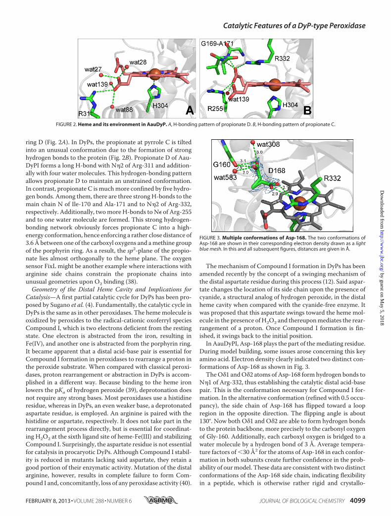
During model building, some issues arose concerning this keyamino acid. Electron density clearly indicated two distinct con-formations of Asp-168 as shown in Fig. 3.TheO�1 andO�2 atoms of Asp-168 form hydrogen bonds to
N�1 of Arg-332, thus establishing the catalytic distal acid-basepair. This is the conformation necessary for Compound I for-mation. In the alternative conformation (refined with 0.5 occu-pancy), the side chain of Asp-168 has flipped toward a loopregion in the opposite direction. The flipping angle is about130°. Now both O�1 and O�2 are able to form hydrogen bondsto the protein backbone, more precisely to the carbonyl oxygenof Gly-160. Additionally, each carboxyl oxygen is bridged to awater molecule by a hydrogen bond of 3 Å. Average tempera-ture factors of�30 Å2 for the atoms of Asp-168 in each confor-mation in both subunits create further confidence in the prob-ability of ourmodel. These data are consistent with two distinctconformations of the Asp-168 side chain, indicating flexibilityin a peptide, which is otherwise rather rigid and crystallo-
FIGURE 2. Heme and its environment in AauDyP. A, H-bonding pattern of propionate D. B, H-bonding pattern of propionate C.
FIGURE 3. Multiple conformations of Asp-168. The two conformations ofAsp-168 are shown in their corresponding electron density drawn as a lightblue mesh. In this and all subsequent figures, distances are given in Å.
Catalytic Features of a DyP-type Peroxidase
FEBRUARY 8, 2013 • VOLUME 288 • NUMBER 6 JOURNAL OF BIOLOGICAL CHEMISTRY 4099
graphically well defined. The two conformations might beindicative for a functionality related to the swinging mecha-nism proposed by Yoshida et al. (12). Moreover, this allows usto discuss yet another issue, the accessibility of the distal sidecavity.The heme access channel features an overall funnel-like
shape that significantly narrows toward the distal site of theheme (see Fig. 5). It is largely hydrophobic with the exception ofthe distal Asp-168/Arg-332 pair and several hydrophilic resi-dues directly at the channel entry. In BadDyP, the opening tothe binding pocket was reported tomeasure about 3Å, having aroughly circular appearance (Fig. 4A). The above mentionedAsp-168 is part of a strictly conserved tetrade that defines thedistal substrate binding pocket. In one conformation, the sidechain of Asp-168 coincides with that of Asp-171 in BadDyP.Consequently, AauDyPI features a very similar opening with aslightly ellipsoid appearance (Fig. 4B). These are the generalconformations involved in forming Compound I. Fig. 4C showsthe opening to the substrate binding pocket with Asp-168 in itsalternative conformation facing toward the peripheral proteinbackbone. In this situation, the opening is enlarged signifi-cantly, measuring about 6 Å in one dimension and about 3 Å inthe other. Switching the side chain toward the carbonyl oxygenof Gly-160, possibly after Compound I formation, Asp-168might work like a gatekeeper that allows passage of substratemolecules bigger than hydrogen peroxide (i.e. small organiccompounds). Consequently, this would enable the enzyme tooxidize substrates directly inside the heme cavity.Identification of Substrate Oxidation Sites—To explore the
possibility of small substrates being oxidized directly in theheme cavity, we tentatively modeled a Compound I state ofAauDyPI. As described above, Compound I wasmodeled basedon the parameters of the oxoferryl species reported for horse-radish peroxidase (PDB entry 1HCH 27). A guaiacol moleculewas included into this hypothetical structure, a standard sub-strate for peroxidases. The MolDock algorithm of MOLEGROwas used to examine whether the distal substrate bindingpocket is sufficiently spacious to contain themodel compound.The calculations confirmed thatmolecules of this size narrowlyfit in the heme access channel of the enzyme.However, the majority of DyP substrates are considerably
larger than guaiacol. Keeping in mind the nature of CompoundI (involving formation of a radical cation) and widening the
perspective on other peroxidases, we gathered an alternative: asurface-exposed substrate interaction site. Depending on thetype of peroxidase, a one-electron hole migrates, resulting in aprotein-based radical cation (41). Thus, in such a case, a novelprotein-based substrate interaction site is introduced. Thisrequires a long-range electron transfer (LRET) pathway (42)from the porphyrin ring to an appropriate redox active aminoacid residue at the surface of the enzyme. An exposed oxidationsite has been demonstrated extensively in a related hemeenzyme viz. lignin peroxidase, where a hydroxylated trypto-phan residue carries out the electronwithdrawal from substratemolecules (43–45). In an electron density map of lignin perox-idase from Phanerochaete chrysosporium, strong electron den-sity showed a hydroxylation at the C� position of the surface-exposed Trp-171. It was assumed that this covalentmodification was the result of a reaction of water or oxygenwith a radical cation intermediate at Trp-171. Because theC�-hydroxylation was absent in a crystal structure of pristinelignin peroxidase (i.e. enzyme before its first enzymatic turn-over), it must have originated from an autocatalytic processduring the first catalytic cycle. Trp-171 mutants (W171F,W171S)were shown to be completely inactive, confirmingTrp-171 as the peripheral substrate oxidation site (44, 45) for sub-strates like veratryl alcohol. In a similar way, a tyrosine on theenzyme surface acts as the substrate interaction site in ligninperoxidase from Trametes cervina (26).Because AauDyPI is able to degrade bulky substrates such as
Reactive Blue 5 (46), it seems obvious to identify those redox-active residues (i.e. Trp, Tyr) in AauDyPI that might act as sur-face-exposed oxidation sites (Fig. 5). Because no post-transla-tional changes that would directly indicate such redox-activeresidues could be detected duringmodel building, we could notidentify a surface-exposed substrate interaction site from thecrystal structure. To address this issue experimentally, in a firststep, wemeasured enzyme activities after chemicallymodifyingtyrosine and tryptophan residues. For specific modification ofthese amino acids, tetranitromethane and N-bromosuccinim-ide were used, respectively. Although there was no effect on theactivity using N-bromosuccinimide, with tetranitromethane, asignificant decrease to roughly 20% of the original activity with2,2�-azino-bis(3-ethylbenzothiazoline-6-sulfonic acid) as sub-strate was detected, strongly indicating the involvement of atyrosine residue in the catalytic process. To locate this key res-
FIGURE 4. Side chain flipping of Asp-168 and its effects on the accession channel toward the heme plane. A and B, substrate accession channel in BadDyP(A) and AauDyPI (B) with Asp-168 facing to Arg-332. C, AauDyPI with Asp-168 facing to Gly-160.
Catalytic Features of a DyP-type Peroxidase
4100 JOURNAL OF BIOLOGICAL CHEMISTRY VOLUME 288 • NUMBER 6 • FEBRUARY 8, 2013
idue (in AauDyPI, there are seven tyrosines in total), spin-trap-ping experiments were performed followed by mass spectro-metric analysis. Excess proline nitric oxide was used to yieldnitrotyrosine from transient tyrosyl radicals (27); it generatesNO, which acts as an in situ radical scavenger trapping theexpected tyrosyl radicals that were generated by adding perox-yacetic acid to an AauDyPI solution. Nitrosotyrosine thusformed is subsequently oxidized to the stable nitrotyrosine. Themodified enzyme was finally digested proteolytically and sub-jected to LC-MS analysis. Nitrotyrosine was solely detected atresidue 337.Tyr-337 is surface-exposed and located at the beginning of a
large protruding loop comprising 20 amino acids. Its side chainis partially embedded in a hollow formed by side chains of res-idues Leu-149 and Gln-218 and the main chain of residues354–358with the edge of the aromatic ring pointing toward thesolvent. The latter peptide is the end of the aforementionedloop and extends into the inner �-sheet, which is closest to thedistal site of the heme. Tyr-337 forms hydrogen bonds via back-
bone to Gly-356 and a van derWaals contact of its side chain tothe nitrogen atom of Leu-357. Interestingly, the side chain ofLeu-357 is in van der Waals contact (�3.8 Å) with the heme,suggesting a possible LRET pathway. Therefore, potential elec-tron transfer pathways from Tyr-337 to the heme moleculewere analyzedwithHARLEM.The best pathway is shown in theinset of Fig. 5. It proceeds from the benzene ring of Tyr-337 tothe backbone amide nitrogen of Leu-357 involving a through-space jump of 3.3 Å followed by the leucine side chain andanother jump (3.8 Å) fromC�1 of Leu-357 to one of the pyrrolerings. The effective length of this pathway is about 13 Å. Thehydroxyl group Tyr-337 forms a strong hydrogen bond of 2.6 Åwith the carboxylate ofGlu-354. It has been reported that acidicside chains in the vicinity of a tryptophan or a tyrosine stabilizean emerging radical cation (26, 47, 48). Alignments with otherDyPs show that Tyr-337 is a conserved residue in fungal as wellas bacterial DyPs and the related enzymes TyrA from She-wanella oneidensis and EfeB from Escherichia coli but is miss-ing in Cld-like proteins (5).Our experiments provide the first unambiguous identifica-
tion of a surface-exposed oxidation site in the large group ofdye-decolorizing peroxidases. Further investigations includingcrystallization of AauDyPI-substrate complexes and site-di-rected mutagenesis to further substantiate the significance ofTyr-337 and the suggested double responsibility of Asp-168 forthe catalytic cycle are in progress.
Acknowledgments—We gratefully acknowledge the opportunity tocollect diffraction data on the synchrotron beamline ID14-4 at theESRF. We thank the staff for technical support. Dr. Eric Haaf of theproteomics unit of the Zentrum fur Biosystemanalyse (ZBSA) inFreiburg, Germany is thanked for LC-MS experiments.
REFERENCES1. Zamocky, M., and Obinger, C. (2010) Molecular phylogeny of heme per-
oxidases. In: Biocatalysis Based on Heme Peroxidases. (Torres, E., andAyala, M., eds) pp. 7–35, Springer Verlag, Berlin, Germany
2. Welinder, K. G. (1992) Superfamily of plant, fungal and bacterial peroxi-dases. Curr. Opin. Struct. Biol. 2, 388–393
3. Martínez, A. T. (2002)Molecular biology and structure-function of lignin-degrading heme peroxidases. Enzyme Microb. Technol. 30, 425–444
4. Sugano, Y., Muramatsu, R., Ichiyanagi, A., Sato, T., and Shoda, M. (2007)DyP, a unique dye-decolorizing peroxidase, represents a novel heme per-oxidase family. J. Biol. Chem. 282, 36652–36658
5. Goblirsch, B., Kurker, R. C., Streit, B. R., Wilmot, C. M., and DuBois, J. L.(2011) Chlorite dismutases, DyPs, and EfeB: 3 microbial heme enzymefamilies comprise the CDE structural superfamily. J. Mol. Biol. 408,379–398
6. Ahmad, M., Roberts, J. N., Hardiman, E. M., Singh, R., Eltis, L. D., andBugg, T. D. H. (2011) Identification of DypB from Rhodococcus jostiiRHA1 as a lignin peroxidase. Biochemistry 50, 5096–5107
7. Kim, S. J., Ishikawa, K., Hirai, M., and Shoda, M. (1995) Characteristics ofa newly isolated fungus, Geotrichum candidum Dec 1, which decolorizesvarious dyes. J. Ferment. Bioeng. 79, 601–607
8. Kim, S. J., and Shoda, M. (1999) Purification and characterization of anovel peroxidase from Geotrichum candidum Dec 1 involved in decolor-ization of dyes. Appl. Environ. Microbiol. 65, 1029–1035
9. Liers, C., Bobeth, C., Pecyna, M., Ullrich, R., and Hofrichter, M. (2010)DyP-like peroxidases of the jelly fungus Auricularia auricula-judae oxi-dize nonphenolic ligninmodel compounds and high-redox potential dyes.Appl. Microbiol. Biotechnol. 85, 1869–1879
FIGURE 5. Distribution of Trp and Tyr residues in AauDyPI and visualiza-tion of the heme access channel calculated with HOLLOW. The proposedLRET from Tyr-337 to the heme is shown in the inset.
Catalytic Features of a DyP-type Peroxidase
FEBRUARY 8, 2013 • VOLUME 288 • NUMBER 6 JOURNAL OF BIOLOGICAL CHEMISTRY 4101
10. Scheibner, M., Hulsdau, B., Zelena, K., Nimtz, M., de Boer, L., Berger,R. G., and Zorn, H. (2008) Novel peroxidases of Marasmius scorodoniusdegrade �-carotene. Appl. Microbiol. Biotechnol. 77, 1241–1250
11. van Bloois, E., Torres Pazmino, D. E., Winter, R. T., and Fraaije, M. W.(2010) A robust and extracellular heme-containing peroxidase fromTher-mobifida fusca as prototype of a bacterial peroxidase superfamily. Appl.Microbiol. Biotechnol. 86, 1419–1430
12. Yoshida, T., Tsuge, H., Konno, H., Hisabori, T., and Sugano, Y. (2011) Thecatalytic mechanism of dye-decolorizing peroxidase DyP may require theswinging movement of an aspartic acid residue. FEBS J. 278, 2387–2394
13. Poulos, T. L., and Kraut, J. (1980) The stereochemistry of peroxidase ca-talysis. J. Biol. Chem. 255, 8199–8205
14. Sato, T., Hara, S., Matsui, T., Sazaki, G., Saijo, S., Ganbe, T., Tanaka, N.,Sugano, Y., and Shoda, M. (2004) A unique dye-decolorizing peroxidase,DyP, from Thanatephorus cucumeris Dec 1: heterologous expression,crystallization and preliminary X-ray analysis. Acta Crystallogr. D 60,149–152
15. White, T. J., Bruns, T., Lee, S., and Taylor, J. (1990) Amplification anddirect sequencing of fungal ribosomal RNA genes for phylogenetics. In:PCR Protocols – a Guide to Methods and Applications (Innis, M. A., Gel-fand, D. H., Sninsky, J. J., and White, T. J., eds) pp. 315–322, AcademicPress, San Diego, CA
16. McCarthy, A. A., Brockhauser, S., Nurizzo, D., Theveneau, P., Mairs, T.,Spruce, D., Guijarro, M., Lesourd,M., Ravelli, R. B. G., andMcSweeney, S.(2009) A decade of user operation on themacromolecular crystallographyMAD beamline ID14–4 at the ESRF. J. Synchrotron Radiat. 16, 803–812
17. Kabsch, W. (2010) XDS. Acta Crystallogr. D 66, 125–13218. Kabsch, W. (2010) Integration, scaling, space-group assignment, and
post-refinement. Acta Crystallogr. D 66, 133–14419. Vagin, A., and Teplyakow, A. (1997)MOLREP: an automated program for
molecular replacement. J. Appl. Crystallogr. 30, 1022–102520. McCoy, A. J., Grosse-Kunstleve, R.W., Storoni, L. C., andRead, R. J. (2005)
Likelihood-enhanced fast translation functions. Acta Crystallogr. D 61,458–464
21. Stein, N. (2008) CHAINSAW: a program for mutating pdb files used astemplates in molecular replacement. J. Appl. Crystallogr. 41, 641–643
22. Cowtan, K. (2010) Recent development in classical density modification.Acta Crystallogr. D 66, 470–478
23. Emsley, P., andCowtan, K. (2004)Coot:model-building tool formoleculargraphics. Acta Crystallogr. D 60, 2126–2132
24. Skubak, P., Murshudov, G. N., and Pannu, N. S. (2004) Direct incorpora-tion of experimental phase information in model refinement. Acta Crys-tallogr. D 60, 2196–2201
25. Inokuchi, N., Takahashi, T., Yoshimoto, A., and Irie, M. (1982)N-Bromo-succinimide oxidation of a glucoamylase from Aspergillus saitoi.J. Biochem. 91, 1661–1668
26. Miki, Y., Calvino, F. R., Pogni, R., Giansanti, S., Ruiz-Duenas, F. J., Mar-tínez, M. J., Basosi, R., Romero, A., and Martínez, A. T. (2011) Crystallo-graphic, kinetic, and spectroscopic study of the first ligninolytic peroxi-dase presenting a catalytic tyrosine. J. Biol. Chem. 286, 15525–15534
27. Zhao, X., Girotto, S., Yu, S., and Magliozzo, R. S. (2004) Evidence forradical formation at Tyr-353 inMycobacterium tuberculosis catalase-per-oxidase (KatG). J. Biol. Chem. 279, 7606–7612
28. Prytkova, T. R., Kurnikov, I. V., and Beratan, D. N. (2005) Ab initio basedcalculations of electron-transfer rates in metalloproteins. J. Phys. Chem. B109, 1618–1625
29. Ho, B. K., and Gruswitz, F. (2008) HOLLOW: generating accurate repre-sentations of channel and interior surfaces in molecular structures. BMCStruct. Biol. 8, 49
30. Berglund, G. I., Carlsson, G. H., Smith, A. T., Szoke, H., Henriksen, A., andHajdu, J. (2002) The catalytic pathway of horseradish peroxidase at highresolution. Nature 417, 463–468
31. Thomsen, R., and Christensen, M. H. (2006) MolDock: a new technique
for high-accuracy molecular docking. J. Med. Chem. 49, 3315–332132. DeLano, W. L. (2010) The PyMOL Molecular Graphics System, version
1.3r1, Schrodinger, LLC, New York33. Potterton, E., McNicholas, S., Krissinel, E., Cowtan, K., and Noble, M.
(2002) The CCP4 molecular-graphics project. Acta Crystallogr. D 58,1955–1957
34. Lerouge, P., Cabanes-Macheteau, M., Rayon, C., Fischette-Laine, A. C.,Gomord, V., and Faye, L. (1998) N-Glycoprotein biosynthesis in plants:recent developments and future trends. Plant Mol. Biol. 38, 31–48
35. Zubieta, C., Krishna, S. S., Kapoor,M., Kozbial, P.,McMullan, D., Axelrod,H. L., Miller, M. D., Abdubek, P., Ambing, E., Astakhova, T., Carlton, D.,Chiu, H. J., Clayton, T., Deller, M. C., Duan, L., Elsliger, M. A., Feuerhelm,J., Grzechnik, S. K., Hale, J., Hampton, E., Han, G. W., Jaroszewski, L., Jin,K. K., Klock, H. E., Knuth, M. W., Kumar, A., Marciano, D., Morse, A. T.,Nigoghossian, E., Okach, L., Oommachen, S., Reyes, R., Rife, C. L., Schim-mel, P., van den Bedem,H.,Weekes, D.,White, A., Xu, Q., Hodgson, K. O.,Wooley, J., Deacon, A. M., Godzik, A., Lesley, S. A., and Wilson, I. A.(2007) Crystal structures of two novel dye-decolorizing peroxidases reveala �-barrel fold with a conserved heme-binding motif. Proteins 69,223–233
36. Krissinel, E., andHenrick, K. (2004) Secondary-structurematching (SSM),a new tool for fast protein structure alignment in three dimensions. ActaCrystallogr. D 60, 2256–2268
37. Sugano, Y. (2009) DyP-type peroxidases comprise a novel heme peroxi-dase family. Cell Mol. Life Sci. 66, 1387–1403
38. Balland, V., Bouzhir-Sima, L., Anxolabehere-Mallart, E., Boussac, A., Vos,M. H., Liebl, U., and Mattioli, T. A. (2006) Functional implications of thepropionate 7-arginine 220 interaction in the FixLH oxygen sensor fromBradyrhizobium japonicum. Biochemistry 45, 2072–2084
39. Jones, P., and Dunford, H. B. (2005) The mechanism of Compound Iformation revisited. J. Inorg. Biochem. 99, 2292–2298
40. Singh, R., Grigg, J. C., Armstrong, Z., Murphy, M. E. P., and Eltis, L. D.(2012) Distal heme pocket residues of B-type dye-decolorizing peroxi-dases. J. Biol. Chem. 287, 10623–10630
41. Mauro, J.M., Fishel, L. A., Hazzard, J. T.,Meyer, T. E., Tollin, G., Cusanov-ich, M. A., and Kraut, J. (1988) Tryptophan-1913 phenylalanine, a prox-imal-side mutation in yeast cytochrome c peroxidase that strongly affectsthe kinetics of ferrocytochrome c oxidation. Biochemistry 27, 6243–6256
42. Klapper, M. H., and Faraggi, M. (1979) Applications of pulse-radiolysis toprotein chemistry. Q. Rev. Biophys. 12, 465–519
43. Doyle,W.A., Blodig,W., Veitch,N. C., Piontek, K., and Smith, A. T. (1998)Two substrate interaction sites in lignin peroxidase revealed by site-di-rected mutagenesis. Biochemistry 37, 15097–15105
44. Blodig, W., Doyle, W. A., Smith, A. T., Winterhalter, K., Choinowski, T.,and Piontek, K. (1998) Autocatalytic formation of a hydroxy group at C�
of Trp171 in lignin peroxidase. Biochemistry 37, 8832–883845. Blodig,W., Smith, A. T.,Winterhalter, K., and Piontek, K. (1999) Evidence
from spin-trapping for a transient radical on tryptophan residue 171 oflignin peroxidase. Arch. Biochem. Biophys. 370, 86–92
46. Sugano, Y., Matsushima, Y., and Shoda, M. (2006) Complete decoloriza-tion of the anthraquinone dye Reactive blue 5 by the concerted action oftwo peroxidases from Thanatephorus cucumeris Dec 1. Appl. Microbiol.Biotechnol. 73, 862–871
47. Piontek, K., Smith, A. T., and Blodig, W. (2001) Lignin peroxidase struc-ture and function. Biochem. Soc. Trans. 29, 111–116
48. Smith, A. T., Doyle, W. A., Dorlet, P., and Ivancich, A. (2009) Spectro-scopic evidence for an engineered, catalytically active Trp radical thatcreates the unique reactivity of lignin peroxidase. Proc. Natl. Acad. Sci.U.S.A. 106, 16084–16089
49. Liers, C., Pecyna, M. J., Kellner, H., Worrich, A., Zorn, H., Steffen, K. T.,Hofrichter, M., and Ullrich, R. (2012) Appl. Microbiol. Biotechnol., DOI10.1007/s00253-012-4521-2
Catalytic Features of a DyP-type Peroxidase
4102 JOURNAL OF BIOLOGICAL CHEMISTRY VOLUME 288 • NUMBER 6 • FEBRUARY 8, 2013