44
Flavin Redox Switching and Proline • Flavins and redox switching • Electrochemical Methods June 16, 2011 • Proline Utilization A (PutA)

Flavin Redox Switching and Proline
• Flavins and redox switching
• Electrochemical Methods
June 16, 2011
• Proline Utilization A (PutA)
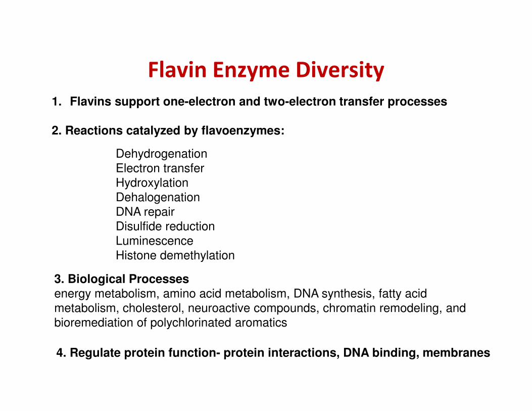
Flavin Enzyme Diversity
1. Flavins support one-electron and two-electron transfer processes
2. Reactions catalyzed by flavoenzymes:
Dehydrogenation
Electron transfer
Hydroxylation
Dehalogenation
DNA repairDNA repair
Disulfide reduction
Luminescence
Histone demethylation
3. Biological Processesenergy metabolism, amino acid metabolism, DNA synthesis, fatty acid
metabolism, cholesterol, neuroactive compounds, chromatin remodeling, and
bioremediation of polychlorinated aromatics
4. Regulate protein function- protein interactions, DNA binding, membranes

Discovery of flavin mononucleotide
(FMN) by a Swedish biochemist
Showed the biochemical basis for riboflavin as a vitamin
O O
N
N
N
N
NH2FAD
Nobel Prize in Physiology or Medicine 1955
Axel Hugo Theodor TheorellN
N
NH
NH3C
H3C
O
O
CH2
CHHO
CHHO
CH
H2C
HO
O P O P O
O-
O O
O-
CH2O
H
OH OH
H
Nobelprize.org
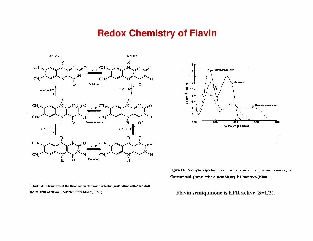
Redox Chemistry of Flavin
Flavin semiquinone is EPR active (S=1/2).

Flavin Redox Switch

ExamplesNifL
Azotobacter vinelandii
PutAEscherichia coli
Vivid (VVD)Neurospora
Circadian clock
ox ox
Nitrogen fixation Proline metabolism
red
red
+ WC-1
WC-1Inhibitory complex
dimer
[carotenoid biosynthesis]
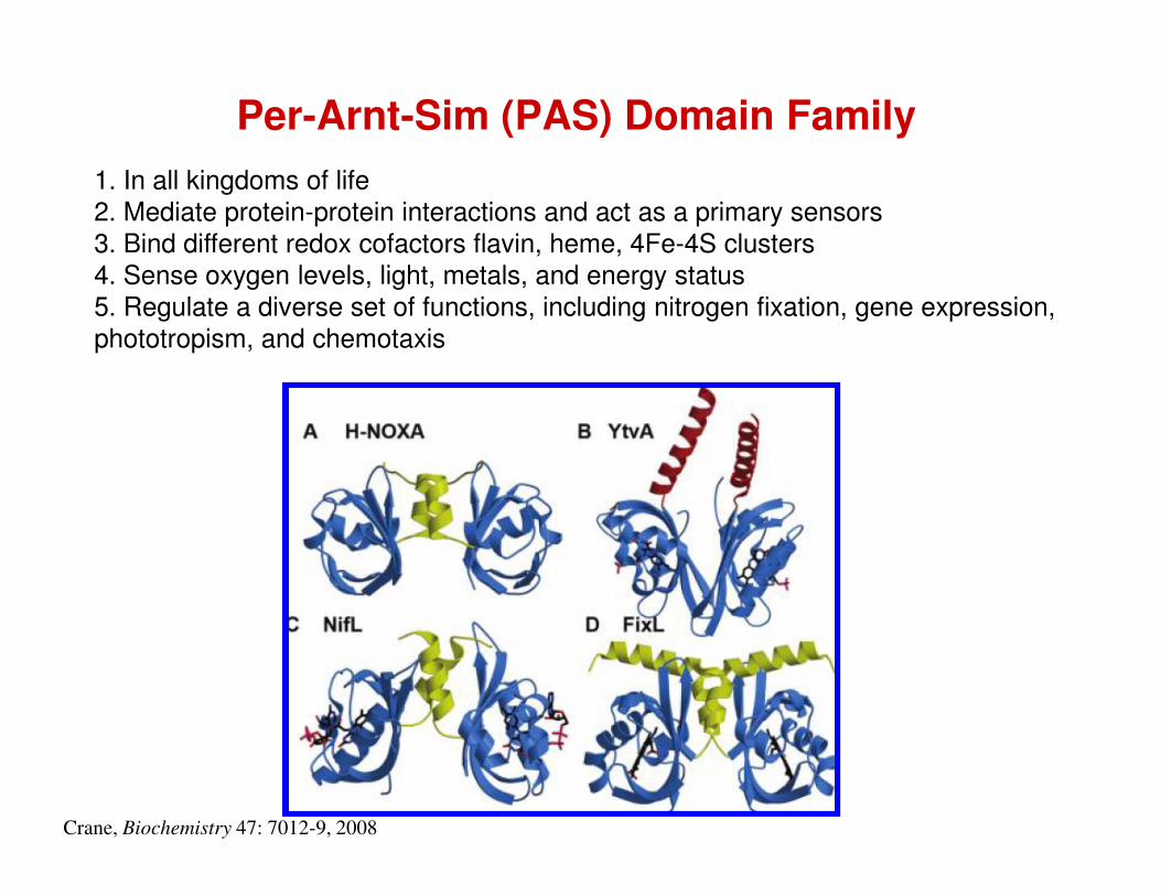
Per-Arnt-Sim (PAS) Domain Family1. In all kingdoms of life
2. Mediate protein-protein interactions and act as a primary sensors
3. Bind different redox cofactors flavin, heme, 4Fe-4S clusters
4. Sense oxygen levels, light, metals, and energy status
5. Regulate a diverse set of functions, including nitrogen fixation, gene expression,
phototropism, and chemotaxis
Crane, Biochemistry 47: 7012-9, 2008
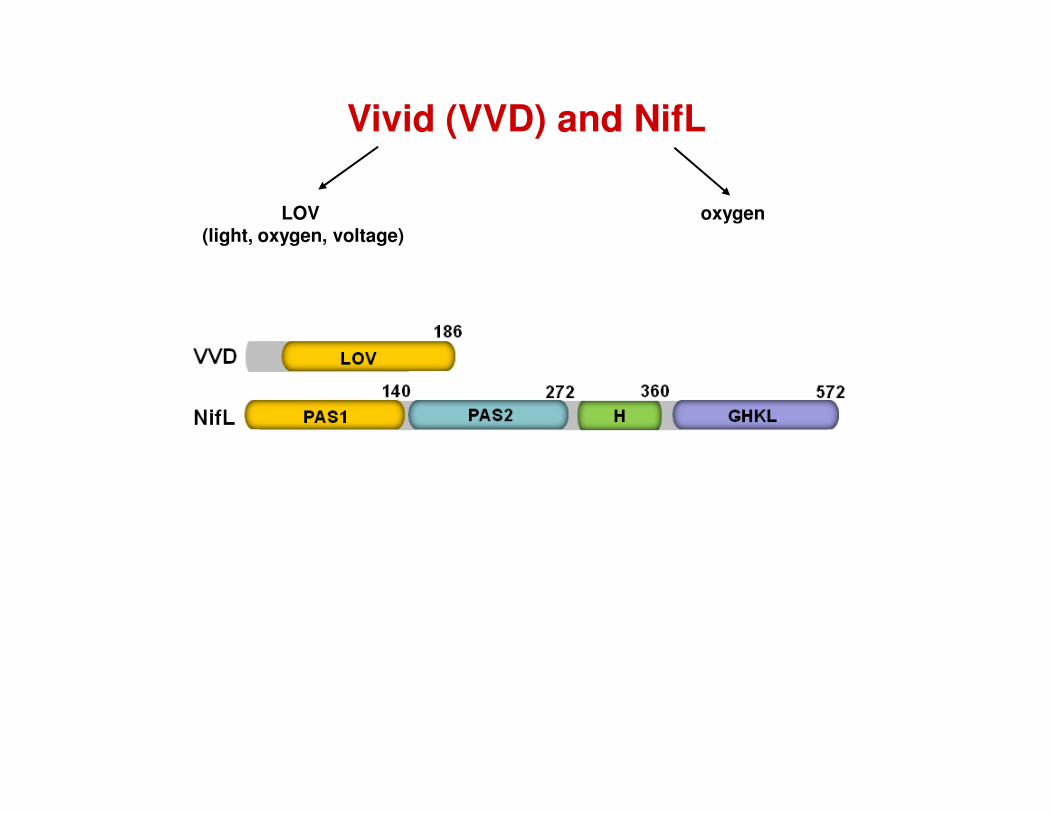
LOV (light, oxygen, voltage)
Vivid (VVD) and NifL
oxygen

VVD
N
N
NH
N
O
O
R
Cys-SH
N
N
NH
N
O
O
R
Cys-S-
N
N
NH
N
O
O
R
H
Moffat, Biochemistry 46: 3614-23, 2007
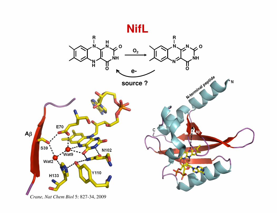
NifL
N
N
NH
N
O
O
R
N
N
NH
N
O
O
R
H
H
O2
(
e-
source ?
Crane, Nat Chem Biol 5: 827-34, 2009
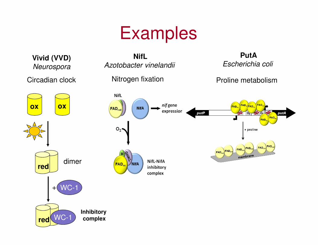
ExamplesNifL
Azotobacter vinelandii
PutAEscherichia coli
Vivid (VVD)Neurospora
Circadian clock
ox ox
Nitrogen fixation Proline metabolism
red
red
+ WC-1
WC-1Inhibitory complex
dimer
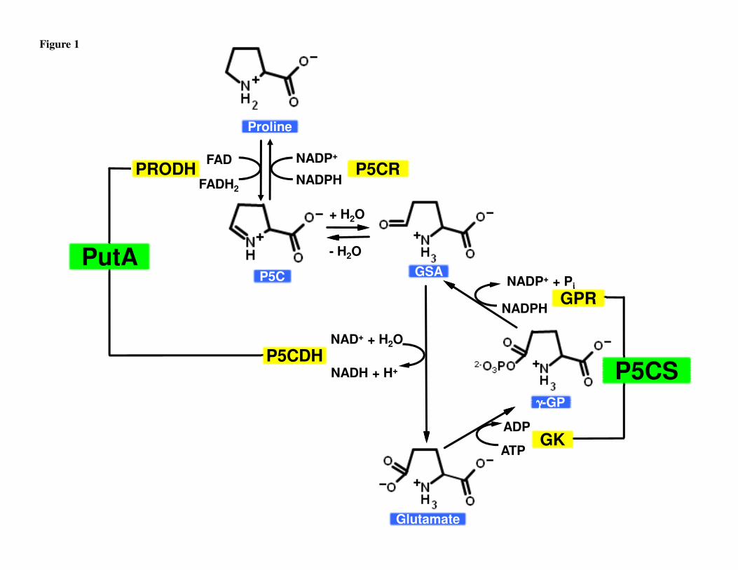
NADP+ + Pi
- H2O
ProlineProline
P5C P5C
P5CR P5CR FAD
NADPH
NADP+
PRODH PRODH
GSA GSA
FADH2
PutAPutA
+ H2O
Figure 1
NADP + Pi
γγγγ-GP γγγγ-GP
ADP
ATP
NADPH
Glutamate Glutamate
P5CDH P5CDH P5CS P5CS
GPR GPR
GK GK
NAD+ + H2O
NADH + H+

Proline Metabolism
PRODH
Ornithine
CitrullineArginineMitochondrion
Cytosol
Urea cycle
P5CDH
OAT
Glutamate
P5C ProlineP5CR
PRODH
NH2
COO-+
NH
COO-+
ProlineP5C
NADPH NADP+
P5CS
P5CDH
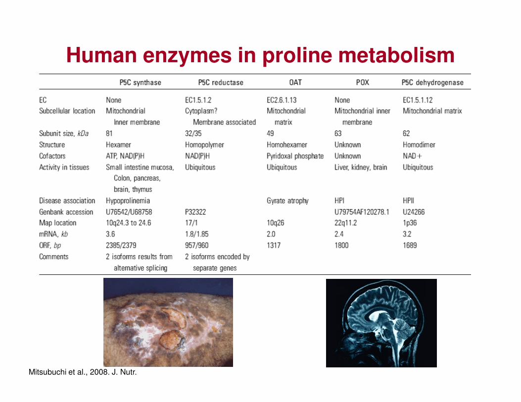
Human enzymes in proline metabolism
Mitsubuchi et al., 2008. J. Nutr.
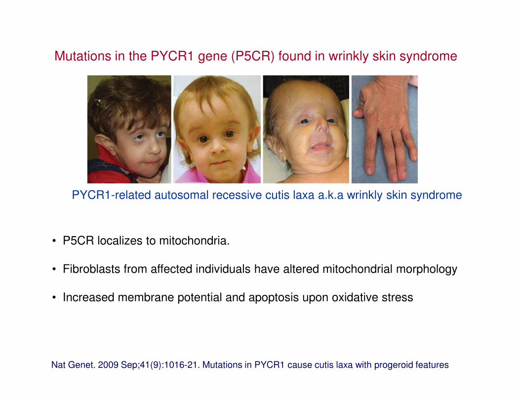
PYCR1-related autosomal recessive cutis laxa a.k.a wrinkly skin syndrome
Mutations in the PYCR1 gene (P5CR) found in wrinkly skin syndrome
Nat Genet. 2009 Sep;41(9):1016-21. Mutations in PYCR1 cause cutis laxa with progeroid features
PYCR1-related autosomal recessive cutis laxa a.k.a wrinkly skin syndrome
• P5CR localizes to mitochondria.
• Fibroblasts from affected individuals have altered mitochondrial morphology
• Increased membrane potential and apoptosis upon oxidative stress
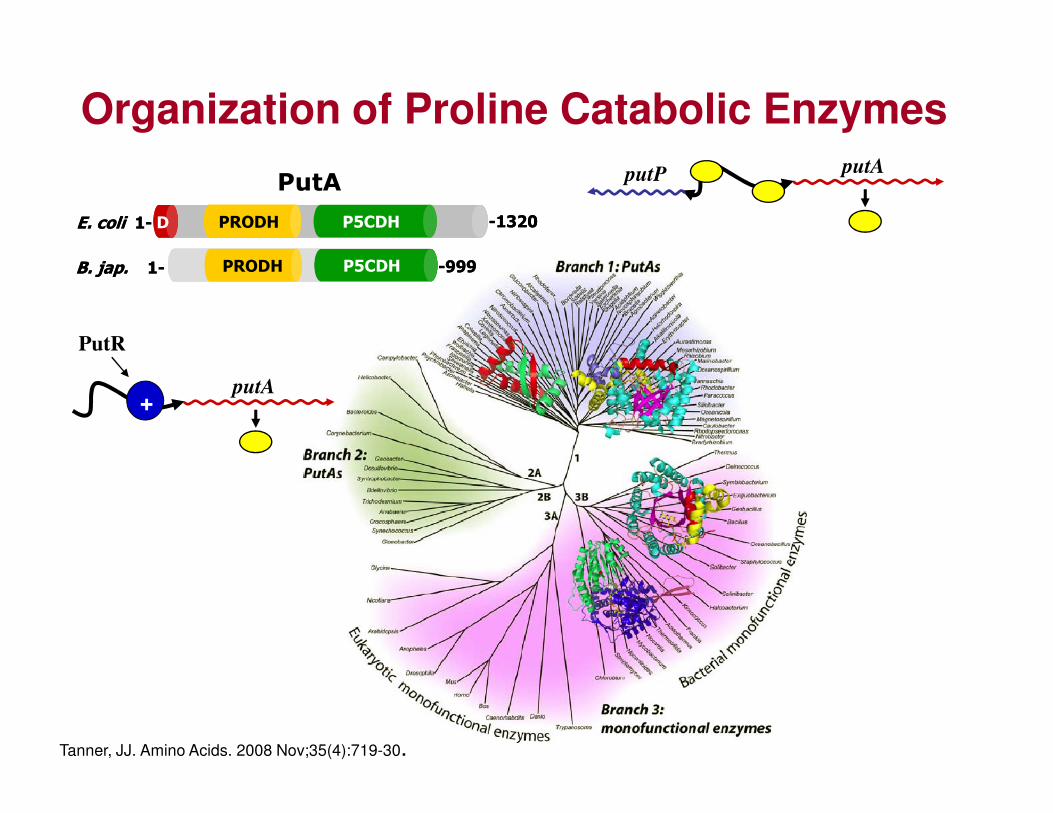
Organization of Proline Catabolic Enzymes
PutA
PRODH P5CDHD
PRODH P5CDH
E. coli 1-
B. jap. 1-
-1320
-999
PRODH P5CDHD
PRODH P5CDH
E. coli 1-
B. jap. 1-
-1320
-999
putP putA
PutR
putA+
Tanner, JJ. Amino Acids. 2008 Nov;35(4):719-30.
+
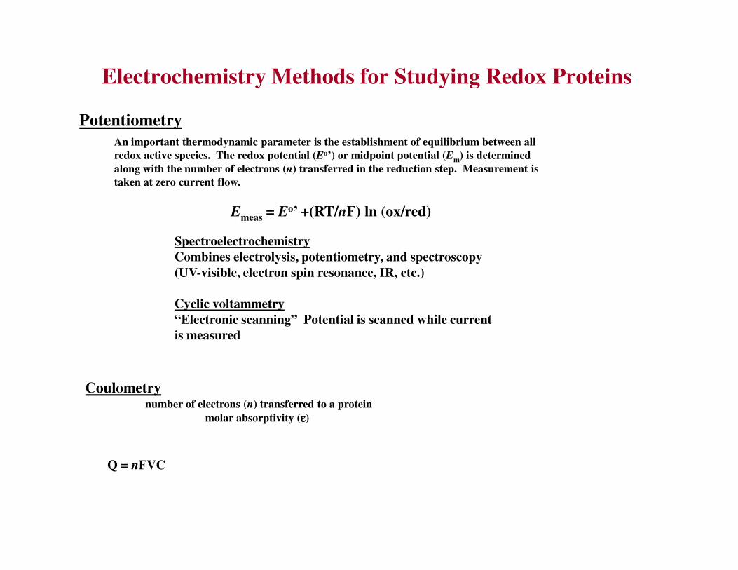
Electrochemistry Methods for Studying Redox Proteins
Potentiometry
An important thermodynamic parameter is the establishment of equilibrium between all
redox active species. The redox potential (Eo’) or midpoint potential (Em) is determined
along with the number of electrons (n) transferred in the reduction step. Measurement is
taken at zero current flow.
Emeas = Eo’ +(RT/nF) ln (ox/red)
Spectroelectrochemistry
Combines electrolysis, potentiometry, and spectroscopy
(UV-visible, electron spin resonance, IR, etc.)
Coulometrynumber of electrons (n) transferred to a protein
molar absorptivity (εεεε)
Q = nFVC
Cyclic voltammetry
“Electronic scanning” Potential is scanned while current
is measured

Prosthetic Groups are buried in ProteinsFAD
Space-Filling
Model

Electrochemical Methods
Emeas=
m(free)+ ln [ox]
[red]RTnF
E
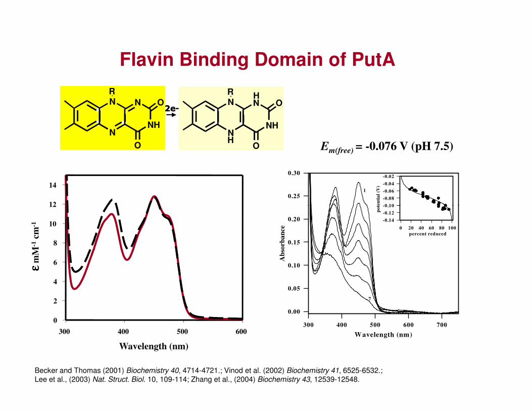
Flavin Binding Domain of PutA
12
14
0.25
0.30
po
ten
tia
l (V
)
-0.10
-0.08
-0.06
-0.04
-0.02
1
2e-
N
N
NH
N
O
OR
H
H
N
N
NH
N
O
OR
2e-
N
N
NH
N
O
OR
H
H
N
N
NH
N
O
OR
Em(free) = -0.076 V (pH 7.5)
Wavelength (nm)
300 400 500 600
0
2
4
6
8
10
12
εε εεm
M-1
cm-1
Becker and Thomas (2001) Biochemistry 40, 4714-4721.; Vinod et al. (2002) Biochemistry 41, 6525-6532.; Lee et al., (2003) Nat. Struct. Biol. 10, 109-114; Zhang et al., (2004) Biochemistry 43, 12539-12548.
Wavelength (nm)
300 400 500 600 700
Ab
sorb
an
ce
0.00
0.05
0.10
0.15
0.20
percent reduced
0 20 40 60 80 100
po
ten
tia
l (V
)
-0.14
-0.12
-0.10
7
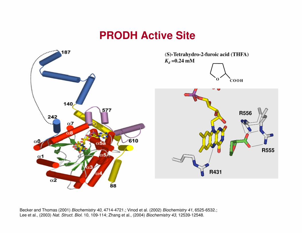
PRODH Active Site
(S)-Tetrahydro-2-furoic acid (THFA)
Kd =0.24 mM
O CO O H
R556
Becker and Thomas (2001) Biochemistry 40, 4714-4721.; Vinod et al. (2002) Biochemistry 41, 6525-6532.; Lee et al., (2003) Nat. Struct. Biol. 10, 109-114; Zhang et al., (2004) Biochemistry 43, 12539-12548.
R555
R431

Structures of PutA
E. coli
PRODH P5CDHD1- -1320PRODH P5CDHD1- -1320
P5CDH
PRODH PRODH
P5CDH
PRODH PRODH
PRODH P5CDH1- -999PRODH P5CDH
B. jap.B. jap.
1- -999
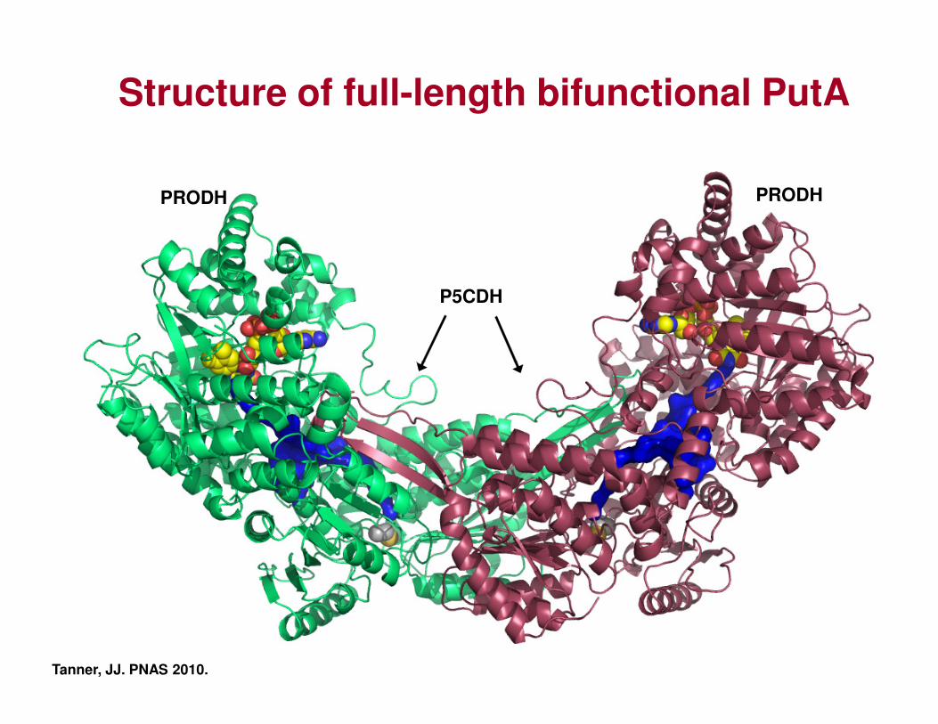
Structure of full-length bifunctional PutA
P5CDH
PRODH PRODH
P5CDH
PRODH PRODH
Tanner, JJ. PNAS 2010.

Strategy for testing channeling
P5CDH
PRODH
P5C
GSAGlu
NADHNAD+
P5CDH P5CDH
wt BjPutA
Non-channeling BjPutA variants
Pro
PRODH
P5CDH P5CDH P5CDH P5CDH
Non-channeling BjPutA variants
C792A R456M

Evidence for channeling in BjPutA
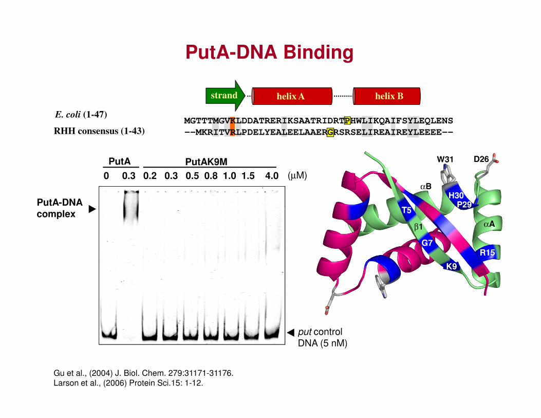
PutA-DNA Binding
PutA
0 0.3 0.2 0.3 0.5 0.8 1.0 1.5 4.0PutAK9M
E. coli (1-47)
RHH consensus (1-43)
helix A helix Bstrand
MGTTTMGVKLDDATRERIKSAATRIDRTPHWLIKQAIFSYLEQLENS
--MKRITVRLPDELYEALEELAAERGRSRSELIREAIREYLEEEE--
(µM)
PutA-DNA complex
put control DNA (5 nM)
complex
Gu et al., (2004) J. Biol. Chem. 279:31171-31176.
Larson et al., (2006) Protein Sci.15: 1-12.

[PutA, µµµµM] 0 0.25 0 0.25 0.5 0.9
wt DNA ∆∆∆∆12345 DNA
PutA-
Identification of DNA Binding Sites
GTTGCA GTCATA
- + - + - + - + - + - +∆5 ∆4 ∆3 ∆2 ∆1 WTEcPutA52
(500 nM)
DNA-Binding
Sites
putP putA1 2 3 4 5
put
control
DNA,
2 nM
PutA-
DNA
complex
put DNA
(2 nM)
PutA52-DNA
Complexes
Zhou et al., J Mol Biol. 2008 Aug 1;381(1):174-88.
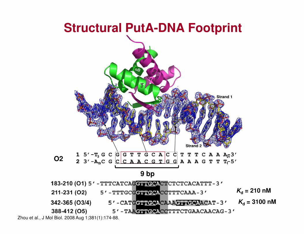
Structural PutA-DNA Footprint
9 bp
Kd = 210 nM
Kd = 3100 nM
O2
Zhou et al., J Mol Biol. 2008 Aug 1;381(1):174-88.
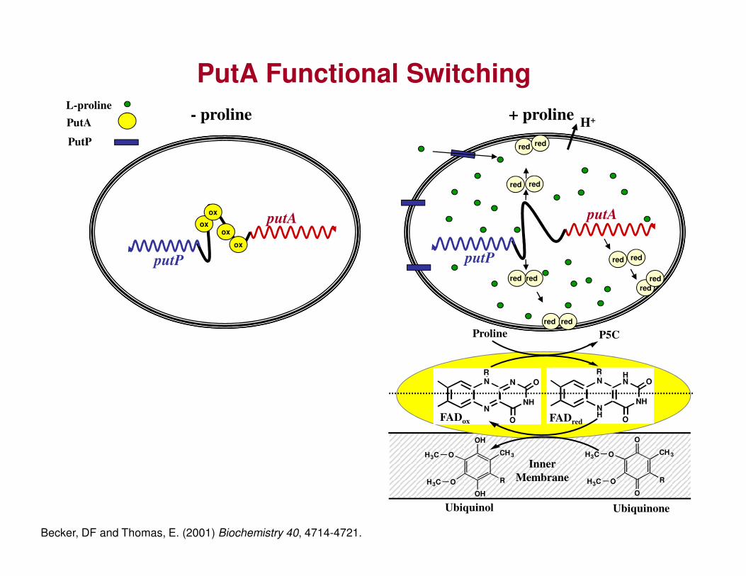
PutA Functional SwitchingL-proline
PutP
PutA
red red
H+
red red
red redputP
putA
redred
red red
ox
ox
ox
ox
putP
putA
- proline + proline
red red
red
UbiquinoneUbiquinol
O
O
CH 3
R
O
O
H3C
H3C
OH
OH
CH 3
R
O
O
H3C
H3C
NH
N
NH
HN
O
OR
N
N
NH
N
O
OR
FADox FADred
P5CProline
Inner
Membrane
Becker, DF and Thomas, E. (2001) Biochemistry 40, 4714-4721.
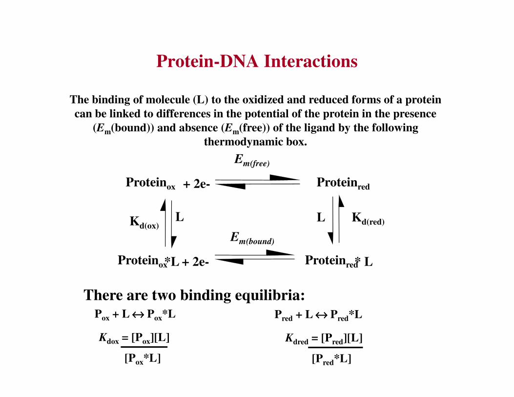
Protein-DNA Interactions
The binding of molecule (L) to the oxidized and reduced forms of a protein
can be linked to differences in the potential of the protein in the presence
(Em(bound)) and absence (Em(free)) of the ligand by the following
thermodynamic box.
Em(free)
+ 2e-Proteinox Proteinred
Kd(ox)L L
Em(bound)
Kd(red)
*L + 2e-Proteinox * L Proteinred
There are two binding equilibria:Pox + L ↔↔↔↔ Pox*L Pred + L ↔↔↔↔ Pred*L
Kdox = [Pox][L]
[Pox*L]
Kdred = [Pred][L]
[Pred*L]

PutA Redox Properties in the Presence
of put Intergenic DNA
0.10
0.15
0.20
percent reduced0 20 40 60 80 100
pote
nti
al
(V)
-0.14
-0.12
-0.10
-0.08
-0.06
-0.04
-0.02
1
Ab
sorb
an
ce
2 e-PutAox PutAred
Em = -76 mV
Em(bound) = -0.086 V (pH 7.5)
Wavelength (nm)
300 400 500 600 700
0.00
0.05
0.10
8
Wavelength (nm)
Ab
sorb
an
ce
PutAox-DNA
Kd = 45 nM DNA DNA
2 e-
2 e-
PutAred-DNA
Em = -86 mV
Kd = 98 nM
Becker and Thomas. (2001) Biochemistry 40, 4714-4721.
Only ~ 2-fold increase in Kd

Proline Dependent Binding to Lipid Bilayers (Surface Plasmon Resonance Study)
Res
pon
se (
RU
)
Time (s)
40
60
80
100
120
PutA + proline (5 mM)
Res
pon
se (
RU
)
-100 0 100 200 300 400 500
-40
-20
0
20
Time (s)
PutA (ox)
Zhang and Becker (2004) Biochemistry 43, 13165-13174.; Zhang et al., (2007) Biochemistry 46:483-91.
Sensorgrams of oxidized PutA (20 nM) and PutA (20 nM) with 5 mM
proline binding on E. coli polar extract lipids. The arrows indicate the
starting and ending of injection of protein sample. Buffer: 10 mM
HEPES, 150 mM NaCl, pH 7.4
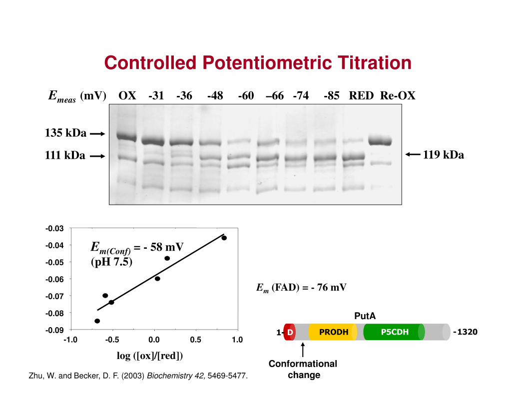
Controlled Potentiometric Titration
Emeas (mV) OX -31 -36 -48 -60 –66 -74 -85 RED Re-OX
135 kDa
119 kDa111 kDap
ote
nti
al
(V)
log ([ox]/[red])
-1.0 -0.5 0.0 0.5 1.0-0.09
-0.08
-0.07
-0.06
-0.05
-0.04
-0.03
Em(Conf) = - 58 mV
(pH 7.5)
Em (FAD) = - 76 mV
Zhu, W. and Becker, D. F. (2003) Biochemistry 42, 5469-5477.
PRODH P5CDHD1- -1320
PutA
- -
Conformational change

Trp 211Ser 216Arg234
Flu
ore
scen
ce in
ten
sity
(au
)
150
200
250
oxB 1
2
Fl (
au)
0.08
0.12
0.16
Kinetics of Conformational Change
PRODH P5CDHD1- -1320
E. coli
- -
Wavelength (nm)
300 350 400 450 500F
luo
resc
ence
inte
nsi
ty (
au)
0
50
100
150
red 0 2 4 6 8 100.04
Time (s)
0.6 s-1
Zhu and Becker, (2005) Biochemistry 44, 12297.
E-FADox + S E-FADoxS E-FADredPslow
DCPIPoxDCPIPred
fast
fast
E*-FADredP membrane
binding
55 s-1

FAD Conformational Changes
THFA-Bound Dithionite reduced crystal
Zhang et al., (2007) Biochemistry 46:483-91.
Kinetic Properties of Reconstituted and Mutant PutA Enzymes
Enzyme Km (mM)
kcat (s-1)
kcat/Km (s-1M-1)
Wild-type 100 8 80
5-deaza-FAD NA (< 1 %) NA
R431M 174 1.2 7
2-deoxy-FAD 103 4 39
R556M > 1 M 1.4 < 1.4
a Parameters were estimated by best-fit analysis to the Michaelis-Menten equation.

N(5) Position is Critical
putC:lacZ
PutA + pro
- PutA
PutA R431M
Zhang et al., (2007) Biochemistry 46:483-91.
.25
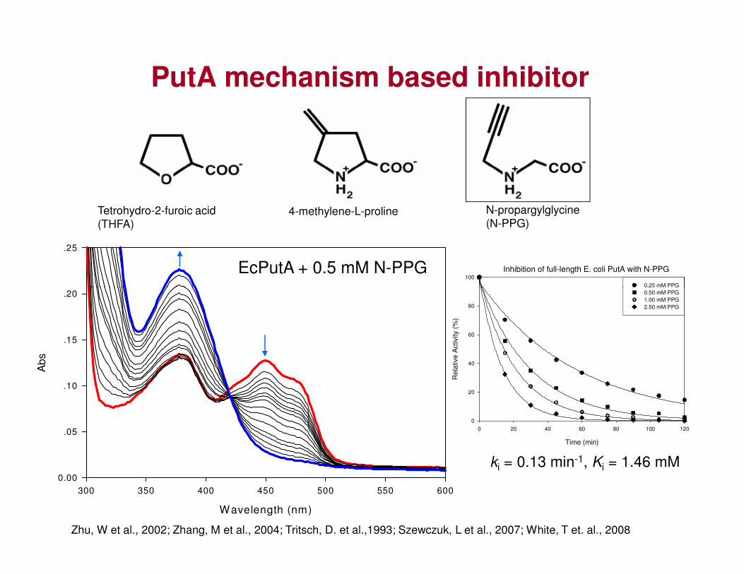
PutA mechanism based inhibitor
4-methylene-L-proline N-propargylglycine
(N-PPG)Tetrohydro-2-furoic acid
(THFA)
EcPutA + 0.5 mM N-PPG Inhibition of full-length E. coli PutA with N-PPG100
0.25 mM PPG
Wavelength (nm)
300 350 400 450 500 550 600
Ab
s
0.00
.05
.10
.15
.20
Zhu, W et al., 2002; Zhang, M et al., 2004; Tritsch, D. et al.,1993; Szewczuk, L et al., 2007; White, T et. al., 2008
Time (min)
0 20 40 60 80 100 120
Re
lative A
ctivity (
%)
0
20
40
60
80
0.25 mM PPG
0.50 mM PPG
1.00 mM PPG
2.50 mM PPG
ki = 0.13 min-1, Ki = 1.46 mM

Structure of PPG inactivated PutA86-630
• Covalent linkage formed between K329, N-PPG and N5 of FAD,
• Flavin isoalloxazine butterfly bending
• 2-OH’ group of ribityl moiety rotates 90°C and forms new H-bond to N1-FAD
Lys3
29

Proposed mechanism of inactivation
SDS treated

PPG mimics proline reduced PutA
M
200 kD
150 kD
100 kD
PutA OX PRO THFA PPG
119 kD
Limited proteolysis Liposome binding
75 kD
50 kD
90 kD

Oxidized and PPG Structures
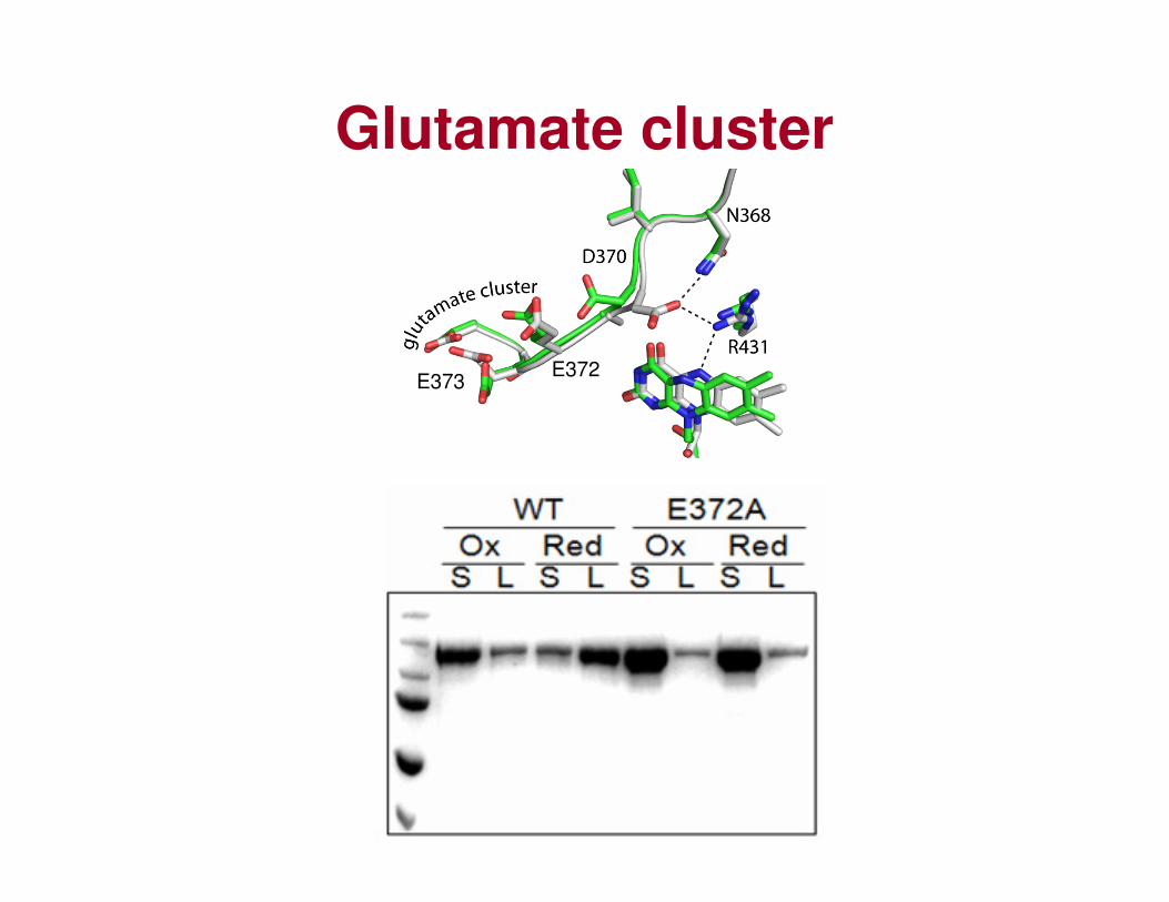
Glutamate cluster
E372E373

C-terminal membrane binding domain ?
E.coli AR----GESNILLERLYIERSLSVNTAAAGGNASLMTIG- 1320 E.coli AR----GESNILLERLYIERSLSVNTAAAGGNASLMTIG- 1320
S.typhimurium AR----GESNILLERLYIERSLSVNTAAAGGNASLMTIG- 1319
K.pneumoniae AR----GETNLLLERLYIERSLSVNTAAAGA--------- 1312
Y.enterocolitica AR----GETNILLERLLIEHSLSVNTAAAGGNASLMTIG- 1323
P.luminescens AR----GETNLLLERLLHERSLSINTAAAGGNASLMTIG- 1326
P.syringae SH----GETNVPLERLVIERALSVNTAAAGGNASLMTIG- 1300
P.putida SS----GDHQIALERLVIERAVSVNTAAAGGNASLMTIG- 1317
R.solanacearum PH----GGQGLALERLLIERSLSVNTAAAGGNASLMTIG- 1325
B.pertussis SADALAAGASYAPDRLLAERSISVNTAAAGGNASLMTIG- 1273
B.japonicum -----------------TEQTVTINTAAAGGNAALLAGEE 999
~ 30 Å
~ 15 ÅYL L RL V AS
hydrocarbon core
polar solvent
inte
rfac
e
Zhou et al., Amino Acids. 2008 Nov;35(4):711-8.
PutA(1-1308)DNA binding (yes)
PRODH activity (yes)
P5CDH Activity (yes)
Substrate Channeling (yes)
Membrane binding (NO)
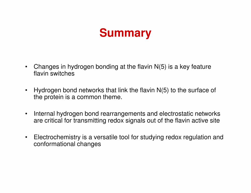
Summary
• Changes in hydrogen bonding at the flavin N(5) is a key feature flavin switches
• Hydrogen bond networks that link the flavin N(5) to the surface of the protein is a common theme.the protein is a common theme.
• Internal hydrogen bond rearrangements and electrostatic networks are critical for transmitting redox signals out of the flavin active site
• Electrochemistry is a versatile tool for studying redox regulation and conformational changes


![Complementing Nanoscale Galvanic Exchange with Redox ... · [2] Galvanic exchange is a classic electrochemical redox process thermodynamically driven by the difference in the redox](https://static.documents.pub/doc/80x56/5f6916c0235e487474390740/complementing-nanoscale-galvanic-exchange-with-redox-2-galvanic-exchange-is.jpg)
![Supplementary Informations Calix[4]phyrin based redox ... · 1 Supplementary Informations Calix[4]phyrin based redox architectures: towards new molecular tools for electrochemical](https://static.documents.pub/doc/80x56/5fe0a043f1a067315a77d374/supplementary-informations-calix4phyrin-based-redox-1-supplementary-informations.jpg)


