Sensors and Actuators A 150 (2009) 280–285 Contents lists available at ScienceDirect Sensors and Actuators A: Physical journal homepage: www.elsevier.com/locate/sna Flexible and disposable immunosensors based on layer-by-layer self-assembled carbon nanotubes and biomolecules Miao Lu, Dongjin Lee, Wei Xue, Tianhong Cui ∗ Department of Mechanical Engineering, University of Minnesota, Twin Cities, 111 Church Street SE, Minneapolis, MN 55455, USA article info Article history: Received 7 September 2008 Received in revised form 23 December 2008 Accepted 24 December 2008 Available online 9 January 2009 Keywords: Single-walled carbon nanotube (SWNT) Layer-by-layer (LbL) self-assembly Immunosensor MEMS abstract A low-cost, flexible, and disposable immunosensor is presented in this paper. The single-walled carbon nanotubes (SWNTs) and biomolecules are self-assembled between two micro-patterned electrodes. The immuno-chip acts as a platform of a horseradish peroxidase (HRP) labeled sandwiched Enzyme-Linked ImmunoSorbent Assay (ELISA). The pH change induced by the biochemical reactions influences the electri- cal conductance of SWNT. A detection resolution of 0.4 ng/ml (2.5 pM) for normal rabbit immunoglobulin G (IgG) is demonstrated. The new fabrication technique and the HRP labeled detection protocol can be extended to the recognition of other antigens for critical applications to clinical diagnosis, food toxin detection, and environment monitoring. Published by Elsevier B.V. 1. Introduction Immunosensors transduce antigen–antibody interactions directly into physical signals, very suitable for applications includ- ing genomics, food toxin detection, and environmental monitoring. Immunosensors have been a main focus of extensive research on bimolecular recognition based on the capability of antibodies to bind various important antigens with high affinity and specificity. Labeled and label-free immunosensor are the two main cate- gories of the reported immunochips. Label-free immunosensors benefit from higher resolution, but rely on special process and suffer from poor yield and repeatability [1]. Currently, for most applications immunosensors use a label to increase the sensitiv- ity of detection. Enzyme labels are very valuable because they can be used to efficiently catalyze the conversion of a substance into a detectable product, and the number of detectable molecules can be exponentially higher than the number of antigens. The most commonly observed form of detection is fluorescence due to its high sensitivity and stability. Although some efforts have been made to realize a compact and rapid detection system [2], fluorescence-labeled immunosensors are still too complex to be used. Electrochemical immunosensors have received much attention in on-site applications because they provide a simple, ∗ Corresponding author. Tel.: +1 612 626 1636; fax: +1 612 625 6069. E-mail address: [email protected](T. Cui). inexpensive, and accurate measurement of antigens based on either potential, current, capacitance, or conductance change caused by a specific bio-recognition reaction [3]. However, the current electro- chemical immunosensors under investigation are somehow fragile, expensive or non-biocompatible due to the material nature and fabrication complexity. Single-walled carbon nanotubes (SWNTs) are one of the most promising candidates for the development of immunosensors due to their unique and well-defined electrical and mechanical prop- erties [4,5]. Yu et al. recently reported a sandwich assay on a CNT forest electrode, and such a system could detect prostate specific antigen (PSA) at a level of 4 pg/ml [6]. Cui et al. reported a electro- chemical immunosensor using gold nanoparticles (GNPs)/carbon nanotubes (CNTs) hybrids platform with a horseradish peroxi- dase (HRP)-functionalized gold nanoparticle label for the sensitive detection of human IgG (HIgG) with a detection limit of 40 pg/ml [7], but the reported CNT based immunosensors are still suffered from expensive process and poor stability. In general, the electrical performance of the CNT films will drift with the microfluidic shock after the immersing of the CNT film in biological solutions. To overcome the above hurdles, a low-cost, flexible, and dis- posable HRP-labeled immunosensor platform is presented [8].A polymethylmethacrylate (PMMA) dielectric layer was coated on the surface of the self-assembled carbon nanotube composite film to shield the microfluidic shock and ion penetration in the substrate solution. The conductance change of the CNT film with different concentrations of target antigen is demonstrated. 0924-4247/$ – see front matter. Published by Elsevier B.V. doi:10.1016/j.sna.2008.12.021
Transcript
Fsa
MD
a
ARR2AA
KSLIM
1
diIbb
gbsaicicmtb[ta
0d
Sensors and Actuators A 150 (2009) 280–285
Contents lists available at ScienceDirect
Sensors and Actuators A: Physical
journa l homepage: www.e lsev ier .com/ locate /sna
lexible and disposable immunosensors based on layer-by-layerelf-assembled carbon nanotubesnd biomolecules
iao Lu, Dongjin Lee, Wei Xue, Tianhong Cui ∗
epartment of Mechanical Engineering, University of Minnesota, Twin Cities, 111 Church Street SE, Minneapolis, MN 55455, USA
r t i c l e i n f o
rticle history:eceived 7 September 2008eceived in revised form3 December 2008
a b s t r a c t
A low-cost, flexible, and disposable immunosensor is presented in this paper. The single-walled carbonnanotubes (SWNTs) and biomolecules are self-assembled between two micro-patterned electrodes. Theimmuno-chip acts as a platform of a horseradish peroxidase (HRP) labeled sandwiched Enzyme-LinkedImmunoSorbent Assay (ELISA). The pH change induced by the biochemical reactions influences the electri-
ccepted 24 December 2008vailable online 9 January 2009
cal conductance of SWNT. A detection resolution of 0.4 ng/ml (2.5 pM) for normal rabbit immunoglobulinG (IgG) is demonstrated. The new fabrication technique and the HRP labeled detection protocol can beextended to the recognition of other antigens for critical applications to clinical diagnosis, food toxindetection, and environment monitoring.
Published by Elsevier B.V.
EMS
. Introduction
Immunosensors transduce antigen–antibody interactionsirectly into physical signals, very suitable for applications includ-
ng genomics, food toxin detection, and environmental monitoring.mmunosensors have been a main focus of extensive research onimolecular recognition based on the capability of antibodies toind various important antigens with high affinity and specificity.
Labeled and label-free immunosensor are the two main cate-ories of the reported immunochips. Label-free immunosensorsenefit from higher resolution, but rely on special process anduffer from poor yield and repeatability [1]. Currently, for mostpplications immunosensors use a label to increase the sensitiv-ty of detection. Enzyme labels are very valuable because theyan be used to efficiently catalyze the conversion of a substancento a detectable product, and the number of detectable moleculesan be exponentially higher than the number of antigens. Theost commonly observed form of detection is fluorescence due
o its high sensitivity and stability. Although some efforts have
een made to realize a compact and rapid detection system2], fluorescence-labeled immunosensors are still too complexo be used. Electrochemical immunosensors have received muchttention in on-site applications because they provide a simple,
924-4247/$ – see front matter. Published by Elsevier B.V.oi:10.1016/j.sna.2008.12.021
inexpensive, and accurate measurement of antigens based on eitherpotential, current, capacitance, or conductance change caused by aspecific bio-recognition reaction [3]. However, the current electro-chemical immunosensors under investigation are somehow fragile,expensive or non-biocompatible due to the material nature andfabrication complexity.
Single-walled carbon nanotubes (SWNTs) are one of the mostpromising candidates for the development of immunosensors dueto their unique and well-defined electrical and mechanical prop-erties [4,5]. Yu et al. recently reported a sandwich assay on a CNTforest electrode, and such a system could detect prostate specificantigen (PSA) at a level of 4 pg/ml [6]. Cui et al. reported a electro-chemical immunosensor using gold nanoparticles (GNPs)/carbonnanotubes (CNTs) hybrids platform with a horseradish peroxi-dase (HRP)-functionalized gold nanoparticle label for the sensitivedetection of human IgG (HIgG) with a detection limit of 40 pg/ml[7], but the reported CNT based immunosensors are still sufferedfrom expensive process and poor stability. In general, the electricalperformance of the CNT films will drift with the microfluidic shockafter the immersing of the CNT film in biological solutions.
To overcome the above hurdles, a low-cost, flexible, and dis-posable HRP-labeled immunosensor platform is presented [8]. A
polymethylmethacrylate (PMMA) dielectric layer was coated on thesurface of the self-assembled carbon nanotube composite film toshield the microfluidic shock and ion penetration in the substratesolution. The conductance change of the CNT film with differentconcentrations of target antigen is demonstrated.
M. Lu et al. / Sensors and Actuators A 150 (2009) 280–285 281
catal
2
bbi
tbaahamod
Fpw
Fig. 1. (a) The structure of SWNT immunosensor, (b) HRP
. Immunosensing principle
The self-assembled immunosensor platform in this paper isased on the principle of sandwiched Enzyme-Linked ImmunoSor-ent Assay (ELISA) with an incorporation of new nano-materials
nto the structure design, as shown in Fig. 1(a).The capturing antibody is immobilized on the solid support, and
he antigen is added to specifically bind to the immobilized anti-ody. The labeled secondary antibody is allowed to bind with thentigen and to make the sandwich structure. Two types of antibodyre required to have different epitopes on the antigen in order not to
inder the binding with each other. The secondary antibody used ishorseradish peroxidase (HRP) conjugated antibody. When 200 �lixture of ascorbic acid and hydrogen peroxide (H2O2) was dipped
n the surface of the chip (Fig. 1(c)), the ascorbic acid is oxidized toehydroascorbic acid (Fig. 1(b)) by the catalytic behavior of HRP as
ig. 2. The process flow of the biochip: (a) an Omnicoat layer and a SU8-3050 layer were satterned. (c) The wafer was immersed in developer overnight to peel off the SU8-3050 layere self-assembled and patterned by lift-off process. (e) PMMA 300 nm thick was spun a
yzed ascorbic acid oxidization, and (c) testing apparatus.
shown in following reactions:
HRP + H2O2 → CompoundI + H2O (i)
2CompoundI + AscorbicAcid
→ 2CompoundII + DehydroascorbicAcid (ii)
2CompoundII + AscorbicAcid
→ HRP + H O + DehydroascorbicAcid (iii)
2
Early study shows that the majority carriers in the layer-by-layer self-assembled SWNT films are holes because SWNT is ap-type material [5]. The structure, as shown in Fig. 1, is a p-typeion sensitive FET. The conductance in the SWNT channel will be
pin-coated and UV exposed on a Si/SiO2 wafer. (b) Cr/Au layers were deposited ander 150 �m thick. (d) Two bi-layers of (PDDA/PSS) and five bi-layers of (PDDA/SWNT)nd patterned as a passivation layer.
282 M. Lu et al. / Sensors and Actuators A 150 (2009) 280–285
F right:( surfa
coostom
3
3
CtSp(P0m1C(drca
3
b
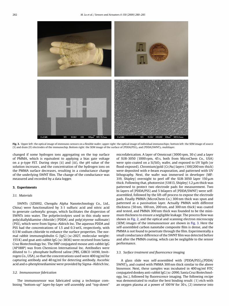
ig. 3. Upper left: the optical image of immnuno-sensors on a flexible wafer; upperS) and drain (D) electrodes of the immunochip. Bottom right: the SEM image of the
hanged if some hydrogen ions aggregating on the top surfacef PMMA, which is equivalent to applying a bias gate voltagen a p-type FET. During steps (ii) and (iii), the pH value of theolution increases, and the concentration of the hydrogen ions onhe PMMA surface decreases, resulting in a conductance changef the underlying SWNT film. The change of the conductance waseasured and recorded by a data logger.
. Experiments
.1. Materials
SWNTs (SZS002, Chengdu Alpha Nanotechnology Co., Ltd.,hina) were functionalized by 3:1 sulfuric acid and nitric acido generate carboxylic groups, which facilitates the dispersion ofWNTs into water. The polyelectrolytes used in this study wereoly(diallyldiamine chloride) (PDDA) and poly(styrene sulfonate)PSS), which were from Sigma–Aldrich Inc. The aqueous PDDA andSS had the concentrations of 1.5 and 0.3 wt%, respectively, with.5 M sodium chloride to enhance the surface properties. The nor-al rabbit immunoglobulin G (IgG) (sc-2027, molecular weight:
55 K) and goat anti-rabbit IgG (sc-3836) were received from Santaruz Biotechnology Inc. The HRP conjugated mouse anti-rabbit IgGAP188P) was from Chemicon International Inc. Antibodies wereiluted to 1× phosphate buffered saline (PBS, GIBCO 14190, Invit-ogen Co., USA), so that the concentrations used were 400 ng/ml forapturing antibody and 40 ng/ml for detecting antibody. Ascorbiccid and o-phenylenediamine were provided by Sigma–Aldrich Inc.
.2. Immunosensor fabrication
The immunosensor was fabricated using a technique com-ining “bottom-up” layer-by-layer self-assembly and “top-down”
the optical image of individual immunochips; bottom left: the SEM image of sourcece of (PDDA/PSS)2 and (PDDA/SWNT)5 multilayer.
microfabrication. A layer of Omnicoat (3000 rpm, 30 s) and a layerof SU8-3050 (1000 rpm, 45 s, both from MicroChem Co., USA)were spin-coated on a Si/SiO2 wafer, and exposed to UV ligth (orflood-exposed). Chromium/gold (Cr/Au) layers (100/200 nm thick)were deposited with e-beam evaporation, and patterned with UVlithography. Next, the wafer was immersed in developer (MF-319, Shipley) overnight to peel off the SU8-3050 layer 150 �mthick. Following that, photoresist (S1813, Shipley) 1.2 �m thick waspatterned to protect two electrode pads for measurement. Twobi-layers of (PDDA/PSS) and 5 bilayers of (PDDA/SWNT) were self-assembled, followed by the lift-off process to expose the electrodepads. Finally PMMA (MicroChem Co.) 300 nm thick was spun andpatterned as a passivation layer. Actually PMMA with differentthickness (50 nm, 100 nm, 200 nm, and 300 nm thick) was coatedand tested, and PMMA 300 nm thick was founded to be the mini-mum thickness to ensure a negligible leakage. The process flow wasshown in Fig. 2, and the optical and scanning electron microscopy(SEM) images of the immunosensor are shown in Fig. 3. Here theself-assembled carbon nanotube composite film is dense, and thePMMA is not found to penetrate through the film. Experimentally asmall conductance difference of the SWNT film was detected beforeand after the PMMA coating, which can be negligible to the sensorperformance.
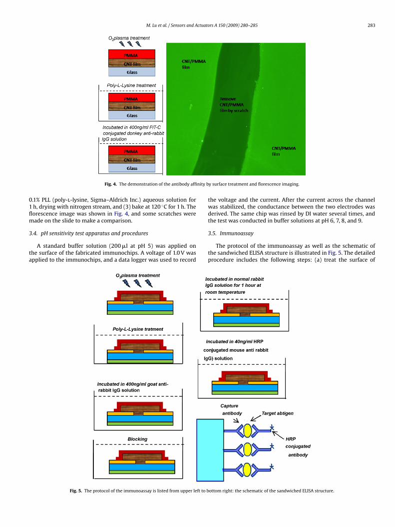
3.3. Surface treatment and fluorescence imaging
A glass slide was self-assembled with (PDDA/PSS)2(PDDA/SWNT)5 and coated with PMMA 300 nm thick similar to the above
biosensor. Next, these samples was incubated in 400 ng/ml FITCconjugated donkey anti-rabbit IgG (sc-2090, Santa Cruz Biotechnol-ogy, Inc.), followed by fluorescence imaging. The following recipewas demonstrated to realize the best binding result: (1) etch withan oxygen plasma at a power of 100 W for 30 s, (2) immerse into
M. Lu et al. / Sensors and Actuators A 150 (2009) 280–285 283
nity b
01flm
3
ta
Fig. 4. The demonstration of the antibody affi
.1% PLL (poly-l-lysine, Sigma–Aldrich Inc.) aqueous solution forh, drying with nitrogen stream, and (3) bake at 120 ◦C for 1 h. Theorescence image was shown in Fig. 4, and some scratches wereade on the slide to make a comparison.
.4. pH sensitivity test apparatus and procedures
A standard buffer solution (200 �l at pH 5) was applied onhe surface of the fabricated immunochips. A voltage of 1.0 V waspplied to the immunochips, and a data logger was used to record
Fig. 5. The protocol of the immunoassay is listed from upper left to b
y surface treatment and florescence imaging.
the voltage and the current. After the current across the channelwas stabilized, the conductance between the two electrodes wasderived. The same chip was rinsed by DI water several times, andthe test was conducted in buffer solutions at pH 6, 7, 8, and 9.
3.5. Immunoassay
The protocol of the immunoassay as well as the schematic ofthe sandwiched ELISA structure is illustrated in Fig. 5. The detailedprocedure includes the following steps: (a) treat the surface of
ottom right: the schematic of the sandwiched ELISA structure.
284 M. Lu et al. / Sensors and Actuators A 150 (2009) 280–285
Fig. 6. pH-dependent conductance of the flexible SWNT immunosensor.
Fn
i(gftsisfI
Fc
ig. 7. The conductance changes of the devices with different coating. At the begin-ing, the devices were immersed into the substrate solution.
mmunosensors with oxygen plasma at a power of 100 W for 30 s;b) immerse into 0.1% PLL aqueous solution for 1 h, dry with nitro-en stream, and bake at 120 ◦C for 1 h; (c) incubate immunochipsor overnight at 4 ◦C in goat anti-rabbit IgG solution at a concen-ration of 400 ng/ml; (d) rinse with PBS solution 3 times using ahaker (100 rpm, 19 mm circle) for 10 min each time; (e) immerse
nto 3% bovine serum albumin (BSA, Sigma–Aldrich Inc.) blockingolution for 3 h at room temperature and rinse as (d); (f) incubateor 1 h at room temperature in the target antigen (normal rabbitgG) diluted into 1% BSA/PBS solution to make 0, 0.4, 4, 40, and
ig. 8. The normalized conductance of the immunosensors at different antigen con-entrations.
Fig. 9. The calibration curve of SWNT conductance drifts at different antigen con-centrations.
400 ng/ml, respectively, followed by rinse as in step (d); (g) incu-bate for 1 h at room temperature in the detecting antibody (HRPconjugated mouse anti-rabbit IgG) solution diluted into 1% BSA/PBSsolutions to be 40 ng/ml, followed by rinsing.
3.6. Immunosensing test apparatus and procedures
The mixture of 1.0 mM ascorbic acid and 1.0 mM o-phenylenediamine was produced by diluting to PB2 solution(1 mM Na-phosphate buffer, pH 6.0, with 15 mM NaCl). The roleof o-phenylenediamine is to expedite the HRP catalyzed reactions.The substrate solution of 100 �l was applied on the surface ofimmunochips. It was observed that the drops can cover an area ofabout 1 cm × 0.5 cm on the sensor surface. Because the dimensionsof the SWNT membrane on the immunosensor is 10 �m long,and 1000 �m wide, the solution was found to cover the SWNTschannel completely even after 20 min exposed at atmosphere inour experiment. Here the SWNT membrane was set to be apartfrom the surface of the solution drops, and surface tension orMaragori effect is found to be negligible to the measurement noise.
Next, 1.0 V voltage was applied, and a data logger was used torecord the voltage and the current across two electrodes. After thecurrent across the channel was stabilized, hydrogen peroxide solu-tion (3.4 �l, a concentration of 0.3%) was carefully added into thesubstrate solution to minimize the shock. If HRP labeled antibodyexists, dehydroascorbic acid would form. The conversion of ascorbicacid into dehydroascorbic acid during the enzymatically catalyzedH2O2 reduction causes a local pH shift, which is detected by theconductance change of the SWNT film.
4. Results and discussion
4.1. pH Dependence
Fig. 6 shows the relationship between conductance of the SWNTfilm and pH of the buffer solutions. The conductance increases withpH value. As a possible explanation, the hydrogen ion concentra-tion decreases with the pH value which gradually changes from5 to 9. This contributes to an equivalent negative bias applied tothe gate dielectric layer of SWNT field-effect transistors. Martel
et al. reported in 1998 that the carrier transport through SWNTstreated by the H2SO4/H2O2 solution is dominated by holes at roomtemperature, and it appears to be diffusive rather than ballistic[9]. Cui et al. found that the layer-by-layer self-assembled SWNTfilm treated by H2SO4/HNO3 solution operated in the accumulation
ctuato
mcfigcftmc
4
uSwitstilsu
4
trfivw
asfHcip
sbgar
[
[
[
[
[
[
[
M. Lu et al. / Sensors and A
ode with holes as the majority carriers, and behaved as a p-hannel metal–oxide–semiconductor FET [5]. Therefore, the SWNTlm is supposed to act as a p-type ion sensitive FET. A negativeate bias increases the density of holes in the underlying p-typearbon nanotube channel, thus increasing the conductance. Thisact suggests that the SWNT film can play a role of electrochemicalransducer, which converts pH value into electrical signal. Further-
ore, the biochemical reactions that include pH changes can beharacterized by the SWNT film.
.2. Sensing capability
Fig. 7 shows the results of conductance measurement of a reg-lar immunosensor, compared to the control devices without theWNT film or without the immobilized antibody. The control deviceithout the SWNT film had relatively low and constant conductiv-
ty although it was subjected to the substrate solution. Withouthe immobilized antibody, the conductance of the chip decreasedince the surface pH value will change form about 7.4 (PBS solu-ion) to below 5 in the ascorbic acid solution. Due to the pHncrease in the HRP catalyzed reaction, the conductance of regu-ar chips with the antibody and the SWNT film slightly decreasedince the surface pH changes from 7.4 to about 6.0 in tens of min-tes.
.3. Detection range
Dozens of immunosensors were tested in different antigen solu-ion with concentrations ranging from 0.4 to 400 ng/ml. The typicalesults are shown in Fig. 8, where the conductance of the SWNTlm was normalized by its original stable value to reduce individualariation. As a result, the conductance of the SWNT film increasesith the increasing antigen concentration.
The low concentration of antigen makes a small number ofntigen molecules bound to capturing antibody, which enables amall number of HRP labeled antibody to bind to antigen. There-ore, a low concentration of antigen means a low concentration ofRP, and slows down the reaction of ascorbic acid oxidation. Thisreates a larger conductivity decrease of the SWNT film, result-ng in a lower pH value of the substrate solution after a certaineriod.
The detection limitation is mainly determined by the non-
pecific bound HRP labeled antibody, which played a role ofackground noise in this immunoassay. To minimize the back-round signal from non-specific binding, we used the chips withntigen coating at zero concentration as the control chips. The validesults was recorded only when the control chips showed a rea-
[
[
rs A 150 (2009) 280–285 285
sonable conductance change which was significantly different fromthat of antigen coated chips.
Fig. 9 shows the calibration curve of the immunosensor. Thedetection limit of 0.4 ng/ml was demonstrated. We also tested theantigen concentration less than 0.4 ng/ml. However, the resultswere hardly identifiable from the control chips with 0 ng/ml con-centration.
5. Conclusions
A flexible and disposable immunosensing device through thedetection of conductance shifts of a self-assembled SWNT filmwas investigated. The sensor platform allows generic applicabilityby varying the antigen–antibody system developed on the flexi-ble immunochips, and could be extended to the detection of manyother biomolecules. Future improvements will be focused on highersensitivity, lower detection limit, and integration with microfluidicdetective systems.
Acknowledgement
This work is partially supported by DARPA MEMS/NEMS Funda-mental Research Program through the MF3 Center.
References
1] G. Zheng, F. Patolsky, Y. Cui, W.U. Wang, C.M. Lieber, Multiplexed electrical detec-tion of cancer markers with nanowire sensor arrays, Nature Biotechnol. 23 (2005)1294–1301.
2] S. Lai, S. Wang, J. Luo, L. James Lee, S.T. Yang, J. Marc, Madou, Design of a compactdisk-like microfluidic platform for enzyme-linked immunosorbent assay, Anal.Chem. 76 (2004) 1832–1837.
3] C. Fernandez-Sanchez, C.J. McNeil, K. Rawson, O. Nilsson, Disposable noncom-petitive immunosensor for free and total prostate-specific antigen based oncapacitance measurement, Anal. Chem. 76 (2004) 5649–5656.
4] K. Maehashi, T. Katsura, K. Kerman, Y. Takamura, K. Matsumoto, E. Tamiya, Label-free protein biosensor based on aptamer-modified carbon nanotube field-effecttransistors, Anal. Chem. 79 (2007) 782–787.
5] W. Xue, Y. Liu, T. Cui, High-mobility transistors based on nanoassembled car-bon nanotube semiconducting layer and SiO2 nanoparticle dielectric layer, Appl.Phys. Lett. 89 (2006) 163512–163514.
6] X. Yu, B. Munge, V. Patel, G. Jensen, A. Bhirde, J.D. Gong, S.N. Kim, J. Gillespie,J.S. Gutkind, F. Papadimitrakopoulos, J.F. Rusling, Carbon nanotube amplificationstrategies for highly sensitive immuno-detection of cancer biomarkers, J. Am.Chem. Soc. 128 (2006) 11199–11205.
7] R.J. Cui, H.P. Huang, Z.Z. Yin, D. Gao, J.J. Zhu, Horseradish peroxidase-functionalized gold nanoparticle label for amplified immunoanalysis based ongold nanoparticles/carbon nanotubes hybrids modified biosensor, Biosens. Bio-
electron. 23 (11) (2008) 1666–1673.
8] M. Lu, D. Lee, W. Xue, T. Cui, Flexible and disposable immunosensors based onlayer-by-layer self-assembled carbon nanotubes and biomolecules, IEEE MEMS2008, pp. 188–191, Tucson, USA, 13–17 January 2008.
9] R. Martel, T. Schmidt, H.R. Shea, T. Hertel, P. Avouris, Single- and multi-wall carbonnanotube field-effect transistors, Appl. Phys. Lett. 73 (1998) 2447.