Page 1
Cancers 2013, 5, 1676-1690; doi:10.3390/cancers5041676
cancers ISSN 2072-6694
www.mdpi.com/journal/cancers
Review
Circulating Tumor Cells in Prostate Cancer
Brian Hu 1, Holly Rochefort
2 and Amir Goldkorn
3,*
1 Institute of Urology, University of Southern California, 1441 Eastlake Avenue, Suite 7416,
Los Angeles, CA 90033, USA; E-Mail: [email protected] 2
Department of Surgery, University of Southern California, 1520 San Pablo Street, HCT 4300,
Los Angeles, CA 90033, USA; E-Mail: [email protected] 3
Department of Internal Medicine and Norris Comprehensive Cancer Center, University of Southern
California Keck School of Medicine, 1441 Eastlake Avenue, Suite 3440, Los Angeles, CA 90033, USA
* Author to whom correspondence should be addressed; E-Mail: [email protected] ;
Tel.: +1-323-442-7721.
Received: 25 September 2013; in revised form: 20 November 2013 / Accepted: 25 November 2013 /
Published: 4 December 2013
Abstract: Circulating tumor cells (CTCs) can provide a non-invasive, repeatable snapshot
of an individual patient’s tumor. In prostate cancer, CTC enumeration has been extensively
studied and validated as a prognostic tool and has received FDA clearance for use in
monitoring advanced disease. More recently, CTC analysis has been shifting from
enumeration to more sophisticated molecular characterization of captured cells, which
serve as a “liquid biopsy” of the tumor, reflecting molecular changes in an individual’s
malignancy over time. Here we will review the main CTC studies in advanced and
localized prostate cancer, highlighting the important gains as well as the challenges posed
by various approaches, and their implications for advancing prostate cancer management.
Keywords: circulating tumor cell; prostate cancer; personalized medicine
1. Introduction
The observation that cancer cells can be found in circulating blood was expounded by Ashworth in
1869 [1]. He drew cancer cells found in the saphenous vein, hoping that this finding would “throw
some light upon the mode of origin of multiple tumours”. Since Ashworth’s discovery over 100 years
ago, the mechanisms behind cancer metastasis still remain unclear. What has recently improved
OPEN ACCESS
Page 2
Cancers 2013, 5 1677
significantly has been the ability to isolate and study circulating tumor cells (CTCs). These advances
have uncovered a field of rich potential, introducing a noninvasive method to accurately monitor and
characterize solid malignancies.
CTCs provide a window into the understanding of metastasis, which can have a profound impact on
prostate cancer (PCa). PCa is unique in its high mortality due to metastatic disease combined with a
high prevalence of localized disease that does not uniformly lead to death. Incurable and unpredictable
metastasis has led to overtreatment of localized disease and undue morbidity. Therefore, better
prediction and knowledge of the biology of metastasis can improve care across the spectrum of PCa.
This paper will examine the biology of CTCs and different modalities for isolation. We will discuss
the rationale for their use and review the current literature specific to PCa.
2. Biology of CTCs
Metastasis is a multi-step process, involving cellular changes that allow for separation from the
primary tumor, intravasation into circulation, survival, and proliferation in a different location. CTCs,
cells found in the blood that are shed from the primary tumor or a metastatic deposit, play a key role in
the hematogenous spread of a malignancy [2–4]. They are rare, with approximately one CTC found
per every billion normal cells in a patient with known metastatic cancer [5]. The majority of CTCs are
cleared from circulation; however, some can deposit in the bone marrow, termed disseminated tumor
cells or seed other sites of metastasis. Interestingly, CTCs can reseed the organ of origin, expressing
factors that lead to accelerated tumor growth and angiogenesis [6,7]. CTCs have been found in
clusters, potentially representing tumor emboli or a product of intravascular proliferation [8,9]. The
exact significance of CTC clusters is unclear, though one study has shown their different gene
expression profiles when compared to individual CTCs [10].
Much of the pathology of metastasis is unclear, but emerging data points to epithelial mesenchymal
transition (EMT) being involved [11,12]. EMT is the process in which adherent epithelial cells gain
migratory and invasive properties. Through this process, cells are able to break through the basement
membrane, separate from the tumor, and survive in circulation. The importance of EMT is bolstered by
the fact that CTCs demonstrate gains in mesenchymal markers [13]. This process can be reversed,
termed mesenchymal epithelial transition, when CTCs may attach and proliferate at a metastatic site [14].
The tumorigenic potential of CTCs has been examined. Carvalho et al. isolated CTCs from men with
castration-resistant prostate cancer (CRPC) and did not observe any tumor growth when the cells were
implanted in mice [15]. This suggests that the majority of human CTCs have little ability to form a
tumor using standard xenograft models; there is, therefore, a small subset of aggressive cells that are
likely required for metastasis, suggesting heterogeneity of CTC populations.
3. CTC Isolation
The biology and clinical implications of CTCs are highly dependent upon the techniques used to
isolate them (Figure 1). Each modality has strengths and limitations in terms of the sensitivity, purity
and ability to perform further testing on the cells [16]. Additionally, as there is heterogeneity among
CTCs, different enrichment techniques can yield different subpopulations of cells, a fact that impacts
further molecular characterization.
Page 3
Cancers 2013, 5 1678
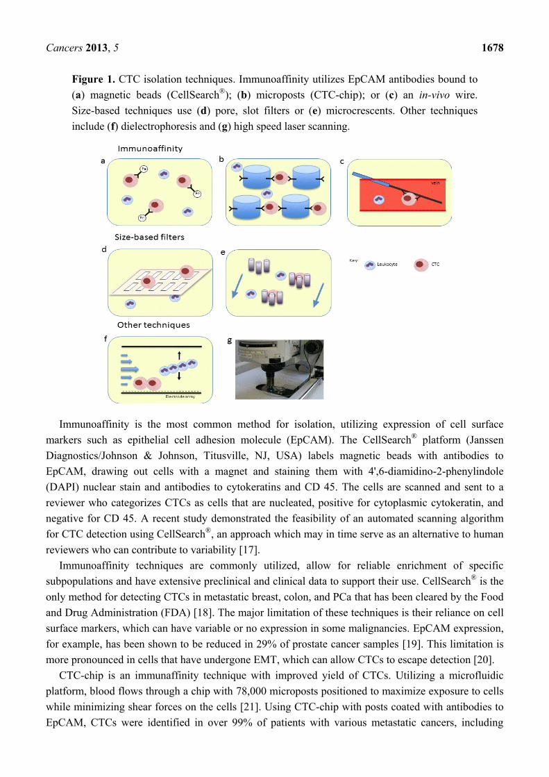
Figure 1. CTC isolation techniques. Immunoaffinity utilizes EpCAM antibodies bound to
(a) magnetic beads (CellSearch®
); (b) microposts (CTC-chip); or (c) an in-vivo wire.
Size-based techniques use (d) pore, slot filters or (e) microcrescents. Other techniques
include (f) dielectrophoresis and (g) high speed laser scanning.
Immunoaffinity is the most common method for isolation, utilizing expression of cell surface
markers such as epithelial cell adhesion molecule (EpCAM). The CellSearch®
platform (Janssen
Diagnostics/Johnson & Johnson, Titusville, NJ, USA) labels magnetic beads with antibodies to
EpCAM, drawing out cells with a magnet and staining them with 4',6-diamidino-2-phenylindole
(DAPI) nuclear stain and antibodies to cytokeratins and CD 45. The cells are scanned and sent to a
reviewer who categorizes CTCs as cells that are nucleated, positive for cytoplasmic cytokeratin, and
negative for CD 45. A recent study demonstrated the feasibility of an automated scanning algorithm
for CTC detection using CellSearch®
, an approach which may in time serve as an alternative to human
reviewers who can contribute to variability [17].
Immunoaffinity techniques are commonly utilized, allow for reliable enrichment of specific
subpopulations and have extensive preclinical and clinical data to support their use. CellSearch®
is the
only method for detecting CTCs in metastatic breast, colon, and PCa that has been cleared by the Food
and Drug Administration (FDA) [18]. The major limitation of these techniques is their reliance on cell
surface markers, which can have variable or no expression in some malignancies. EpCAM expression,
for example, has been shown to be reduced in 29% of prostate cancer samples [19]. This limitation is
more pronounced in cells that have undergone EMT, which can allow CTCs to escape detection [20].
CTC-chip is an immunaffinity technique with improved yield of CTCs. Utilizing a microfluidic
platform, blood flows through a chip with 78,000 microposts positioned to maximize exposure to cells
while minimizing shear forces on the cells [21]. Using CTC-chip with posts coated with antibodies to
EpCAM, CTCs were identified in over 99% of patients with various metastatic cancers, including
Page 4
Cancers 2013, 5 1679
prostate. The CTC-chip proved sensitive in PCa, identifying CTCs in seven out of seven men with
localized disease.
Size-based filters do not rely on cell surface markers but rather on the fact that CTCs are typically
larger than other blood cells. Morphometric analyses have demonstrated a spectrum of CTC size
depending on the cell line of origin with breast and prostate CTCs typically smaller than CTCs from
cervical and liver cancer [22]. All CTCs, however, were significantly larger than white blood cells.
Described techniques include filtration through pores, slots or microcrescents [23,24]. These
techniques capture a broader spectrum of cells irrespective of cell surface markers and the cells are not
bound by antibodies, helping with further analysis. On the other hand, size-based enrichment has
potential drawbacks. CTCs may escape capture due to smaller size; conversely, cells that are captured still
require subsequent “positive identification” using various immunofluorescent stains (e.g., cytokeratins).
There are a multitude of other techniques to isolate CTCs. Dielectrophoresis separates cells through
an electromagnetic method, taking advantage of cells having different polarizable properties [25]. It
has been combined with immunoaffinity assays to work synergistically [26]. High speed laser scanning
identifies CTCs after staining and is based upon a cell’s morphologic and fluorescence patterns [27].
Another technology, termed NanoVelcro-Chip, utilizes EpCAM antibody-coated silicon wires and has
been shown to be easily replicated between different facilities [28]. The addition of laser capture
microdissection to this platform allows for pure CTC isolation and sequencing [29]. Still another group
utilized a three dimensional tumor cell culture on a microfluidic platform has been shown to
successfully culture prostate cancer cells spiked into blood [30]. Lastly, an in vivo EpCAM antibody-coated
wire placed in a patient’s vein for 30 minutes has shown promising results with regard to CTC capture
in breast and lung cancer [31].
4. Rationale for CTCs in Prostate Cancer
CTC analysis, in many ways, is ideally suited to PCa. To date, CTCs have been most extensively
studied and qualified in advanced disease. In castration resistant PCa (CRPC), PSA lacks accuracy in
reflecting disease burden and hence is not used as a valid endpoint for clinical trials [32]. Therefore,
clinicians are often left with symptoms, imaging or even bone biopsies to define progression or
response to treatment. CTC analysis can fulfill the role of a biomarker by serving as an accurate,
noninvasive index of disease that can be followed at multiple time points. As a reliable marker, CTCs
can be used in clinical trials as an intermediate endpoint, helping expedite the testing of future
therapeutics [33].
Another potential application for CTCs in PCa, though not as developed, may be their use to
differentiate true localized PCa from occult disseminated disease. Imaging studies lack accuracy in this
setting and nomograms utilizing clinicopathologic factors are far from definitive [34]. Currently, the
most reliable method for determining micrometastases is with surgical lymphadenectomy which may
not necessarily reflect hematogenous disease [35]. CTCs can potentially identify patients with early
systemic disease, allowing these patients more aggressive, upfront treatment.
Perhaps most importantly, CTC analysis is rapidly shifting away from simple enumeration and
towards molecular characterization. PCa is a heterogeneous disease with different molecular drivers.
Selection of treatment for CRPC, however, is largely empiric. CTC analysis (i.e., protein and mRNA
Page 5
Cancers 2013, 5 1680
expression, DNA mutations or translocations) can characterize a patient’s cancer at different points
and in response to different treatments. This may eventually allow clinicians to predict a patient’s
response or lack of response to different therapies based on a CTC molecular signature.
5. CTCs in Advanced Prostate Cancer
In an early study of men with metastatic PCa, CellSearch® detected ≥2 CTCs per 7.5 mL blood in 57%
of men [36]. In patients with CRPC, >2 CTCs were found in 75% of men with the CellSearch
platform [37]. Higher CTC counts were seen in patients with bone metastases and those who had prior
chemotherapy, a finding corroborated by Scher et al. who demonstrated 81% of men with CRPC who
had prior docetaxel chemotherapy had evaluable CTCs during a phase 3 study of abiraterone acetate
(AA) [38].
CTCs were initially studied in CRPC, where enumeration emerged as an important prognostic tool.
Danila et al. examined CTC number in relation to survival and found that a higher baseline CTC count
was significantly associated with worse survival [37]. De Bono et al. studied CTC counts in 231 men
with CRPC before and after chemotherapy and found that men with baseline >5 CTCs per 7.5 mL
blood had a significantly shorter overall survival (OS) (11.5 vs. 21.7 months, p < 0.0001) compared to
men with ≤5 CTCs [18]. Changes in a patient’s CTC count after treatment were also associated with
survival. Patients who experienced a change from favorable (≤5) to unfavorable (>5) CTC count had a
significantly worse survival when compared to those with continuously favorable CTC counts (>26 vs.
9.3 months, p < 0.0001). Conversely, those patients who had a CTC count that decreased with
treatment to ≤5 had an almost 15 month improvement in OS (21.3 vs. 6.8 months, p < 0.0001)
compared to those with continuously unfavorable CTC counts. Additionally, CTC count was superior
to PSA in predicting survival. These findings ultimately lead to the FDA clearing CellSearch®
for
evaluation of metastatic CRPC.
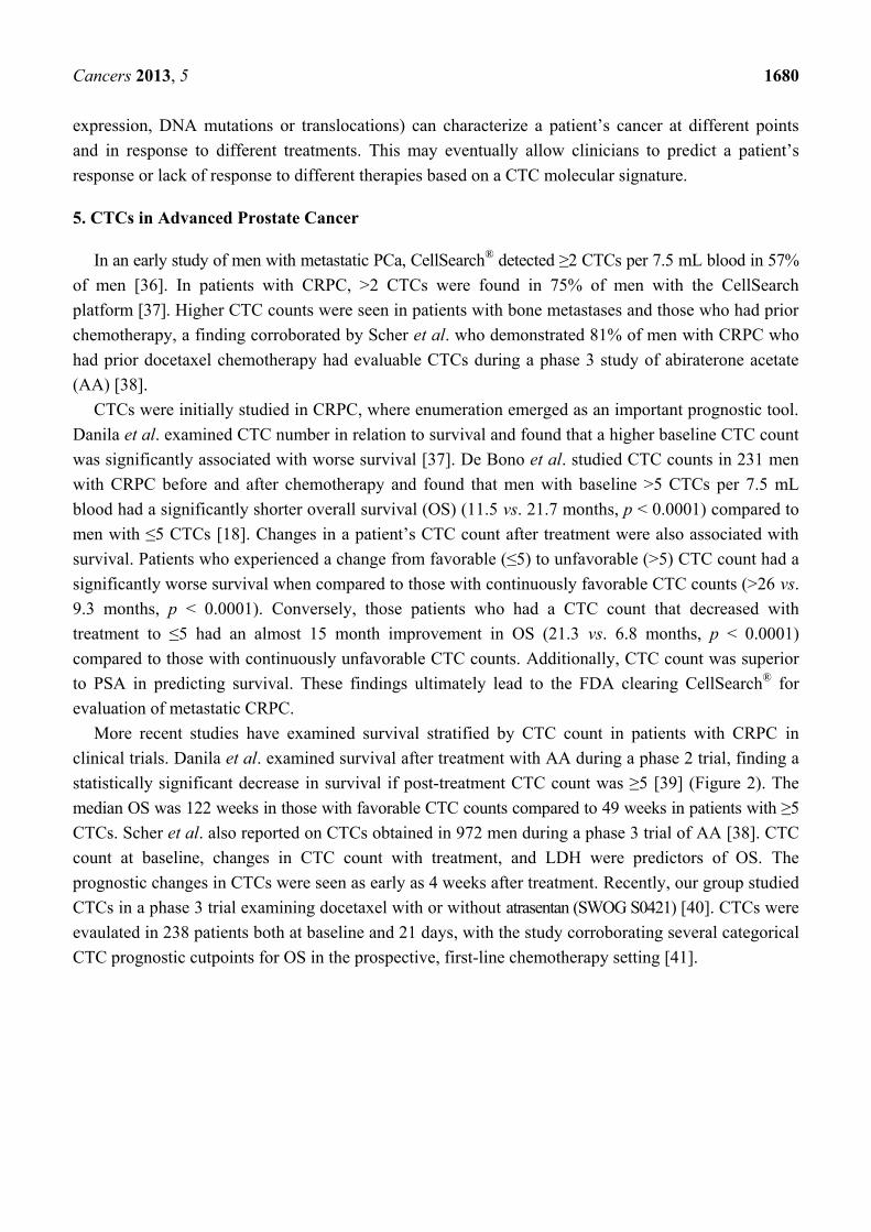
More recent studies have examined survival stratified by CTC count in patients with CRPC in
clinical trials. Danila et al. examined survival after treatment with AA during a phase 2 trial, finding a
statistically significant decrease in survival if post-treatment CTC count was ≥5 [39] (Figure 2). The
median OS was 122 weeks in those with favorable CTC counts compared to 49 weeks in patients with ≥5
CTCs. Scher et al. also reported on CTCs obtained in 972 men during a phase 3 trial of AA [38]. CTC
count at baseline, changes in CTC count with treatment, and LDH were predictors of OS. The
prognostic changes in CTCs were seen as early as 4 weeks after treatment. Recently, our group studied
CTCs in a phase 3 trial examining docetaxel with or without atrasentan (SWOG S0421) [40]. CTCs were
evaulated in 238 patients both at baseline and 21 days, with the study corroborating several categorical
CTC prognostic cutpoints for OS in the prospective, first-line chemotherapy setting [41].
Page 6
Cancers 2013, 5 1681
Figure 2. CTC count and survival in castration-resistant prostate cancer. Probability of
survival stratified by post-treatment CTC count. CTC count ≥5 at 4 weeks was associated
with shorter overall survival compared with patients with CTC counts of <5 (reprinted
from [39] with permission of Elsevier).
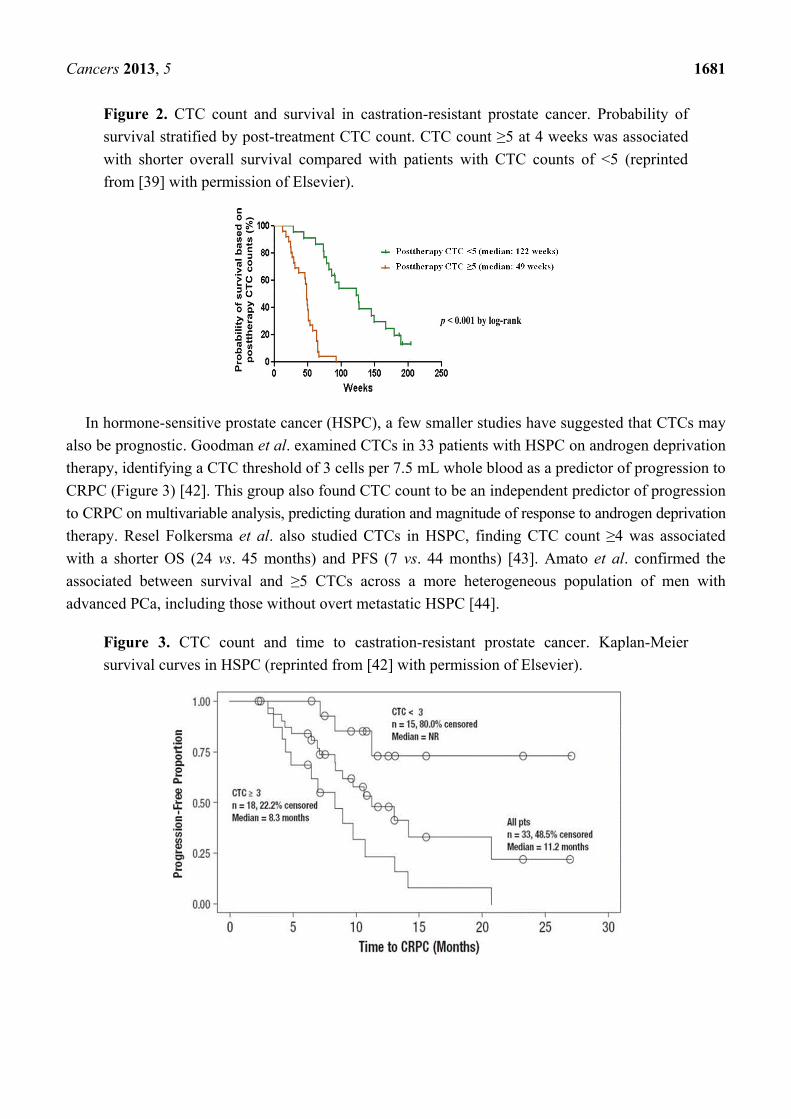
In hormone-sensitive prostate cancer (HSPC), a few smaller studies have suggested that CTCs may
also be prognostic. Goodman et al. examined CTCs in 33 patients with HSPC on androgen deprivation
therapy, identifying a CTC threshold of 3 cells per 7.5 mL whole blood as a predictor of progression to
CRPC (Figure 3) [42]. This group also found CTC count to be an independent predictor of progression
to CRPC on multivariable analysis, predicting duration and magnitude of response to androgen deprivation
therapy. Resel Folkersma et al. also studied CTCs in HSPC, finding CTC count ≥4 was associated
with a shorter OS (24 vs. 45 months) and PFS (7 vs. 44 months) [43]. Amato et al. confirmed the
associated between survival and ≥5 CTCs across a more heterogeneous population of men with
advanced PCa, including those without overt metastatic HSPC [44].
Figure 3. CTC count and time to castration-resistant prostate cancer. Kaplan-Meier
survival curves in HSPC (reprinted from [42] with permission of Elsevier).
Page 7
Cancers 2013, 5 1682
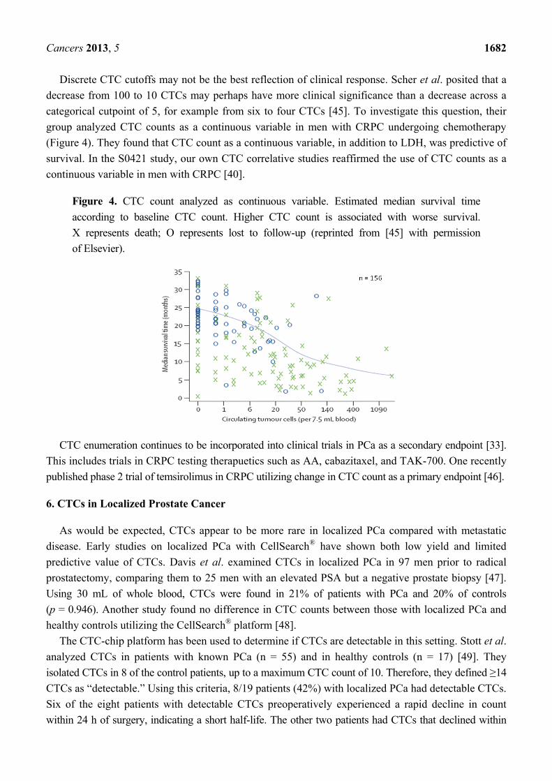
Discrete CTC cutoffs may not be the best reflection of clinical response. Scher et al. posited that a
decrease from 100 to 10 CTCs may perhaps have more clinical significance than a decrease across a
categorical cutpoint of 5, for example from six to four CTCs [45]. To investigate this question, their
group analyzed CTC counts as a continuous variable in men with CRPC undergoing chemotherapy
(Figure 4). They found that CTC count as a continuous variable, in addition to LDH, was predictive of
survival. In the S0421 study, our own CTC correlative studies reaffirmed the use of CTC counts as a
continuous variable in men with CRPC [40].
Figure 4. CTC count analyzed as continuous variable. Estimated median survival time
according to baseline CTC count. Higher CTC count is associated with worse survival.
X represents death; O represents lost to follow-up (reprinted from [45] with permission
of Elsevier).
CTC enumeration continues to be incorporated into clinical trials in PCa as a secondary endpoint [33].
This includes trials in CRPC testing therapuetics such as AA, cabazitaxel, and TAK-700. One recently
published phase 2 trial of temsirolimus in CRPC utilizing change in CTC count as a primary endpoint [46].
6. CTCs in Localized Prostate Cancer
As would be expected, CTCs appear to be more rare in localized PCa compared with metastatic
disease. Early studies on localized PCa with CellSearch®
have shown both low yield and limited
predictive value of CTCs. Davis et al. examined CTCs in localized PCa in 97 men prior to radical
prostatectomy, comparing them to 25 men with an elevated PSA but a negative prostate biopsy [47].
Using 30 mL of whole blood, CTCs were found in 21% of patients with PCa and 20% of controls
(p = 0.946). Another study found no difference in CTC counts between those with localized PCa and
healthy controls utilizing the CellSearch®
platform [48].
The CTC-chip platform has been used to determine if CTCs are detectable in this setting. Stott et al.
analyzed CTCs in patients with known PCa (n = 55) and in healthy controls (n = 17) [49]. They
isolated CTCs in 8 of the control patients, up to a maximum CTC count of 10. Therefore, they defined ≥14
CTCs as “detectable.” Using this criteria, 8/19 patients (42%) with localized PCa had detectable CTCs.
Six of the eight patients with detectable CTCs preoperatively experienced a rapid decline in count
within 24 h of surgery, indicating a short half-life. The other two patients had CTCs that declined within
Page 8
Cancers 2013, 5 1683
3 months. Of all 19 patients with localized PCa, six had transient increases in CTC values postoperatively,
but the clinical significance of these detectable postoperative CTCs in that cohort was not clear.
Others have utilized RT-PCR for prostate-specific genes from peripheral whole blood as a surrogate
for measuring CTCs. Yates et al. demonstrated that the presence of perioperative PSA and prostate-specific
membrane antigen (PSMA) mRNA improves nomogram prediction of PCa recurrence after radical
prostatectomy [50]. Eschwège et al. prospectively compared recurrence free survival between patients
with detectable (n = 57) and undetectable (n = 53) PSA and PSMA genes in patients undergoing
radical prostatectomy. Those with undetectable preoperative mRNA of these prostate-specific genes
had a significantly better recurrence free survival (69.6 vs. 36.2 months, p < 0.0001).
Giesing et al. examined antioxidant genes from CTC clusters with a mesh filtration platform in
patients with an elevated PSA [51]. They found mRNA levels of three antioxidant genes (GPX1,
SOD2, TXNRD1) correlated with the detection of primary PCa. Additionally, continued overexpression of
these antioxidant genes predicted disease recurrence after radical prostatectomy.
CTC count after salvage radiotherapy for localized PCa was studied by Lowes et al. [52]. CTCs
were detectable in 19/26 patients (73%) prior to radiotherapy. Radiotherapy did not significantly
reduce the CTC counts. Patients with good biochemical response were more likely to have decreases in
CTC count compared to non-responders, though this did not reach statistical significance.
Interestingly, there is evidence that even in the setting of localized PCa, CTCs can deposit in the
bone marrow, sometimes referred to as disseminated tumor cells (DTCs) [53]. Berg et al. evaluated
bone marrow aspirates from 272 patients prior to definitive radiotherapy [54]. DTCs were found in
18% of patients and their presence was associated with worse Gleason score and an increased risk of
distant metastasis (28% vs. 9%, p = 0.03). A follow-up study by Lilleby et al. identified DTCs in 14%
of patients prior to radiotherapy and in 19% of patients after radiotherapy for cT1-3N0M0 disease [55].
The presence of these pretreatment DTCs was the only independent predictor of cancer-specific
survival and OS on multivariable analysis. Kollerman et al. found that the presence of DTCs in
patients treated with neoadjuvant androgen deprivation prior to radical prostatectomy was an
independent marker for biochemical relapse [56]. Collectively, these studies suggest that DTCs may,
in some cases, constitute occult micrometastatic deposits which may clinically manifest as recurrent
disease, i.e., failure of definitive local therapy.
Though not as robust as the data in advanced PCa, the evidence that CTCs have a role in localized
disease continues to accumulate. CTCs may prove useful in differentiating the truly aggressive forms
of localized PCa at high risk for recurrence, allowing for multimodal treatment in those select patients.
7. CTCs to Characterize Prostate Cancer
Perhaps more promising than enumeration is the ability to study CTCs in order to molecularly
characterize a cancer. The ability to take a “liquid biopsy” at different time points in treatment creates
opportunities for therapeutic decisions informed by the specific phenotype of a patient’s cancer,
moving closer towards the goal of personalized cancer therapy. Molecular characterization of CTCs,
however, represents new challenges. For example, whereas white blood cell contamination may be
tolerable when detecting (“yes” or “no”) the presence of disease-related genetic aberrations (e.g.,
TMPRSS2-ERG fusion product), moving towards more quantitative analyses (e.g., mRNA transcript
Page 9
Cancers 2013, 5 1684
levels of gene expression) necessitates ultra-pure CTC samples. Technological improvements have
helped overcome some of these obstacles, leading to early successes in genomic and transcriptomic
profiling of CTCs, sometimes with as little as one cell [57–60].
Our group demonstrated that targeted next generation sequencing of CTCs is possible, sometimes in
patients with little evidence of clinical disease (PSA values <1 ng/mL) [61]. This sequencing was able
to detect single nucleotide variants (SNVs) in men with HSPC. SNVs have already been shown to be
associated with changes in PCa clinical outcomes [62]. Additional profiling was shown to be feasible
by Shaffer et al., who examined epidermal growth factor receptor expression, chromosome ploidy, and
androgen receptor (AR) gene amplification in CTCs [63].
Giesing et al. performed molecular phenotyping of PCa CTCs with direct links to clinical
outcomes [10]. They tested functional gene targets as biomarkers for disease, analyzing 23 targets and
identifying overexpression of five genes (SOD2, GPX1, AR, cyclin B and bFGF) that predicted
clinical metastases. Three of these genes were able to predict bone metastases independent of
pathologic or treatment-related variables.
Given the important role of EMT in CTCs and metastasis, Chen et al. examined EMT-specific
genetic profiles [64]. They analyzed CTCs from eight patients with advanced PCa with real time PCR
for 84 EMT-related and reference genes. They identified a heterogeneous pattern of expression of
EMT-related genes and found that CTCs frequently lost epithelial characteristics. A subset of the
EMT-related genes was more frequently expressed in CRPC compared to HSPC.
The TMPRSS2-ERG gene fusion has been implicated in prostate carcinogenesis and castration-resistant
growth through disruption of AR signaling and promoting cellular de-differentiation [65]. Attard et al.
examined expression of the ERG oncogene in patients with CRPC in a clinical trial for AA [66]. They
found that ERG status was correlated with PSA decline after treatment with AA. They also found that
CTC ERG overexpression persisted in CRPC, with the expression status mirroring that of the original
prostate biopsy. This data suggests that the most commonly found tumor on transrectal biopsy is the
one that results in hematogenous metastases. This is a notable finding since PCa can be multifocal with
clonal heterogeneity and differences in ERG gene expression [67,68]. The importance of ERG was
further studies by Danila et al. to determine if the TMPRSS2-ERG gene fusion could be used as a
marker for sensitivity to AA [39]. In men with CRPC, the gene fusion was found in 15 of 41 (37%)
CTC samples. Its presence, however, did not predict PSA decline or response to treatment.
AR and its signaling axis have been examined in CTCs. Miyamoto et al. utilized single cell
immunofluorescence to determine the degree of AR signaling within a CTC [69]. They found that
first-line androgen deprivation therapy resulted in a transition from an AR-on to AR-off signature.
Second-line hormonal therapy in CRPC resulted in more varied changes to the AR signatures. Jiang et al.
examined mutations in AR, an established escape mechanism to CRPC [70]. They obtained CTCs in
men with CRPC, amplified AR by PCR, and detected mutations in 57% of men. Leversha et al.
utilized fluorescence in-situ hybridization in the CTCs of 77 men with CRPC, finding AR gene
amplification, another mechanism for castration-resistant growth, in 38% of men [71]. These types of
molecular characterization of AR may soon lead to alterations in treatments based upon an individual’s
unique AR signature.
In addition to genetic analyses, other molecular characterizations have been performed, such as
quantification of telomerase activity. Activation of telomerase, the enzyme that lengthens telomeres to
Page 10
Cancers 2013, 5 1685
protect chromosomes, is an important cancer marker in >90% of malignancies [72]. We demonstrated
the feasibility of measuring telomerase activity in CTCs, which can act as a functional cancer cell
assay across various malignancies [23]. We transitioned this approach to a large PCa phase 3 clinical
trial, and showed for the first time in a large prospective setting that a CTC-derived biomarker
(telomerase) could be prognostic of OS in a significant subset of men [40,41].
8. Conclusions
The role of CTCs in PCa is rapidly evolving. CTCs provide a window into the hematogenous
spread of cancer and can drastically improve oncologic understanding and patient care. In metastatic
PCa, CTC enumeration is an accurate method for monitoring disease and has been used in clinical
trials as an intermediate endpoint. CTCs can be detected in localized disease and hold the potential to
detect early metastasis. Perhaps the most exciting feature of CTCs is that they provide a platform for
noninvasive, repeated inquiries into a cancer’s molecular behavior, ultimately enabling individualized,
adaptive and more effective management of PCa over time.
Acknowledgments
The authors would like to thank David Quinn for his thoughtful feedback and discussions regarding
this manuscript. This work was supported in part by CA014089-38.
Conflicts of Interest
The authors declare no conflicts of interest.
References
1. Ashworth, T. A case of cancer in which cells similar to those in the tumours were seen in the
blood after death. Aust. Med. J. 1869, 14, 146–147.
2. Muller, V.; Stahmann, N.; Riethdorf, S.; Rau, T.; Zabel, T.; Goetz, A.; Janicke, F.; Pantel, K.
Circulating tumor cells in breast cancer: Correlation to bone marrow micrometastases,
heterogeneous response to systemic therapy and low proliferative activity. Clin. Cancer Res.
2005, 11, 3678–3685.
3. Fehm, T.; Sagalowsky, A.; Clifford, E.; Beitsch, P.; Saboorian, H.; Euhus, D.; Meng, S.;
Morrison, L.; Tucker, T.; Lane, N.; et al. Cytogenetic evidence that circulating epithelial cells in
patients with carcinoma are malignant. Clin. Cancer Res. 2002, 8, 2073–2084.
4. Arya, M.; Bott, S.R.; Shergill, I.S.; Ahmed, H.U.; Williamson, M.; Patel, H.R. The metastatic
cascade in prostate cancer. Surg. Oncol. 2006, 15, 117–128.
5. Maheswaran, S.; Haber, D.A. Circulating tumor cells: A window into cancer biology and
metastasis. Curr. Opin. Genet. Dev. 2010, 20, 96–99.
6. Kim, M.Y.; Oskarsson, T.; Acharyya, S.; Nguyen, D.X.; Zhang, X.H.; Norton, L.; Massague, J.
Tumor self-seeding by circulating cancer cells. Cell 2009, 139, 1315–1326.
7. Pantel, K.; Brakenhoff, R.H.; Brandt, B. Detection, clinical relevance and specific biological
properties of disseminating tumour cells. Nat. Rev. Cancer 2008, 8, 329–340.
Page 11
Cancers 2013, 5 1686
8. Stott, S.L.; Hsu, C.H.; Tsukrov, D.I.; Yu, M.; Miyamoto, D.T.; Waltman, B.A.; Rothenberg, S.M.;
Shah, A.M.; Smas, M.E.; Korir, G.K.; et al. Isolation of circulating tumor cells using a
microvortex-generating herringbone-chip. Proc. Natl. Acad. Sci. USA 2010, 107, 18392–18397.
9. Brandt, B.; Junker, R.; Griwatz, C.; Heidl, S.; Brinkmann, O.; Semjonow, A.; Assmann, G.;
Zanker, K.S. Isolation of prostate-derived single cells and cell clusters from human peripheral
blood. Cancer Res. 1996, 56, 4556–4561.
10. Giesing, M.; Driesel, G.; Molitor, D.; Suchy, B. Molecular phenotyping of circulating tumour
cells in patients with prostate cancer: Prediction of distant metastases. BJU Int. 2012, 110,
E1202–E1211.
11. De Craene, B.; Berx, G. Regulatory networks defining EMT during cancer initiation and
progression. Nat. Rev. Cancer 2013, 13, 97–110.
12. Bednarz-Knoll, N.; Alix-Panabieres, C.; Pantel, K. Plasticity of disseminating cancer cells in
patients with epithelial malignancies. Cancer Metastasis Rev. 2012, 31, 673–687.
13. Gradilone, A.; Raimondi, C.; Nicolazzo, C.; Petracca, A.; Gandini, O.; Vincenzi, B.; Naso, G.;
Agliano, A.M.; Cortesi, E.; Gazzaniga, P. Circulating tumour cells lacking cytokeratin in breast
cancer: The importance of being mesenchymal. J. Cell Mol. Med. 2011, 15, 1066–1070.
14. Thiery, J.P.; Acloque, H.; Huang, R.Y.; Nieto, M.A. Epithelial-mesenchymal transitions in
development and disease. Cell 2009, 139, 871–890.
15. Carvalho, F.L.; Simons, B.W.; Antonarakis, E.S.; Rasheed, Z.; Douglas, N.; Villegas, D.; Matsui, W.;
Berman, D.M. Tumorigenic potential of circulating prostate tumor cells. Oncotarget 2013, 4,
413–421.
16. Joosse, S.A.; Pantel, K. Biologic challenges in the detection of circulating tumor cells.
Cancer Res. 2013, 73, 8–11.
17. Ligthart, S.T.; Coumans, F.A.; Bidard, F.C.; Simkens, L.H.; Punt, C.J.; de Groot, M.R.; Attard, G.;
de Bono, J.S.; Pierga, J.Y.; Terstappen, L.W. Circulating tumor cells count and morphological
features in breast, colorectal and prostate cancer. PLoS One 2013, 8, e67148.
18. De Bono, J.S.; Scher, H.I.; Montgomery, R.B.; Parker, C.; Miller, M.C.; Tissing, H.; Doyle, G.V.;
Terstappen, L.W.; Pienta, K.J.; Raghavan, D. Circulating tumor cells predict survival benefit from
treatment in metastatic castration-resistant prostate cancer. Clin. Cancer Res. 2008, 14, 6302–6309.
19. Went, P.T.; Lugli, A.; Meier, S.; Bundi, M.; Mirlacher, M.; Sauter, G.; Dirnhofer, S. Frequent
EpCam protein expression in human carcinomas. Hum. Pathol. 2004, 35, 122–128.
20. Gorges, T.M.; Tinhofer, I.; Drosch, M.; Rose, L.; Zollner, T.M.; Krahn, T.; von Ahsen, O.
Circulating tumour cells escape from EpCam-based detection due to epithelial-to-mesenchymal
transition. BMC Cancer 2012, 12, e178.
21. Nagrath, S.; Sequist, L.V.; Maheswaran, S.; Bell, D.W.; Irimia, D.; Ulkus, L.; Smith, M.R.;
Kwak, E.L.; Digumarthy, S.; Muzikansky, A.; et al. Isolation of rare circulating tumour cells in
cancer patients by microchip technology. Nature 2007, 450, 1235–1239.
22. Vona, G.; Sabile, A.; Louha, M.; Sitruk, V.; Romana, S.; Schutze, K.; Capron, F.; Franco, D.;
Pazzagli, M.; Vekemans, M.; et al. Isolation by size of epithelial tumor cells: A new method for
the immunomorphological and molecular characterization of circulatingtumor cells. Am. J.
Pathol. 2000, 156, 57–63.
Page 12
Cancers 2013, 5 1687
23. Xu, T.; Lu, B.; Tai, Y.C.; Goldkorn, A. A cancer detection platform which measures telomerase
activity from live circulating tumor cells captured on a microfilter. Cancer Res. 2010, 70, 6420–6426.
24. Coumans, F.A.; van Dalum, G.; Beck, M.; Terstappen, L.W. Filter characteristics influencing
circulating tumor cell enrichment from whole blood. PLoS One 2013, 8, e61770.
25. Gascoyne, P.R.; Noshari, J.; Anderson, T.J.; Becker, F.F. Isolation of rare cells from cell mixtures
by dielectrophoresis. Electrophoresis 2009, 30, 1388–1398.
26. Huang, C.; Liu, H.; Bander, N.H.; Kirby, B.J. Enrichment of prostate cancer cells from blood cells
with a hybrid dielectrophoresis and immunocapture microfluidic system. Biomed. Microdevices
2013, 15, 941–948.
27. Scholtens, T.M.; Schreuder, F.; Ligthart, S.T.; Swennenhuis, J.F.; Tibbe, A.G.; Greve, J.;
Terstappen, L.W. Celltracks TDI: An image cytometer for cell characterization. Cytometry A
2011, 79, 203–213.
28. Lu, Y.T.; Zhao, L.; Shen, Q.; Garcia, M.A.; Wu, D.; Hou, S.; Song, M.; Xu, X.; Ouyang, W.H.;
Ouyang, W.W.; et al. Nanovelcro chip for CTC enumeration in prostate cancer patients. Methods
2013, 64, 144–152.
29. Zhao, L.; Lu, Y.T.; Li, F.; Wu, K.; Hou, S.; Yu, J.; Shen, Q.; Wu, D.; Song, M.; Ouyang, W.H.; et al.
High-purity prostate circulating tumor cell isolation by a polymer nanofiber-embedded microchip
for whole exome sequencing. Adv. Mater. 2013, doi:10.1002/adma.201205237.
30. Bichsel, C.A.; Gobaa, S.; Kobel, S.; Secondini, C.; Thalmann, G.N.; Cecchini, M.G.; Lutolf, M.P.
Diagnostic microchip to assay 3D colony-growth potential of captured circulating tumor cells.
Lab Chip 2012, 12, 2313–2316.
31. Saucedo-Zeni, N.; Mewes, S.; Niestroj, R.; Gasiorowski, L.; Murawa, D.; Nowaczyk, P.; Tomasi, T.;
Weber, E.; Dworacki, G.; Morgenthaler, N.G.; et al. A novel method for the in vivo isolation of
circulating tumor cells from peripheral blood of cancer patients using a functionalized and
structured medical wire. Int. J. Oncol. 2012, 41, 1241–1250.
32. Fleming, M.T.; Morris, M.J.; Heller, G.; Scher, H.I. Post-therapy changes in PSA as an outcome
measure in prostate cancer clinical trials. Nat. Clin. Pract. Oncol. 2006, 3, 658–667.
33. United States National Institutes of Health. Clinicaltrials.gov. Available online: http://www.
clinicaltrials.gov (accessed on 9 Semptember 2013).
34. Hovels, A.M.; Heesakkers, R.A.; Adang, E.M.; Jager, G.J.; Strum, S.; Hoogeveen, Y.L.;
Severens, J.L.; Barentsz, J.O. The diagnostic accuracy of CT and MRI in the staging of pelvic
lymph nodes in patients with prostate cancer: A meta-analysis. Clin. Radiol. 2008, 63, 387–395.
35. Saitoh, H.; Yoshida, K.; Uchijima, Y.; Kobayashi, N.; Suwata, J.; Kamata, S. Two different
lymph node metastatic patterns of a prostatic cancer. Cancer 1990, 65, 1843–1846.
36. Allard, W.J.; Matera, J.; Miller, M.C.; Repollet, M.; Connelly, M.C.; Rao, C.; Tibbe, A.G.; Uhr, J.W.;
Terstappen, L.W. Tumor cells circulate in the peripheral blood of all major carcinomas but not in
healthy subjects or patients with nonmalignant diseases. Clin. Cancer Res. 2004, 10, 6897–6904.
37. Danila, D.C.; Heller, G.; Gignac, G.A.; Gonzalez-Espinoza, R.; Anand, A.; Tanaka, E.; Lilja, H.;
Schwartz, L.; Larson, S.; Fleisher, M.; et al. Circulating tumor cell number and prognosis in
progressive castration-resistant prostate cancer. Clin. Cancer Res. 2007, 13, 7053–7058.
Page 13
Cancers 2013, 5 1688
38. Scher, H.I.; Heller, G.; Molina, A.; Kheoh, T.S.; Attard, G.; Moreira, J.; Sandhu, S.K.; Parker, C.;
Logothetis, C.; McCormack, R.T.; et al. Evaluation of Circulating Tumor Cell (CTC)
Enumeration as an Efficacy Response Biomarker of Overall Survival (OS) in Metastatic
Castration-Resistant Prostate Cancer (mCRPC): Planned Final Analysis (FA) of COU-AA-301, a
Randomized Double-Blind, Placebo-Controlled Phase III Study of Abiraterone Acetate (AA) plus
Low-Dose Prednisone (P) Post Docetaxel. In Proceedings of 2011 ASCO Annual Meeting,
Chiacago, IL, USA, 3–7 June 2011.
39. Danila, D.C.; Anand, A.; Sung, C.C.; Heller, G.; Leversha, M.A.; Cao, L.; Lilja, H.; Molina, A.;
Sawyers, C.L.; Fleisher, M.; et al. TMPRSS2-ERG status in circulating tumor cells as a predictive
biomarker of sensitivity in castration-resistant prostate cancer patients treated with abiraterone
acetate. Eur. Urol. 2011, 60, 897–904.
40. Quinn, D.I.; Tangen, C.M.; Hussain, M.; Lara, P.N., Jr.; Goldkorn, A.; Moinpour, C.M.;
Garzotto, M.G.; Mack, P.C.; Carducci, M.A.; Monk, J.P.; et al. Docetaxel and atrasentan versus
docetaxel and placebo for men with advanced castration-resistant prostate cancer (swog s0421): A
randomised phase 3 trial. Lancet Oncol. 2013, 14, 893–900.
41. Goldkorn, A.; Vogelzang, N.J.; Fink, L.M.; Ely, B.; Quinn, D.I.; Tangen, C.M.; Tai, Y.C.;
Twardowski, P.; van Veldhuizen, P.J.; Agarwal, N.; et al. Circulating Tumor Cell (CTC) Counts
and CTC Telomerase Activity (TA) as Prognotic Markers of Overall Survival (OS) in SWOG
S0421: Docetaxelwith or Without Atrasentan for Metastatic Castration-Resistant Prostate Cancer
(mCRPC). In Proceedings of Markers in Cancer Joint Meeting of the American Society of
Clinical Oncology (ASCO), European Organization for Research and Treatment of Cancer
(EORTC) and the National Cancer Institute (NCI), Hollywood, FL, USA, 20 October 2012.
42. Goodman, O.B., Jr.; Symanowski, J.T.; Loudyi, A.; Fink, L.M.; Ward, D.C.; Vogelzang, N.J.
Circulating tumor cells as a predictive biomarker in patients with hormone-sensitive prostate
cancer. Clin. Genitourin. Cancer 2011, 9, 31–38.
43. Resel, F.L.; San, J.M.L.; Galante, R.I.; Moreno, S.J.; Olivier, G.C. Prognostic significance of
circulating tumor cell count in patients with metastatic hormone-sensitive prostate cancer.
Urology 2012, 80, 1328–1332.
44. Amato, R.J.; Melnikova, V.; Zhang, Y.; Liu, W.; Saxena, S.; Shah, P.K.; Jensen, B.T.; Torres, K.E.;
Davis, D.W. Epithelial cell adhesion molecule-positive circulating tumor cells as predictive
biomarker in patients with prostate cancer. Urology 2013, 81, 1303–1307.
45. Scher, H.I.; Jia, X.; de Bono, J.S.; Fleisher, M.; Pienta, K.J.; Raghavan, D.; Heller, G. Circulating
tumour cells as prognostic markers in progressive, castration-resistant prostate cancer: A
reanalysis of IMMC38 trial data. Lancet Oncol. 2009, 10, 233–239.
46. Armstrong, A.J.; Shen, T.; Halabi, S.; Kemeny, G.; Bitting, R.L.; Kartcheske, P.; Embree, E.;
Morris, K.; Winters, C.; Jaffe, T.; et al. A phase II trial of temsirolimus in men with castration-
resistant metastatic prostate cancer. Clin. Genitourin. Cancer 2013, 11, 397–406.
47. Davis, J.W.; Nakanishi, H.; Kumar, V.S.; Bhadkamkar, V.A.; McCormack, R.; Fritsche, H.A.;
Handy, B.; Gornet, T.; Babaian, R.J. Circulating tumor cells in peripheral blood samples from
patients with increased serum prostate specific antigen: Initial results in early prostate cancer.
J. Urol. 2008, 179, 2187–2191.
Page 14
Cancers 2013, 5 1689
48. Thalgott, M.; Rack, B.; Maurer, T.; Souvatzoglou, M.; Eiber, M.; Kress, V.; Heck, M.M.;
Andergassen, U.; Nawroth, R.; Gschwend, J.E.; et al. Detection of circulating tumor cells in
different stages of prostate cancer. J. Cancer Res. Clin. Oncol. 2013, 139, 755–763.
49. Stott, S.L.; Lee, R.J.; Nagrath, S.; Yu, M.; Miyamoto, D.T.; Ulkus, L.; Inserra, E.J.; Ulman, M.;
Springer, S.; Nakamura, Z.; et al. Isolation and characterization of circulating tumor cells from
patients with localized and metastatic prostate cancer. Sci. Transl. Med. 2010, 2, 25ra23.
50. Yates, D.R.; Roupret, M.; Drouin, S.J.; Comperat, E.; Ricci, S.; Lacave, R.; Sebe, P.;
Cancel-Tassin, G.; Bitker, M.O.; Cussenot, O. Quantitative RT-PCR analysis of PSA and
prostate-specific membrane antigen mRNA to detect circulating tumor cells improves recurrence-free
survival nomogram prediction after radical prostatectomy. Prostate 2012, 72, 1382–1388.
51. Giesing, M.; Suchy, B.; Driesel, G.; Molitor, D. Clinical utility of antioxidant gene expression
levels in circulating cancer cell clusters for the detection of prostate cancer in patients with
prostate-specific antigen levels of 4–10 ng/mL and disease prognostication after radical
prostatectomy. BJU Int. 2010, 105, 1000–1010.
52. Lowes, L.E.; Lock, M.; Rodrigues, G.; D'Souza, D.; Bauman, G.; Ahmad, B.; Venkatesan, V.;
Allan, A.L.; Sexton, T. Circulating tumour cells in prostate cancer patients receiving salvage
radiotherapy. Clin. Transl. Oncol. 2012, 14, 150–156.
53. Riethdorf, S.; Wikman, H.; Pantel, K. Review: Biological relevance of disseminated tumor cells
in cancer patients. Int. J. Cancer Suppl. 2008, 123, 1991–2006.
54. Berg, A.; Berner, A.; Lilleby, W.; Bruland, O.S.; Fossa, S.D.; Nesland, J.M.; Kvalheim, G.
Impact of disseminated tumor cells in bone marrow at diagnosis in patients with nonmetastatic
prostate cancer treated by definitive radiotherapy. Int. J. Cancer Suppl. 2007, 120, 1603–1609.
55. Lilleby, W.; Stensvold, A.; Mills, I.G.; Nesland, J.M. Disseminated tumor cells and their
prognostic significance in nonmetastatic prostate cancer patients. Int. J. Cancer Suppl. 2013, 133,
149–155.
56. Kollermann, J.; Weikert, S.; Schostak, M.; Kempkensteffen, C.; Kleinschmidt, K.; Rau, T.;
Pantel, K. Prognostic significance of disseminated tumor cells in the bone marrow of prostate
cancer patients treated with neoadjuvant hormone treatment. J. Clin. Oncol. 2008, 26, 4928–4933.
57. Cann, G.M.; Gulzar, Z.G.; Cooper, S.; Li, R.; Luo, S.; Tat, M.; Stuart, S.; Schroth, G.; Srinivas, S.;
Ronaghi, M.; et al. mRNA-Seq of single prostate cancer circulating tumor cells reveals recapitulation
of gene expression and pathways found in prostate cancer. PLoS One 2012, 7, e49144.
58. Sieuwerts, A.M.; Kraan, J.; Bolt-de Vries, J.; van der Spoel, P.; Mostert, B.; Martens, J.W.;
Gratama, J.W.; Sleijfer, S.; Foekens, J.A. Molecular characterization of circulating tumor cells in
large quantities of contaminating leukocytes by a multiplex real-time PCR. Breast Cancer Res.
Treat. 2009, 118, 455–468.
59. Welty, C.J.; Coleman, I.; Coleman, R.; Lakely, B.; Xia, J.; Chen, S.; Gulati, R.; Larson, S.R.;
Lange, P.H.; Montgomery, B.; et al. Single cell transcriptomic analysis of prostate cancer cells.
BMC Mol. Biol. 2013, 14, e6.
60. Magbanua, M.J.; Sosa, E.V.; Scott, J.H.; Simko, J.; Collins, C.; Pinkel, D.; Ryan, C.J.; Park, J.W.
Isolation and genomic analysis of circulating tumor cells from castration resistant metastatic
prostate cancer. BMC Cancer 2012, 12, e78.
Page 15
Cancers 2013, 5 1690
61. Stephen, V.L.; Paul, W.; Strauss, W.; Xu, Y.; Xu, T.; Pinski, J.K.; Dorff, T.B.; Quinn, D.I.;
Triche, T.J.; Winer-Jones, J.; et al. Targeted next-generation sequencing (NGS) of circulating tumor
cells (CTCs) in hormone-sensitive prostate cancer (HSPC). J. Clin. Oncol. 2013, 31, abstr 11040.
62. Tsuchiya, N.; Matsui, S.; Narita, S.; Kamba, T.; Mitsuzuka, K.; Hatakeyama, S.; Horikawa, Y.;
Inoue, T.; Saito, S.; Ohyama, C.; et al. Distinct cancer-specific survival in metastatic prostate
cancer patients classified by a panel of single nucleotide polymorphisms of cancer-associated
genes. Genes Cancer 2013, 4, 54–60.
63. Shaffer, D.R.; Leversha, M.A.; Danila, D.C.; Lin, O.; Gonzalez-Espinoza, R.; Gu, B.; Anand, A.;
Smith, K.; Maslak, P.; Doyle, G.V.; et al. Circulating tumor cell analysis in patients with
progressive castration-resistant prostate cancer. Clin. Cancer Res. 2007, 13, 2023–2029.
64. Chen, C.L.; Mahalingam, D.; Osmulski, P.; Jadhav, R.R.; Wang, C.M.; Leach, R.J.; Chang, T.C.;
Weitman, S.D.; Kumar, A.P.; Sun, L.; et al. Single-cell analysis of circulating tumor cells
identifies cumulative expression patterns of EMT-related genes in metastatic prostate cancer.
Prostate 2013, 73, 813–826.
65. Yu, J.; Yu, J.; Mani, R.S.; Cao, Q.; Brenner, C.J.; Cao, X.; Wang, X.; Wu, L.; Li, J.; Hu, M.; et al.
An integrated network of androgen receptor, polycomb, and TMPRSS2-ERG gene fusions in
prostate cancer progression. Cancer Cell 2010, 17, 443–454.
66. Attard, G.; Swennenhuis, J.F.; Olmos, D.; Reid, A.H.; Vickers, E.; A’Hern, R.; Levink, R.;
Coumans, F.; Moreira, J.; Riisnaes, R.; et al. Characterization of ERG, AR and PTEN gene status
in circulating tumor cells from patients with castration-resistant prostate cancer. Cancer Res.
2009, 69, 2912–2918.
67. Mehra, R.; Han, B.; Tomlins, S.A.; Wang, L.; Menon, A.; Wasco, M.J.; Shen, R.; Montie, J.E.;
Chinnaiyan, A.M.; Shah, R.B. Heterogeneity of TMPRSS2 gene rearrangements in multifocal
prostate adenocarcinoma: Molecular evidence for an independent group of diseases. Cancer Res.
2007, 67, 7991–7995.
68. Schoenborn, J.R.; Nelson, P.; Fang, M. Genomic profiling defines subtypes of prostate cancer
with the potential for therapeutic stratification. Clin. Cancer Res. 2013, 19, 4058–4066.
69. Miyamoto, D.T.; Lee, R.J.; Stott, S.L.; Ting, D.T.; Wittner, B.S.; Ulman, M.; Smas, M.E.; Lord, J.B.;
Brannigan, B.W.; Trautwein, J.; et al. Androgen receptor signaling in circulating tumor cells as a
marker of hormonally responsive prostate cancer. Cancer Discov. 2012, 2, 995–1003.
70. Jiang, Y.; Palma, J.F.; Agus, D.B.; Wang, Y.; Gross, M.E. Detection of androgen receptor
mutations in circulating tumor cells in castration-resistant prostate cancer. Clin. Chem. 2010, 56,
1492–1495.
71. Leversha, M.A.; Han, J.; Asgari, Z.; Danila, D.C.; Lin, O.; Gonzalez-Espinoza, R.; Anand, A.;
Lilja, H.; Heller, G.; Fleisher, M.; et al. Fluorescence in situ hybridization analysis of circulating
tumor cells in metastatic prostate cancer. Clin. Cancer Res. 2009, 15, 2091–2097.
72. Shay, J.W.; Bacchetti, S. A survey of telomerase activity in human cancer. Eur. J. Cancer 1997,
33, 787–791.
© 2013 by the authors; licensee MDPI, Basel, Switzerland. This article is an open access article
distributed under the terms and conditions of the Creative Commons Attribution license
(http://creativecommons.org/licenses/by/3.0/).