The Journal of Clinical Investigation RESEARCH ARTICLE 3027 jci.org Volume 125 Number 8 August 2015 Introduction Atherosclerotic disease remains the leading cause of death in the world (1). Development of atherosclerotic plaque is driven by dynamic interactions among the lipid milieu, fluid shear forces, cells of the vascular wall, and cells recruited from the circulation. Endothelial cells, monocytes, T cells, and platelets have all been implicated in atherogenesis (2–4). Recent studies have described the role of platelet activation in the early stages of atherogenesis via transient endothelial and mononuclear interactions and in situ release of mitogens and chemoattractants (5, 6). Activated plate- lets provide cholesterol that is scavenged by macrophages to form foam cells (7, 8). Studies in the last 4 decades have established the role of shear stress–induced platelet activation as well as suscep- tibility to atherosclerosis. Atherosclerosis predominates at branch points and areas of disturbed flow in human arteries, while regions encountering stable flow are more resistant to plaque formation (9–15). Further, a partial carotid ligation surgery induced robust atherosclerosis within 2 weeks, demonstrating that disturbed flow directly causes atherosclerosis under hyperlipidemic condi- tions (16). Central to this concept is the insight that flow-regulated genes, microRNAs, and epigenomic changes initiate programs that trigger activated or quiescent endothelial phenotypes and, in turn, susceptibility to or protection from atherosclerosis (4, 17–21). The enzyme ectonucleotide tri(di)phosphohydrolase-1 (ENT- PD1, also known as CD39) represents the main control system for blood fluidity and is a principal regulator of platelet and leukocyte activation in vivo. This enzyme, expressed on the surface of leu- kocytes and endothelial cells, is responsible for serial phosphohy- drolysis of extracellular ATP to ADP and further to AMP, by virtue of its ecto-ATPase and ecto-ADPase activity (22–24). Since ADP is a potent platelet agonist via P2Y 12 receptors, endothelial CD39- mediated dissipation of ADP serves as a critical negative regula- tor of platelet activation and recruitment (22–27). By metaboliz- ing extracellular ATP and ADP, CD39 provides natural protection from nucleotides released from the injured vasculature or acti- vated platelets. Here, we elucidate what we believe to be a previ- ously unrecognized role for CD39 as an endogenous regulator of endovascular purine levels, serving as a modulator of key cellular drivers of atherosclerosis. Moreover, we demonstrate that CD39 expression is modulated by vascular fluid phase forces. Results CD39 deficiency enhances atherosclerotic lesion formation in ApoE-defi- cient mice. Expression of CD39 changes dynamically with a cell’s envi- ronment (28, 29). This phenomenon initially led us to examine the presence of CD39 in regions susceptible to atherosclerosis. Immu- nofluorescent staining of the apolipoprotein E–deficient (Apoe –/– ) The ability of cells to detect and respond to nucleotide signals in the local microenvironment is essential for vascular homeostasis. The enzyme ectonucleotide tri(di)phosphohydrolase-1 (ENTPD1, also known as CD39) on the surface of leukocytes and endothelial cells metabolizes locally released, intravascular ATP and ADP, thereby eliminating these prothrombotic and proinflammatory stimuli. Here, we evaluated the contribution of CD39 to atherogenesis in the apolipoprotein E–deficient (ApoE-deficient) mouse model of atherosclerosis. Compared with control ApoE-deficient animals, plaque burden was markedly increased along with circulating markers of platelet activation in Cd39 +/– Apoe –/– mice fed a high-fat diet. Plaque analysis revealed stark regionalization of endothelial CD39 expression and function in Apoe –/– mice, with CD39 prominently expressed in atheroprotective, stable flow regions and diminished in atheroprone areas subject to disturbed flow. In mice, disturbed flow as the result of partial carotid artery ligation rapidly suppressed endothelial CD39 expression. Moreover, unidirectional laminar shear stress induced atheroprotective CD39 expression in human endothelial cells. CD39 induction was dependent upon the vascular transcription factor Krüppel-like factor 2 (KLF2) binding near the transcriptional start site of CD39. Together, these data establish CD39 as a regionalized regulator of atherogenesis that is driven by shear stress. Flow-dependent expression of ectonucleotide tri(di) phosphohydrolase-1 and suppression of atherosclerosis Yogendra Kanthi, 1,2 Matthew C. Hyman, 3 Hui Liao, 1 Amy E. Baek, 1,4 Scott H. Visovatti, 1 Nadia R. Sutton, 1 Sascha N. Goonewardena, 1,2 Mithun K. Neral, 5 Hanjoong Jo, 6 and David J. Pinsky 1,4 1 Department of Internal Medicine, Cardiovascular Medicine, University of Michigan, Ann Arbor, Michigan, USA. 2 Cardiovascular Medicine, VA Ann Arbor Healthcare System, Ann Arbor, Michigan, USA. 3 Department of Internal Medicine, Cardiovascular Medicine, University of Pennsylvania, Philadelphia, Pennsylvania, USA. 4 Department of Molecular and Integrative Physiology, University of Michigan, Ann Arbor, Michigan, USA. 5 Department of Orthopaedic Surgery, University Hospitals Case Medical Center, Cleveland, Ohio, USA. 6 Department of Internal Medicine, Cardiology, Emory University, and Coulter Department of Biomedical Engineering at Georgia Tech and Emory University, Atlanta, Georgia, USA. Authorship note: Yogendra Kanthi and Matthew C. Hyman contributed equally to this work. Conflict of interest: David J. Pinsky is an inventor on a patent related to CD39 owned by Columbia University that is not currently licensed (USPTO 6867177). Submitted: October 14, 2014; Accepted: May 21, 2015. Reference information: J Clin Invest. 2015;125(8):3027–3036. doi:10.1172/JCI79514.
Transcript
The Journal of Clinical Investigation R e s e a R c h a R t i c l e
3 0 2 7jci.org Volume 125 Number 8 August 2015
IntroductionAtherosclerotic disease remains the leading cause of death in the world (1). Development of atherosclerotic plaque is driven by dynamic interactions among the lipid milieu, fluid shear forces, cells of the vascular wall, and cells recruited from the circulation. Endothelial cells, monocytes, T cells, and platelets have all been implicated in atherogenesis (2–4). Recent studies have described the role of platelet activation in the early stages of atherogenesis via transient endothelial and mononuclear interactions and in situ release of mitogens and chemoattractants (5, 6). Activated plate-lets provide cholesterol that is scavenged by macrophages to form foam cells (7, 8). Studies in the last 4 decades have established the role of shear stress–induced platelet activation as well as suscep-tibility to atherosclerosis. Atherosclerosis predominates at branch points and areas of disturbed flow in human arteries, while regions encountering stable flow are more resistant to plaque formation (9–15). Further, a partial carotid ligation surgery induced robust atherosclerosis within 2 weeks, demonstrating that disturbed flow directly causes atherosclerosis under hyperlipidemic condi-tions (16). Central to this concept is the insight that flow-regulated
genes, microRNAs, and epigenomic changes initiate programs that trigger activated or quiescent endothelial phenotypes and, in turn, susceptibility to or protection from atherosclerosis (4, 17–21).
The enzyme ectonucleotide tri(di)phosphohydrolase-1 (ENT-PD1, also known as CD39) represents the main control system for blood fluidity and is a principal regulator of platelet and leukocyte activation in vivo. This enzyme, expressed on the surface of leu-kocytes and endothelial cells, is responsible for serial phosphohy-drolysis of extracellular ATP to ADP and further to AMP, by virtue of its ecto-ATPase and ecto-ADPase activity (22–24). Since ADP is a potent platelet agonist via P2Y12 receptors, endothelial CD39-mediated dissipation of ADP serves as a critical negative regula-tor of platelet activation and recruitment (22–27). By metaboliz-ing extracellular ATP and ADP, CD39 provides natural protection from nucleotides released from the injured vasculature or acti-vated platelets. Here, we elucidate what we believe to be a previ-ously unrecognized role for CD39 as an endogenous regulator of endovascular purine levels, serving as a modulator of key cellular drivers of atherosclerosis. Moreover, we demonstrate that CD39 expression is modulated by vascular fluid phase forces.
ResultsCD39 deficiency enhances atherosclerotic lesion formation in ApoE-defi-cient mice. Expression of CD39 changes dynamically with a cell’s envi-ronment (28, 29). This phenomenon initially led us to examine the presence of CD39 in regions susceptible to atherosclerosis. Immu-nofluorescent staining of the apolipoprotein E–deficient (Apoe–/–)
The ability of cells to detect and respond to nucleotide signals in the local microenvironment is essential for vascular homeostasis. The enzyme ectonucleotide tri(di)phosphohydrolase-1 (ENTPD1, also known as CD39) on the surface of leukocytes and endothelial cells metabolizes locally released, intravascular ATP and ADP, thereby eliminating these prothrombotic and proinflammatory stimuli. Here, we evaluated the contribution of CD39 to atherogenesis in the apolipoprotein E–deficient (ApoE-deficient) mouse model of atherosclerosis. Compared with control ApoE-deficient animals, plaque burden was markedly increased along with circulating markers of platelet activation in Cd39+/–Apoe–/– mice fed a high-fat diet. Plaque analysis revealed stark regionalization of endothelial CD39 expression and function in Apoe–/– mice, with CD39 prominently expressed in atheroprotective, stable flow regions and diminished in atheroprone areas subject to disturbed flow. In mice, disturbed flow as the result of partial carotid artery ligation rapidly suppressed endothelial CD39 expression. Moreover, unidirectional laminar shear stress induced atheroprotective CD39 expression in human endothelial cells. CD39 induction was dependent upon the vascular transcription factor Krüppel-like factor 2 (KLF2) binding near the transcriptional start site of CD39. Together, these data establish CD39 as a regionalized regulator of atherogenesis that is driven by shear stress.
Flow-dependent expression of ectonucleotide tri(di)phosphohydrolase-1 and suppression of atherosclerosisYogendra Kanthi,1,2 Matthew C. Hyman,3 Hui Liao,1 Amy E. Baek,1,4 Scott H. Visovatti,1 Nadia R. Sutton,1 Sascha N. Goonewardena,1,2 Mithun K. Neral,5 Hanjoong Jo,6 and David J. Pinsky1,4
1Department of Internal Medicine, Cardiovascular Medicine, University of Michigan, Ann Arbor, Michigan, USA. 2Cardiovascular Medicine, VA Ann Arbor Healthcare System, Ann Arbor, Michigan, USA. 3Department of Internal Medicine, Cardiovascular Medicine, University of Pennsylvania, Philadelphia, Pennsylvania, USA. 4Department of Molecular and Integrative Physiology, University of Michigan,
Ann Arbor, Michigan, USA. 5Department of Orthopaedic Surgery, University Hospitals Case Medical Center, Cleveland, Ohio, USA. 6Department of Internal Medicine, Cardiology, Emory University,
and Coulter Department of Biomedical Engineering at Georgia Tech and Emory University, Atlanta, Georgia, USA.
Authorship note: Yogendra Kanthi and Matthew C. Hyman contributed equally to this work.Conflict of interest: David J. Pinsky is an inventor on a patent related to CD39 owned by Columbia University that is not currently licensed (USPTO 6867177).Submitted: October 14, 2014; Accepted: May 21, 2015.Reference information: J Clin Invest. 2015;125(8):3027–3036. doi:10.1172/JCI79514.
The Journal of Clinical Investigation R e s e a R c h a R t i c l e
3 0 2 8 jci.org Volume 125 Number 8 August 2015
platelet activation (Figure 3, A and B, and refs. 26, 30, 31). Hemi-zygous deficiency of CD39 did not confer desensitization to ADP stimulation. Interestingly, these mice had enhanced responses to ADP agonism. This increased sensitivity was particularly pro-nounced at low concentrations of ADP stimulation. At higher con-centrations of ADP, total aggregation of platelets in whole blood of Cd39+/+ and Cd39+/– mice was similar, implying that the maximal aggregation threshold of the 2 genotypes is not different. Instead, there is a saturable difference in sensitivity to ADP stimulation with enhanced platelet aggregation in CD39 haploinsufficiency at a low dose of ADP stimulation. In hyperlipidemia, the activation sensitiv-ity of platelets from Cd39+/– Apoe–/– mice persisted, though maximal aggregation was achieved at an even lower dose of ADP than that in normolipidemic counterparts (Figure 3C). Hyperlipidemia has been previously shown to enhance platelet reactivity (32). These ex vivo data support the hypothesis that CD39 haploinsufficiency may promote atherosclerosis by exaggerating platelet activation.
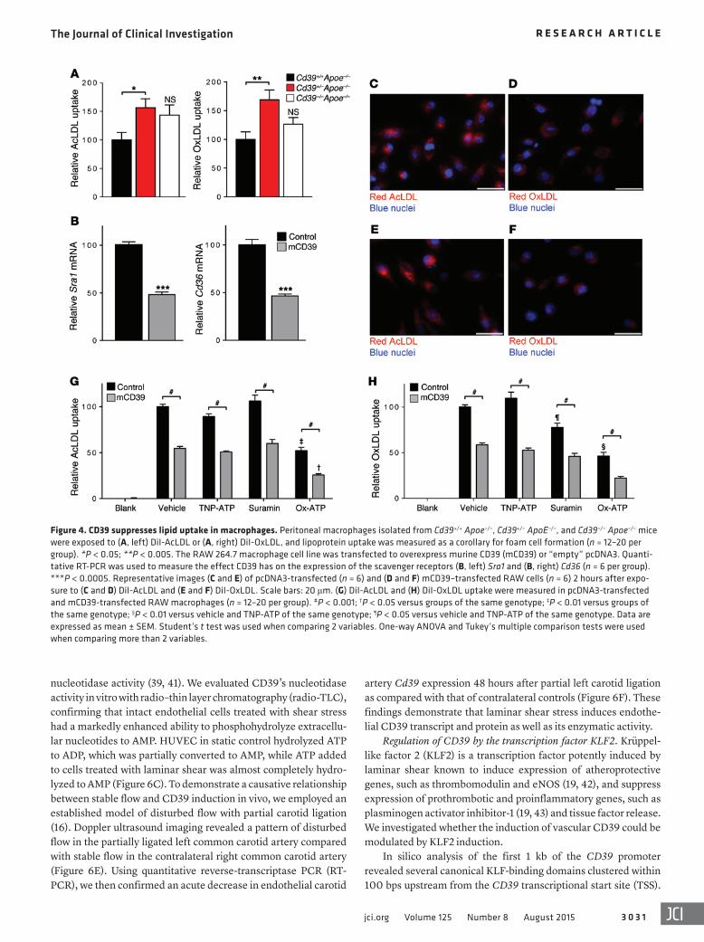
Enhanced lipid uptake in CD39-deficient macrophages. Macro-phages exert a central role in atherogenesis development by their migration, lipid uptake, foam cell formation, and proliferation (33). Previous studies have shown dysregulated migration in CD39-deficient macrophages (31, 34). Assessment of plaque lesions in Cd39+/– Apoe–/– and Cd39–/– Apoe–/– mice did not show appreciably altered macrophage numbers when compared with controls.
Although CD39 haplodeficiency confers a proatherogenic phenotype, the atherosclerotic burden seen in Cd39+/– Apoe–/– mice might not be explained by macrophage number. Modulation of foam cell formation has profound effects on the formation of aortic plaque without altering plaque macrophage content (35). We inves-tigated lipid uptake in macrophages deficient in CD39. Peritoneal macrophages from CD39 hemizygous mice took up significantly greater acetylated LDL (AcLDL) and oxidized LDL (OxLDL) than those from Apoe–/– controls (Figure 4A). Concordantly, CD39 over-expression in murine macrophages (31) suppressed expression of the scavenger receptors Sra1 and Cd36 (Figure 4B) and reduced both AcLDL and OxLDL uptake (Figure 4, C–F). To test the hypoth-esis that CD39-mediated dissipation of ATP reduces stimulation of ATP-sensitive receptors and thereby mitigates lipid scaveng-ing, we employed a pharmacologic approach to selectively inhibit
mouse aortas revealed a stark regionalization of CD39 expression. Robust CD39 expression was present on endothelial cells overlying regions of the vascular wall protected from atherosclerosis, whereas CD39 was significantly diminished over areas prone to atherosclero-sis (Figure 1, A and B). Cerium chloride staining of ATPase activity demonstrated a concordant loss of CD39 activity at these sites rela-tive to healthy vasculature in the same mouse (Figure 1, C and D).
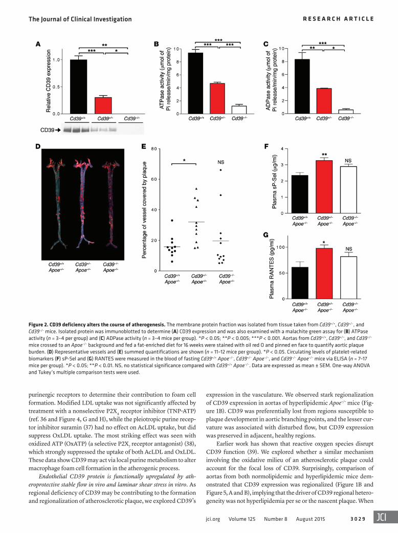
To assess whether loss of CD39 alters the course of atheroscle-rosis, Cd39–/– mice were crossed onto the hyperlipidemic Apoe–/– background to generate Cd39+/+ Apoe–/–, Cd39+/– Apoe–/–, and Cd39–/– Apoe–/– mice. Cd39+/– Apoe–/– mice were haploinsufficient with respect to CD39 expression, as enzymatic activity decreased in a dose-dependent manner with allele copy number (Figure 2, A–C). After 16 weeks of an atherogenic diet, mice missing only 1 allele of CD39 (Cd39+/– Apoe–/–) developed atherosclerotic lesions that were 2-fold larger than those of their Cd39+/+ Apoe–/– counterparts (Fig-ure 2, D and E) (n = 11–12, P < 0.005). Total absence of CD39, how-ever, did not alter total plaque burden relative to controls. In all experiments, heart rate, blood pressure, and cholesterol levels did not vary significantly between control and CD39-deficient mice (Supplemental Table 1; supplemental material available online with this article; doi:10.1172/JCI79514DS1).
Effect of CD39 deficiency on platelet activation and reactivity. We next investigated whether platelet activation is altered in CD39-deficient mice. We examined the composition of atherosclerotic plaques in CD39-deficient mice. Platelet remnants identified by the constitutive surface marker CD41/glycoprotein IIb appeared to localize to the surface and leading edge of atherosclerotic plaques. Platelet quantification revealed Cd39+/– Apoe–/– mice showed a ten-dency for platelet deposition in their atherosclerotic plaques as compared with controls, although this did not meet statistical sig-nificance (Supplemental Figure 1). We next examined circulating platelet-derived factors and observed a concordant increase in cir-culating soluble P-selectin (sP-Sel) and RANTES, at 39% and 60% higher concentrations, respectively, in the plasma of Cd39+/– Apoe–/– mice compared with Cd39+/+ Apoe–/– controls (Figure 2, F and G).
To examine platelet reactivity in CD39-deficient and hyperlip-idemic mice, we used whole blood aggregometry. Consistent with previous studies, Cd39–/– mice were desensitized to ADP-mediated
Figure 1. CD39 function and expression over atherosclerotic plaque. Coronal sections of an Apoe–/– aortic arch with atherosclerotic plaque are shown via (A) bright-field and (B) immunofluorescent microscopy. CD39 is shown in red and nuclei are blue (n = 3 mice). Scale bars: 25 μm. Transmis-sion electron micrographs of cerium chloride–stained ATPase activity in Apoe–/– aorta at sites of (C) healthy and (D) atherosclerotic vascular wall. Black cerium precipitate denotes sites of ATPase activity (arrowheads). n = 3 mice, representative images shown. Original magnification, ×34,000.
The Journal of Clinical Investigation R e s e a R c h a R t i c l e
3 0 2 9jci.org Volume 125 Number 8 August 2015
expression in the vasculature. We observed stark regionalization of CD39 expression in aortas of hyperlipidemic Apoe–/– mice (Fig-ure 1B). CD39 was preferentially lost from regions susceptible to plaque development in aortic branching points, and the lesser cur-vature was associated with disturbed flow, but CD39 expression was preserved in adjacent, healthy regions.
Earlier work has shown that reactive oxygen species disrupt CD39 function (39). We explored whether a similar mechanism involving the oxidative milieu of an atherosclerotic plaque could account for the focal loss of CD39. Surprisingly, comparison of aortas from both normolipidemic and hyperlipidemic mice dem-onstrated that CD39 expression was regionalized (Figure 1B and Figure 5, A and B), implying that the driver of CD39 regional hetero-geneity was not hyperlipidemia per se or the nascent plaque. When
purinergic receptors to determine their contribution to foam cell formation. Modified LDL uptake was not significantly affected by treatment with a nonselective P2X4 receptor inhibitor (TNP-ATP) (ref. 36 and Figure 4, G and H), while the pleiotropic purine recep-tor inhibitor suramin (37) had no effect on AcLDL uptake, but did suppress OxLDL uptake. The most striking effect was seen with oxidized ATP (OxATP) (a selective P2X7 receptor antagonist) (38), which strongly suppressed the uptake of both AcLDL and OxLDL. These data show CD39 may act via local purine metabolism to alter macrophage foam cell formation in the atherogenic process.
Endothelial CD39 protein is functionally upregulated by ath-eroprotective stable flow in vivo and laminar shear stress in vitro. As regional deficiency of CD39 may be contributing to the formation and regionalization of atherosclerotic plaque, we explored CD39’s
Figure 2. CD39 deficiency alters the course of atherogenesis. The membrane protein fraction was isolated from tissue taken from Cd39+/+, Cd39+/–, and Cd39–/– mice. Isolated protein was immunoblotted to determine (A) CD39 expression and was also examined with a malachite green assay for (B) ATPase activity (n = 3–4 per group) and (C) ADPase activity (n = 3–4 mice per group). *P < 0.05; **P < 0.005; ***P < 0.001. Aortas from Cd39+/+, Cd39+/–, and Cd39–/– mice crossed to an Apoe–/– background and fed a fat-enriched diet for 16 weeks were stained with oil red O and pinned en face to quantify aortic plaque burden. (D) Representative vessels and (E) summed quantifications are shown (n = 11–12 mice per group). *P < 0.05. Circulating levels of platelet-related biomarkers (F) sP-Sel and (G) RANTES were measured in the blood of fasting Cd39+/+ Apoe–/–, Cd39+/– Apoe–/–, and Cd39–/– Apoe–/– mice via ELISA (n = 7–17 mice per group). *P < 0.05; **P < 0.01. NS, no statistical significance compared with Cd39+/+ Apoe–/–. Data are expressed as mean ± SEM. One-way ANOVA and Tukey’s multiple comparison tests were used.
The Journal of Clinical Investigation R e s e a R c h a R t i c l e
3 0 3 0 jci.org Volume 125 Number 8 August 2015
significantly diminished activity in areas of turbulent blood flow. We extended these observations by exploring fluid shear stress as a regulator of CD39 expression in vitro using a cone-plate viscom-eter to treat endothelial cells with laminar shear stress. Antiath-erogenic, unidirectional laminar shear (15 dynes/cm2) induced a significant increase in transcripts of CD39 in human aortic endo-thelial cells (HAECs) and HUVECs (Figure 6A), but not of other ectonucleotidases expressed at the endothelial cell surface (ref. 40 and Supplemental Figure 2). We observed a corresponding increase in aortic and umbilical vein endothelial CD39 protein expression when compared with no-flow static controls (Figure 6B). Our group and others have previously demonstrated that CD39 undergoes posttranslational modification that modifies its
examining the branch points of the aortic arch, we noted a defined and consistent pattern of diminished CD39 expression at branch points in the aorta, but robustly expressed distal to the branch point. Similar patterns of expression with diminished expression at branch points in the vasculature are found in endothelial genes regulated by fluid shear stress. This led us to test the hypothesis that Cd39 is a shear stress–responsive gene, with areas of laminar fluid flow induc-ing and disturbed fluid flow suppressing CD39 expression.
To determine whether the regionalized vascular CD39 expres-sion was enzymatically active, we evaluated CD39’s ATPase activ-ity in aortic sections using a cerium chloride assay. Electron micro-graphs of the aortic branch points showed strong ATPase activity (Figure 5C) in areas associated with high laminar shear (9) and
Figure 3. Modulation of whole blood aggregometry by CD39. Whole blood was drawn from Cd39+/+, Cd39+/–, and Cd39–/– mice to test for ADP sensitivity. (A) Representative aggregometry responses were taken at different concentrations of ADP stimulation (n = 3–9 per group). Summed aggregometry data are shown for (B) normolipidemic (n = 3–9 per group) and (C) hyperlipidemic Cd39+/+, Cd39+/–, and Cd39–/– mice (n = 3–9 per group). *P < 0.05; **P < 0.01. Data are expressed as mean ± SEM. One-way ANOVA and Tukey’s multiple comparison tests were used.
The Journal of Clinical Investigation R e s e a R c h a R t i c l e
3 0 3 1jci.org Volume 125 Number 8 August 2015
artery Cd39 expression 48 hours after partial left carotid ligation as compared with that of contralateral controls (Figure 6F). These findings demonstrate that laminar shear stress induces endothe-lial CD39 transcript and protein as well as its enzymatic activity.
Regulation of CD39 by the transcription factor KLF2. Krüppel-like factor 2 (KLF2) is a transcription factor potently induced by laminar shear known to induce expression of atheroprotective genes, such as thrombomodulin and eNOS (19, 42), and suppress expression of prothrombotic and proinflammatory genes, such as plasminogen activator inhibitor-1 (19, 43) and tissue factor release. We investigated whether the induction of vascular CD39 could be modulated by KLF2 induction.
In silico analysis of the first 1 kb of the CD39 promoter revealed several canonical KLF-binding domains clustered within 100 bps upstream from the CD39 transcriptional start site (TSS).
nucleotidase activity (39, 41). We evaluated CD39’s nucleotidase activity in vitro with radio–thin layer chromatography (radio-TLC), confirming that intact endothelial cells treated with shear stress had a markedly enhanced ability to phosphohydrolyze extracellu-lar nucleotides to AMP. HUVEC in static control hydrolyzed ATP to ADP, which was partially converted to AMP, while ATP added to cells treated with laminar shear was almost completely hydro-lyzed to AMP (Figure 6C). To demonstrate a causative relationship between stable flow and CD39 induction in vivo, we employed an established model of disturbed flow with partial carotid ligation (16). Doppler ultrasound imaging revealed a pattern of disturbed flow in the partially ligated left common carotid artery compared with stable flow in the contralateral right common carotid artery (Figure 6E). Using quantitative reverse-transcriptase PCR (RT-PCR), we then confirmed an acute decrease in endothelial carotid
Figure 4. CD39 suppresses lipid uptake in macrophages. Peritoneal macrophages isolated from Cd39+/+ Apoe–/–, Cd39+/– ApoE–/–, and Cd39–/– Apoe–/– mice were exposed to (A, left) DiI-AcLDL or (A, right) DiI-OxLDL, and lipoprotein uptake was measured as a corollary for foam cell formation (n = 12–20 per group). *P < 0.05; **P < 0.005. The RAW 264.7 macrophage cell line was transfected to overexpress murine CD39 (mCD39) or “empty” pcDNA3. Quanti-tative RT-PCR was used to measure the effect CD39 has on the expression of the scavenger receptors (B, left) Sra1 and (B, right) Cd36 (n = 6 per group). ***P < 0.0005. Representative images (C and E) of pcDNA3-transfected (n = 6) and (D and F) mCD39–transfected RAW cells (n = 6) 2 hours after expo-sure to (C and D) DiI-AcLDL and (E and F) DiI-OxLDL. Scale bars: 20 μm. (G) DiI-AcLDL and (H) DiI-OxLDL uptake were measured in pcDNA3-transfected and mCD39-transfected RAW macrophages (n = 12–20 per group). #P < 0.001; †P < 0.05 versus groups of the same genotype; ‡P < 0.01 versus groups of the same genotype; §P < 0.01 versus vehicle and TNP-ATP of the same genotype; ¶P < 0.05 versus vehicle and TNP-ATP of the same genotype. Data are expressed as mean ± SEM. Student’s t test was used when comparing 2 variables. One-way ANOVA and Tukey’s multiple comparison tests were used when comparing more than 2 variables.
The Journal of Clinical Investigation R e s e a R c h a R t i c l e
3 0 3 2 jci.org Volume 125 Number 8 August 2015
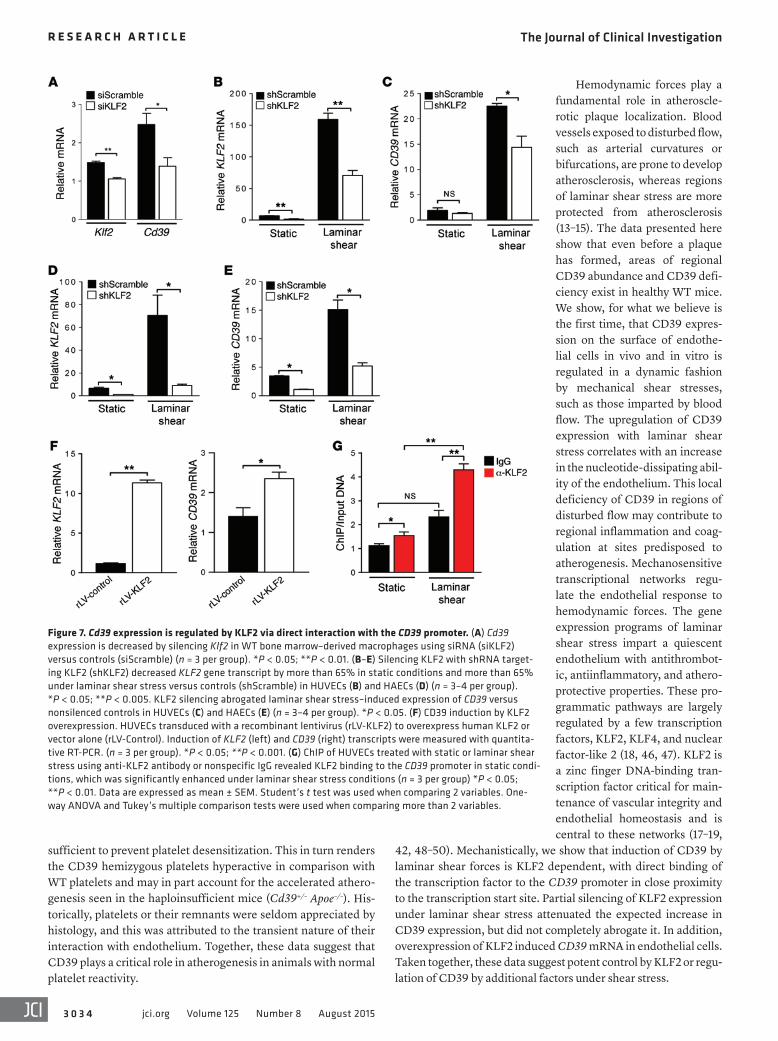
To investigate whether KLF2 could regulate CD39 expression, we silenced KLF2 in WT bone marrow–derived macrophages and noted that even a modest, 29% reduction in Klf2 expression resulted in a significant, 44% decrease in Cd39 expression (Figure 7A). To determine whether this regulatory effect persists across different vascular cell types, we then silenced KLF2 in endothelial cells. Reduction of KLF2 expression by 75% led to a nonsignificant decrease in CD39 expression under static shear conditions (Fig-ure 7, B and C). However, under conditions of laminar shear stress, we observed a 55% decrease in KLF2 expression under laminar shear conditions with shKLF2 transduction, leading to a signifi-cant, 36% suppression of endothelial CD39 gene expression when compared with transduction with a nontargeting sequence (Figure 7, B and C). We next sought to examine whether overexpression of KLF2 could induce CD39 expression. Transducing endothelial cells with a lentivirus to overexpress KLF2 resulted in a concordant increase in CD39 transcript (Figure 7D). This implies KLF2 plays a critical role in driving the basal and shear-induced upregulation of CD39 expression in macrophages and the endothelium.
KLF2 regulates CD39 expression by direct binding to the CD39 pro-moter. With the observation that KLF2 regulates CD39 expression, we next examined whether KLF2’s transcriptional control occurs via direct binding to the CD39 promoter. We focused on the candi-date sites between the TSS and 100 bp upstream of the TSS. ChIP using an anti-KLF2 or nonspecific IgG antibody was performed on HUVECs exposed to 48 hours of laminar or static shear stress. Using quantitative RT-PCR analysis, we determined that KLF2 is bound to the CD39 promoter in static conditions when compared with IgG control antibody and this binding was significantly enhanced with arterial levels of shear stress (Figure 7E). These results suggest that induction of CD39 expression under arterial laminar shear stress
conditions is transcriptionally regulated by direct binding of the transcription factor KLF2 to the CD39 promoter.
DiscussionThis study presents what we believe is the first direct evidence linking CD39 expression to atherosclerotic plaque formation via increased platelet reactivity and foam cell formation. This finding is complemented by the identification of a regionalized pattern of endothelial CD39 expression, driven by fluid phase shear stress, within the aorta. Furthermore, we show that induction of CD39 is regulated by the highly conserved transcription factor KLF2, sug-gesting an endogenous system of atheroprotection.
Our findings help elucidate a potential mechanism based upon purinergic signaling leading to plaque formation. Humans with coro-nary atherosclerosis have been shown to have abnormal ADPase and ATPase activity (44). Under normal conditions, CD39 is a primary regulator of vascular homeostasis due to its avid catabolism of ATP and ADP (22, 23, 26, 30). Oxidative stress is a factor in the milieu of a plaque that can also affect CD39 by suppressing enzymatic activ-ity independently of CD39 expression (39). Free radicals and TNF-α both play a role in the development of atherosclerotic plaque and are both known to suppress the activity of endothelial CD39. The loss of CD39 expression over plaque (Figure 1, A–D) may synergize with the local oxidative environment to further suppress regional CD39 function. The end result would be an atherosclerotic plaque virtu-ally devoid of ectoapyrase activity on its surface (Figure 5C). Conse-quently, when CD39 expression is reduced by only 50%, atheroscle-rotic plaque burden is more than doubled (Figure 2, D and E).
These focal losses of CD39 as seen over atherosclerotic plaque could be of great consequence with respect to atherosclerosis. Pre-vious studies have shown the critical role of CD39 in autoregula-
Figure 5. Regional expression of CD39 in the murine aorta. Coronal sections of a WT aorta. (A) Analysis of the WT aortic arch showed a stark regionalization of CD39 expression (red) and a consis-tent pattern centering on aortic branch points. (B) The same image is overlaid with bright-field imaging of the region. Original magnification, ×20. Arrowhead designates innominate artery ostium. Arrow designates left common carotid artery ostium. Dashed arrow designates left subclavian artery ostium. n = 3 mice. Transmission electron micrographs show cerium chloride staining for ATPase activ-ity in the luminal face of the left common carotid artery branch (C, left) proximal to the carotid ostium, where disturbed flow predominates and (C, right) distal to the carotid ostium where blood flow is stable. Black cerium precipitate (denoted by arrowheads) identifies sites of ATPase activity. Original magnification, ×19,000. n = 5 mice, representative images shown.
The Journal of Clinical Investigation R e s e a R c h a R t i c l e
3 0 3 3jci.org Volume 125 Number 8 August 2015
tion of leukocyte trafficking into regions of inflammation (31, 45). Simultaneously, deficiency of CD39 predisposes macrophages to markedly increase uptake of OxLDL and AcLDL due to overstimu-lation by purines, which could lead to augmented foam cell forma-tion. Concordantly, CD39 overexpression decreased macrophage expression of the scavenger receptors CD36 and SRA-1, the latter of which is thought to be a critical mediator of lipid influx into mac-rophages and progression of atherosclerosis (33). Circulating acti-vated platelets are known to accelerate plaque formation in Apoe–/– mice (5). Though the combination of platelets desensitized from excess extracellular nucleotide stimulation and enhanced foam cell
formation does not increase atherosclerosis in Cd39–/– Apoe–/– mice, the summation of exaggerated platelet reactivity and foam cell for-mation in mice hemizygous for CD39 (Cd39+/– Apoe–/–) is associated with a profound increase in atherosclerotic plaque burden.
The results presented here and elsewhere (26, 30, 31) indicate that in the complete absence of CD39, platelet P2Y12 receptors are overwhelmed by ambient nucleotides and become desensitized to further stimulation. Mice in which platelets are desensitized (Cd39–/–) exhibit significantly blunted platelet reactivity to ago-nist challenge and a concordant blunting of atherogenesis. Our data show that partial expression of CD39 in mice (Cd39+/–) is
Figure 6. Laminar shear stress induces endothelial CD39 expression and nucleotidase activity in vitro and in vivo. HUVECs and HAECs were exposed to laminar shear stress (15 dynes/cm2) for 48 hours and then assessed for (A) CD39 transcript expression, as measured by quantitative RT-PCR (n = 3–6 per group), and (B) protein expression, as measured by immunoblotting (n = 3–6 per group). Samples exposed to shear stress were compared with static con-trols. *P < 0.05; **P < 0.005. TLC demonstrated (C, representative plots, and D) enhanced CD39-mediated phosphohydrolysis of radiolabeled nucleotides by intact endothelial cells following laminar shear stress versus static conditions (n = 3–6 per group). *P < 0.05. Partial ligation of left common carotid artery in mice causes disturbed flow and reduced endothelial CD39 expression. Representative ultrasound demonstrating flow velocity profiles in the right (RCA) and left (LCA) common carotid artery revealing (E) disturbed flow (right) 48 hours following partial LCA ligation with diastolic flow reversal in the LCA (indicated by arrow), while contralateral RCA flow remains stable (left) (n = 3 mice). (F) Carotid endothelial expression of Klf2 and Cd39 is decreased under disturbed flow following partial carotid ligation (n = 3 mice) *P < 0.05, Student’s t test. Data are expressed as mean ± SEM.
The Journal of Clinical Investigation R e s e a R c h a R t i c l e
3 0 3 4 jci.org Volume 125 Number 8 August 2015
Hemodynamic forces play a fundamental role in atheroscle-rotic plaque localization. Blood vessels exposed to disturbed flow, such as arterial curvatures or bifurcations, are prone to develop atherosclerosis, whereas regions of laminar shear stress are more protected from atherosclerosis (13–15). The data presented here show that even before a plaque has formed, areas of regional CD39 abundance and CD39 defi-ciency exist in healthy WT mice. We show, for what we believe is the first time, that CD39 expres-sion on the surface of endothe-lial cells in vivo and in vitro is regulated in a dynamic fashion by mechanical shear stresses, such as those imparted by blood flow. The upregulation of CD39 expression with laminar shear stress correlates with an increase in the nucleotide-dissipating abil-ity of the endothelium. This local deficiency of CD39 in regions of disturbed flow may contribute to regional inflammation and coag-ulation at sites predisposed to atherogenesis. Mechanosensitive transcriptional networks regu-late the endothelial response to hemodynamic forces. The gene expression programs of laminar shear stress impart a quiescent endothelium with antithrombot-ic, antiinflammatory, and athero-protective properties. These pro-grammatic pathways are largely regulated by a few transcription factors, KLF2, KLF4, and nuclear factor-like 2 (18, 46, 47). KLF2 is a zinc finger DNA-binding tran-scription factor critical for main-tenance of vascular integrity and endothelial homeostasis and is central to these networks (17–19,
42, 48–50). Mechanistically, we show that induction of CD39 by laminar shear forces is KLF2 dependent, with direct binding of the transcription factor to the CD39 promoter in close proximity to the transcription start site. Partial silencing of KLF2 expression under laminar shear stress attenuated the expected increase in CD39 expression, but did not completely abrogate it. In addition, overexpression of KLF2 induced CD39 mRNA in endothelial cells. Taken together, these data suggest potent control by KLF2 or regu-lation of CD39 by additional factors under shear stress.
sufficient to prevent platelet desensitization. This in turn renders the CD39 hemizygous platelets hyperactive in comparison with WT platelets and may in part account for the accelerated athero-genesis seen in the haploinsufficient mice (Cd39+/– Apoe–/–). His-torically, platelets or their remnants were seldom appreciated by histology, and this was attributed to the transient nature of their interaction with endothelium. Together, these data suggest that CD39 plays a critical role in atherogenesis in animals with normal platelet reactivity.
Figure 7. Cd39 expression is regulated by KLF2 via direct interaction with the CD39 promoter. (A) Cd39 expression is decreased by silencing Klf2 in WT bone marrow–derived macrophages using siRNA (siKLF2) versus controls (siScramble) (n = 3 per group). *P < 0.05; **P < 0.01. (B–E) Silencing KLF2 with shRNA target-ing KLF2 (shKLF2) decreased KLF2 gene transcript by more than 65% in static conditions and more than 65% under laminar shear stress versus controls (shScramble) in HUVECs (B) and HAECs (D) (n = 3–4 per group). *P < 0.05; **P < 0.005. KLF2 silencing abrogated laminar shear stress–induced expression of CD39 versus nonsilenced controls in HUVECs (C) and HAECs (E) (n = 3–4 per group). *P < 0.05. (F) CD39 induction by KLF2 overexpression. HUVECs transduced with a recombinant lentivirus (rLV-KLF2) to overexpress human KLF2 or vector alone (rLV-Control). Induction of KLF2 (left) and CD39 (right) transcripts were measured with quantita-tive RT-PCR. (n = 3 per group). *P < 0.05; **P < 0.001. (G) ChIP of HUVECs treated with static or laminar shear stress using anti-KLF2 antibody or nonspecific IgG revealed KLF2 binding to the CD39 promoter in static condi-tions, which was significantly enhanced under laminar shear stress conditions (n = 3 per group) *P < 0.05; **P < 0.01. Data are expressed as mean ± SEM. Student’s t test was used when comparing 2 variables. One-way ANOVA and Tukey’s multiple comparison tests were used when comparing more than 2 variables.
The Journal of Clinical Investigation R e s e a R c h a R t i c l e
3 0 3 5jci.org Volume 125 Number 8 August 2015
dal branches of the left carotid artery (left external carotid, internal carotid, and occipital artery) were ligated, leaving the superior thyroid artery intact. Disturbed flow was confirmed with ultrasonography and endothelial RNA harvested for gene-expression analysis. For detailed information, see Supplemental Methods.
ChIP assay. HUVECs were treated with laminar shear stress or stat-ic conditions for 48 hours. Equal aliquots of isolated crosslinked chro-matin were subjected to immunoprecipitation with rabbit polyclonal anti-human KLF2 antibody or with negative control rabbit IgG. DNA was isolated and used as a template for the PCR to amplify the CD39 promoter sequence containing the KLF2 consensus binding site closest to the TSS using quantitative RT-PCR in 3 separate experiments. Quan-titative RT-PCR was performed with Brilliant SYBR green mix.
Animal breeding and experimental protocol. For detailed informa-tion, see Supplemental Methods.
Statistics. Values are expressed as mean ± SEM, with the number of experiments performed provided in the figure legends. Data were analyzed using GraphPad Prism 6.05. For experiments in which 2 vari-ables were compared, unpaired 2-tailed Student’s t tests were used with Welch’s correction. For experiments in which more than 2 variables were compared, 1-way ANOVA was used with Tukey’s post hoc analysis to test for significant differences. P < 0.05 was considered significant.
Study approval. All animal experiments were conducted in accordance with NIH guidelines for the use of live animals and were approved by the Institutional Animal Care and Use Committee at the University of Michigan.
AcknowledgmentsWe thank Sandeep Kumar, Chanwoo Kim, Wakako Takabe, Dor-othy Sorenson, and the University of Michigan Cardiovascular Physiology Phenotyping Core, Vector, Imaging, and Hybridoma Cores for expert technical assistance. D.J. Pinsky was supported by grant funding from the NIH (HL127151, NS087147) and the A. Alfred Taubman Medical Research Institute. D.J. Pinsky received additional funding support from the J. Griswold Ruth MD & Mar-gery Hopkins Ruth Professorship. H. Jo was supported by grant funding from the NIH (HL119798). Y. Kanthi and N.R. Sutton were supported by training grants from the NIH (T32HL007853). S.H. Visovatti and S.N. Goonewardena were supported by grant funding from the National Heart, Lung, and Blood Institute of the NIH (K08HL119623 and K08HL123621, respectively). M.C. Hyman and A.E. Baek were supported in part by predoctoral fel-lowship grants from the American Heart Association. The Uni-versity of Michigan Physiology Phenotyping Core was supported by grant funding from the NIH (OD016502) and the Frankel Cardiovascular Center.
Address correspondence to: David J. Pinsky, 7240 Medical Science Research Building III, 1150 West Medical Center Drive, Ann Arbor, Michigan 48109, USA. Phone: 734.936.3500; E-mail: [email protected].
In conclusion, we have uncovered an important role for extracellular nucleotide dissipation by CD39 in the development of atherogenesis. These experiments further identify a regulato-ry mechanism of endothelial and macrophage CD39 expression by KLF2. It will be important to determine whether manipula-tion of KLF2 represents a novel way to harness the therapeutic potential of CD39.
MethodsTissue harvesting, preparation, oil red O staining, and immunostaining. For detailed information, see Supplemental Methods.
Cerium chloride staining of ATPase activity. Apoe–/– mice (26 weeks old) were euthanized prior to cerium chloride staining of the aorta to measure liberation of inorganic phosphate following ATP application as previously described (51). For detailed information, see Supplemental Methods.
Cell culture, cell and tissue protein isolation, immunoblotting, histol-ogy, RNA analysis, and measurement of plasma cytokines and lipids. For detailed information, see Supplemental Methods.
Measurement of CD39 function. Apyrase activity of the purified mouse membrane fraction was assayed with a Malachite Green Phos-phate Assay Kit (BioAssay Systems). Radio-TLC was used to assess CD39 enzymatic function of live HUVECs as previously described (52). Equal numbers of cells were mixed with 1.0 mM [8-14C]ATP (ARC) in the presence of 286 μM AOPCP (Sigma-Aldrich), a CD73 inhibitor to arrest conversion of AMP to adenosine, and incubated at 37°C for 40 minutes. Reactions were stopped using 8 M formic acid, spotted onto silica gel TLC plates (Invitrogen), and nucleotides were separated by TLC. For detailed information, see Supplemental Methods.
Whole blood aggregometry, LDL uptake assay. For detailed informa-tion, see Supplemental Methods.
KLF2 gene-silencing and overexpression studies. shRNA oligonucle-otides were generated against KLF2 (shKLF2) (50) and a nonspecific control (shScramble) (19) (Invitrogen). Lentivirus was produced and titrations performed by the University of Michigan Vector Core. Pri-mary HUVECs and HAECs (PCS-100-011, ATCC) were transduced with shKLF2 or shScramble for 48 hours prior to treatment with lami-nar shear stress. Bone marrow–derived macrophages were transfected with siRNA (Invitrogen) against KLF2 (siKLF2) or a scramble sequence (siScramble) as previously described (53). Human KLF2 cDNA (Ori-gene) was inserted into a lentivirus plasmid and pseudovirus with or without the cDNA insert generated using Lenti-vpak Packaging Kit (Origene) according to manufacturer protocol. Titrations were per-formed according to manufacturer protocol. For detailed information, see Supplemental Methods.
In vitro shear stress studies. Cells were exposed to static shear con-ditions or arterial levels of laminar shear stress using a cone-and-plate shear apparatus, as modified for a 60-mm tissue culture dish (Corn-ing) (54). For detailed information, see Supplemental Methods.
Partial carotid ligation to induce disturbed flow. Disturbed carotid flow was induced in 8- to 10-week-old male C57BL/6 mice (Jackson Laboratories) as previously described (16, 21). Briefly, 3 of 4 cau-
1. Lozano R, et al. Global and regional mortality from 235 causes of death for 20 age groups in 1990 and 2010: a systematic analysis for the Global Burden of Disease Study 2010. Lancet. 2012;380(9859):2095–2128.
2. von Hundelshausen P, et al. RANTES deposition by platelets triggers monocyte arrest on inflamed and atherosclerotic endothelium. Circulation. 2001;103(13):1772–1777.
3. Koltsova EK, et al. Dynamic T cell-APC interac-
tions sustain chronic inflammation in atheroscle-rosis. J Clin Invest. 2012;122(9):3114–3126.
4. Dunn J, et al. Flow-dependent epigenetic DNA methylation regulates endothelial gene expression and atherosclerosis. J Clin Invest.
The Journal of Clinical Investigation R e s e a R c h a R t i c l e
3 0 3 6 jci.org Volume 125 Number 8 August 2015
2014;124(7):3187–3199. 5. Huo Y, et al. Circulating activated platelets exac-
erbate atherosclerosis in mice deficient in apoli-poprotein E. Nat Med. 2003;9(1):61–67.
6. Schober A, et al. Deposition of platelet RANTES triggering monocyte recruitment requires P-selectin and is involved in neointima for-mation after arterial injury. Circulation. 2002;106(12):1523–1529.
7. Mendelsohn ME, Loscalzo J. Role of platelets in cholesteryl ester formation by U-937 cells. J Clin Invest. 1988;81(1):62–68.
8. Kockx MM, et al. Phagocytosis and macro-phage activation associated with hemorrhagic microvessels in human atherosclerosis. Arterio-scler Thromb Vasc Biol. 2003;23(3):440–446.
9. Suo J, Ferrara DE, Sorescu D, Guldberg RE, Taylor WR, Giddens DP. Hemodynamic shear stresses in mouse aortas: implications for atherogenesis. Arterioscler Thromb Vasc Biol. 2007;27(2):346–351.
10. Iiyama K, et al. Patterns of vascular cell adhesion molecule-1 and intercellular adhesion molecule-1 expression in rabbit and mouse atherosclerotic lesions and at sites predisposed to lesion forma-tion. Circ Res. 1999;85(2):199–207.
11. Nakashima Y, Raines EW, Plump AS, Breslow JL, Ross R. Upregulation of VCAM-1 and ICAM-1 at atherosclerosis-prone sites on the endothelium in the ApoE-deficient mouse. Arterioscler Thromb Vasc Biol. 1998;18(5):842–851.
12. Sorescu GP, et al. Bone morphogenic protein 4 produced in endothelial cells by oscillatory shear stress induces monocyte adhesion by stimulating reactive oxygen species production from a nox1-based NADPH oxidase. Circ Res. 2004;95(8):773–779.
13. Zarins CK, Giddens DP, Bharadvaj BK, Sottiurai VS, Mabon RF, Glagov S. Carotid bifurcation ath-erosclerosis. Quantitative correlation of plaque localization with flow velocity profiles and wall shear stress. Circ Res. 1983;53(4):502–514.
14. Ku DN, Giddens DP, Zarins CK, Glagov S. Pulsa-tile flow and atherosclerosis in the human carotid bifurcation. Positive correlation between plaque location and low oscillating shear stress. Arterio-sclerosis. 1985;5(3):293–302.
15. Glagov S, Zarins C, Giddens DP, Ku DN. Hemo-dynamics and atherosclerosis. Insights and per-spectives gained from studies of human arteries. Arch Pathol Lab Med. 1988;112(10):1018–1031.
16. Nam D, et al. Partial carotid ligation is a model of acutely induced disturbed flow, leading to rapid endothelial dysfunction and ath-erosclerosis. Am J Physiol Heart Circ Physiol. 2009;297(4):H1535–H1543.
17. Atkins GB, et al. Hemizygous deficiency of Kruppel-like factor 2 augments experimental atherosclerosis. Circ Res. 2008;103(7):690–693.
18. SenBanerjee S, et al. KLF2 Is a novel transcrip-tional regulator of endothelial proinflammatory activation. J Exp Med. 2004;199(10):1305–1315.
19. Lin Z, et al. Kruppel-like factor 2 (KLF2) regu-lates endothelial thrombotic function. Circ Res. 2005;96(5):e48–e57.
20. Ni CW, et al. Discovery of novel mechanosensi-tive genes in vivo using mouse carotid artery
endothelium exposed to disturbed flow. Blood. 2010;116(15):e66–e73.
21. Son DJ, et al. The atypical mechanosensitive microRNA-712 derived from pre-ribosomal RNA induces endothelial inflammation and athero-sclerosis. Nat Commun. 2013;4:3000.
22. Kaczmarek E, et al. Identification and character-ization of CD39/vascular ATP diphosphohydro-lase. J Biol Chem. 1996;271(51):33116–33122.
23. Marcus AJ, et al. The endothelial cell ecto-ADPase responsible for inhibition of platelet function is CD39. J Clin Invest. 1997;99(6):1351–1360.
24. Marcus AJ, et al. Role of CD39 (NTPDase-1) in thromboregulation, cerebroprotection, and cardioprotection. Semin Thromb Hemost. 2005;31(2):234–246.
25. Gayle RB 3rd, et al. Inhibition of platelet function by recombinant soluble ecto-ADPase/CD39. J Clin Invest. 1998;101(9):1851–1859.
26. Pinsky DJ, et al. Elucidation of the thromboregu-latory role of CD39/ectoapyrase in the ischemic brain. J Clin Invest. 2002;109(8):1031–1040.
27. Remijn JA, et al. Role of ADP receptor P2Y(12) in platelet adhesion and thrombus formation in flowing blood. Arterioscler Thromb Vasc Biol. 2002;22(4):686–691.
28. Kittel A, Kiss AL, Mullner N, Matko I, Sperlagh B. Expression of NTPDase1 and caveolins in human cardiovascular disease. Histochem Cell Biol. 2005;124(1):51–59.
29. Kohler D, et al. CD39/ectonucleoside triphos-phate diphosphohydrolase 1 provides myocardial protection during cardiac ischemia/reperfusion injury. Circulation. 2007;116(16):1784–1794.
30. Enjyoji K, et al. Targeted disruption of cd39/ATP diphosphohydrolase results in disordered hemostasis and thromboregulation. Nat Med. 1999;5(9):1010–1017.
31. Hyman MC, et al. Self-regulation of inflammatory cell trafficking in mice by the leukocyte surface apy-rase CD39. J Clin Invest. 2009;119(5):1136–1149.
32. Podrez EA, et al. Platelet CD36 links hyperlipid-emia, oxidant stress and a prothrombotic pheno-type. Nat Med. 2007;13(9):1086–1095.
33. Robbins CS, et al. Local proliferation dominates lesional macrophage accumulation in atheroscle-rosis. Nat Med. 2013;19(9):1166–1172.
34. Goepfert C, et al. Disordered cellular migration and angiogenesis in cd39-null mice. Circulation. 2001;104(25):3109–3115.
35. Ricci R, et al. Requirement of JNK2 for scavenger receptor A-mediated foam cell formation in ath-erogenesis. Science. 2004;306(5701):1558–1561.
36. Sakaki H, Fujiwaki T, Tsukimoto M, Kawano A, Harada H, Kojima S. P2X4 receptor regulates P2X7 receptor-dependent IL-1β and IL-18 release in mouse bone marrow-derived dendritic cells. Bio-chem Biophys Res Commun. 2013;432(3):406–411.
37. Hoyle CH, Knight GE, Burnstock G. Suramin antagonizes responses to P2-purinoceptor ago-nists and purinergic nerve stimulation in the guinea-pig urinary bladder and taenia coli. Br J Pharmacol. 1990;99(3):617–621.
38. Wang X, et al. P2X7 receptor inhibition improves recovery after spinal cord injury. Nat Med. 2004;10(8):821–827.
39. Robson SC, et al. Loss of ATP diphosphohydro-
lase activity with endothelial cell activation. J Exp Med. 1997;185(1):153–163.
40. Sevigny J, et al. Differential catalytic properties and vascular topography of murine nucleoside triphosphate diphosphohydrolase 1 (NTPDase1) and NTPDase2 have implications for thrombo-regulation. Blood. 2002;99(8):2801–2809.
41. Baek AE, Kanthi Y, Sutton NR, Liao H, Pinsky DJ. Regulation of ecto-apyrase CD39 (ENTPD1) expression by phosphodiesterase III (PDE3). FASEB J. 2013;27(11):4419–4428.
42. Parmar KM, et al. Integration of flow-dependent endothelial phenotypes by Kruppel-like factor 2. J Clin Invest. 2006;116(1):49–58.
43. Topper JN, Cai J, Falb D, Gimbrone MA, Gimbro-ne MA Jr. Identification of vascular endothelial genes differentially responsive to fluid mechani-cal stimuli: cyclooxygenase-2, manganese super-oxide dismutase, and endothelial cell nitric oxide synthase are selectively up-regulated by steady laminar shear stress. Proc Natl Acad Sci U S A. 1996;93(19):10417–10422.
44. El-Omar MM, et al. The ratio of ADP- to ATP-ectonucleotidase activity is reduced in patients with coronary artery disease. Thromb Res. 2005;116(3):199–206.
45. Reutershan J, Vollmer I, Stark S, Wagner R, Ngamsri KC, Eltzschig HK. Adenosine and inflammation: CD39 and CD73 are critical mediators in LPS-induced PMN trafficking into the lungs. FASEB J. 2009;23(2):473–482.
46. Fledderus JO, et al. KLF2 primes the antioxi-dant transcription factor Nrf2 for activation in endothelial cells. Arterioscler Thromb Vasc Biol. 2008;28(7):1339–1346.
47. Hamik A, et al. Kruppel-like factor 4 regu-lates endothelial inflammation. J Biol Chem. 2007;282(18):13769–13779.
48. Atkins GB, Jain MK. Role of Kruppel-like tran-scription factors in endothelial biology. Circ Res. 2007;100(12):1686–1695.
49. Mahabeleshwar GH, et al. The myeloid transcrip-tion factor KLF2 regulates the host response to polymicrobial infection and endotoxic shock. Immunity. 2011;34(5):715–728.
50. Sen-Banerjee S, et al. Kruppel-like factor 2 as a novel mediator of statin effects in endothelial cells. Circulation. 2005;112(5):720–726.
51. Kittel A. Lipopolysaccharide treatment modifies pH- and cation-dependent ecto-ATPase activ-ity of endothelial cells. J Histochem Cytochem. 1999;47(3):393–400.
52. Visovatti SH, Hyman MC, Bouis D, Neubig R, McLaughlin VV, Pinsky DJ. Increased CD39 nucleotidase activity on microparticles from patients with idiopathic pulmonary arterial hypertension. PLoS One. 2012;7(7):e40829.
54. Rhee WJ, Ni CW, Zheng Z, Chang K, Jo H, Bao G. HuR regulates the expression of stress-sensitive genes and mediates inflammatory response in human umbilical vein endothelial cells. Proc Natl Acad Sci U S A. 2010;107(15):6858–6863.


![[XLS] · Web viewTARAPUR GOPALPURGHAT NADIA 741122 PMS/07-08/22898 MITHUN MANDAL NAZRUL MANDAL BIDUPUR SARKAR PARA MURSHIDABAD 742305 PMS/07-08/22899 MITHUN MONDAL LOTIMUDDIN MONDLA](https://static.documents.pub/doc/80x56/5afda3cf7f8b9a256b8be4c2/xls-viewtarapur-gopalpurghat-nadia-741122-pms07-0822898-mithun-mandal-nazrul.jpg)


