Leishmania Typing with Fluorogenic Assays • JID 2005:192 (15 August) • 685 MAJOR ARTICLE Fluorogenic Assay for Molecular Typing of the Leishmania donovani Complex: Taxonomic and Clinical Applications Kelly Wilber Quispe-Tintaya, 1,2 Thierry Laurent, 1 Saskia Decuypere, 1 Mallorie Hide, 3 Anne-Laure Ban ˜ uls, 3 Simonne De Doncker, 1 Suman Rijal, 4 Carmen Can ˜ avate, 5 Lenea Campino, 6 and Jean-Claude Dujardin 1 1 Laboratory of Molecular Parasitology, Prins Leopold Instituut voor Tropische Geneeskunde, Antwerp, and 2 Unite ´ de Recherche en Biologie Mole ´culaire, Faculte ´s Notre Dame de la Paix, Namur, Belgium; 3 Ge ´ne ´tique et Evolution des Maladies Infectieuses, Unite ´ Mixte Recherche Centre National de Recherche Scientifique/Institut de Recherche pour le De ´veloppement 2724, Montpellier, France; 4 B. P. Koirala Institute for Health Sciences, Dharan, Nepal; 5 Centro Nacional de Microbiologia, Instituto de Salud Carlos III, Madrid, Spain; 6 Unidade de Leishmanioses/Centro de Mala ´ria Doenc ¸as Tropicais, Instituto de Higiene e Medicina Tropical, Lisbon, Portugal We describe a new fluorogenic assay for the identification of species and intraspecies groups within the Leishmania donovani complex. The assay combined (1) 2 polymerase chain reactions targeting the 2 cysteine proteinase b isogenes and (2) a fluorescence-resonance energy transfer/melting curve analysis of the poly- morphisms within a 31-nt region. All strains within the L. donovani complex were distinguished from L. tropica, L. major, and L. aethiopica, and 5 distinct groups were identified within the L. donovani complex. Discrepancies were observed with the present taxonomy on the basis of isoenzyme analysis and concerned East African strains, which suggests the need for a systematic reevaluation of the taxonomy. The capacity to type parasites directly from clinical samples was demonstrated with blood and bone marrow samples. This rapid and high-throughput alternative for molecular diagnosis and epidemiological studies of visceral leish- maniasis could be adapted for use with other Leishmania species. Leishmaniasis affects 88 countries and 12 million peo- ple, and 350 million people are estimated to be at risk for developing the disease. Visceral leishmaniasis (VL), which is lethal if untreated, is the most severe mani- festation of the disease, and an estimated 500,000 new cases occur each year [1]. VL is (re-)emerging and spreading worldwide, because of several risk factors, such as drug resistance, HIV coinfection, human mi- grations, and environmental changes [2]. Monitoring this expansion is essential for disease control and re- quires molecular epidemiological tools that allow for Received 8 February 2005; accepted 22 March 2005; electronically published 6 July 2005. Potential conflicts of interest: none reported. Financial support: European Commission (contracts QLK2-CT-2001-01810 and ICA4-CT-2001-10076). Reprints or correspondence: Prof. Jean-Claude Dujardin, Prins Leopold Instituut voor Tropische Geneeskunde, Molecular Parasitology, Nationalest. 155, B-2000 Ant- werp, Belgium ([email protected]). The Journal of Infectious Diseases 2005; 192:685–92 2005 by the Infectious Diseases Society of America. All rights reserved. 0022-1899/2005/19204-0018$15.00 rapid identification of Leishmania species and intra- species groups. Indeed, VL is caused by several species of the Leish- mania donovani complex that differ markedly in their epidemiological characteristics. L. infantum is the eti- ological agent of zoonotic VL in Europe, Africa, China, and Latin America (in the latter region, it is called L. chagasi). L. donovani causes an anthroponotic form of the disease and is primarily restricted to East Africa and the Indian subcontinent, where 50% of the VL cases reported worldwide occur [1]. L. archibaldi produces zoonotic leishmaniasis in East Africa [3, 4]. The present reference method for the identification of Leishmania species is multilocus enzyme electrophoresis (MLEE) [3]. This method is isolation and cultivation dependent and is limited to specialized centers. Therefore, several polymerase chain reaction (PCR)–based methods have been developed for parasite genotyping: PCR amplifi- cation followed by restriction fragment–length poly- morphism (PCR-RFLP) analysis [5–8], single-stranded conformation polymorphism (SSCP) analysis [9], or
Simonne De Doncker,1 Suman Rijal,4 Carmen Canavate,5 Lenea Campino,6 and Jean-Claude Dujardin1
1Laboratory of Molecular Parasitology, Prins Leopold Instituut voor Tropische Geneeskunde, Antwerp, and 2Unite de Recherche en BiologieMoleculaire, Facultes Notre Dame de la Paix, Namur, Belgium; 3Genetique et Evolution des Maladies Infectieuses, Unite Mixte Recherche CentreNational de Recherche Scientifique/Institut de Recherche pour le Developpement 2724, Montpellier, France; 4B. P. Koirala Institute for HealthSciences, Dharan, Nepal; 5Centro Nacional de Microbiologia, Instituto de Salud Carlos III, Madrid, Spain; 6Unidade de Leishmanioses/Centrode Malaria Doencas Tropicais, Instituto de Higiene e Medicina Tropical, Lisbon, Portugal
We describe a new fluorogenic assay for the identification of species and intraspecies groups within theLeishmania donovani complex. The assay combined (1) 2 polymerase chain reactions targeting the 2 cysteineproteinase b isogenes and (2) a fluorescence-resonance energy transfer/melting curve analysis of the poly-morphisms within a 31-nt region. All strains within the L. donovani complex were distinguished from L.tropica, L. major, and L. aethiopica, and 5 distinct groups were identified within the L. donovani complex.Discrepancies were observed with the present taxonomy on the basis of isoenzyme analysis and concernedEast African strains, which suggests the need for a systematic reevaluation of the taxonomy. The capacity totype parasites directly from clinical samples was demonstrated with blood and bone marrow samples. Thisrapid and high-throughput alternative for molecular diagnosis and epidemiological studies of visceral leish-maniasis could be adapted for use with other Leishmania species.
Leishmaniasis affects 88 countries and 12 million peo-
ple, and 350 million people are estimated to be at risk
for developing the disease. Visceral leishmaniasis (VL),
which is lethal if untreated, is the most severe mani-
festation of the disease, and an estimated 500,000 new
cases occur each year [1]. VL is (re-)emerging and
spreading worldwide, because of several risk factors,
such as drug resistance, HIV coinfection, human mi-
grations, and environmental changes [2]. Monitoring
this expansion is essential for disease control and re-
quires molecular epidemiological tools that allow for
Received 8 February 2005; accepted 22 March 2005; electronically published6 July 2005.
Potential conflicts of interest: none reported.Financial support: European Commission (contracts QLK2-CT-2001-01810 and
ICA4-CT-2001-10076).Reprints or correspondence: Prof. Jean-Claude Dujardin, Prins Leopold Instituut
The Journal of Infectious Diseases 2005; 192:685–92� 2005 by the Infectious Diseases Society of America. All rights reserved.0022-1899/2005/19204-0018$15.00
rapid identification of Leishmania species and intra-
species groups.
Indeed, VL is caused by several species of the Leish-
mania donovani complex that differ markedly in their
epidemiological characteristics. L. infantum is the eti-
ological agent of zoonotic VL in Europe, Africa, China,
and Latin America (in the latter region, it is called L.
chagasi). L. donovani causes an anthroponotic form of
the disease and is primarily restricted to East Africa and
the Indian subcontinent, where 50% of the VL cases
reported worldwide occur [1]. L. archibaldi produces
zoonotic leishmaniasis in East Africa [3, 4]. The present
reference method for the identification of Leishmania
species is multilocus enzyme electrophoresis (MLEE)
[3]. This method is isolation and cultivation dependent
and is limited to specialized centers. Therefore, several
polymerase chain reaction (PCR)–based methods have
been developed for parasite genotyping: PCR amplifi-
cation followed by restriction fragment–length poly-
686 • JID 2005:192 (15 August) • Quispe-Tintaya et al.
Table 1. Leishmania strains used in this study.
Strain, international codeCountryof origin Zymodeme
L. infantumMHOM/FR/1978/LEM75 France MON1MHOM/FR/1995/LPN114(LEM3001) France MON1MHOM/ES/1993/PM1(LEM2608) Spain MON1MHOM/FR/1997/LSL29(LEM3420) France MON1MHOM/ES/1986/BCN16(LEM1078) Spain MON1MHOM/PT/2000/IMT260(LEM3975) Portugal MON1MHOM/FR/1996/LEM3249 France MON29MHOM/ES/1991/LEM2298 Spain MON183MHOM/SD/1982/GILANIa Sudan MON30MHOM/FR/1980/LEM189 France MON11MHOM/SD/62/3Sa Sudan MON81MHOM/ES/88/LLM175 Spain MON198MHOM/ES/92/LLM373 Spain MON199MHOM/IT/94ISS1036 Italy MON228MHOM/IT/93/ISS800 Italy MON188MHOM/SD/97/LEM3472a Sudan MON267
L. donovaniMHOM/IN/00/DEVI(LEM138) India MON2MHOM/IN/1996/THAK35(LEM3178) India MON2MHOM/ET/00/HUSSEN Ethiopia LON42MCAN/SD/2000/LEM3946 Sudan MON274
L. archibaldiMHOM/ET/1972/GEBRE1 Ethiopia MON82MHOM/SD/97/LEM3429 Sudan MON257MHOM/SD/97/LEM3463 Sudan MON258
L. tropica, MHOM/SU/74/K27 Russia …L. aethiopica, MHOM/ET/72/L100 Ethiopia MON14L. major, MHOM/SU/73/5-ASKH Russia MON4
NOTE. Taxonomic classification of these strains was based on multilocusenzyme electrophoresis typing.
a The taxonomic status of the zymodeme (L. infantum or L. donovani) isgiven in Discussion.
sequence analysis [10, 11]. These methods allow a direct anal-
ysis of host samples, but they involve several steps after PCR,
so use of them increases the amount of necessary equipment,
the time needed for the analysis, the intensity of the labor, and
the risk of DNA contamination [12, 13]. Furthermore, these
methods generally do not allow high-throughput applications.
Fluorogenic hybridization probes offer an attractive comple-
ment to PCR: they require only a single piece of equipment (a
real-time PCR thermal cycler), and they offer the possibility
for rapid and high-throughput analyses with high sensitivity
[13]. In Leishmania species fluorogenic assays have so far been
used primarily for detection and quantification of the patho-
gens [14, 15] or discrimination of Leishmania complexes [12].
One study mentions the discrimination of species [14], but
validation of the method was limited to 1 strain/species.
In the present study, we developed a fluorogenic PCR assay
for typing parasites of the L. donovani complex. The cysteine
proteinase b (cpb) gene was selected as the target, for several
reasons. First, this gene is repeated, which is important for the
sensitivity of PCR detection. Second, the repeats are polymor-
phic [6] and are organized into 2 major groups of isogenes
according to species [11]: A, B, C, or D (A–D) isogenes and
E or F (E/F) isogenes. A physical map of the locus indicates
that A–D isogenes are quite similar to each other, whereas A–
D isogenes show 76%–78% identity with E/F isogenes [16].
Third, in a previous work, we showed that PCR-RFLP analysis
of this locus was able to discriminate species and geographical
populations within the L. donovani complex [6]. The discrim-
inatory power of our fluorogenic assay was tested on a panel
of strains representative of the L. donovani complex, and the
assay was applied to clinical samples.
MATERIALS AND METHODS
Leishmania strains and samples. Twenty-three Leishmania
strains belonging to the L. donovani complex (L. infantum, L.
donovani, and L. archibaldi) were selected on the basis of their
different geographical origins and their classification in various
zymodemes. Even though the taxonomic status of some zymo-
demes is under debate (see Discussion), we refer throughout
this work to the MLEE-based species identification. Three
strains of other Old World Leishmania species (L. tropica, L.
aethiopica, and L. major) were used as out groups (table 1).
Clinical samples from patients with confirmed VL were ob-
tained from the B. P. Koirala Institute of Health Sciences
(Dharan, Nepal) and the Instituto de Salud Carlos III (Madrid,
Spain). Clinical samples from dogs were obtained from the
Instituto de Higiene e Medicina Tropical (Lisbon, Portugal).
Informed consent was obtained from patients or their parents
or guardians, the human-experimentation guidelines of the Prins
Leopold Instituut voor Tropische Geneeskunde (Antwerp, Bel-
gium) were followed, and ethical clearance was obtained from
the review boards of all institutions that provided samples. Eight
human bone marrow aspirates (BMAs; collected on EDTA), 2
human venous blood samples (180 mL; collected on EDTA and
mixed with an equal volume of AS1 buffer [Qiagen]), and 3
spleen aspirates from dogs were obtained. DNA was extracted
using the QIAamp DNA Mini Kit, for BMAs and spleen as-
pirates, or the QIAamp DNA Blood Mini Kit (both from Qia-
gen), for venous blood samples, in accordance with the man-
ufacturer’s instructions. The detection threshold of our method
was defined by processing healthy human blood containing
known amounts of parasites (in concentrations ranging from
to 1 L. donovani MHOM/SD/00/1S promastigote/18041 � 10
mL of blood) by use of the same procedures.
Primers/probe design. Sequences of the cpb gene were ob-
tained from GenBank (accession numbers AF004592, AF309626,
U43706, AF309627, and AF217087). These sequences were used
to design the PCR primers and the probe (table 2) with the
program Primer Premier (version 5.0; Premier Biosoft Interna-
Table 2. Oligonucleotides used for heminested polymerase chain reaction amplification and fluorogenic assay of cysteine proteinaseb A–D (cpbA–D) and E/F (cbpE/F) isogenes of Leishmania strains.
Primer/probe SequenceGenBank accession no.
(nucleotide position)
7forUniv cpbA–D and cpbE/F sense primera 5′-TGaGGTTCCGTACTGGGTG-3′ AF004592 (1245–1263)Arev8 cpbA–D reverse primera,b 5′-GGACCAAAGCAATGAGGG-3′ AF004592 (1581–1598)Dos12.1 cpbE/F reverse primera,b 5′-AGCATCACTGTCCcGCATG-3′ AF309627 (1070–1088)PanchoCPB cpbA–D and cpbE/F sense primerb 5′-GGCGAtAAGGGtTACGTGC-3′ AF004592 (1291–1309)PdCPB probe 5′-CACGCGTTCAGCCCCATGACCACGCGCACGT-3′ AF004592 (1304–1334)
NOTE. Nucleotide changes introduced to avoid secondary structures are in lowercase letters; bold type indicates nucleotides labeled with ROX (PanchoCPB)or FAM (PdCPB).
a Used in the first PCR run.b Used in the second PCR run.
Figure 1. Fluorescence-resonance energy transfer/melting curve analysis of cysteine proteinase b A–D (A) and E/F (B) isogenes of Leishmaniastrains. The change in the amount of fluorescence (in relative fluorescence units [RFU]) for each polymerase chain reaction product and probe wasplotted against the temperature (T), and its negative derivative (�d) appeared as a positive peak (melting peak). The thick horizontal line denotes thethreshold for background fluorescence, and the curve entirely below that line denotes the results for the nontemplate control. T1–6 denote the differentmelting peaks used to characterize the strains.
tional). Two heminested PCR assays were developed: 1 for the
amplification of cpbA–D and 1 for the amplification of cpbE/F.
cpbA–D copies were amplified using the primer sets 7forUniv
and Arev8 (first run) and PanchoCPB and Arev8 (second run).
cpbE/F copies were amplified using the primer sets 7forUniv and
Dos12.1 (first run) and PanchoCPB and Dos12.1 (second run).
PCR assay and fluorescence-resonance energy transfer/melt-
ing curve analysis (FRET/MCA). The PCR assay and the
688 • JID 2005:192 (15 August) • Quispe-Tintaya et al.
Table 3. Partial sequences of the cysteine proteinase b A–D (cpbA–D) and E/F (cbpE/F)variants of Leishmania strains by melting temperature (Tm) of the corresponding plasmid.
NOTE. T1–6 denote the different melting peaks used to characterize the strains. Underlined nucleotidesshow regions of mismatch.
FRET/MCA were performed on the iCycler (Bio-Rad). The first
round of each PCR was performed with a 50-mL PCR reaction
mix containing 20–50 ng of DNA, 1� PCR buffer, 1.0 mmol/
L (final concentration) MgCl2, 800 mmol/L dNTPs, 300 nmol/
limits of flocculation each primer, and 1.5 U of Taq DNA poly-
merase (Eurogentec). The second round of each PCR was per-
formed with a 50-mL PCR reaction mix containing 1 mL of PCR
product from the first round, 1� iQ supermix buffer (Bio-
Rad), 500 nmol/L sense primer, and 100 nmol/L antisense
primer. The same thermal cycling parameters were used for the
first and second rounds of PCR: initial denaturation at 95�C
for 5 min; 35 cycles consisting of denaturation at 95�C for 30
s, annealing at 55�C for 1 min, and extension at 72�C for 40
s; and a final extension at 72�C for 8 min. After the second
round of PCR, the probe was added to a final concentration
of 200 nmol/L for use in the FRET/MCA. The FRET/MCA was
performed as follows: 95�C for 10 min, 65�C for 5 min and 10
s, and 50 cycles of 10 s during which the temperature was
increased by 0.7�C. The assays were performed in duplicate to
verify the reproducibility of the results. During the FRET/MCA,
the change in the amount of fluorescence (in relative fluores-
cence units) for each PCR product and probe was plotted
against the temperature (melting curve), and its negative de-
rivative appeared as a positive peak (melting peak). A coefficient
of variation (cv; ) was calculated for es-cv p 100 � SD/mean
timating the variation of the melting temperature (Tm) between
different strains of the same species or group of parasites.
RESULTS
FRET/MCA of cpb in Leishmania strains. The FRET/MCA of
cpbA–D and cpbE/F was performed for the 26 Leishmania strains.
Experiments on reproducibility showed that, for a given sam-
ple, the interexperimental SD for Tm was !0.4�C. The 3 species
not in the L. donovani complex (L. tropica, L. aethiopica, and L.
major) showed an amplification product during the PCR only
for cpbA–D and a single melting peak characterized by a mean
Tm of 74.0�C (hereafter “T1”; cv between strains, 0.4%); there
was no amplification product during the PCR for cpbE/F, because
that isogene is present in only the L. donovani complex [11]. All
23 strains of the L. donovani complex had a positive signal in
both cpb assays. In the assay targeting cpbA–D, a single melting
peak characterized each strain, and 3 different values for Tm were
observed (figure 1A). All East African strains showed a mean Tm
that was undistinguishable from that of the out groups: 73.8�C
(T1; cv, 0.4%). Two other groups were distinguished: Indian
strains with a mean Tm of 81.8�C (hereafter, “T2”; cv, 0.0%) and
European strains with a mean Tm of 83.0�C (hereafter, “T3”; cv,
0.5%). In the assay targeting cpbE/F, a single melting peak also
characterized each strain, and 3 different values for Tm were
observed (figure 1B). Three groups were distinguished: (1) East
African strains of zymodemes LON42, MON274, MON257,
MON258, and MON267 and Indian strains with a mean Tm of
74.2�C (hereafter, “T4”; cv, 0.4%); (2) European strains of zy-
modemes MON1, MON11, MON183, MON188, MON198,
MON199, and MON228 and East African strains of zymo-
demes MON30, MON81, and MON82 with a mean Tm of 80.2�C
(hereafter, “T5”; cv, 0.4%); and (3) the European strain of
zymodeme MON29 with a mean Tm of 83.0�C (hereafter, “T6”).
When the results of the 2 assays were combined, (1) all strains
of the L. donovani complex could be distinguished from the
group formed by L. tropica, L. aethiopica, and L. major, and
(2) 5 groups were observed within the L. donovani complex.
FRET/MCA of cloned cpb sequence variants. To better un-
derstand the profiles observed in Leishmania strains, we cloned
and sequenced cpb amplicons corresponding to the 6 melting
peaks described above. Then, the FRET/MCA was performed
on PCR products of the corresponding plasmids. For the cpbA–
D plasmids, 3 sequence variants were observed in the region
targeted by the probe. Their Tm values corresponded with a
difference of !0.4�C, compared with those observed in Leish-
mania strains, and varied—as expected—with the number of
nucleotide mismatches with the probe (table 3): 83.3�C (pLG1,
no mismatch), 81.8�C (pLG10, 2 mismatches), and 73.8�C
(pLG11, 3 mismatches). For cpbE/F plasmids, 3 sequence var-
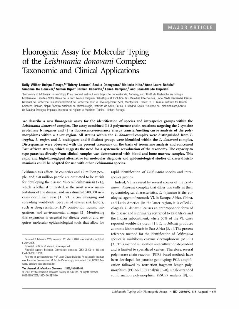
Figure 2. Fluorescence-resonance energy transfer/melting curve analysis of the cysteine proteinase b A–D (A and C ) and E/F isogenes (B and D ) of Leishmania strains in clinical samples from Nepal(A and B ) and Europe (C and D ). The change in the amount of fluorescence (in relative fluorescence units [RFU]) for each PCR product and probe was plotted against the temperature (T), and its negativederivative (�d) appeared as a positive peak (melting peak). The thick horizontal line denotes the threshold for background fluorescence, and the curve entirely below that line denotes the results forthe nontemplate control. T2–5 denote the different melting peaks used to characterize the strains.
690 • JID 2005:192 (15 August) • Quispe-Tintaya et al.
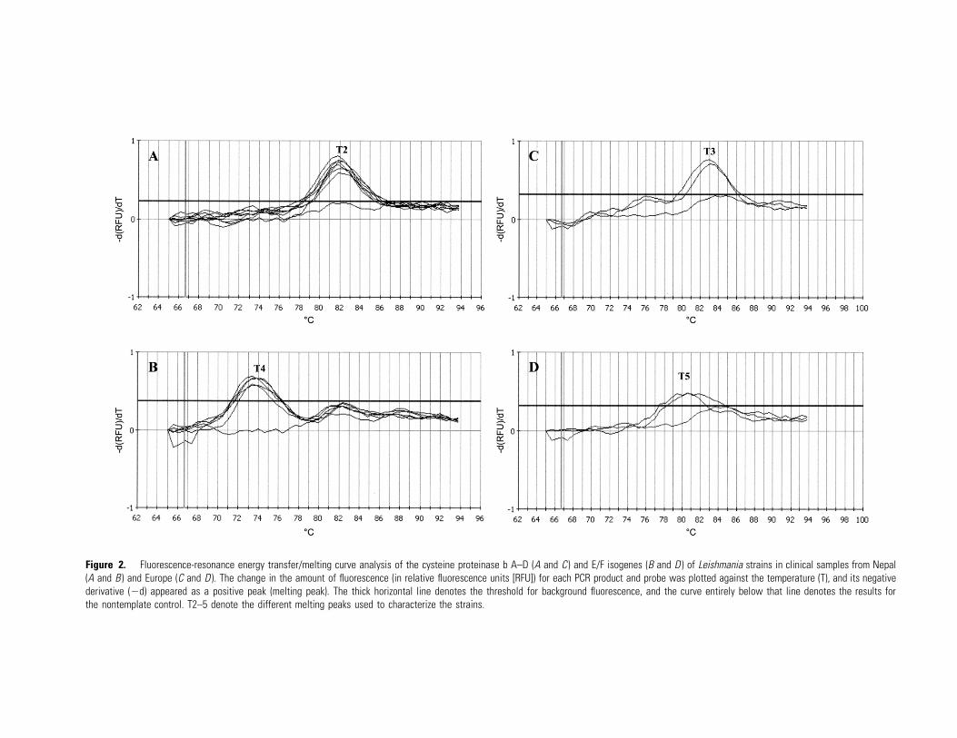
Figure 3. Classification of the 6 different Leishmania groups on the basis of fluorescence-resonance energy transfer/melting curve analysis ofcysteine proteinase b A–D (cpbA–D) and E/F (cpbE/F) isogenes. T1–6 denote the different melting peaks used to characterize the strains. PCR,polymerase chain reaction.
iants were observed in the region targeted by the probe, and
the same relationship between Tm values and nucleotide mis-
matches was observed. Notably, for the region targeted by the
probe, 2 of the 3 cpbA–D sequence variants (characterized by
T1 and T3) corresponded to 1 of the sequence variants ob-
served in cpbE/F (characterized by T4 and T6).
Detection threshold and clinical samples. In healthy hu-
man blood containing known amounts of L. donovani pro-
mastigotes, a positive signal was obtained in the FRET/MCA
until a concentration of 0.5 and 5 parasites/mL of blood was
used, respectively, in the assays targeting cpbA–D and cpbE/F.
This result was expected, because there are more copies of
cpbA–D than cpbE/F [16]. DNA extracted from clinical sam-
ples—5 BMAs and 2 venous blood samples from Nepalese
patients, 3 BMAs from Spanish patients, and 3 spleen aspirates
from Portuguese dogs, all with confirmed VL—were used to
test the capability of both assays to perform a direct charac-
terization, without isolation and culture, of the parasites. For
the 13 samples, a positive signal was obtained in both assays.
For the Nepalese samples, the same strain was observed, and
it was found to be similar to Indian L. donovani strains (figure
2A and 2B): Tm of 81.9�C and 73.65�C for cpbA–D and cpbE/
F, respectively. For the Spanish and Portuguese samples, the
strain was found to be similar to L. infantum strains of the
Europe I group (figure 2C and 2D): Tm of 83.0�C and 80.6�C
for cpbA–D and cpbE/F, respectively.
DISCUSSION
In the present study, we developed a new fluorogenic probe-
based PCR assay for the identification of species and intraspe-
cies groups within the L. donovani complex. The assay combines
(1) 2 PCRs targeting members of the 2 different cpb isogenes
(A–D and E/F) and (2) the FRET/MCA of the polymorphism
within a 31-nt region common to both cpb isogenes. With-
in the present samples, we observed, by FRET/MCA, 3 melting
peaks for each cpb isogene, and we confirmed, by sequencing,
that they corresponded to different variants of the 31-nt region,
some of which were shared in cpbA–D and cpbE/F isogenes.
The study of each of the cpbA–D and cpbE/F isogenes allowed
us to distinguish groups of strains, but the highest resolution
was obtained when the results of both analyses were combined.
Six different groups were found: (1) L. tropica, L. aethiopica,
and L. major; (2) European strains classified as L. infantum zy-
modemes MON1, MON11, MON183, MON188, MON198,
MON199, and MON228; (3) European strains classified as L.
infantum zymodeme MON29; (4) East African strains classified
as L. infantum zymodemes MON30 and MON81 and L. archi-
baldi zymodeme MON82; (5) East African strains classified as
L. infantum zymodeme MON267, L. donovani zymodemes LON42
and MON274, and L. archibaldi zymodemes MON257 and
MON258; and (6) Indian strains classified as L. donovani zy-
Discrepancies were thus observed between the present tax-
onomy, which is based on MLEE, and our results, and these
concerned East African strains of the L. donovani complex. Sim-
ilar discrepancies that concerned the same strains and the same
endemic region have been reported. Random amplified poly-
morphic DNA analysis and PCR-RFLP analysis of intragenic and
intergenic regions of cpb and gp63 genes [6] and ribosomal in-
ternal transcribed spacers and miniexon genes [7] showed that
strains classified as zymodeme MON30 did not cluster with L.
infantum and did not support the status of zymodeme MON82
as a separate species (L. archibaldi). The convergent results ob-
tained with non-MLEE markers strengthen the need for a sys-
tematic reevaluation of the L. donovani complex that would in-
clude epidemiological and clinical aspects [17].
Species delineation within the L. donovani complex is rela-
tively difficult in East Africa, which is thought to be the origin
of VL [18] and where a broad continuum of genetic diversity
can be observed in wild-type strains [6]. Our data fit perfectly
with this perception of the genus’s evolution, and they provide
evidence for the following series of events in cpb gene variation.
If L. tropica, L. major, and L. aethiopica are members of the
out group (cpbA–D [T1 type]), the members of East African
groups I and II are closest to it genetically on the basis of their
cpbA–D type (also T1), but diverge from it on the basis of their
cpbE/F type (T5 and T4, respectively). From these 2 groups, 2
major branches would stem: (1) the European strains of L.
infantum (group I), which are similar to the members of East
African group I on the basis of their cpbE/F type (T5) but are
different on the basis of their cpbA–D type (T3), and (2) the
Indian strains of L. donovani, which are similar to the members
of East African group II on the basis of their cpbE/F type (T4)
but are different on the basis of their cpbA–D type (T2). Ac-
cording to our data, the zymodeme MON29 (Europe group II
of L. infantum) would branch from the European strains of
the L. infantum cluster: identical cpbA–D type (T3) but different
cpbE/F type (T6). This observation is particularly interesting
from a clinical point of view, because zymodeme MON29 par-
asites are generally associated with benign cutaneous lesions
[19]. The difference described here should be validated using
more strains of zymodeme MON29. This difference might be
particularly relevant when considering the important role that
the cpb gene plays in Leishmania virulence [16, 20].
Finally, the capacity to type the parasites directly in clinical
samples was tested with venous blood and BMAs from Nepa-
lese and Spanish patients and spleen aspirates from Portuguese
dogs, all with confirmed VL. In all samples, typing of the par-
asites was possible and revealed the presence of parasites simi-
lar to either Indian strains of L. donovani (Nepal) or European
strains (group II) of L. infantum. A more extensive study to
test the sensitivity of our assay, particularly for samples with
very low parasite loads, is required. In the present format of
the FRET/MCA, the parasite detection threshold—without spe-
cies typing—for which cpbA–D isogene analysis was sufficient,
was 0.5 parasites/mL of blood. With respect to the typing thresh-
old, for which cpbA–D and cpbE/F isogene analysis is required,
a higher parasite load was needed (5 parasites/mL of blood),
because of the limiting effect of the single-copy cpbE/F isogene.
These results are dependent on the performance of the PCR
assay itself, which could be further optimized. However, the
DNA extraction and concentration method also played a major
role. We used the QIAamp DNA Blood Mini Kit, which requires
a starting volume of 180 mL of blood, and other methods that
use larger volumes of blood or buffy coat are likely to increase
both detection and typing thresholds [21, 22].
The FRET/MCA is a rapid, high-throughput method that
can be directly applied in any laboratory equipped with a PCR
setup that allows for the detection of fluorogenic probes. As
such, our method represents a significant alternative for the
molecular typing of Leishmania species that can be used in
diagnostic and epidemiological studies. We also confirm that
cpb genes are adequate targets for the FRET/MCA. These genes
have also been shown to be very informative in the genetic
characterization of neotropical Leishmania species [23]. Fur-
ther research might include the development of a FRET/MCA
that is applicable in the context of New World leishmaniasis.
Acknowledgment
We thank Xavier De Bolle for his critical reading of the manuscript.
References
1. Desjeux P. Leishmaniasis public health aspects and control. Clin Der-matol 1996; 14:417–23.
2. Desjeux P. The increase in risk factors for leishmaniasis worldwide.Trans R Soc Trop Med Hyg 2001; 95:239–43.
3. Rioux JA, Lanotte G, Serres E, Pratlong F, Bastien P, Perieres J. Tax-onomy of Leishmania. Use of isoenzymes. Suggestions for a new clas-sification. Ann Parasitol Hum Comp 1990; 65:111–25.
4. Dereure J, Boni M, Pratlong F, et al. Visceral leishmaniasis in Sudan:first identifications of Leishmania from dogs. Trans R Soc Trop MedHyg 2000; 94:154–5.
5. Guerbouj S, Victoir K, Guizani I, et al. Gp63 gene polymorphism andpopulation structure of Leishmania donovani complex: influence of thehost selection pressure? Parasitology 2001; 122:25–35.
6. Quispe Tintaya KW, Ying X, Dedet JP, et al. Antigen genes for molecularepidemiology of leishmaniasis: polymorphism of cysteine proteinase band surface metalloprotease glycoprotein 63 in the Leishmania donovanicomplex. J Infect Dis 2004; 189:1035–43.
7. Mauricio IL, Stothard JR, Miles MA. Leishmania donovani complex:genotyping with the ribosomal internal transcribed spacer and themini-exon. Parasitology 2004; 128:263–7.
8. Marfurt J, Niederwieser I, Makia ND, et al. Diagnostic genotyping ofOld and New World Leishmania species by PCR-RFLP. Diagn MicrobiolInfect Dis 2003; 46:115–24.
9. Lewin S, Schonian G, El Tai N, et al. Strain typing in Leishmaniadonovani by using sequence-confirmed amplified region analysis. IntJ Parasitol 2002; 32:1267–76.
10. El Tai NO, El Fari M, Mauricio I, et al. Leishmania donovani: intra-
692 • JID 2005:192 (15 August) • Quispe-Tintaya et al.
specific polymorphisms of Sudanese isolates revealed by PCR-basedanalyses and DNA sequencing. Exp Parasitol 2001; 97:35–44.
11. Hide M. Variabilite pathogenique du complexe Leishmania donovani,agent de la leishmaniose viscerale: etude comparative des caracteresbiologiques, genetiques et d’expression genique [PhD thesis]. Mont-pellier, France: University of Montpellier, 2004.
12. Schulz A, Mellenthin K, Schonian G, et al. Detection, differentiation,and quantitation of pathogenic Leishmania organisms by a fluorescenceresonance energy transfer-based real-time PCR assay. J Clin Microbiol2003; 41:1529–35.
13. Bell AS, Ranford-Cartwright LC. Real-time quantitative PCR in par-asitology. Trends Parasitol 2002; 18:337–42.
14. Nicolas L, Milon G, Prina E. Rapid differentiation of Old World Leish-mania species by LightCycler polymerase chain reaction and meltingcurve analysis. J Microbiol Methods 2002; 51:295–9.
15. Mary C, Faraut F, Lascombe L, et al. Quantification of Leishmaniainfantum DNA by a real-time PCR assay with high sensitivity. J ClinMicrobiol 2004; 42:5249–55.
16. Mundodi V, Somanna A, Farrell P, Gedamu L. Genomic organizationand functional expression of differentially regulated cysteine proteasegenes of Leishmania donovani complex. Gene 2002; 282:257–65.
17. Tibayrenc M. Genetic epidemiology of parasitic protozoa and other
infectious agents: the need for an integrated approach. Int J Parasitol1998; 28:85–104.
18. Pratlong F, Dereure J, Bucheton B, et al. Sudan: the possible originalfocus of visceral leishmaniasis. Parasitology 2001; 122:599–605.
19. Pratlong F, Rioux JA, Marty P, et al. Isoenzymatic analysis of 712 strainsof Leishmania infantum in the south of France and relationship of en-zymatic polymorphism to clinical and epidemiological features. J ClinMicrobiol 2004; 42:4077–82.
20. Coombs GH, Mottram JC. Parasite proteinases and amino acid me-tabolism: possibilities for chemotherapeutic exploitation. Parasitology1997; 114:S61–80.
21. Lachaud L, Chabbert E, Dubessay P, et al. Comparison of varioussample preparation methods for PCR diagnosis of visceral leishmaniasisusing peripheral blood. J Clin Microbiol 2001; 39:613–7.
22. Avila HA, Sigman DS, Cohen LM, et al. Polymerase chain reactionamplification of Trypanosoma cruzi kinetoplast minicircle DNA isolatedfrom whole blood lysates: diagnosis of chronic Chagas’ disease. MolBiochem Parasitol 1991; 48:211–21.
23. Garcia L, Kindt A, Quispe-Tintaya KW, et al. American tegumentaryleishmaniasis: antigen-gene polymorphism, taxonomy and clinical ple-omorphism. Infect Genet Evol 2005; 5:109–16.