1 Fluoroscopic Imaging Equipment Guidelines for Detector Input Dose Settings and Image Optimization Phil Rauch Henry Ford Health Systems Detroit, MI 2009 AAPM Meeting-Phil Rauch Entrance Air Kerma for 22 cm FOV (Measured at Interventional Ref Point) 0 20 40 60 80 100 120 140 4 6 8 10 Nominal Phantom Thickness (inches) Patient Entrance Air Kerma Rate (mGy/min) RELATIVE EERD vs kVp 80% 90% 100% 110% 120% 130% 140% 150% 160% 170% 180% 190% 200% 210% 40 50 60 70 80 90 100 110 kVp Detector Exposure Rate Normalized to Minimum Shimadzu Bransist - 22 cm FOV Direct Flat Panel Detector (Se/TFT) Philips Xper - 22 cm FOV Indirect Flat Panel Detector (CsI(Tl)/TFT) Siemens Artis Zee - 42 cm FOV Indirect Flat Panel Detector (CsI(Tl)/TFT) Siemens Siregraph - 22 cm FOV X-ray Image Intensifier Detector 2009 AAPM Meeting-Phil Rauch Fluoroscopy Evaluation Patient Dose Image Quality 2009 AAPM Meeting-Phil Rauch Dose Reduction vs Image Quality Dose reduction depends on…. ….technology ….proper equipment design ….proper set up of equipment parameters ….proper utilization of the equipment ….knowledge and skill of the radiologist 2009 AAPM Meeting-Phil Rauch Image quality depends on…. ….technology ….proper equipment design ….proper set up of equipment parameters ….proper utilization of the equipment ….knowledge and skill of the radiologist Dose Reduction vs Image Quality
Transcript
1
Fluoroscopic Imaging EquipmentGuidelines for Detector Input Dose Settings
and Image Optimization
Phil RauchHenry Ford Health Systems
Detroit, MI2009 AAPM Meeting-Phil Rauch
Entrance Air Kerma for 22 cm FOV(Measured at Interventional Ref Point)
0
20
40
60
80
100
120
140
46
810Nominal Phantom Thickness (inches)
Pa
tien
t E
ntr
ance
Air
Ker
ma
Ra
te (
mG
y/m
in)
No Grid-70 kV 10R AF 2kW 4ms/7msmicrofocusNote: Thi s one at 40 nGy/pulse and 10 pps100kV 10R A F 2kW smallfocus
No Grid-70kV 10R A F 2kW 4ms/7msmicrofocusNote: Thi s one at 23 nGy/pulse and 30 pps80kV 10R 0302 3kW smallfocusNote: Thi s one at 36 nGy/pulse and 15 ppsso meas. Dose was mul tiplied by 2Intervent ional IQ 2
80kV 10R 0302 2kW smallfocusNote: Thi s one at 36 nGy/pulse and 15 ppsso meas. Dose was mul tiplied by 270kV 10R A F 2kW 5ms/8ms microfocus
80kV 10R A F 2kW smallfocus
80kV 10R 02 3kW small focus
Intervent ional IQ 1 (no DHHS)
70kV 10R A F 2kW microfocus
70kV Service 02
70kV 10R A F 3kW 6ms/9ms small focus
70kV 10R A F 3kW 8ms/12ms smallfocus
70kV 10R 02 3kW small focus
70kV 10R 02 2kW small focus
70kV 20R A F 3kW smallfocus
70kV 10R A F 3kW smallfocus
70kV 20R A F 3kW 8ms/12ms smallfocus
70kV Service 00
RELATIVE EERD vs kVp
80%
90%
100%
110%
120%
130%
140%
150%
160%
170%
180%
190%
200%
210%
40 50 60 70 80 90 100 110
kVp
Det
ecto
r E
xpo
sure
Rat
e
No
rmal
ized
to
Min
imu
m
Shimadzu Bransist - 22 cm FOV Direct
Flat Panel Detector (Se/TFT)
Philips Xper - 22 cm FOV Indirect Flat
Panel Detector (CsI(Tl)/TFT)
Siemens Artis Zee - 42 cm FOV Indirect
Flat Panel Detector (CsI(Tl)/TFT)
Siemens Siregraph - 22 cm FOV X-ray
Image Intensifier Detector
2009 AAPM Meeting-Phil Rauch
Fluoroscopy Evaluation
�Patient Dose�Image Quality
2009 AAPM Meeting-Phil Rauch
Dose Reduction vs Image Quality
�Dose reduction depends on….….technology….proper equipment design….proper set up of equipment parameters….proper utilization of the equipment….knowledge and skill of the radiologist
2009 AAPM Meeting-Phil Rauch
� Image quality depends on….….technology….proper equipment design….proper set up of equipment parameters….proper utilization of the equipment….knowledge and skill of the radiologist
Dose Reduction vs Image Quality
2
2009 AAPM Meeting-Phil Rauch
What is the exam protocol?
What is the Patient Dose?
2009 AAPM Meeting-Phil Rauch
What is the Exam Protocol?
2009 AAPM Meeting-Phil Rauch
Entrance Air Kerma for 22 cm FOV(Measured at Interventional Ref Point)
0
20
40
60
80
100
120
140
4 6 8 10
Nominal Phantom Thickness (inches)
Pat
ien
t E
ntra
nce
Air
Ker
ma
Rat
e (m
Gy/
min
)
No Grid-70 kV 10R AF 2kW 4ms/7msmicrofocusNote: This one at 40 nGy/pulse and 10 pps100kV 10R AF 2kW smallfocus
No Grid-70kV 10R AF 2kW 4ms/7msmicrofocusNote: This one at 23 nGy/pulse and 30 pps80kV 10R 0302 3kW smallfocusNote: This one at 36 nGy/pulse and 15 ppsso meas. Dose was multiplied by 2Interventional IQ 2
80kV 10R 0302 2kW smallfocusNote: This one at 36 nGy/pulse and 15 ppsso meas. Dose was multiplied by 270kV 10R AF 2kW 5ms/8ms microfocus
80kV 10R AF 2kW smallfocus
80kV 10R 02 3kW smallfocus
Interventional IQ 1 (no DHHS)
70kV 10R AF 2kW microfocus
70kV Service 02
70kV 10R AF 3kW 6ms/9ms smallfocus
70kV 10R AF 3kW 8ms/12ms smallfocus
70kV 10R 02 3kW smallfocus
70kV 10R 02 2kW smallfocus
70kV 20R AF 3kW smallfocus
70kV 10R AF 3kW smallfocus
70kV 20R AF 3kW 8ms/12ms smallfocus
70kV Service 00
FDA Limit
Patient dose (and image quality) are highly dependent on the exam protocol setting
2009 AAPM Meeting-Phil Rauch
Entrance Air Kerma for 22 cm FOV(Measured at Interventional Ref Point)
0
20
40
60
80
100
120
140
4 6 8 10
Nominal Phantom Thickness (inches)
Pat
ien
t E
ntra
nce
Air
Ker
ma
Rat
e (m
Gy/
min
)
No Grid-70 kV 10R AF 2kW 4ms/7msmicrofocusNote: This one at 40 nGy/pulse and 10 pps100kV 10R AF 2kW smallfocus
No Grid-70kV 10R AF 2kW 4ms/7msmicrofocusNote: This one at 23 nGy/pulse and 30 pps80kV 10R 0302 3kW smallfocusNote: This one at 36 nGy/pulse and 15 ppsso meas. Dose was multiplied by 2Interventional IQ 2
80kV 10R 0302 2kW smallfocusNote: This one at 36 nGy/pulse and 15 ppsso meas. Dose was multiplied by 270kV 10R AF 2kW 5ms/8ms microfocus
80kV 10R AF 2kW smallfocus
80kV 10R 02 3kW smallfocus
Interventional IQ 1 (no DHHS)
70kV 10R AF 2kW microfocus
70kV Service 02
70kV 10R AF 3kW 6ms/9ms smallfocus
70kV 10R AF 3kW 8ms/12ms smallfocus
70kV 10R 02 3kW smallfocus
70kV 10R 02 2kW smallfocus
70kV 20R AF 3kW smallfocus
70kV 10R AF 3kW smallfocus
70kV 20R AF 3kW 8ms/12ms smallfocus
70kV Service 00
Low doses for this protocol may be appropriate for pediatric imaging
FDA Limit
3
2009 AAPM Meeting-Phil Rauch
Entrance Air Kerma for 22 cm FOV(Measured at Interventional Ref Point)
0
20
40
60
80
100
120
140
4 6 8 10
Nominal Phantom Thickness (inches)
Pat
ien
t E
ntra
nce
Air
Ker
ma
Rat
e (m
Gy/
min
)
No Grid-70 kV 10R AF 2kW 4ms/7msmicrofocusNote: This one at 40 nGy/pulse and 10 pps100kV 10R AF 2kW smallfocus
No Grid-70kV 10R AF 2kW 4ms/7msmicrofocusNote: This one at 23 nGy/pulse and 30 pps80kV 10R 0302 3kW smallfocusNote: This one at 36 nGy/pulse and 15 ppsso meas. Dose was multiplied by 2Interventional IQ 2
80kV 10R 0302 2kW smallfocusNote: This one at 36 nGy/pulse and 15 ppsso meas. Dose was multiplied by 270kV 10R AF 2kW 5ms/8ms microfocus
80kV 10R AF 2kW smallfocus
80kV 10R 02 3kW smallfocus
Interventional IQ 1 (no DHHS)
70kV 10R AF 2kW microfocus
70kV Service 02
70kV 10R AF 3kW 6ms/9ms smallfocus
70kV 10R AF 3kW 8ms/12ms smallfocus
70kV 10R 02 3kW smallfocus
70kV 10R 02 2kW smallfocus
70kV 20R AF 3kW smallfocus
70kV 10R AF 3kW smallfocus
70kV 20R AF 3kW 8ms/12ms smallfocus
70kV Service 00
Settings optimized for low dose pediatric imaging may not be appropriate for adults
FDA Limit
2009 AAPM Meeting-Phil Rauch
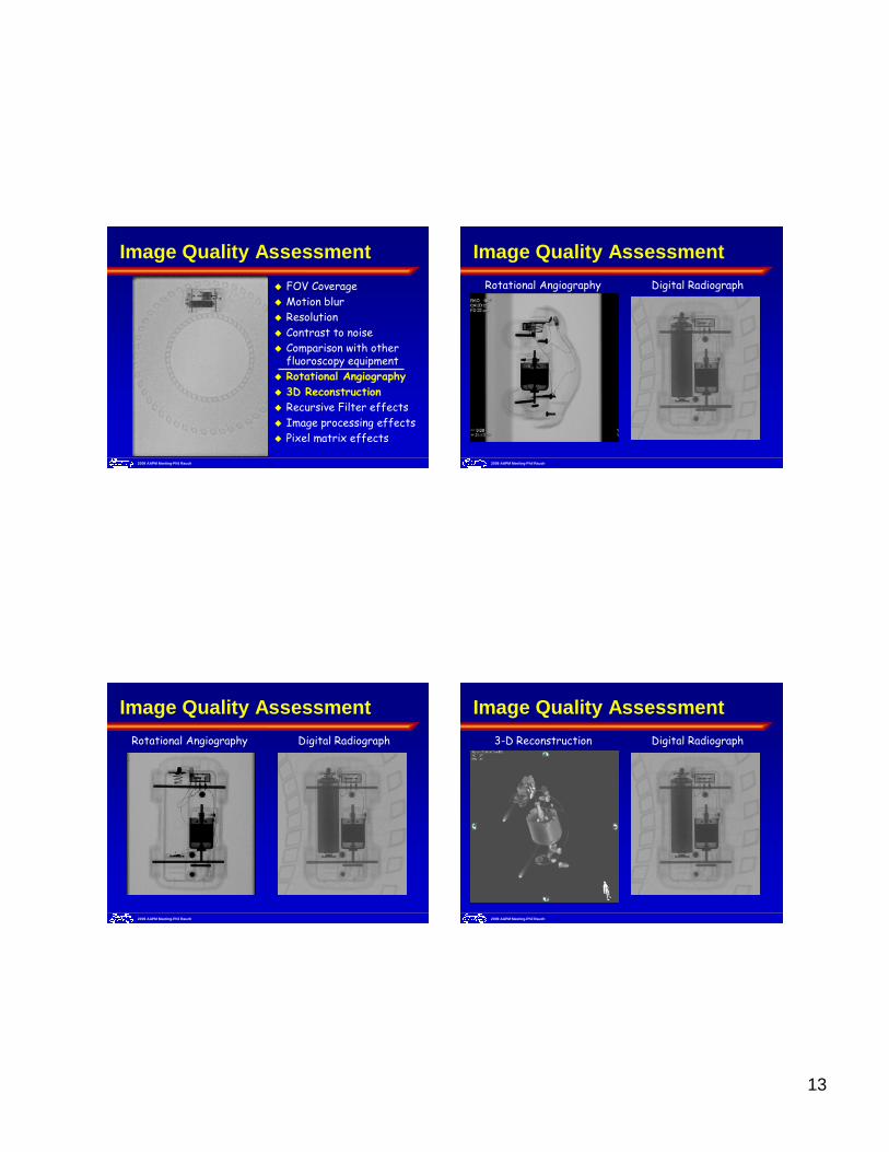
Image Quality Assessment
2009 AAPM Meeting-Phil Rauch
Image Quality Assessment
Contrast
Noise
Sharpness
Saturation
Artifacts
Descriptors
2009 AAPM Meeting-Phil Rauch
Image Quality Assessment
Edge Detection
Pattern Recognition
Comparison
Assessment
Relevance
Conclusion
Evaluation(HVS)
4
2009 AAPM Meeting-Phil Rauch
Is Pulse Width Important?
2009 AAPM Meeting-Phil Rauch
Is Pulse Width Important?
2009 AAPM Meeting-Phil Rauch
Fluoroscopic Image Quality
Blur (Fram
e
integratio
n
Time)
Image Intensifier
Flat Panel
2009 AAPM Meeting-Phil Rauch
Exposure ON-time – Pulsed vs Cont
Pulsed Fluoro*(30 pulses per sec)
*Displayed at 7.5 fps
Continuous Fluoro*(30 video frames per sec)
*Displayed at 7.5 fps
5
2009 AAPM Meeting-Phil Rauch
Fluoroscopic Image Quality
Frame
integration
time
LAG(Signal retention)
Image Intensifier
Flat Panel
2009 AAPM Meeting-Phil Rauch
Fluoroscopic Image Quality
Frame
integration
time
LAG(Signal retention)
Recursive filter setting
Image Intensifier
Flat Panel
2009 AAPM Meeting-Phil Rauch
Blur versus Recursive Filtering
Continuous Fluoroscopy Pulsed FluoroscopyWithout Recursive Filtering With Recursive Filtering
2009 AAPM Meeting-Phil Rauch
Blur versus Recursive Filtering
Continuous Fluoroscopy Pulsed FluoroscopyWithout Recursive Filtering With Recursive Filtering
6
2009 AAPM Meeting-Phil Rauch
Fluoroscopic Image Quality
Frame
integration
time
LAG(Signal retention)
Recursive filter settingPerc
eptual
integrat
ion
Image Intensifier
Flat Panel
2009 AAPM Meeting-Phil Rauch
Where are the 3’s
Perceptual Integration
2009 AAPM Meeting-Phil Rauch
Perceptual Integration
30 Frames/sec
Where are the 3’s
2009 AAPM Meeting-Phil Rauch
Perceptual Integration
8 Frames/sec
Where are the 3’s
7
†EERD = Entrance Exposure Rate to Detector
Beam Alignment
Geometry
SID SSD
FDA Max
FDA 21CFR
Normal Level
Minimum Filtration
Spatial Beam
Shaping
FOVElectronic Zoom
Measurement
Iris (Optical)
Slow Scan
Normal Scan
Video Camera
Detector Characteristics
Display Characteristics
Spatial Resolution
Anti-scatter Grid
Contrast Resolution
Temporal Resolution
Noise
MTF
X-ray Tube
kV-mA Power Curves
X-ray Generator
Continuous Fluoroscopy
Pulsed Fluoroscopy
Fluoroscopy Waveform
Fluoroscopy ON-Time
Last Image Hold
High Level
Pulse Width
Collimation without
Radiation
Pulse Rate
Acquisition Type
Image Size
Wedge Filter
Scatter to Primary Ratio
Dynamic Range
DQE
Luminance
Contrast
Gamma
Iris (Lead)
Spectral Beam
Shaping
Grid Control
Dose Mode
SNR
Patient Size
Spectral Filtration
Patient Exposure
Image Quality
EERD†
kV
2009 AAPM Meeting-Phil Rauch
Generator Fluoroscopy Technique
Generator Fluoroscopy Power Curves
60
70
80
90
100
110
120
130
0.0 0.5 1.0 1.5 2.0 2.5 3.0 3.5 4.0
mA
kVp
Cont. Low - 30 (video) fps
2009 AAPM Meeting-Phil Rauch
Generator Fluoroscopy Technique
Generator Fluoroscopy Power Curves
60
70
80
90
100
110
120
130
0.0 0.5 1.0 1.5 2.0 2.5 3.0 3.5 4.0
mA
kVp Cont. Norm - 30 (video) fps
Cont. Low - 30 (video) fps
2009 AAPM Meeting-Phil Rauch
Generator Fluoroscopy Technique
Generator Fluoroscopy Power Curves
60
70
80
90
100
110
120
130
0.0 0.5 1.0 1.5 2.0 2.5 3.0 3.5 4.0
mA
kVp
Pulsed - 30 pps
Cont. Norm - 30 (video) fps
Cont. Low - 30 (video) fps
8
2009 AAPM Meeting-Phil Rauch
Generator Fluoroscopy Technique
Generator Fluoroscopy Power Curves
50
60
70
80
90
100
0 5 10 15 20 25 30 35 40 45
mA
kVp
N o rm a l D o s e P uls e d - 3 .7 5 pp s / 0 .2 m m C u S pe c tra l F ilte r
2009 AAPM Meeting-Phil Rauch
Generator Fluoroscopy Technique
Generator Fluoroscopy Power Curves
50
60
70
80
90
100
0 5 10 15 20 25 30 35 40 45
mA
kVp
Hig h D o s e P u ls e d - 3 .7 5 p ps / 0 .2 m m C u S p e c t ra l F ilte r
N o rm a l D o s e P uls e d - 3 .7 5 pp s / 0 .2 m m C u S pe c tra l F ilt e r
2009 AAPM Meeting-Phil Rauch
Generator Fluoroscopy Technique
Generator Fluoroscopy Power Curves
50
60
70
80
90
100
0 5 10 15 20 25 30 35 40 45
mA
kVp
N o rm a l D o s e P u ls e d - 15 p ps / 0 .2 m m C u S p e c t ra l F ilte r
2009 AAPM Meeting-Phil Rauch
Generator Fluoroscopy Technique
Generator Fluoroscopy Power Curves
50
60
70
80
90
100
0 5 10 15 20 25 30 35 40 45
mA
kVp
H ig h P uls e d - 15 pp s / 0 .2 m m C u S pe c t ra l F ilte r
N o rm a l D o s e P u ls e d - 15 p ps / 0 .2 m m C u S p e c t ra l F ilte r
9
2009 AAPM Meeting-Phil Rauch
Generator Fluoroscopy Technique
Generator Fluoroscopy Power Curves
50
60
70
80
90
100
0 5 10 15 20 25 30 35 40 45
mA
kVp
Normal Dose Continuous / 0.2mm Cu Spectral Filter
2009 AAPM Meeting-Phil Rauch
Generator Fluoroscopy Technique
Generator Fluoroscopy Power Curves
50
60
70
80
90
100
0 5 10 15 20 25 30 35 40 45
mA
kVp
High Dose Continuous / 0.2mm Cu Spectral Filter)
Normal Dose Continuous / 0.2mm Cu Spectral Filter
2009 AAPM Meeting-Phil Rauch
Entrance Exposure Rate to Detector
Solid state detectors must be positioned outside the automatic dose rate measurement field
2009 AAPM Meeting-Phil Rauch
EERD Test Conditions
RELATIVE EERD vs kVp
80%
90%
100%
110%
120%
130%
140%
150%
160%
170%
180%
190%
200%
210%
40 50 60 70 80 90 100 110
kVp
Det
ecto
r E
xpos
ure
Rat
e N
orm
aliz
ed to
Min
imum
Siemens Siregraph - 22 cm FOV X-rayImage Intensifier Detector
10
2009 AAPM Meeting-Phil Rauch
EERD Test Conditions
RELATIVE EERD vs kVp
80%
90%
100%
110%
120%
130%
140%
150%
160%
170%
180%
190%
200%
210%
40 50 60 70 80 90 100 110
kVp
Det
ecto
r E
xpos
ure
Rat
e N
orm
aliz
ed to
Min
imum Siemens Artis Zee - 42 cm FOV Indirect
Flat Panel Detector (CsI(Tl)/TFT)
Siemens Siregraph - 22 cm FOV X-rayImage Intensifier Detector
2009 AAPM Meeting-Phil Rauch
EERD Test Conditions
RELATIVE EERD vs kVp
80%
90%
100%
110%
120%
130%
140%
150%
160%
170%
180%
190%
200%
210%
40 50 60 70 80 90 100 110
kVp
Det
ecto
r E
xpos
ure
Rat
e N
orm
aliz
ed to
Min
imum
Philips Xper - 22 cm FOV Indirect FlatPanel Detector (CsI(Tl)/TFT)
Siemens Artis Zee - 42 cm FOV IndirectFlat Panel Detector (CsI(Tl)/TFT)
Siemens Siregraph - 22 cm FOV X-rayImage Intensifier Detector
2009 AAPM Meeting-Phil Rauch
EERD Test Conditions
RELATIVE EERD vs kVp
80%
90%
100%
110%
120%
130%
140%
150%
160%
170%
180%
190%
200%
210%
40 50 60 70 80 90 100 110
kVp
Det
ecto
r E
xpos
ure
Rat
e N
orm
aliz
ed to
Min
imum
Shimadzu Bransist - 22 cm FOV DirectFlat Panel Detector (Se/TFT)
Philips Xper - 22 cm FOV Indirect FlatPanel Detector (CsI(Tl)/TFT)
Siemens Artis Zee - 42 cm FOV IndirectFlat Panel Detector (CsI(Tl)/TFT)
Siemens Siregraph - 22 cm FOV X-rayImage Intensifier Detector
……can use 2x these values if minimum 0.2 mm Cubeam filtration is utilized
2009 AAPM Meeting-Phil Rauch
Detector Input Dose Rates (XRII)
• Low Dose Mode• Cont. Fluoroscopy• 30 cm FOV
Detector Input Dose/Fr?1x 1.5x 3.0x
2009 AAPM Meeting-Phil Rauch
Detector Input Dose Rates (XRII)
• Low Dose Mode• Cont. Fluoroscopy• 30 cm FOV
Detector Input Dose/Fr?1x 1.5x 3.0x
Detector dose scales inversely as either the ratio of the FOV, or as the ratio squared
40 cm 30 cm 22 cm 17 cm
2009 AAPM Meeting-Phil Rauch
Video Frame Rate
What is the video frame rate in the U.S.A.?
Ans: 30 video frames per second
18
2009 AAPM Meeting-Phil Rauch
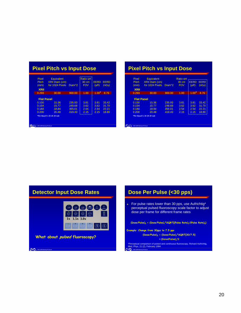
� Rauch's 30-30-30 Rule:� For a nominal 30 cm image intensifier field of view (FOV),
Pulse rate of 30 pps, Set the EERD to 30 µR/sec
� For other FOV's, scale by either the ratio of the FOV (auto optical lens aperture) or the square of the ratio
� For spectral beam filtration…� 0.1 mm Cu equivalent, multiply by 1.41
� 0.2 mm Cu equivalent, multiply by 2
Detector Input Dose Rates (XRII)
For flat panel, special consideration is required!!
2009 AAPM Meeting-Phil Rauch
Image Intensifier
Flat Panel
Detector Input Dose Rates (FP)
Detector Metrics(Fluoroscopy Doses)
Adapted from Koch, A., Macherel, J.M., Wirth, T., de Groot, P., Ducourant, T., Couder, D., Moy, J.P., & Calais, E. (2001). Detective quantum efficiency of an X-ray image intensifier chain as a benchmark for amorphous silicon flat panel detectors, Proc. SPIE, 4320, 115-20
2009 AAPM Meeting-Phil Rauch
Image Intensifier
Flat Panel
Detector Input Dose Rates (FP)
Detector Metrics(Fluoroscopy Doses)
“The minimum operating dose level is defined by the magnitude of the noise arising in the AM array and readout electronics. This typically sets the lower operating limit for detector dose at 20-50 nGy.”Cowen AR et al., Solid-state, flat-panel, digital radiography detectors and their physical imaging characteristics, Clin Radiol (2008)
2009 AAPM Meeting-Phil Rauch
Pixel Pitch – Air Kerma per Pixel
Pixel Pitch
For the transition from XRII to Flat Panel, the required EERD can be estimated by determining the air kerma required for the XRII when it is operating at the same pixel pitch at that for the flat panel.
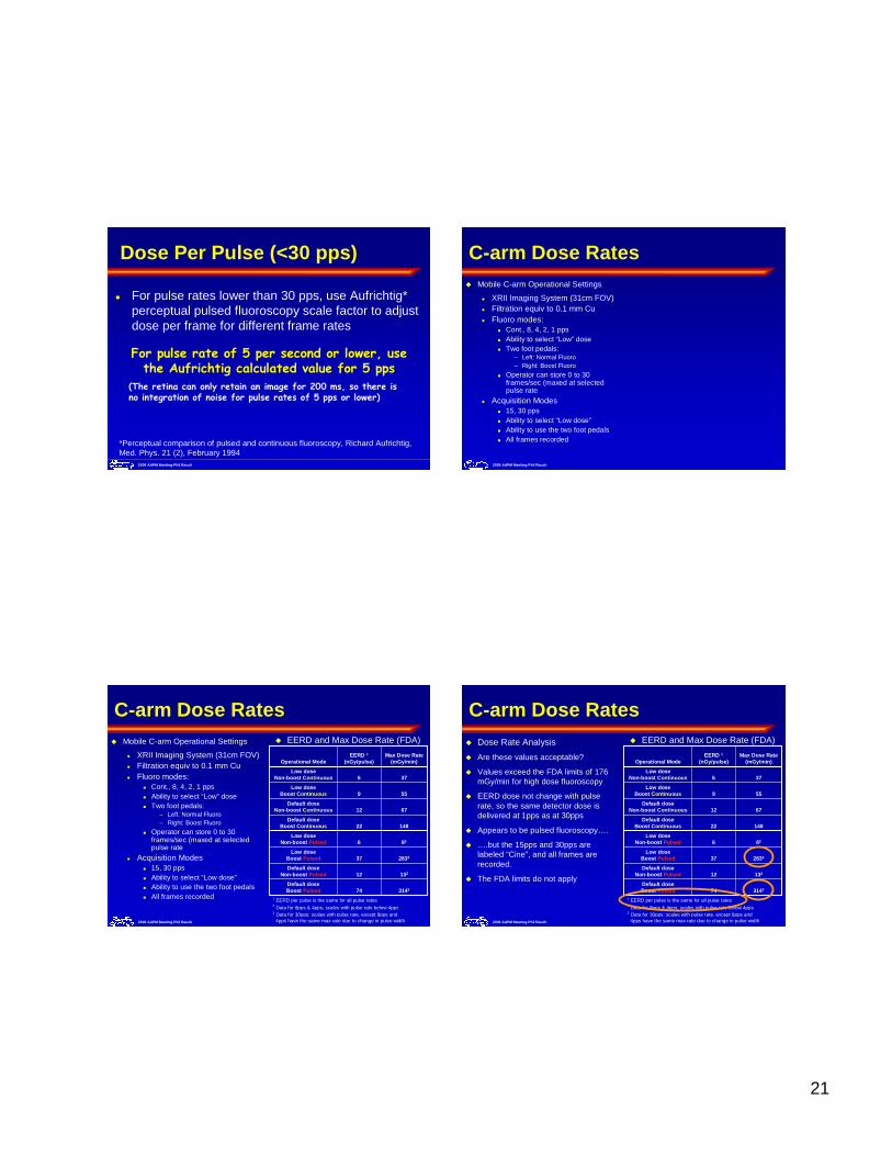
� For pulse rates lower than 30 pps, use Aufrichtig* perceptual pulsed fluoroscopy scale factor to adjust dose per frame for different frame rates
*Perceptual comparison of pulsed and continuous fluoroscopy, Richard Aufrichtig, Med. Phys. 21 (2), February 1994
Dose Per Pulse (<30 pps)
For pulse rate of 5 per second or lower, use the Aufrichtig calculated value for 5 pps
(The retina can only retain an image for 200 ms, so there is no integration of noise for pulse rates of 5 pps or lower)
C-arm Dose Rates
2009 AAPM Meeting-Phil Rauch
� Mobile C-arm Operational Settings
� XRII Imaging System (31cm FOV)� Filtration equiv to 0.1 mm Cu� Fluoro modes:
� Cont., 8, 4, 2, 1 pps� Ability to select “Low” dose� Two foot pedals:
– Left: Normal Fluoro– Right: Boost Fluoro
� Operator can store 0 to 30 frames/sec (maxed at selected pulse rate
� Acquisition Modes� 15, 30 pps� Ability to select “Low dose”� Ability to use the two foot pedals� All frames recorded
C-arm Dose Rates
314374Default dose Boost Pulsed
13212Default dose
Non-boost Pulsed
283337Low dose
Boost Pulsed
826Low dose
Non-boost Pulsed
14822Default dose
Boost Continuous
6712Default dose
Non-boost Continuous
559Low dose
Boost Continuous
376Low dose
Non-boost Continuous
Max Dose Rate(mGy/min)
EERD 1(nGy/pulse)Operational Mode
1 EERD per pulse is the same for all pulse rates2 Data for 8pps & 4pps; scales with pulse rate below 4pps3 Data for 30pps; scales with pulse rate, except 8pps and 4pps have the same max rate due to change in pulse width2009 AAPM Meeting-Phil Rauch
� Mobile C-arm Operational Settings
� XRII Imaging System (31cm FOV)� Filtration equiv to 0.1 mm Cu� Fluoro modes:
� Cont., 8, 4, 2, 1 pps� Ability to select “Low” dose� Two foot pedals:
– Left: Normal Fluoro– Right: Boost Fluoro
� Operator can store 0 to 30 frames/sec (maxed at selected pulse rate
� Acquisition Modes� 15, 30 pps� Ability to select “Low dose”� Ability to use the two foot pedals� All frames recorded
� EERD and Max Dose Rate (FDA)
C-arm Dose Rates
314374Default dose Boost Pulsed
13212Default dose
Non-boost Pulsed
283337Low dose
Boost Pulsed
826Low dose
Non-boost Pulsed
14822Default dose
Boost Continuous
6712Default dose
Non-boost Continuous
559Low dose
Boost Continuous
376Low dose
Non-boost Continuous
Max Dose Rate(mGy/min)
EERD 1(nGy/pulse)Operational Mode
1 EERD per pulse is the same for all pulse rates2 Data for 8pps & 4pps; scales with pulse rate below 4pps3 Data for 30pps; scales with pulse rate, except 8pps and 4pps have the same max rate due to change in pulse width2009 AAPM Meeting-Phil Rauch
� Dose Rate Analysis
� Are these values acceptable?
� Values exceed the FDA limits of 176 mGy/min for high dose fluoroscopy
� EERD dose not change with pulse rate, so the same detector dose is delivered at 1pps as at 30pps
� Appears to be pulsed fluoroscopy….
� ….but the 15pps and 30pps are labeled “Cine”, and all frames are recorded.
� The FDA limits do not apply
� EERD and Max Dose Rate (FDA)
22
Entrance Exposure Rate to Detector
� EERD and Max Dose Rate (FDA)
314374Default dose Boost Pulsed
13212Default dose
Non-boost Pulsed
283337Low dose
Boost Pulsed
826Low dose
Non-boost Pulsed
14822Default dose
Boost Continuous
6712Default dose
Non-boost Continuous
559Low dose
Boost Continuous
376Low dose
Non-boost Continuous
Max Dose Rate(mGy/min)
EERD 1(nGy/pulse)Operational Mode
1 EERD per pulse is the same for all pulse rates2 Data for 8pps & 4pps; scales with pulse rate below 4pps3 Data for 30pps; scales with pulse rate, except 8pps and 4pps have the same max rate due to change in pulse width2009 AAPM Meeting-Phil Rauch
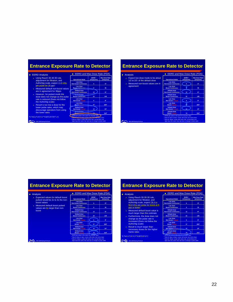
� EERD Analysis� Using Rauch 30-30-30 rule,
adjustment for filtration, and Aufrichtig scale, expect 11.6 nGy per pulse for 30 pps†
� Measured default non-boost values are in agreement for 30pps
� However, for pulsed mode the dose does not change as the pulse rate is reduced (Does not follow the Aufrichtig scale)
� Result is too low a dose for the lower pulse rates, which may discourage operators from using the lower rates
†8.76nGy*(30/31)2*SQRT(30/30)*1.41
Entrance Exposure Rate to Detector
� EERD and Max Dose Rate (FDA)
314374Default dose Boost Pulsed
13212Default dose
Non-boost Pulsed
283337Low dose
Boost Pulsed
826Low dose
Non-boost Pulsed
14822Default dose
Boost Continuous
6712Default dose
Non-boost Continuous
559Low dose
Boost Continuous
376Low dose
Non-boost Continuous
Max Dose Rate(mGy/min)
EERD 1(nGy/pulse)Operational Mode
1 EERD per pulse is the same for all pulse rates2 Data for 8pps & 4pps; scales with pulse rate below 4pps3 Data for 30pps; scales with pulse rate, except 8pps and 4pps have the same max rate due to change in pulse width2009 AAPM Meeting-Phil Rauch
� Analysis� Expect low dose mode to be about
1/2 to 2/3 of the default dose
� Measured non-boost values are in agreement
Entrance Exposure Rate to Detector
� EERD and Max Dose Rate (FDA)
314374Default dose Boost Pulsed
13212Default dose
Non-boost Pulsed
283337Low dose
Boost Pulsed
826Low dose
Non-boost Pulsed
14822Default dose
Boost Continuous
6712Default dose
Non-boost Continuous
559Low dose
Boost Continuous
376Low dose
Non-boost Continuous
Max Dose Rate(mGy/min)
EERD 1(nGy/pulse)Operational Mode
1 EERD per pulse is the same for all pulse rates2 Data for 8pps & 4pps; scales with pulse rate below 4pps3 Data for 30pps; scales with pulse rate, except 8pps and 4pps have the same max rate due to change in pulse width2009 AAPM Meeting-Phil Rauch
� Analysis� Expected values for default boost
pulsed would be 2x to 3x the non-boost values
� Measured default boost pulsed values are 6x larger than non-boost
Entrance Exposure Rate to Detector
� EERD and Max Dose Rate (FDA)
314374Default dose Boost Pulsed
13212Default dose
Non-boost Pulsed
283337Low dose
Boost Pulsed
826Low dose
Non-boost Pulsed
14822Default dose
Boost Continuous
6712Default dose
Non-boost Continuous
559Low dose
Boost Continuous
376Low dose
Non-boost Continuous
Max Dose Rate(mGy/min)
EERD 1(nGy/pulse)Operational Mode
1 EERD per pulse is the same for all pulse rates2 Data for 8pps & 4pps; scales with pulse rate below 4pps3 Data for 30pps; scales with pulse rate, except 8pps and 4pps have the same max rate due to change in pulse width2009 AAPM Meeting-Phil Rauch
� Analysis� Using Rauch 30-30-30 rule,
adjustment for filtration, and Aufrichtig scale, expect 28.4 to 56.8 nGy per pulse for boost at 5 pps or lower†
� Measured default boost value is much larger than this estimate
� Furthermore, the dose does not change as the pulse rate is increased (Does not follow the Aufrichtig scale)
� Result is much larger than necessary doses for the higher frame rates
†8.76nGy*(30/31)2*SQRT(30/5)*2
23
2009 AAPM Meeting-Phil Rauch
Rauch’s 30-30-30 Rule
� Use the previous type analysis to determine how the vendor specified or measured EERD compares with the expected values
� Use the rule to compare measured values with the expected change in EERD whenever there are variations in the field of view, filtration, or pulse rate
2009 AAPM Meeting-Phil Rauch
Image Quality: What is it?
� “Image quality depends only on intrinsic, objective physical characteristics of an imaging system, and can be measured independently of an observer”
� “Image quality is whatever the observer says it is (i.e., it is a subjective perception of the image, ‘in the eye of the beholder’)”
Definitions courtesy Ralph Schaetzing, Agfa Corp.
2009 AAPM Meeting-Phil Rauch
Human Visual Perception
Fluoroscopic Image Quality
Biliary endoscopic catheterization
Mobile C-arm
Gastroenterology clinic (No X-ray techs)
1024 x 1280 x 12 bit image
Secondary capture of fluoro frame
24
Observations
Mottled
Blurred, probably due to long pulse width
Low dynamic range
Lack of detail
Inappropriate anatomical setting
No x-ray technique or image processing information available in the DICOM metadata