28
4/30/2009 1 FN PARALYSIS HEMIFACIAL SPASM
4/30/2009
1
FN PARALYSIS
HEMIFACIAL SPASM
4/30/2009
2
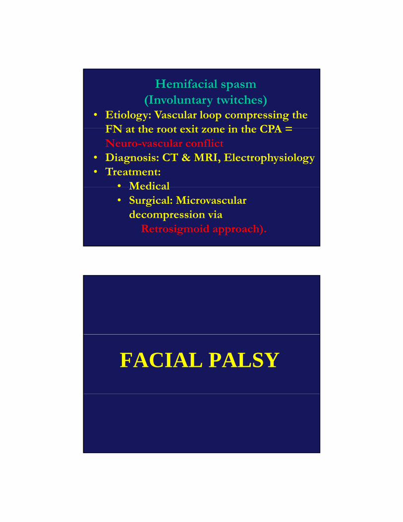
Hemifacial spasm (Involuntary twitches)
• Etiology: Vascular loop compressing the FN at the root exit zone in the CPA =FN at the root exit zone in the CPA = Neuro-vascular conflict
• Diagnosis: CT & MRI, Electrophysiology • Treatment:
• MedicalMedical• Surgical: Microvascular
decompression via Retrosigmoid approach).
FACIAL PALSY
4/30/2009
3
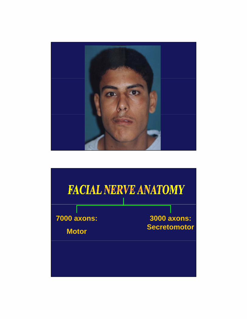
7000 axons:
Motor
3000 axons: Secretomotor
4/30/2009
4
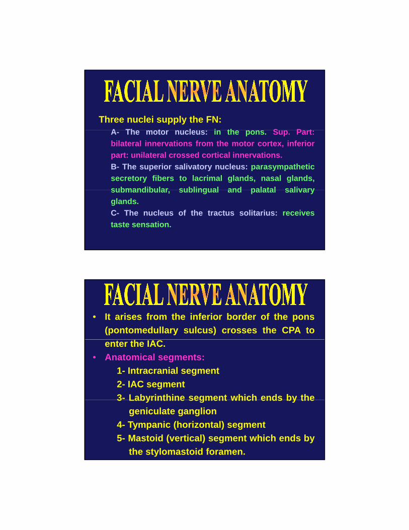
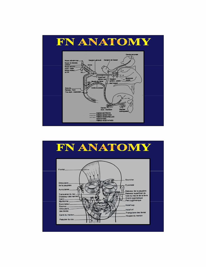
Three nuclei supply the FN:A Th t l i th S P tA- The motor nucleus: in the pons. Sup. Part:bilateral innervations from the motor cortex, inferiorpart: unilateral crossed cortical innervations.B- The superior salivatory nucleus: parasympatheticsecretory fibers to lacrimal glands, nasal glands,submandibular sublingual and palatal salivarysubmandibular, sublingual and palatal salivaryglands.C- The nucleus of the tractus solitarius: receivestaste sensation.
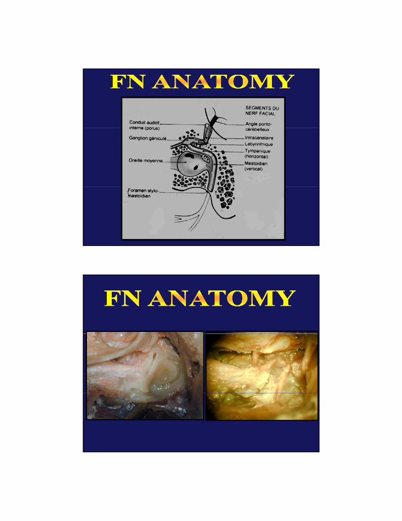
• It arises from the inferior border of the pons(pontomedullary sulcus) crosses the CPA toenter the IAC.
• Anatomical segments:1- Intracranial segment2- IAC segment3- Labyrinthine segment which ends by the3 Labyrinthine segment which ends by the
geniculate ganglion4- Tympanic (horizontal) segment5- Mastoid (vertical) segment which ends by
the stylomastoid foramen.
4/30/2009
5
4/30/2009
6
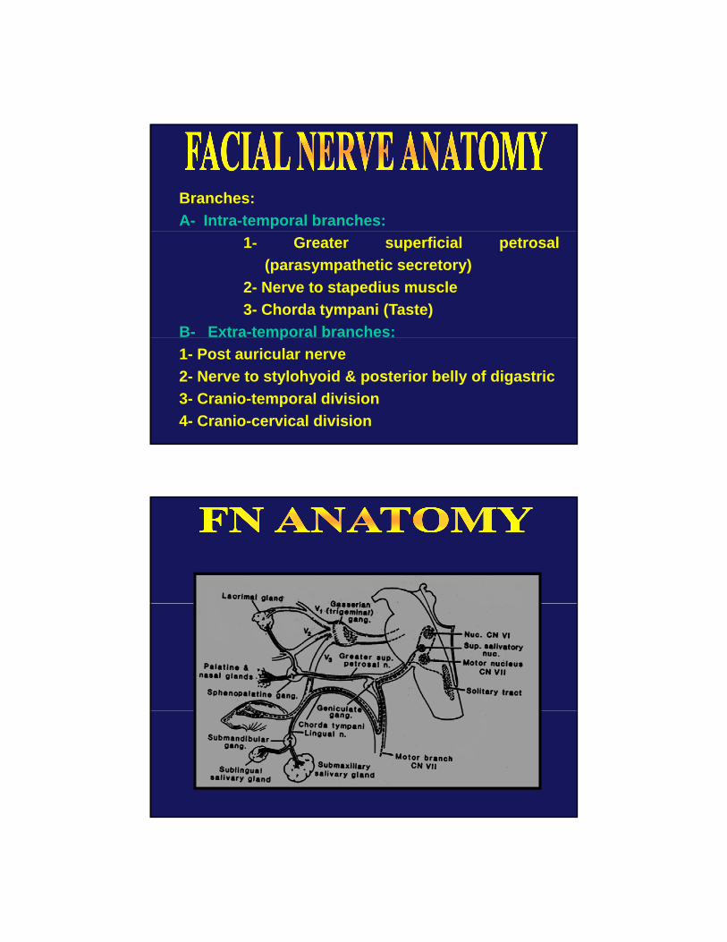
Branches:A- Intra-temporal branches:
1- Greater superficial petrosal(parasympathetic secretory)
2- Nerve to stapedius muscle3- Chorda tympani (Taste)
B- Extra-temporal branches:p1- Post auricular nerve2- Nerve to stylohyoid & posterior belly of digastric3- Cranio-temporal division4- Cranio-cervical division
4/30/2009
7
4/30/2009
8
4/30/2009
9
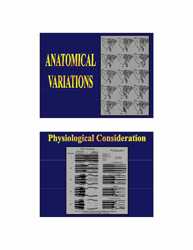
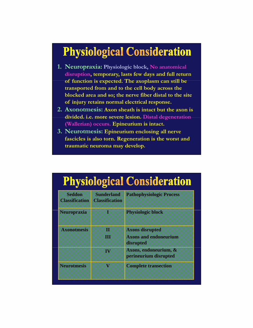
1. Neuropraxia: Physiologic block, No anatomical disruption, temporary, lasts few days and full return of f nction is e pected The a oplasm can still beof function is expected. The axoplasm can still be transported from and to the cell body across the blocked area and so; the nerve fiber distal to the site of injury retains normal electrical response.
2. Axonotmesis: Axon sheath is intact but the axon is divided i e more severe lesion Distal degenerationdivided. i.e. more severe lesion. Distal degeneration (Wallerian) occurs. Epineurium is intact.
3. Neurotmesis: Epineurium enclosing all nerve fascicles is also torn. Regeneration is the worst and traumatic neuroma may develop.
Seddon Classification
Sunderland Classification
Pathophysiologic Process
Neuropraxia I Physiologic block
Axonotmesis IIIII
Axons disruptedAxons and endoneurium disrupted
IV Axons, endoneurium, & perineurium disrupted
Neurotmesis V Complete transection
4/30/2009
10
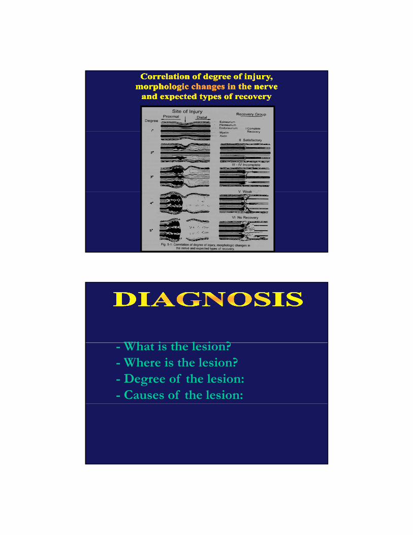
Wh i h l i ?- What is the lesion? - Where is the lesion? - Degree of the lesion: - Causes of the lesion:
4/30/2009
11
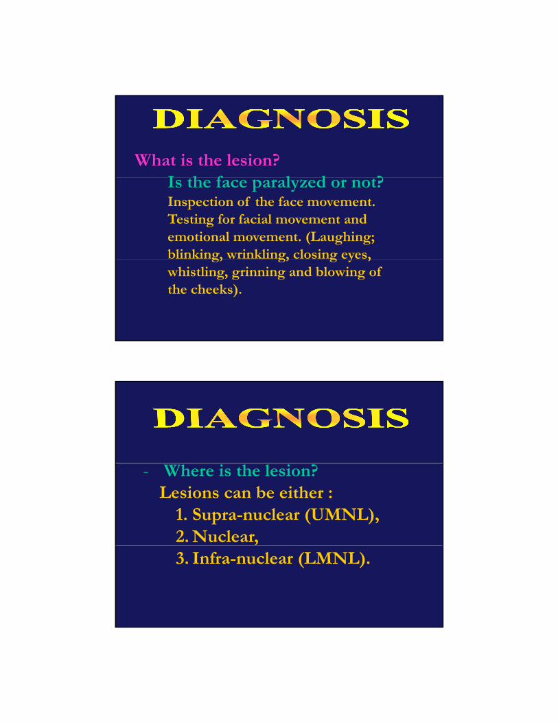
What is the lesion? I h f l d ?Is the face paralyzed or not? Inspection of the face movement. Testing for facial movement and emotional movement. (Laughing; blinking, wrinkling, closing eyes, g, g, g y ,whistling, grinning and blowing of the cheeks).
- Where is the lesion? Lesions can be either :
1. Supra-nuclear (UMNL),2. Nuclear,3. Infra-nuclear (LMNL).
4/30/2009
12
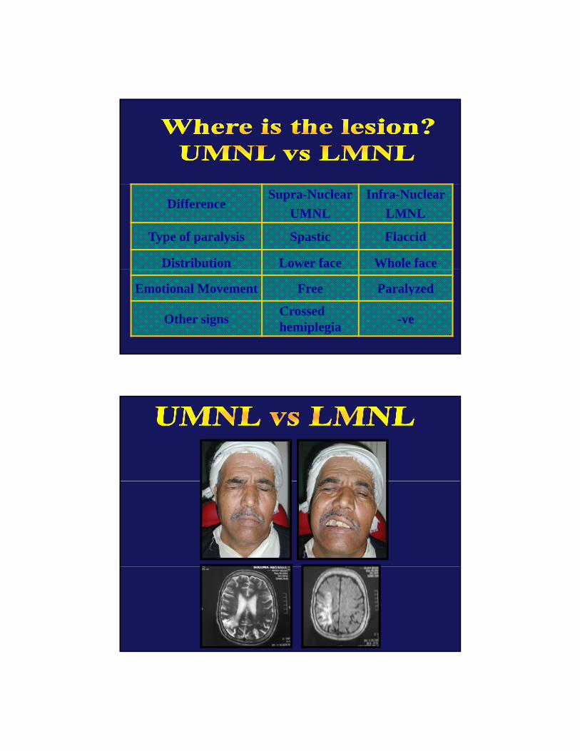
DifferenceSupra-Nuclear
UMNLInfra-Nuclear
LMNL
Type of paralysis Spastic Flaccid
Distribution Lower face Whole face
Emotional Movement Free Paralyzed
Other signs Crossed hemiplegia -ve
4/30/2009
13
Where is the lesion?
i- Nuclear lesions are associated by crossed hemiplegia, crossed hemianesthesia, other cranial nerves palsies specially VI. Lacrimation salivation and taste are normalLacrimation, salivation and taste are normal.
ii- LMNL : FN may be affected at different sites of its course.
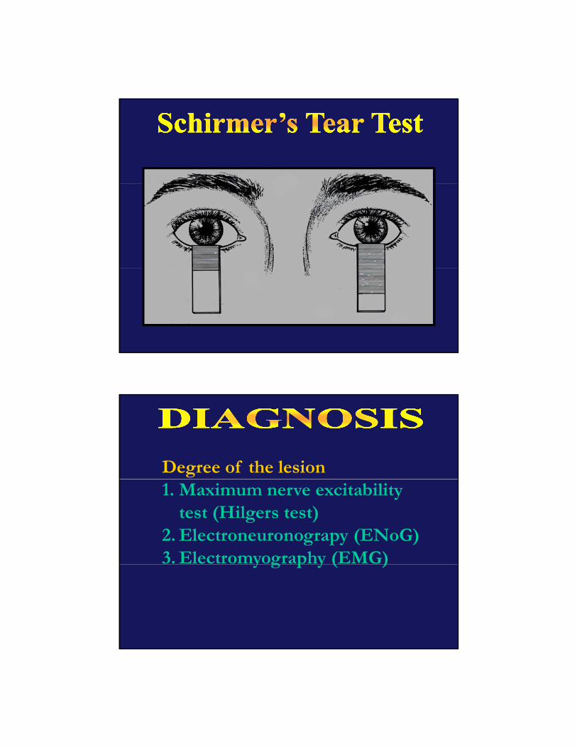
1 Schirmer’s tear test1. Schirmer s tear test2. Stapedius reflex3. Evaluation of Taste4. Submandibular salivary flow
4/30/2009
14
Degree of the lesion 1. Maximum nerve excitability
test (Hilgers test)2. Electroneuronograpy (ENoG)3. Electromyography (EMG)y g p y ( )
4/30/2009
15
1- Maximum nerve excitability test (Hilger’s test): The response on the involved side istest): The response on the involved side is compared to the normal side. The idea is that the distal segment of a completely cut nerve may continue to conduct when stimulated by a faradic current (distal to the cut) for as long as 24-72 hours. The response then disappear due to Wallerian degeneration. Ideally testing is started on the third post onset day.
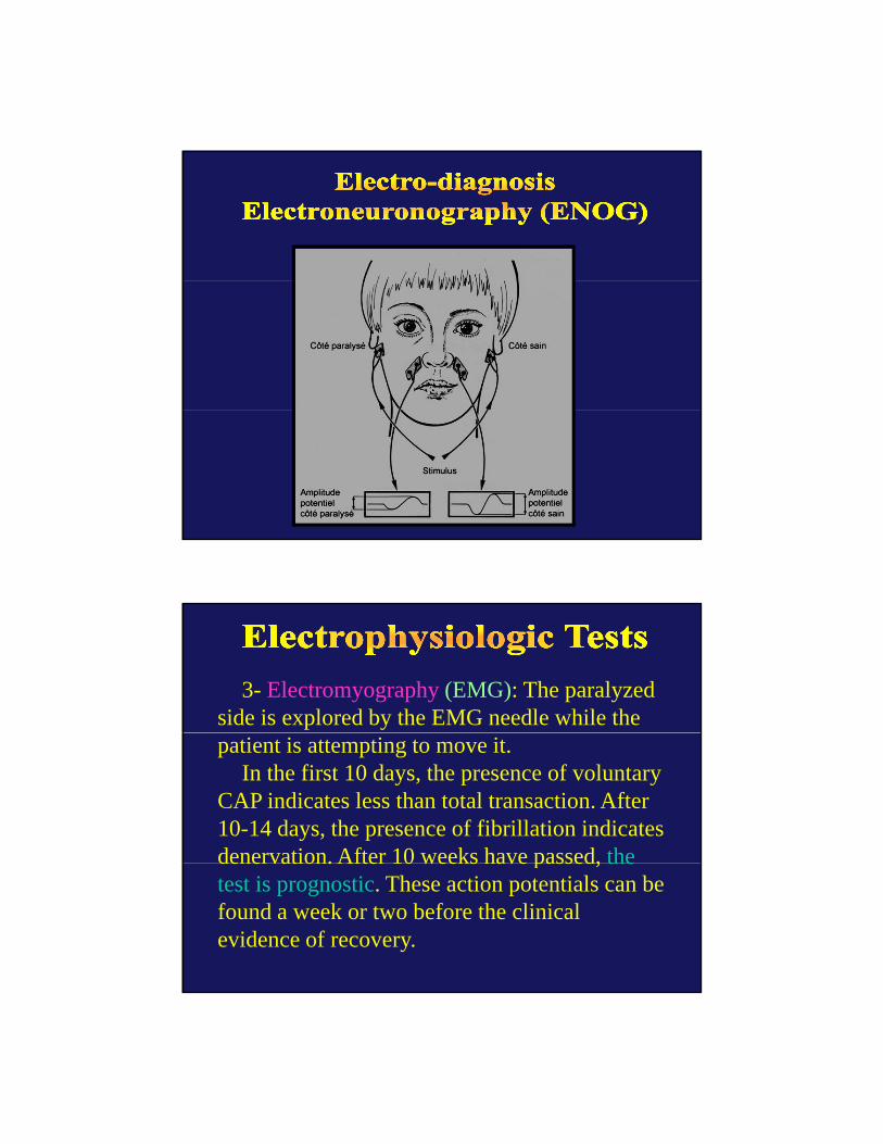
2- Electroneuronography (ENOG) =g p y ( )Evoked electromyography: Similar to maximum stimulation test in which the evoked CAP are recorded. The height of the action potential is compared with the good side and a percentage comparison is made It is apercentage comparison is made. It is a quantitative test. Degeneration of more than 90% means recovery is poor.
4/30/2009
16
3- Electromyography (EMG): The paralyzed side is explored by the EMG needle while the patient is attempting to move it.
In the first 10 days, the presence of voluntary CAP indicates less than total transaction. After 10-14 days, the presence of fibrillation indicates denervation. After 10 weeks have passed, thedenervation. After 10 weeks have passed, the test is prognostic. These action potentials can be found a week or two before the clinical evidence of recovery.
4/30/2009
17
Supra-nuclear or nuclear: Hemorrhage; vascular; encephalitis; tumors, cerebral palsy; …….
Infra-nuclear:Infra-nuclear: 1. Congenital: poor facial muscular development2. Inflammatory: Malignant ext. otitis; AOM, Ch.OM,
cholesteatoma, Herpes zoster oticus (Ramasy Hunt syndrome), spirochetes (Lyme disease)
3. Traumatic: Fracture base, forceps delivery,4. Toxic: Diphteria, tetanus,..p , ,5. Metabolic: Diabetes6. Neoplastic: 1ry tumor: FN neuroma; 2ry tumors of middle
ear and parotid and CPA.7. Iatrogenic: mastoid, parotid, CPA surgery8. Idiopathic: Bell’s palsy
A- Complications due to FN paralysis:
1- Drooling
2 Eye complication: exposure keratitis; corneal ulcer; pan2- Eye complication: exposure keratitis; corneal ulcer; pan-ophthalmitis.
3- Facial contractures (fixed shortening of the muscles)
4- Psychological disturbance, depression, and suicidal tendencies.
B- Complications due to abnormal regeneration
1. Facial tics and spasms: due to short-circuiting between nerve fibersp g
2. Abnormal mass movement of the face: Synkinesis.
3. Crocodile tears: due to misdirected salivation fibers to the lacrimal gland.
4. Frey’s syndrome: facial flushing and sweating over the parotid during mastication = misdirected salivary fibers to the sweat glands.
4/30/2009
18
1- Treatment of the cause2- Treatment of the complication: eg. Gold weight for protection
of the cornea; Facial plastic surgery for the contractures.3 T f h f i l l i b f i l i i3- Treatment of the facial paralysis by facial re-animation
surgery meant to:a- Restoration of anatomical integrity of the FN. Eg. FN decompression; anastomosis and grafting. May be indicated in Bell’s palsy, Herpes zoster oticus, acute or chronic otitis media, trauma, and tumorsb- Restoration of the functional integrity of the neuromuscular system of the face: includes: i. nerve substitution technique e.g. Facial-hypoglossal anastomosis; and ii. muscle substitution techniques e.g. Masseter or temporalis muscle transfer.
4- Facial rehabilitation: e.g. Massage, heat and facial exercises.
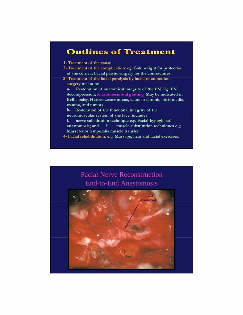
Facial Nerve ReconstructionEnd-to-End Anastomosis
Line of Anastomosis
FN-Pr.
Tumor bed
FN-d.
4/30/2009
19
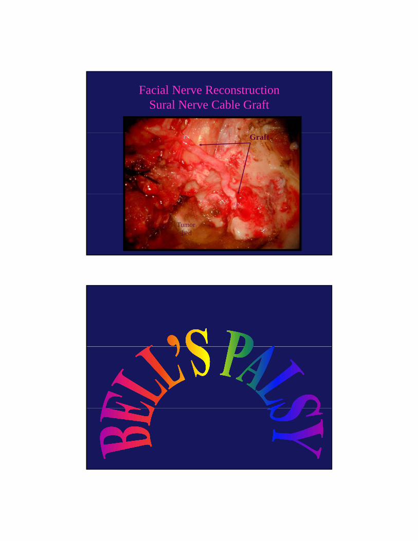
Facial Nerve ReconstructionSural Nerve Cable Graft
GraftFN
FN
Tumor bed
4/30/2009
20
Defined as the sudden occurrence of a flaccid
facial paralysis (LMNL) in an apparentlyfacial paralysis (LMNL) in an apparently
healthy individual. It is the commonest cause
of intratemporal causes of facial paralysis.
Incidence: 1/5000 per year- Incidence: 1/5000 per year.
It is the most common form of FN paralysis.
It is attributed to sudden exposure to cold.
- Etiology: Due to VIRAL causes (Activation of
Herpes Simplex)Herpes Simplex).
- Pathogenesis: Segmental demyelination followed by
2ry edema and compression.
- Clinical picture: Sudden onset of FN paralysis of LMN
type. Pain in the ear is noted in over half of patients.
Recovery starts even without treatment in most
cases within few days up to 3 weeks.
4/30/2009
21
P i 71% l t 25%- Prognosis: 71% complete recovery; 25%
recovers with slight sequalae; and 4% with
severe sequelae. Not a single patient who
does not show some recoverydoes not show some recovery.
- Diagnosis: It’s a diagnosis of exclusion.
A- Conservative treatment is helpful in 96% of cases.
1. Zovirax (800 mg 5 x daily for ONE week) + Cortisone
therapy
2. Physiotherapy treatment: massage, heat, and facial
exercises.
3. Dark eye glasses and artificial tears. Protection of the eye.
B- Surgical treatment: For complete paralysis with more than 90%
degeneration by ENOG within 6 days: Decompression of the FN
(specially the entry of the nerve into the Fallopian canal)
through Middle Cranial Fossa.
4/30/2009
22
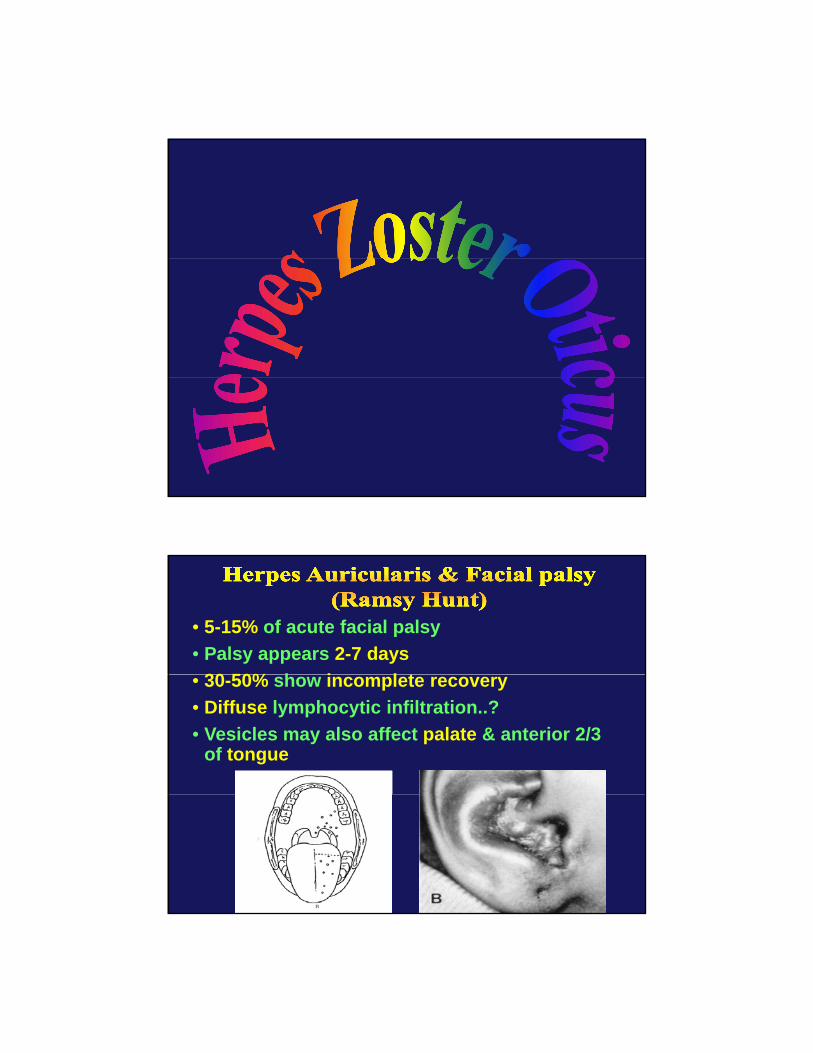
• 5-15% of acute facial palsy• Palsy appears 2-7 days
30 50% h i l t• 30-50% show incomplete recovery• Diffuse lymphocytic infiltration..?• Vesicles may also affect palate & anterior 2/3
of tongue
4/30/2009
23
• The FN appears to be more severelypp ydamaged in herpes zoster oticusthan with idiopathic facial palsy.Only 30% of patients will recovernormal function, and thus 70%normal function, and thus 70%experience some sequelae.
Ramsay Hunt Syndrome consists ofRamsay Hunt Syndrome consists of
• Facial paralysis;
• Auditory and vestibular dysfunctionassociated with
• Painful herpetic vesicles of the auricleand the EAC.
4/30/2009
24
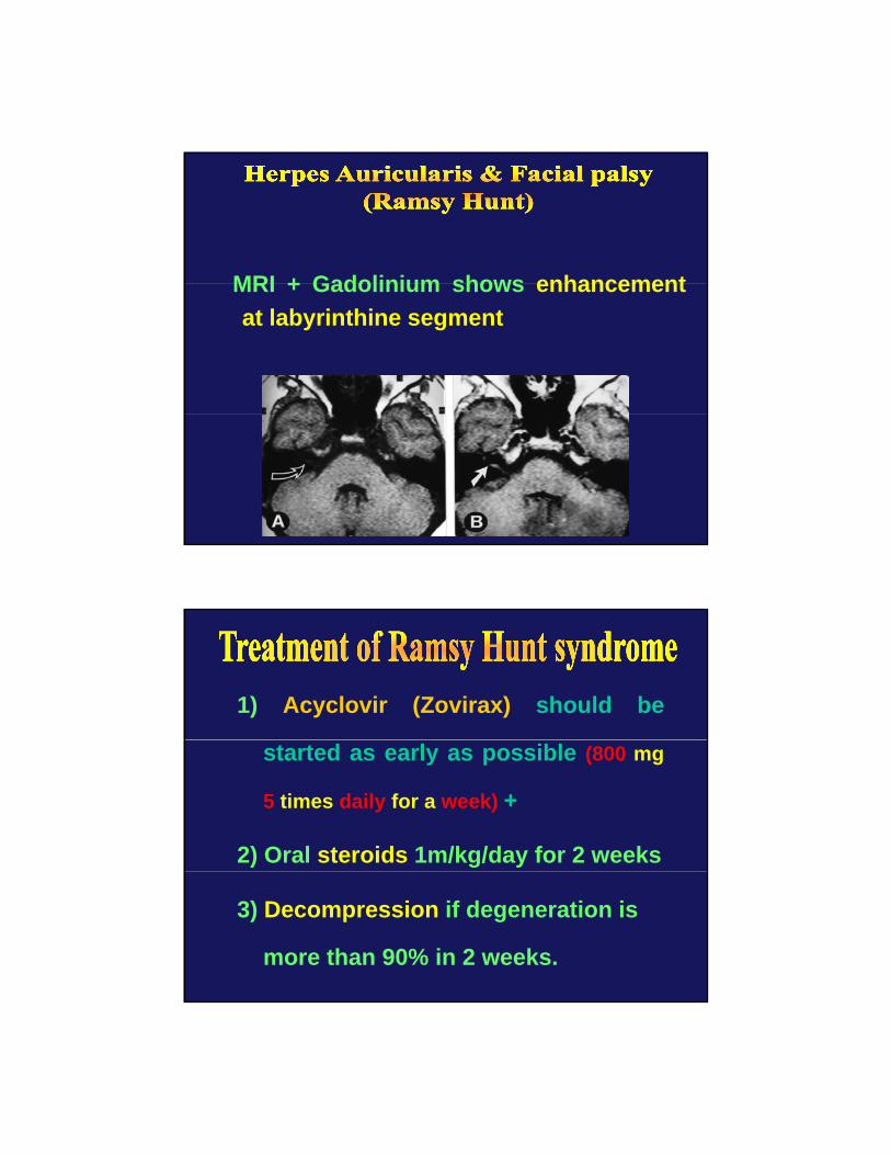
MRI + Gadolinium shows enhancementMRI + Gadolinium shows enhancementat labyrinthine segment
1) Acyclovir (Zovirax) should be
started as early as possible (800 mg
5 times daily for a week) +
2) Oral steroids 1m/kg/day for 2 weeks
3) Decompression if degeneration is
more than 90% in 2 weeks.
4/30/2009
25
1 Accidental: eg Fracture of the1. Accidental: eg. Fracture of thepetrous temporal bone:
2. Iatrogenic (Unintentional surgicalinjury)j y)
3. Surgical (unavoidable injuryduring surgery)
4/30/2009
26
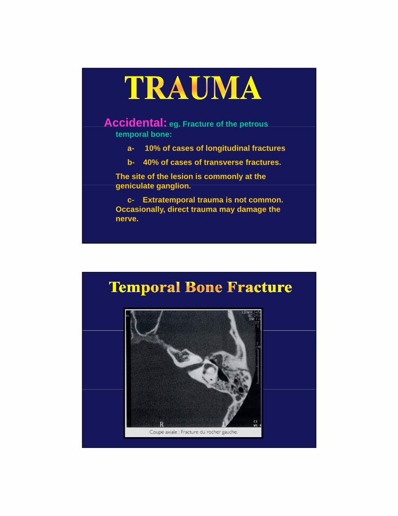
Accidental: eg. Fracture of the petrous Accidental: eg. Fracture of the petrous temporal bone:
a- 10% of cases of longitudinal fractures
b- 40% of cases of transverse fractures.
The site of the lesion is commonly at the geniculate gangliongeniculate ganglion.
c- Extratemporal trauma is not common. Occasionally, direct trauma may damage the nerve.
4/30/2009
27
Iatrogenic (Unintentional surgical injury)ga- Incision of a postauricular abscess in infant.
Incision must be placed high.
b- Classical cortical & radical mastoidectomies
c- Congenital ear surgery
d S f CPAd- Surgery of CPA.
Intraoperative Facial Nerve Monitoring (IOM) is of great help to prevent surgical trauma during difficult otological/neurotological surgery.
Surgical (unavoidable injury during surgery) g ( j y g g y)Impossible to preserve the FN during surgery as in some cases of malignancies, Tumors of the CPA, extensive cholesteatomas, …
Management: Diagnosis:. electric testing;Management: Diagnosis:. electric testing; Medical steroids; Surgical Exploration..

4/30/2009
28


