Lipid Composition of the Zoospores of Blastocladiella emersonrii"G. L. MILLS AND E. C. CANTINO
Department of Botany and Plant Pathology, Michigan State University, East Lansing, Michigan 48824
Received for publication 11 January 1974
The zoospores of Blastocladiella emersonii, when derived from cultures grownon solid media, contain about 11% total lipid. This lipid was separatedchromatographically on silicic acid into neutral lipid (46.6%), glycolipid (15.8%),and phospholipid (37.6%). Each class was fractionated further on columns ofsilicic acid, Florisil, or diethylaminoethyl-cellulose, and monitored by thin-layerchromatography. Triglycerides were the major neutral lipids, mono- anddiglycosyldiglycerides were the major glycolipids, and phosphatidylcholine andphosphatidylethanolamine were the major phospholipids. Other neutral lipidsand phospholipids detected were: hydrocarbons, free fatty acids, free sterols,sterol esters, diglycerides, monoglycerides, lysophosphatidylcholine, lysophos:phatidylethanolamine, phosphatidic acid, phosphatidylserine, and phos-phatidylinositol. Palmitic, palmitoleic, stearic, oleic, y-linolenic, and arachi-donic acids were the most frequently occurring fatty acids. When B. emersoniiwas grown in "C-labeled liquid media, lipid again accounted for 11% of bothmature plants and zoospores released from them. The composition of the lipidextracted from such plants and spores was also the same; however, it differedmarkedly from that of the lipid in spores harvested from solid media, consistingof 28.3% neutral lipid, 12.0% glycolipid, and 59.7% phospholipid. The majorlipids were again triglycerides for neutral lipids, mono- and diglycosyldiglyce-rides for glycolipids, and phosphatidyl choline and phosphatidylethanolaminefor phospholipids.
Except for a paper by Katsura (21), cited byGay et al. (12), which we have not seen,apparently the only available data on the lipidcontent of fungal zoospores, as estimated bydirect chemical analysis, are to be found in tworecent reports on the water mold Blastocladiellaemersonii (5). The first of these (37) establishedchanges in the quantity of lipid/cell duringendogenous metabolism of swimming zoo-spores; the second (36) provided a preliminarydescription of changes in lipid composition afterzoospore germination. We have been investigat-ing the lipids in certain organelles in thesezoospores (for a review of their structure, seereference 40), for which the composition of totalcell lipid constitutes an essential referencepoint. In this communication, we characterizethe whole spore lipid of B. emersonii, andprovide some comparative information aboutthe lipid content of the plants from which suchspores are derived.
' Dedicated to Frederick K. Sparrow, Jr., in the year of hisretirement as Professor of Botany in' the University ofMichigan, and in honor of his distinguished service to thefield of mycology.
MATERIALS AND METHODSProduction of zoospores on solid media. The
original strain of B. emersonii (5) was grown onpeptone-yeast extract-glucose (Difco; PYG) agar byinoculating with 4 x 10⁵ spores per standard petriplate and culturing at 22 C in the dark. Zoosporeswere obtained about 24 h later from first-generationplants by flooding each plate with 5 ml of water andfiltering 15 min later. After population densities wereestablished with a model B Coulter counter, thezoospores were sedimented at 1,000 x g for 5 min.Under these conditions, the yield was approximately 4x 10⁹ zoospores per 100 plates.Production of plants and zoospores in liquid
media. PYG broth cultures were prepared, inocu-lated, and induced to release zoospores at 22 C, by themethod of Myers and Cantino (26). [1,2-'4C]sodiumacetate (New England Nuclear Corp.; 50 pCi) wasadded 9 h after inoculation, the final concentrationbeing 5 x 10-I M. For studies of zoosporangial lipid,thalli were harvested either just before zoosporecleavage or after zoospores had been cleaved but notreleased (ca. 23 h after. inoculation); for studies ofzoospore lipid, spores were collected about 1 h later.
Extraction of lipid. Spore pellets were washedwith water, sedimented, sonically treated (30 s, 80W), and extracted at room temperature with chloro-
form-methanol (2: 1, vol/vol) overnight. Additionalextractions did not increase yields. The spore homoge-nate. was filtered through a coarse, fritted-glassBiichner funnel, and the filtrate was evaporated todryness under N2. Nonlipid contaminants were re-
moved with Sephadex G-25 by the method of Rouserand Fleischer (30).Column chromatography. Lipids were fraction-
ated into neutral, glyco-, and phospholipids on acti-vated silicic acid (100 mesh, Mallinckrodt ChemicalCo., St. Louis, Mo.). Neutral lipids were separatedfurther on silicic acid after removal of free fatty acids(FFA; 9), or on 7% hydrated Florisil (6). Glycolipidswere separated into individual components with Flo-risil (27), and phospholipids were fractionated on
diethylaminoethyl (DEAE)-cellulose (Sigma Chemi-cal Co., St. Louis, Mo.) by the method of Rouser et al.(31).
Thin-layer and paper chromatography. Thin-layer chromatography (TLC) was used to check thepurity of column fractions. Neutral lipids were sepa-
rated with petroleum ether-diethyl ether-acetic acid(80:20: 1, vol/vol) by using ITLC-SG chromatographymedia (Gelman Instrument Co., Ann Arbor, Mich.).Phospholipids were chromatographed on the same
media with isopropanol-ammonium hydroxide(100:7, vol/vol); glycolipids were separated with thesame solvent on ITLC-SA media. Phospholipids were
also chromatographed two-dimensionally on Redi-Coats (Supelco Inc., Bellefonte, Pa.) by using chloro-form-methanol-ammonium hydroxide (60:25:5, vol/vol) in the first direction and chloroform-acetone-methanol-acetic acid-water (3:4:1:1:0.5, vol/vol) inthe second direction.
Lipid components were visualized with ultravioletlight, 12 vapor, or a saturated solution of K2CrO4 in70% (vol/vol) H2SO4 (34). Specific sprays includedSbCl, for sterols and sterol esters (34), 0.2% ninhydrinin butanol for free amino groups, Dragendorff reagentfor the detection of choline-containing compounds(34), and the reagent of Dittmer and Lester (8) for P.Glycolipids were detected with orcinol (34), phenol-H2SO4 (15), or diphenylamine (34).
Descending chromatography of water-soluble hy-drolysis products was carried out on Whatman no. 1paper. Glycerol phosphate esters were resolved withphenol-water (100: 38, vol/vol) (45), and the phos-phate groups were detected by the salicylsulfonicacid-FeCls procedure (41) or with acid molybdate(17). Glycolipid hydrolysis products were chromato-graphed with N-propanol-ammonium hydroxide-water (6:3: 1, vol/vol) or ethyl acetate-pyridine-water(12:5:4, vol/vol) (19). Ammoniacal AgNO, was usedto detect carbohydrates. The hydrolysis products were
also studied by TLC. Phosphate-impregnatedChromagram sheets (Eastman; 6061 silica gel) were
prepared, spotted, and multideveloped, and sugars
were located thereon, all by the method of Welch andMartin (43).
Hydrolysis procedures. Glycerol phosphate esterswere prepared by deacylating the phospholipids in 0.2N methanolic NaOH for 15 min at room temperature(20). The solution was neutralized with Dowex-50
(H+) after partitioning with chloroform and water.The water-soluble hydrolysis products were concen-trated almost to dryness under a stream of N2 at 40 C,and used for chromatography. Phospholipids werealso deacylated by mild alkaline hydrolysis at 0 C(45). Glycolipids were hydrolized in 2 N HCl for 2 h at100 C. The hydrolysate was extracted three timeswith petroleum ether; the HCl was removed under astream of N2 or by drying the samples over KOHpellets.
Analytical procedures. Lipid-P was determinedafter digestion of samples with 10 N H2S04 by amodification of Bartlett's method (1). Total N wasassayed with the microprocedure of Sloane-Stanley(35) or by direct nesslerization with commercial(Harleco dry pack) Folin Wu (11) reagent. Acyl esterswere estimated by the ferric hydroxamate method(28) with tripalmitin as a standard. Glycerol analyses(16) were based on the determination of formaldehydeproduced by oxidation of glycerol with periodate byusing a-glycerol phosphate as a standard. Totalhexoses were determined with anthrone (44) or thephenol-sulfuric acid method (10). Glucose was alsoanalyzed enzymatically (Glucostat, Worthington Bio-chemical Corp., Freehold, N.J.), and total hexosa-mines were estimated by a modification (9) of theElson-Morgan reaction.Gas chromatography. Fatty acid methyl esters of
the total lipid and the neutral, glyco-, and phospho-lipid fractions were prepared by saponification andextraction of fatty acids (9); the latter were methyl-ated with BF2 (25). Methyl esters were examined witha Packard gas chromatograph model 7300 equippedwith a flame ionization detector. The methyl esterswere separated on a column (0.32 by 200 cm) packedwith 15% Lac-2R-446 on Chromosorb W (80 to 100mesh) operated at 187 C. The carrier gas was He; theinjector port temperature was 193 C; and the detectortemperature was 225 C. The esters were identified bytheir retention times relative to methyl ester stan-dards.
Materials. Phospholipid standards were preparedfrom egg yolks. The phospholipids were extractedwith chloroform-methanol (1:1, vol/vol), separatedfrom neutral lipids by silicic acid chromatography,and then fractionated on DEAE-cellulose. The phos-pholipids were purified further by TLC and comparedwith published data on egg yolk phospholipids (29).Some organic reagents and most solvents were redis-tilled before use. Methyl esters were obtained fromSupelco Inc., Bellefonte, Pa.
RESULTSCharacterization of total lipid in spores
produced on solid media. Lipid extracts werefractionated on silicic acid columns (Table 1).Neutral, glyco-, and phospholipid accounted for46.6, 15.8, and 37.6%, respectively, of the totallipid. The latter constituted 11% of the dryweight of the zoospore.Phospholipid. The phospholipid was frac-
a Percentages were established gravimetricallyafter the lipid had been fractionated on silicic acidcolumns (2 by 8 cm).
' Fractions were eluted successively with 150 ml ofchloroform, 100 ml of acetone, and 150 ml of metha-nol.
C Means and standard deviations for four experi-ments.
purity of each fraction was verified by TLC(Fig. 1). Peak I contained phosphatidylcholine(PC; R, 0.26; all Rf values listed in this reportare average values for many runs) and lysophos-phatidylcholine (LPC; Rf 0.12). Both spots weremolybdate positive and gave reactions for cho-line. Phosphatidylethanolamine (PE; R, 0.72)and lysophosphatidylethanolamine (LPE; R,0.45) were found in peaks II and III, respec-tively. Both spots were ninhydrin and molyb-date positive, as was phosphatidylserine (PS; R,0.00), the only compound in peak V. Peak IVwas due to oxidation products of PE. Peak VIcontained two components, phosphatidic acid(PA; R, 0.78) and phosphatidyl inositol (PI; Rr0.25); both spots for peak VI were molybdate
.
e
;' r- i t iH'So,
a ..
~,. tw *".^lJ
IrI i'
FIG. 1. Representative elution patterns for milligrams dry weight of lipid (right axis) and micrograms of totalP (left axis) obtained by column chromatography of total phospholipid on DEAE-cellulose. The column (2 by 20cm) was loaded with 1,190 ,g of phospholipid-P (obtained by silicic acid chromatographv), and 50-ml fractions(horizontal axis) were collected at a rate of 3 ml/min; 1,163 Mg of phospholipid-P (97.7%o) were recovered. Theelution sequence 1 to 6 (top) was as follows: chloroform-methanol (9:1, vol/vol), chloroform-methanol-aceticacid (7:3:0.002, vol/vol), methanol, chloroform-acetic acid (3:1, vol/vol), acetic acid, chloroform-methanol-ammonium hydroxide (32:8:1, vol/vol). The insert shows the results of TLC of compounds in peaks I to VI,phospholipid being visualized by H2SO4 charring. Both peaks I and VI contained two phospholipid components.Phosphatidyl choline (PC) and phosphatidic acid (PA) were in the peak fractions for I and VI, respectively,whereas lysophosphatidyl choline (LPC) and phosphatidyl inositol (PI) occurred in the correspondingshoulders.
positive and ninhydrin negative. The quantita-tive composition of the total phospholipid isshown in Table 2.The phospholipids were also characterized by
determining their molar ratios of P: acyl es-ters: glycerol: N (Table 3). Theoretical and ac-tual values agreed closely. The high acyl estercontent of LPC was due to contamination withPC.
Glycerol phosphate esters obtained by deacy-lation of the phospholipids were examined bypaper chromatography. Seven compounds weredetected, each one reacting positively to theacid molybdate spray for P. The R, values of theglycerylphosphoryl derivatives correspondedwith published data (20) and those for ourstandards prepared from egg yolks. Two com-pounds (glycerylphosphorylserine and glyceryl-phosphorylethanolamine) were ninhydrin posi-tive; two inositol-containing glycerylphosphorylesters were detected.
Glycolipid. This lipid class was separatedinto five fractions on Florisil; seven differentspots were derived therefrom by TLC (Fig. 2).Fraction I contained one component (Rt 0.86)which represented 10% of the total glycolipid asdetermined gravimetrically. Fractions II and IIIalso contained one component each, both hav-ing an Rt of 0.55; they represented 38 and 32% ofthe total glycolipid, respectively. Another 12%of the glycolipid was present in fraction IV inthe form of three components with R, values of0.31, 0.20, and 0.08. Fraction V had one compo-nent (R, 0.88); it represented 8% of the glyco-lipid.
Results obtained with spray reagents sug-gested that all the foregoing substances wereorcinol and diphenylamine positive except IVaand IVb; the latter were weakly orcinol andmolybdate positive. The components in frac-
a The phospholipids used to establish molar ratioswere taken either from column fractions (where puritywas verified by TLC) or directly from TLC plates.
II III* :S I
I, ,
IV V
C)4W
40~~~~~~~~1~
*~~ br
C;
FIG. 2. Glycolipid fractions collected by Florisilcolumn chromatography (1 by 15 cm) and resolvedfurther by TLC. Fractions I to V were obtained bysuccessive elutions with 50 ml of chloroform-acetone(1:1, vol/vol), acetone, 95% acetone, 90%o acetone, andmethanol. The chromatograms were visualized byH2SO4 charring.
tions II, III, and IVa were also ninhydrin posi-tive.The glycolipid fraction (15.8% of the total
lipid [Table 1]) contained 6.8% of the P in thetotal lipid extract. Analyses by phenol-sulfuricacid, and anthrone methods, with glucose as astandard, suggested that 23% of the glycolipidwas carbohydrate. However, judging by en-zymatic assays with glucose oxidase, only 6% ofthis carbohydrate was actually glucose.
Thin-layer and paper chromatography of theacid hydrolysis products indicated that, in ad-dition to glycerol, several substances that be-
haved like carbohydrates were present in theglycolipid. Fractions I and V appeared to con-tain monoglycosyl diglycerides judging from R,values (Fig. 2) and the fact that each releasedonly one carbohydrate after hydrolysis andTLC. Fractions II and III behaved like diglyco-syl diglycerides chromatographically (Fig. 2),and each released two components, one of thembeing ninhydrin positive. Three carbohydrateswere produced by Fraction IV, one of which wasninhydrin positive.Neutral lipid. This group of lipids was re-
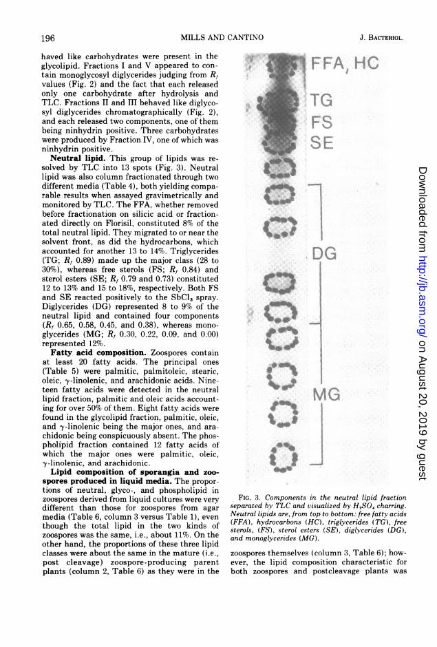
solved by TLC into 13 spots (Fig. 3). Neutrallipid was also column fractionated through twodifferent media (Table 4), both yielding compa-rable results when assayed gravimetrically andmonitored by TLC. The FFA, whether removedbefore fractionation on silicic acid or fraction-ated directly on Florisil, constituted 8% of thetotal neutral lipid. They migrated to or near thesolvent front, as did the hydrocarbons, whichaccounted for another 13 to 14%. Triglycerides(TG; Rf 0.89) made up the major class (28 to30%), whereas free sterols (FS; R, 0.84) andsterol esters (SE; R, 0.79 and 0.73) constituted12 to 13% and 15 to 18%, respectively. Both FSand SE reacted positively to the SbCl, spray.Diglycerides (DG) represented 8 to 9% of theneutral lipid and contained four components(R, 0.65, 0.58, 0.45, and 0.38), whereas mono-glycerides (MG; R, 0.30, 0.22, 0.09, and 0.00)represented 12%.Fatty acid composition. Zoospores contain
at least 20 fatty acids. The principal ones(Table 5) were palmitic, palmitoleic, stearic,oleic, y-linolenic, and arachidonic acids. Nine-teen fatty acids were detected in the neutrallipid fraction, palmitic and oleic acids account-ing for over 50% of them. Eight fatty acids werefound in the glycolipid fraction, palmitic, oleic,and y-linolenic being the major ones, and ara-chidonic being conspicuously absent. The phos-pholipid fraction contained 12 fatty acids ofwhich the major ones were palmitic, oleic,y-linolenic, and arachidonic.Lipid composition of sporangia and zoo-
spores produced in liquid media. The propor-tions of neutral, glyco-, and phospholipid inzoospores derived from liquid cultures were verydifferent than those for zoospores from agarmedia (Table 6, column 3 versus Table 1), eventhough the total lipid in the two kinds ofzoospores was the same, i.e., about 11%. On theother hand, the proportions of these three lipidclasses were about the same in the mature (i.e.,post cleavage) zoospore-producing parentplants (column 2, Table 6) as they were in the
'.0
4*
FIG. 3. Components in the neutral lipid fractionseparated by TLC and visualized by H,SO, charring.Neutral lipids are, from top to bottom: free fatty acids(FFA), hydrocarbons (HC), triglycerides (TG), freesterols, (FS), sterol esters (SE), diglycerides (DG),and monoglycerides (MG).
zoospores themselves (column 3, Table 6); how-ever, the lipid composition characteristic forboth zoospores and postcleavage plants was
different than that of precleavage plants (col-umn 1, Table 6). These conclusions were sub-stantiated by the distribution of radioactivity inthe three lipid classes (columns 4, 5, and 6,Table 6) derived from precleavage plants, post-cleavage plants, and zoospores from liquid cul-tures containing ['IC ]acetate.The three lipid classes derived from zoospores
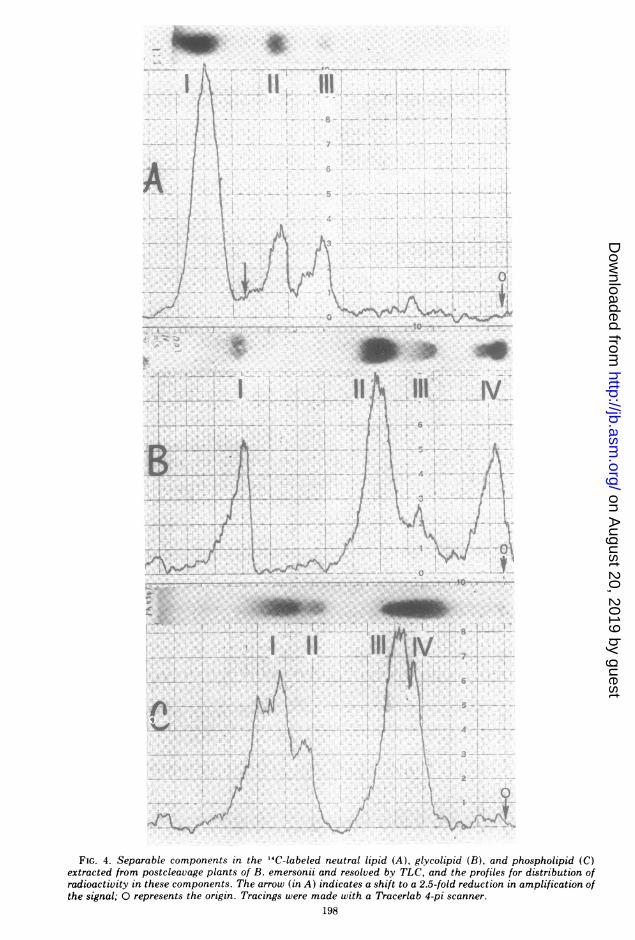
also resembled those from postcleavage plantswhen characterized further by TLC. The resultsobtained by similarly separating the labeledneutral, glyco-, and phospholipid via TLC andthen scanning the chromatograms for radioac-tivity are also delineated (Fig. 4). Most of theradioactivity in the neutral lipid fraction was
associated with TG (A-I). FS (A-II) and SE(A-III) were also major components, whereassmall amounts of label were associated with theMG and DG. Monoglycosyl diglycerides (B-I)and diglycosyl diglycerides (B-II) were themajor components labeled in the glycolipidfractions; peaks B-III and B-IV are mixtures ofglycolipids that were not resolved completely inthis solvent system. PC (C-Ill) and PE (C-I)TABLE 4. Composition of zoospore neutral lipida
aTotal lipid was extracted and separated as before(see Table 1) on silicic acid columns.
b Counted with a Tracerlab Versa/matic scaler.
were the major labeled compounds in the phos-pholipid fraction, LPE (C-Il) and LPC (C-III)also being present. Minor phospholipids werePA, PI, and PS.
DISCUSSIONThe wall-less, motile zoospores of B.
emersonii possess an elaborate, tightly orga-nized, membrane-bound corps of organelles;one especially prominent component is a clusterof discrete lipid globules partially wedged into astructure which, until its function and chemicalcomposition has been at least partially charac-terized, is being called "SB matrix" (40). Thesezoospores can develop along four different mac-rocylic pathways (4), as well as a microcyclicpathway (18), thereby producing plants whichgive rise to new generations of swarm cells.Against this background, we wish to commentbriefly about the extractable lipid produced bythis fungus.Both zoospores and sporulating cells contain
neutral lipids, glycolipids, and phospholipids,which vary in amount and relative proportionsdepending on the developmental stage. Addi-tionally, we have shown in this report that theproportions of these three lipid classes in zoo-spores derived from plate-grown cultures ofordinary colorless (OC) plants differed from thecorresponding amounts in zoospores derivedfrom liquid cultures; this observation is consist-ent with accounts showing that other microor-ganisms also change in lipid composition, asthey adjust to different environmental condi-tions (2, 23). With B. emersonii, the tempera-ture, pH, and composition of the media used forthe two kinds of cultures were similar; however,population densities-especially when consid-ered in relation to the amounts of oxygenapparently available-were quite different. We
FIG. 4. Separable components in the 14C-labeled neutral lipid (A), glycolipid (B), and phospholipid (C)extracted from postcleavage plants of B. emersonii and resolved by TLC, and the profiles for distribution ofradioactivity in these components. The arrow (in A) indicates a shift to a 2.5-fold reduction in amplification ofthe signal; 0 represents the origin. Tracings were made with a Tracerlab 4-pi scanner.
think this difference may account for the corre-sponding dissimilarities in lipid composition ofthe zoospores; our reasoning is as follows.We observed (Table 6), as did Smith and
Silverman (36), that there was about 45 to 50%more neutral lipid in zoospores than in sporulat-ing plants when both were derived from liquidcultures. The transition (precleavage plantspostcleavage plants - free swimming zoo-spores) was associated (Table 6) with a shift inneutral lipid from 16.8% through 24% to 28.3%,respectively. When grown in well-aerated liquidcultures, OC plants increase exponentially inweight up to about the time of sporogenesis (13,22); the corresponding increases in respirationand other evidence for oxidative activity (22,24), including the reutilization of lactic acid (3),suggest that there was a continuously increasingdemand for 02 up to the end of their generationtime. In plate cultures, on the other hand,neither forced aeration nor agitation was em-ployed; the demand for oxygen may have exist-ed, but its availability was undoubtedly re-stricted by various diffusion processes and per-haps other factors; hence, the average 02 up-take per plant was probably very much reduced,whereas lactic acid simultaneously accumu-lated in the medium. This information and thefact that the population density of the plants inplate cultures (ca. 2 x 10⁵ thalli/ml aftersubmersion in flooding water) was about fivetimes greater than that in liquid cultures (ca.0.4 x 105 plants/ml) lead us to suspect thatexogenous oxygen deficiencies and correspond-ing shifts toward fermentative metabolism priorto and during sporogenesis may have beenresponsible, at least in part, for the increasedproportion of neutral lipids in B. emersoniigrown on solid media.The composition of the extractable lipid in
spores derived from plate cultures was alsodifferent than that in spores derived from liquidcultures. In the former, the major lipids werePC (20.3%), TG (13.5%), and diglycosyldiglyce-rides (DGD; 13.3%), whereas PE, FS plus SE,and lysophosphatides (LP) accounted for 6.7,12.2, and 4.9% of the lipid, respectively. In thelatter, PC and PE were the most abundantcomponents, and lesser amounts of TG, DGD,LP, FS, and SE were also present, the phospha-tides constituting 62% of the total lipid. Theseresults are consistent with the phospholipidcontents of zoospores derived from liquid cul-tures as reported by Smith and Silverman (36)and Suberkropp and Cantino (37), i.e., 55% andup to 85% of the extractable lipid, respectively.The latter value is not out of line because it was
unquestionably inflated by its inclusion of gly-colipids; the fractionation techniques used atthat time separated the total lipid into only twoclasses, neutral lipids and polar lipids.Although the quantity of sterols and SE in
the zoospores of B. emersonii seems to exceedthe amount in most other Phycomycetes (42),the fatty acid composition of the B. emersoniizoospores does resemble that of other terrestrialphycomycetes (2, 7, 14, 33, 39). In addition,'y-linolenic acid-once thought (33) to be bothcharacteristic of and limited to the phycomy-cetes, but recently found (32) to occur in otherfungi-was associated predominantly withpolar lipids in the zoospores we analyzed, as itwas in the mixture of variously aged thalliharvested from multiple generation B.emersonii cultures by Sumner (38). However,the average degree of saturation among thefatty acids we extracted from zoospores wasgreater (an approximate estimate can be de-rived from the data in Table 5) than the averagevalue obtained by Sumner (38) for plants. Thisapparent difference between spores and plantsof B. emersonii could, of course, be due simplyto differences in culture conditions rather thanstages in ontogeny; on the other hand, ourresults do agree with Sumner's (38) observa-tions in that more polyunsaturated fatty acidswere associated with polar lipids than withneutral lipids.The glycolipid in B. emersonii seems to be of
an unusual nature. It is also the most homeo-static of the three lipid classes, apparentlybeing stabilized at a level of about 12 to 15% ofthe total lipid whatever the developmentalstage-whether the glycolipid is derived fromzoospores or sporangia, mature or immature, orextracted from plate-grown or liquid-growncultures. This conclusion contrasts sharply withthe results of Smith and Silverman (36), whoconcluded that glycolipid accumulated duringsporulation. Although they did not specify howmany spores were released by their sporulatingplants, the information provided suggests thatthey may have been microcyclic plants similarto the unispored plantlets studied by Hennessyand Cantino (18). The physiological changesoccurring after induction of sporogenesis inlag-phase (microcyclic) germlings and log-phase (macrocyclic) plants are known to showsimilarities but also some differences (18). Per-haps the latter includes lipid composition.Our unpublished data lead us to believe that
the glycolipids are associated with specific or-ganelles in B. emersonii, which could accountfor the stability of this class of lipids. A more
detailed study is underway to identify thenature of these glycolipids and their possiblerelationships to certain functional aspects of thezoospore of B. emersonii.
ACKNOWLEDGMENTSWe thank Ronald Myers for his help, and Michael Klug
and Keller Suberkropp for the use of their gas chromatographand facilities at W. K. Kellogg Biological Station.
This investigation was supported by Public Health Serviceresearch grants AI-01568-16 and AI-01568-17 to E. C. Cantinofrom the National Institute of Allergy and Infectious Dis-eases.
LITERATURE CITED
1. Bartlett, G. R. 1959. Phosphorus assay in column chro-matography. J. Biol. Chem. 234:466-468.
2. Bowman, R. D., and R. 0. Mumma. 1967. The lipids ofPhythium ultimum. Biochim. Biophys. Acta144:501-510.
3. Cantino, E. C. 1965. Intracellular distribution of "Cduring sporogenesis in Blastocladiella emersonii. Effectof light on hemoprotein. Arch. Mikrobiol. 51:42-59.
4. Cantino, E. C. 1966. Morphogenesis in aquatic fungi, p.283-337. In G. C. Ainsworth and A. S. Sussman (ed.),The fungi, Vol. 2. Academic Press Inc., New York.
5. Cantino, E. C., and M. T. Hyatt. 1953. Phenotypic "sex"determination in the life history of a new species ofBlastocladiella, B. emersonii. Antonie van Leeuwen-hoek J. Microbiol. Serol. 19:25-70.
6. Carroll, K. K., and B. Serdarevich. 1967. Lipid chromato-graphic analysis, p. 205-237. Dekker Inc., New York.
7. Chenouda, M. S. 1970. Investigation on lipid contents ofPhycomyces blakesleeanus. J. Gen. Appl. Microbiol.16:501-509.
8. Dittmer, J. C., and R. L. Lester. 1964. A simple, specificspray for the detection of phospholipids on thin-layerchromatograms. J. Lipid Res. 5:126-127.
9. Dittmer, J. C., and M. A. Wells. 1969. Quantitative andqualitative analysis of lipids and lipid components, p.482-530. In J. M. Lowenstein (ed.), Methods in enzy-mology, vol. 14. Academic Press Inc., New York.
10. Dubois, M., K. A. Gilles, J. K. Hamilton, P. A. Rebers,and F. Smith. 1956. Colorimetric method for determi-nation of sugars and related substances. Anal. Chem.28:350-356.
11. Folin, O., and A. Wu. 1919. A system of blood analysis. J.Biol. Chem. 38:81-110.
12. Gay, J. L., A. D. Greenwood, and I. B. Heath. 1971. Theformation and behaviour of vacuoles (vesicles) during,oosphere development and zoospore germination inSaprolegnia. J. Gen. Microbiol. 65:223-241.
13. Goldstein, A., and E. C. Cantino. 1962. Light-stimulatedpolysaccharide and protein synthesis by synchronized,single generations of Blastocladiella emersonii. J. Gen.Microbiol. 28:689-699.
14. Gordon, P. A., P. R. Stewart, and G. D. Clark-Walker.1971. Fatty acid and sterol composition of Mucorgenevensis in relation to dimorphism and anaerobicgrowth. J. Bacteriol. 107:114-120.
15. Gray, G. M. 1965. A comparison of the glycolipids foundin different strains of Ascites tumor cells in mice.Nature (London) 207:505-507.
16. Hanahan, D. J., and J. N. Olley. 1958. Chemical natureof monophosphoinositides. J. Biol. Chem. 231:813-828.
17. Hanes, C. S., and F. A. Isherwood. 1949. Separation ofthe phosphoric esters on the filter paper chromato-gram. Nature (London) 164:1107-1110.
D CANTINO J. BACTERIOL.
18. Hennessy, S. W., and E. C. Cantino. 1972. Lag-phasesporogenesis in Blastocladiella emersonii: induced for-mation of unispored plantlets. Mycologia64:1066-1087.
19. Isherwood, F. A., and M. A. Jermyn. 1951. Relationshipbetween the structure of the simple sugars and theirbehaviour on the paper chromatogram. Biochem. J.48:515-524.
20. Kates, M. 1972. Techniques of lipidology, p. 558-561.North-Holland Publishing Co., Amsterdam.
21. Katsura, K. 1970. Swimming behaviour of Phytophthoracapsici zoospores. In Morphological and related bio-chemical events in host-parasite interaction, p. 20-29.East-west center, Honolulu, Hawaii. The UnitedStates-Japan Cooperative Science program.
22. Khouw, B. T., and H. D. McCurdy. 1969. Tricarboxylicacid cycle enzymes and morphogenesis in Blasto-cladiella emersonii. J. Bacteriol. 99:197-205.
23. Kostiw, L. L., C. W. Boylen, and B. J. Tyson. 1972. Lipidcomposition of growing and starving cells of Arthrobac-ter crystallopoietes. J. Bacteriol. 111:103-111.
24. McCurdy, H. D., Jr., and E. C. Cantino. 1960. Isocitra-tase, glycine-alanine transaminase, and developmentin Blastoclodiella emersonii. Plant Physiol. 35:463-476.
25. Metcalfe, L. D., and A. A. Schmitz. 1961. The rapidpreparation of fatty acid esters for gas chromatographicanalysis. Anal. Chem. 33:363-364.
26. Myers, R. B., and E. C. Cantino. 1971. DNA profile of thespore of Blastocladiella emersonii: evidence for -y-parti-cle DNA. Arch. Mikrobiol. 78:252-267.
27. Radin, N. S. 1969. Florisil chromatography, p. 268-272.In J. M. Lowenstein (ed.), Methods in enzymology, vol.14. Academic Press Inc., New York.
28. Rapport, M. M., and N. Alonzo. 1955. Photometricdetermination of fatty acid ester groups in phospho-lipids. J. Biol. Chem. 217:193-198.
29. Rhodes, D. N., and C. H. Lea. 1957. Phospholipids 4. Onthe composition of hen's egg phospholipids. Biochem.J. 65:526-5'33.
30. Rouser, G., and S. Fleischer. 1965. Isolation, character-ization, and determination of polar lipids of mitochon-dria, p. 392-394. In R. W. Estabrook and M. E.Pullman (ed.), Methods in enzymology, vol. 10. Aca-demic Press Inc., New York.
31. Rouser, G., G. Kritcheresky, A. Yamamoto, G. Simon, C.Galli, and A. J. Bauman. 1969. Diethylaminoethyl andtriethylaminoethyl cellulose column chromatographicprocedures for phospholipids, glycolipids, and pig-ments, p. 272-317. In J. M. Lowenstein (ed.), Methodsin enzymology, vol. 14. Academic Press Inc., New York.
32. Safe, S., and D. Brewer. 1973. Lipid composition ofChaetomium cochliodes: effect of media. Lipid8:311-314.
33. Shaw, R. 1966. The fatty acids of Phycomycete fungi, andthe significance of the -y-linolenic acid component.Comp. Biochem. Physiol. 18:325-331.
34. Skipski, V. P., and M. Barclay. 1969. Thin layer chroma-tography of lipids, p. 541-548. In J. M. Lowenstein(ed.), Methods in enzymology, vol. 14. Academic PressInc., New York.
35. Sloane-Stanley, G. H. 1967. A simple procedure for theestimation of very small amounts of nitrogen in lipids.Biochem. J. 104:293-295.
36. Smith, J. D., and P. M. Silverman. 1973. Lipid turnoverduring mophogenesis in the water mold Blastocladiellaemersonii. Biochem. Biophys. Res. Commun.54:1191-1197.
37. Suberkropp, K. F., and E. C. Cantino. 1973. Utilizationof endogenous reserves by swimming zoospores ofBlastocladiella emersonii. Arch. Mikrobiol.
89:205-221.38. Sumner, J. L. 1970. The fatty acid composition of
Blastocladiella emersonii. Can. J. Microbiol.16:1161-1164.
39. Sumner, J. L., and E. D. Morgan. 1969. The fatty acidcomposition of sporangiospores and vegetative myce-lium of temperature-adapted fungi in the order Muco-rales. J. Gen. Microbiol. 59:215-221.
40. Truesdell, L. C., and E. C. Cantino. 1971. The inductionand early events of germination in the zoospore ofBlastocladiella emersonii, p. 1-44. In A. Monroy and A.A. Moscona (ed.), Current topics in developmentalbiology, vol. 6. Academic Press Inc., New York.
41. Vorbeck, M., and G. V. Marinetti. 1965. Intracellular
B. EMERSONII 201
distribution and characterization of the lipids of Strep-tococcus fecalis (ATCC 9790). Biochemistry 4:296-305.
42. Weete, J. D. 1973. Sterols of the fungi: distribution andbiosynthesis. Phytochemistry 12:1843-1864.
43. Welch, B. L., and N. E. Martin. 1972. Quantitativeanalysis of sugars by densitometric inspection of thin-layer chromatograms: analysis of method. J. Chro-matogr. 72:359-364.
44. Wells, M. A., and J. C. Dittmer. 1963. The use ofSephadex for the removal of non-lipid contaminantsfrom lipid extracts. Biochemistry 2:1259-1263.
45. White, D. C., and F. E. Frerman. 1967. Extraction,characterization, and cellular localization of the lipidsof Staphylococcus aureus. J. Bacteriol. 94:1854-1867.