For Peer Review Tracing propionic acid infused to rat brain via deuterium tagging - further development of a novel rodent model of autism spectrum disorders Journal: Surface and Interface Analysis Manuscript ID: SIA-09-0571.R1 Wiley - Manuscript type: SIMS proceedings paper Date Submitted by the Author: Complete List of Authors: Nie, HY; University of Western Ontario, Surface Science Western Taylor, Roy; University of Western Ontario, Departments of Psychology and Psychiatry Francis, James; University of Western Ontario, Surface Science Western Walzak, Mary Jane; University of Western Ontario, Surface Science Western Lau, Leo; University of Western Ontario, Surface Science Western MacFabe, Derrick; University of Western Ontario, Departments of Psychology and Psychiatry Keywords: ToF-SIMS, Autism spectrum disorders, Propionic acid, Rat brain section, deuterium-tagged PPA infusion, tracing deuterium http://mc.manuscriptcentral.com/sia Surface and Interface Analysis
Transcript
For Peer Review
Tracing propionic acid infused to rat brain via deuterium
tagging - further development of a novel rodent model of autism spectrum disorders
Journal: Surface and Interface Analysis
Manuscript ID: SIA-09-0571.R1
Wiley - Manuscript type: SIMS proceedings paper
Date Submitted by the Author:
Complete List of Authors: Nie, HY; University of Western Ontario, Surface Science Western Taylor, Roy; University of Western Ontario, Departments of Psychology and Psychiatry Francis, James; University of Western Ontario, Surface Science Western Walzak, Mary Jane; University of Western Ontario, Surface Science
Western Lau, Leo; University of Western Ontario, Surface Science Western MacFabe, Derrick; University of Western Ontario, Departments of Psychology and Psychiatry
Tracing propionic acid infused to rat brain via deuterium tagging - further
development of a novel rodent model of autism spectrum disorders H.-Y. Nie,a* A.R. Taylor,b J.T. Francis,a M.J. Walzak,a W.M. Laua and D.F. MacFabeb aSurface Science Western, Room G-1, WSC, The University of Western Ontario, London, Ontario N6A 5B7, Canada bThe Kilee Patchell-Evans Autism Research Group, Departments of Psychology and Psychiatry, Division of Developmental Disabilities, Shulich School of Medicine and Dentistry, The University of Western Ontario, Room 7252, SSC, London, Ontario N6A 5C2, Canada Email: [email protected] Abstract
MacFabe et al. have proposed a novel rat model of autism spectrum disorders (ASDs) with intraventricular infusions of propionic acid (PPA), a gut bacterial metabolic end product and common food preservative, which produces reversible behavioural and electrographic effects coupled with increased oxidative stress and innate neuroinflammatory changes consistent with findings in ASD patients. PPA appears to be a potential environmental trigger linking the disparate behavioural, dietary, gut, metabolic and immune factors implicated in ASD. These previous studies used traditional immunohistochemical and biochemical techniques to examine increased oxidative stress and visualize neurons, reactive astrocytes, and activated microglia in hippocampus and adjacent external capsule white matter from coronally sectioned rat brain. As PPA is also an important metabolic intermediate of fatty acid beta oxidation, we have repeated the experiments with deuterium-tagged PPA (DPPA) and employed ToF-SIMS to trace the deuterium decoration of comparable brain regions ipsilateral to the DPPA infusion. In the present case of revealing DPPA induced changes in brain regions, ToF-SIMS imaging of deuterium confirms the effectiveness of the methodology and the imaging results support the PPA-triggered ASD rodent model. Keywords: ToF-SIMS, Ion image, Deuterium, Deuterated sodium propionate, PPA, Rat brain, PPA infusion, Autism spectrum disorders, Oxidative stress, Neuroinflammation
1. Introduction Autism spectrum disorders (ASDs) are comprised of a family of
neurodevelopmental conditions characterized by language impairment, restricted interests, stereotypic motor behaviours, sensory disturbances and self-injury.[1-7] There is a growing need to develop animal models which can be used to examine putative environmental factors that can link some of the disparate brain, behavioral and systemic abnormalities found in ASD. MacFabe and co-workers have investigated the role of propionic acid (PPA), a major end product of many opportunistic enteric bacteria (i.e. clostridia), particularly involving those implicated in antibiotic induced diarrhea. PPA is also an intermediate of mitochondrial beta oxidation of fatty acids, and a common food preservative of refined wheat and dairy products.[5-8] Brief infusions of PPA into the
cerebral ventricles of adult rats produces ASD like repetitive hyperactive, dystonic, perseverative and impaired social behaviours, basal ganglia spiking and complex partial seizures. Furthermore analysis of brains of PPA treated rats showed evidence of innate neuroinflammation, increased oxidative stress markers (i.e protein carbonylation, lipoperoxidation) and impaired glutathione metabolism, all consistent with findings in patients with ASD.[5-8]
Given the complex biochemical changes found in human ASD and our PPA rodent model, there is a need to develop techniques to simultaneously quantify multiple biochemical metabolic pathways in situ, particularly those involved in oxidative stress and altered lipid metabolism. Many groups have demonstrated that time-of-flight secondary ion mass spectrometry (ToF-SIMS)[9] is a powerful technique to image the distribution of biologically important molecules on the surface of biological tissues and cells.[10-17] In this article, we use deuterium-tagged PPA (DPPA) to demonstrate the ability of ToF-SIMS to trace the chemical by detecting deuterium related species distribution over the DPPA-infused rat brain. The presence of deuterium identifies the pathway of the infused DPPA, from which we are also able to identify ion fragments suggesting oxidative stress and edema. All these results prove the suitability and uniqueness of ToF-SIMS to probe metabolic clues for PPA-triggered ASD-like behavior in this novel animal model. 2. Materials and methods
Deuterated sodium propionate (DPPA, CD3CD2COONa, 98% isotope deuterium) purchased from Cambridge Isotope Laboratories Inc. (Andover, Massachusetts, US) was dissolved in ethanol and cast on a Si substrate as a ToF-SIMS reference for identifying characteristic ion fragments to the chemical.
For the DPPA infusion animal experiment, an adult Long-Evans rat was implanted stereotactically with chronic indwelling intraventricular canullae for direct infusion of 0.26 M DPPA buffered in 0.1 M phosphate buffered saline with pH adjusted to 7.5, as previously described.[3] The DPPA solution was infused into the animal using a surgically implanted abdominal osmotic minipump[18] with a flow rate of 1µl/hr for 7 days. Then the animal was sacrificed by decapitation. Procedures were completed in accordance with guidelines of the Canadian Council on Animal Care (CCAC and approved by the University of Western Ontario Animal Use Committee). The brain was rapidly removed and immediately frozen at −70 ºC with OCT-embedding for cryoprotection. Coronal sections (~5 µm) of brain at the level of dorsal hippocampus and adjacent neocortex and external capsule white matter were cut from the OCT-embedded brains in a cryostat operated at −20 ºC and were placed on positively charged glass slides. Those sectioned rat brain samples on a glass slide were exposed to the ambient air and immediately subjected to ToF-SIMS analysis.
An ION-TOF (Münster, Germany) TOF-SIMS IV equipped with a bismuth liquid metal ion gun was employed in this study. A 25 keV Bi3
+ cluster primary ion beam pulsed at 10 kHz with a pulse width of 12 ns with a target current of 1 pA was used to image the rat brain sample. The secondary ions, either positive or negative, each at a time, were extracted from the sample surface and mass separated and detected via a reflectron type of time of flight analyzer. A pulsed, low energy electron flood was employed to neutralize sample charging caused by the bombardment of the primary ion
beam. Secondary ion mass spectra were collected at an array of 128 × 128 pixels with 128 shots of primary ion beam per pixel over a scanned area of 1.2 cm × 1.2 cm to cover the entire rat brain sample. Ion images are rendered by plotting their individual intensity against the pixels where mass spectra were collected.
3. Results and discussion
3.1 Tracing deuterium on DPPA-infused rat brain sample
Shown in Figure 1 are the most abundant ion fragments observed from a DPPA film cast on a Si substrate. These are D‾ (the centre mass/charge ratio, m/z, measured is 2.014), D+ (2.014) and D2
+ (4.029) in (a), OD‾ (18.008), CD2+ (16.029), CHD2
+ (17.036) and CD3
+ (18.043) in (b), C2D‾ (26.015), C2D3+ (30.043), C2HD4
+ (33.065) and C2D5+
(34.072) in (c), C3D3O2‾ (74.041) and C3D5O2‾ (78.065) in (d). The two dominant ion fragments C2HONa+ (63.993) and C2H2Na2
+ (71.999) shown in Figure 1(d) are associated with sodium (because the chemical we used is a sodium salt of propionic acid). Many of the ion fragments shown here are also observed for deuterated fatty acids and lipids.[19,20]
However, it turned out that most of these characteristic fragments of DPPA shown in Figure 1 are not unique in ToF-SIMS spectra obtained from the DPPA-infused rat brain. Therefore, we need to identify fragments, from the DPPA-infused rat brain, that serve to trace the tagged deuterium. Figure 2(a) shows the total negative secondary ion image on the rat brain sample. This image serves to show the shape of the sectioned rat brain. The DPPA was infused to the hippocampus at the right-hand-side, as indicated by arrow 1 in Figure 2(a). Therefore, the hippocampus on the other side, indicated by arrow 2, can be regarded as a partial control.
By surveying the negative secondary ion images, we found that, as shown in Figure 2(b), D‾ mapping has a clear contrast showing the distribution of deuterium over the right-hand-side hippocampus ipsilateral to DPPA infusion. In contrast, there is no such contrast seen on the contralateral hippocampus. Because the natural abundance of deuterium is 0.012% (detected as D‾) of that of H‾ (1.008), deuterium from DPPA infused to the rat brain can thus be detected once its content is enough to exceed the natural abundance of deuterium.
In order to see how much the D‾/H‾ ratio increases for the DPPA-infused rat brain, we isolated ion mass spectra from the area where D‾ is visible in Figure 2(b), where the D‾/H‾ ratio reaches 0.629% on the area where the D‾ contrast is the strongest. This is around 50 times increase from the natural abundance of deuterium, indicating that it is easy to use D‾ to trace the tagged deuterium in a DPPA-infused rat brain. Of course, the deuterium abundance changes in accordance to the amount of DPPA infused to the brain of the animal model.
In contrast, the amount of deuterium infused to the rat brain was not enough to be detected as D+, where H2
+ (2.016) dominates. Thus, as shown in Figure 2(c), there is no contrast for the species centered at m/z = 2.016 over the same rat brain sectioned film sample, indicating that this peak must be dominated by H2
+ and any signal of D+ originated from the infused DPPA is buried by that of H2
+. It is clear from Figure 2(b) that D‾ defines deuterium distribution in the rat brain,
which must originate initially from the infused DPPA. Based on this, we can identify deuterium-containing species by comparing their images with that of D‾. We have indeed
identified four positive secondary ion fragments that serve to trace the infused-DPPA based on the resemblance of their contrast to that of D‾. These four fragments, as shown in Figure 2(e)-2(h), are assigned to C2HD4
+(33.065), C2D5+(34.072), C3HD5
+ (47.080) and C3H2D5
+ (48.088). Shown in Figure 2(d) is the sum of these four species, which resembles the contrast of D‾ as seen in Figure 2(b).
Shown in Figure 3 are spectra that are (a) collected from the DPPA film cast on a Si substrate and (b) isolated from the DPPA-infused rat brain over the area showing a contrast suggesting the presence of DPPA-related species [Figure 2(e)-2(h)]. As shown in Figure 3(a), C2HD4
+ and C2D5+ are detected from both the reference DPPA film cast on a
Si substrate and the DPPA-infused rat brain sample. Though weak in comparison with many other fragments from the DPPA reference, the usefulness of these two fragments lies in the fact that they are not covered by any other fragments generated from the biological tissues themselves. Moreover, C3HD5
+ and C3H2D5+ are unique to the DPPA-
infused rat brain, indicating that these two fragments have to be related to metabolic products from the DPPA uptake. Further studies are ongoing to determine the identity of these compounds, given the role of PPA and related dietary short chain fatty acids in the modulation of short, medium and long chain fatty acid metabolism via important intermediary molecules such as Coenzyme A and carnitine.[5]
3.2 Changes observed in DPPA-infused hippocampus
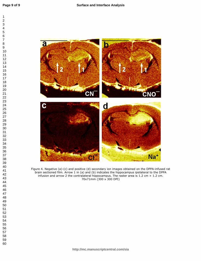
MacFabe et al have observed changes in immunoreactivity in the hippocampus and white matter as a result of PPA treatment. Previous studies have shown an innate neuroinflammatory response, consisting of reactive astrocytes and activated microglia in the white matter area and the hippocampus of PPA treated rodents. This pathological process is known to lead to increased production of cytokines, and reactive oxygen/nitrogen species, leading to increased oxidative stress, all consistent with that found in ASDs.[5-8] Shown in Figure 4 are ion images obtained on a DPPA-infused rat brain. From the ion images shown in Figures 4(a) and 4(b), it is clear that the DPPA treatment has caused significant changes as manifested by an increase in CN‾ (26.003) and CNO‾ (41.997) intensity in the DPPA-infused hippocampus indicated by arrow 1, in comparison with the contralateral hippocampus indicated by arrow 2. CN‾ and CNO‾ may originate from nitrogen-containing molecules and are consistent with increased oxidative stress found in the PPA rodent model and autopsy cases of ASD. The significance of our ToF-SIMS results is that we have been able to simultaneously image multiple markers reflecting increased oxidative stress[1-4] caused by the DPPA infusion, which compliments traditional neuroimmunohistochemical and biochemical techniques.
As shown in Figures 4(c) and 4(d), an increase in Cl‾ (34.968) is observed around the DPPA-infused hippocampus, which, together with a huge increase in Na+ (22.990), is consistent with extracellular edema, reported in MRI brain imaging.[21] In contrast, on the contralateral hippocampus, there is no apparent contrast for Cl‾ and Na+. Along with Na+, K+ (38.964) is another most important alkali metal ion in living organisms. There is no apparent change in K+ intensity (not shown) between the two hippocampal areas ipsilateral and contralateral to the DPPA-infusion side.
With infusion of deuterated sodium propionate (DPPA) to the brain of a rat, we used imaging ToF-SIMS to trace this short chain fatty acid via ion fragments D‾, C2HD4
+ and C2D5
+ that are also detectable from the reference DPPA itself. Another two ion fragments making a similar contrast in the rat brain sample to that of the three fragments are assigned to C3HD5
+ and C3H2D5+, which are found to be unique to DPPA-infused rat
brain. Future analysis of these two fragments may be a clue to the metabolic pathway of the short chain fatty acid in the central nervous system. Over the same anatomical locations (i.e hippocampus and external capsule white matter), we have confirmed ASD associated oxidative stress and edema evidenced by increasing CN‾ and CNO‾ intensity and Cl‾ and Na+ intensity, respectively. This study provides further validity of the PPA rodent model of ASD and the promise of ToF-SIMS imaging as a powerful tool to examine multiple metabolic processes in neuropsychiatric diseases.
Acknowledgement
This research was supported in part by generous contributions from GoodLife Children's Foundation and an Autism Collaboration grant from the Autism Research Institute to DFM. [1] D.L. Vargas, C. Nascimbene, C. Krishnan, A.W. Zimmerman, C.A. Pardo, Ann. Neurol. 2005, 57, 67. [2] M.R. Herbert, Neuroscientist 2005, 11, 417. [3] A. Chauhan, V. Chauhan, Pathophysiology 2006, 13, 171. [4] J. K. Kern, A. M. Jones, J. Toxicol. Env. Heal. B 2006, 9, 485. [5] D.F. MacFabe, D.P. Cain, K. Rodriguez-Capote, A.E. Franklin, J.E. Hoffman, F. Boon, A.R. Taylor, M. Kavaliers, K.-P. Ossenkopp, Behav. Brain Res. 2007, 176, 149. [6] D.F. MacFabe, K. Rodríguez-Capote, J.E. Hoffman, A.E. Franklin, Y. Mohammad-Asef, A.R. Taylor, F. Boon, D.P. Cain, M. Kavaliers, F. Possmayer, K.-P. Ossenkopp, Am. J. Biochem. Biotechnol. 2008, 4, 146 [7] S.R. Shultz, D.F. MacFabe, K.-P. Ossenkopp, S. Scratch, J. Whelan, D.P. Cain, Neuropharmacolgy 2008, 54, 901. [8] S.R. Shultz, D.F. MacFabe, S. Martin, J. Jackson, R. Taylor, F. Boon, K.-P. Ossenkopp, D.P. Cain, Behav. Brain Res. 2009, 200, 33. [9] A. Benninghoven, Angew. Chem. Int. Ed. Engl. 1994, 33, 1023. [10] D. Léonard, H.J. Mathieu, Fresenius J. Anal. Chem. 1999, 365, 3. [11] A.M. Belu, D.J. Graham, D.G. Castner, Biomaterials 2003, 24, 3635. [12] P. Sjövall, J. Lausmaa, B. Johnasson, Anal. Chem. 2004, 76, 4271. [13] D. Touboul, F. Kollmer, E. Niehuis, A. Brunelle, O. Laprévote, J. Am. Soc. Mass Spectrom. 2005, 16, 1608. [14] B. Johansson, Surf. Interface Sci. 2006, 38, 1401. [15] K. Börner, P. Malmberg, J.-E. Månsson, H. Nygren, Int. J. Mass Spectrom. 2007, 260, 128. [16] J.S. Fletcher, A. Henderson, G.X. Biddulph, S. Vaidyanathan, N.P. Lockyer, J.C. Vickerman, Appl. Surf. Sci. 2008, 255, 1264. [17] H.-Y. Nie, J.T. Francis, A.R. Taylor, M.J. Walzak, W.H. Chang, D.F. MacFabe, W.M. Lau, Appl. Surf. Sci. 2008, 255, 1079. [18] S.R. Thornton, F.L. Smith, J. Pharmacol. Exp. Ther. 1997, 281,514. [19] I. Minami, T. Kubo, S. Fujiwara, Y. Ogasawara, H. Nanao, S. Mori, Tribol. Letts. 2005, 20, 287. [20] A. Kunze, P. Sjövall, B. Kasemo, S. Svedhem, J. Am. Chem. Soc. 2009, 131, 2450. [21] T. Gerriets, E. Stolz, M. Walberer, C. Müller, A. Kluge, A. Bachmann, M. Fisher, M. Kaps, G. Bachmann, Stroke 2004, 35, 566.
Figure 1. Negative and positive secondary ion mass spectra for a DPPA (CD3CD2COONa) film cast on a Si substrate. The mass/charge ratio range for the spectra is (a) 0.5-4.5, (b) 15.5-18.5, (c)
25.5-35 and (d) 60-80 m/z, respectively. 80x189mm (1000 x 1000 DPI)
Figure 4. Negative (a)-(c) and positive (d) secondary ion images obtained on the DPPA-infused rat brain sectioned film. Arrow 1 in (a) and (b) indicates the hippocampus ipsilateral to the DPPA infusion and arrow 2 the contralateral hippocampus. The raster area is 1.2 cm × 1.2 cm.
Figure 3. Positive secondary ion mass spectra collected from a DPPA-infused rat brain sample (upper panel) and from a DPPA film cast on a Si substrate, showing (a) fragments existing in the
DPPA film and (b) new species unique in DPPA-infused rat brain.
Figure 4. Negative (a)-(c) and positive (d) secondary ion images obtained on the DPPA-infused rat brain sectioned film. Arrow 1 in (a) and (b) indicates the hippocampus ipsilateral to the DPPA infusion and arrow 2 the contralateral hippocampus. The raster area is 1.2 cm × 1.2 cm.