Rochester Institute of Technology Rochester Institute of Technology RIT Scholar Works RIT Scholar Works Theses 10-3-2018 Forensic Art in Law Enforcement: The Art and Science of the Forensic Art in Law Enforcement: The Art and Science of the Human Head Human Head Kourtnei F. Rodriguez [email protected]Follow this and additional works at: https://scholarworks.rit.edu/theses Recommended Citation Recommended Citation Rodriguez, Kourtnei F., "Forensic Art in Law Enforcement: The Art and Science of the Human Head" (2018). Thesis. Rochester Institute of Technology. Accessed from This Thesis is brought to you for free and open access by RIT Scholar Works. It has been accepted for inclusion in Theses by an authorized administrator of RIT Scholar Works. For more information, please contact [email protected].
Transcript
Rochester Institute of Technology Rochester Institute of Technology
RIT Scholar Works RIT Scholar Works
Theses
10-3-2018
Forensic Art in Law Enforcement: The Art and Science of the Forensic Art in Law Enforcement: The Art and Science of the
Follow this and additional works at: https://scholarworks.rit.edu/theses
Recommended Citation Recommended Citation Rodriguez, Kourtnei F., "Forensic Art in Law Enforcement: The Art and Science of the Human Head" (2018). Thesis. Rochester Institute of Technology. Accessed from
This Thesis is brought to you for free and open access by RIT Scholar Works. It has been accepted for inclusion in Theses by an authorized administrator of RIT Scholar Works. For more information, please contact [email protected].
The face also has a variety of secondary structures that are extremely important to human
identity. These structures are unique to every individual, making no two faces the same. The
secondary structures of the face are the supraorbital ridges (brows), nose, mouth, eyes, cheeks,
chin, and ears. These structures combined with other facial features and characteristics, such as
skin tone, facial hair, and wrinkles, make the face more individualized.
Forensic artists should have a good understanding of rhytide patterns, or wrinkles, when
producing age-progressed drawings. Wrinkles are important facial features that occur naturally in
every human being. The author of a book about the anatomy of the human head wrote, “Wrinkle
patterns are not a random phenomenon, but follow definite routes over the surface of the
face” (Hogarth, 2002). The author continued writing, “Wrinkling of the face occurs for a variety
of reasons. The most common reasons relate to incidental facial activity, such as chewing,
grimacing, winking, pouting, squinting, expressions of pleasure and distaste, and demonstrations
of emotion. Other reasons relate to psychological stress and inner tensions, as well as aging,
muscular flaccidity, or loss of firmness in flesh. Whatever the origin of wrinkles, their
development follows three major patterns: frontal, oblique, and lateral” (Hogarth, 2002).
Understanding these patterns are crucial and help forensic artists create accurate scientific
drawings. It is especially important when working on an age-progressed drawing because the
individual looks more realistic when the wrinkles in the skin appear more natural (see Figure 15,
Figure 16, Figure 17, and Figure 18).
Rodriguez - !25
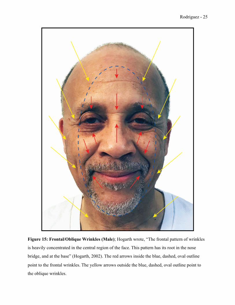
Figure 15: Frontal/Oblique Wrinkles (Male); Hogarth wrote, “The frontal pattern of wrinkles
is heavily concentrated in the central region of the face. This pattern has its root in the nose
bridge, and at the base” (Hogarth, 2002). The red arrows inside the blue, dashed, oval outline
point to the frontal wrinkles. The yellow arrows outside the blue, dashed, oval outline point to
the oblique wrinkles.
Rodriguez - !26
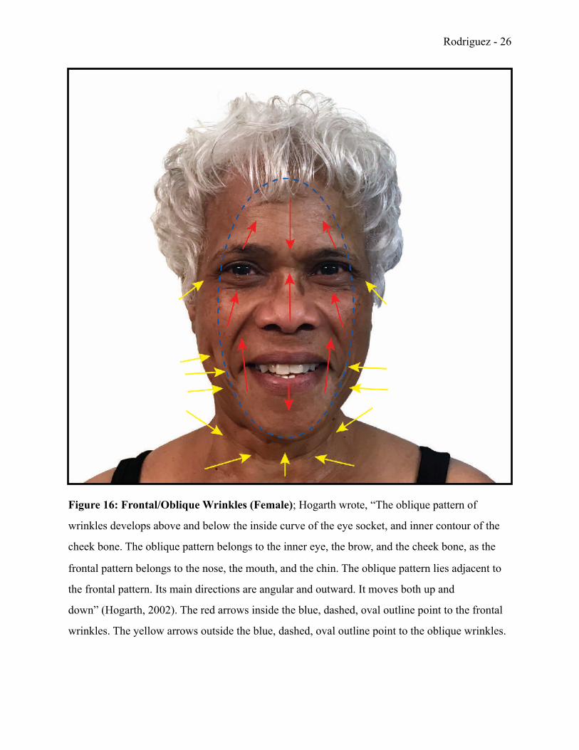
Figure 16: Frontal/Oblique Wrinkles (Female); Hogarth wrote, “The oblique pattern of
wrinkles develops above and below the inside curve of the eye socket, and inner contour of the
cheek bone. The oblique pattern belongs to the inner eye, the brow, and the cheek bone, as the
frontal pattern belongs to the nose, the mouth, and the chin. The oblique pattern lies adjacent to
the frontal pattern. Its main directions are angular and outward. It moves both up and
down” (Hogarth, 2002). The red arrows inside the blue, dashed, oval outline point to the frontal
wrinkles. The yellow arrows outside the blue, dashed, oval outline point to the oblique wrinkles.
Rodriguez - !27
Figure 17: Lateral Wrinkles (Male); Hogarth wrote, “The lateral pattern consists of all the
wrinkles on the side region of the face, neck, and back of the head. This diffuse pattern shows
isolated furrows and rifts which emanate from the outer eye, ear, jaw line, and back of the neck
below the skull base” (Hogarth, 2002). The red arrows outside the blue, dashed, oval outline
point to the frontal wrinkles. The yellow arrows inside the blue, dashed, oval outline point to the
lateral wrinkles. Gravity has shifted the skin downward in these areas.
Rodriguez - !28
Figure 18: Lateral Wrinkles (Female); Muscles are repeatedly extended and compressed,
which causes tension in the muscle fibers. Repetitive facial movements, such as smiling or
frowning, will eventually cause wrinkles to form that can be seen on the surface of the skin. The
woman in this figure informed me that her dimple used to be up higher on the side of her cheek,
but overtime it has shifted downward. The red arrows outside the blue oval with the dashed
outline point to the frontal wrinkles. The yellow arrows inside the blue oval with the dashed
outline point to the lateral wrinkles. Gravity has shifted the skin downward in these areas.
Rodriguez - !29
Unlike the wrinkle patterns discussed earlier, some wrinkle patterns can be random due to
the natural aging of the skin and how it changes with gravity. A book on the anatomy of the
human head noted, “As we age, the flesh loosens and the skin shrivels; the tissues lose their
firmness and elasticity. This creates what we may call sag and shrinkage wrinkles. These
wrinkles are random and unpredictable. They usually sag downward as they are pulled by the
force of gravity” (Hogarth, 2002).
Although gravity may be a huge factor in the formation of wrinkles, doctors have
discovered that shifting bone structure may also be another reason why skin sags. An article from
Duke University Magazine on cranial forms and facial structures concludes, “Researchers at
Duke Medical Center have discovered that changes in the face's underlying bony structure may
be the culprit. And those changes appear to occur more dramatically in women than in
men” (“Cranium Changes,” 2008). This explains why cosmetic procedures such as botulinum
toxin therapy (Botox), which treats various muscle spasms and diseases characterized by
overactive muscles, do not permanently eradicate wrinkles in the skin.
Furthermore, Dr. Michael Richard, assistant clinical professor of ophthalmology and
oculoplastic surgeon at the Duke Eye Center, added, “Our focus has always been on tightening
and lifting the soft tissues, skin, and muscle in an attempt to cosmetically restore patients'
youthful appearance. Based on this information, it might actually be better to restore the
underlying bony framework of the face to its youthful proportions” (“Cranium Changes,” 2008).
By studying computerized tomography (CT) scans, researchers discovered that the structures of
the human skull continued to grow many years after puberty. During these studies, researchers
noticed the frontal bone continued to move forward and the zygomatic bones continued to move
backwards. As these bones moved, the muscles and skin on top of them changed as well. This
caused the overall appearance of the face to be altered. Dr. Richard continued, “The facial bones
also appear to tilt forward as we get older, which causes them to lose support for the overlying
soft tissues and results in more sagging and drooping” (“Cranium Changes,” 2008). Forensic
artists should be aware of this research and remember it when doing age progressions or any
other forensic artwork that may require knowledge of craniofacial growth and development.
Rodriguez - !30
Section II: Body of Work
I-c … The Human Face: Variations by Age, Gender, and Race
The underlying skeletal structures of the human face are unique to each individual. The
face also varies by age, gender, and race. Even monozygotic twins —who are created when a
single egg is fertilized to form one zygote, then split to form two embryos— are never
completely identical. “As the head ages, changes take place in the head proportions, skull
development, and bone articulation in the brow, nose, jaw, and teeth. Growth in flesh, tissue,
skin, lips, ears, eyelids, and hair all contribute to the descriptive aspects of aging” (Hogarth,
2002). These changes contribute to the uniqueness of one’s appearance because the variation of
changes are infinite. Other variables, such as gender and race, also play a huge role in how
people look. I have placed people into seven (7) age categories: babies, children, teenagers, young
adults, middle-aged adults, older adults, and senior adults. In this context, I consider babies to be
newborns, infants, and toddlers between the ages of zero (0) and three (3); children are older kids
between the ages of four (4) and twelve (12); and teenagers are younger persons between the
ages of thirteen (13) and nineteen (19). I have also classified young adults as anyone between the
ages of twenty (20) and thirty-five (35), middle-aged adults between the ages of thirty-six (36)
and fifty (50), older adults between the ages of fifty-one (51) and sixty-four (64), and senior
adults as anyone who is sixty-five (65) years of age or older. Different growth and developmental patterns of the head occur within these 7 age groups.
Through my own personal observation, I noticed how the head of a baby is small, but the skull is
large compared to the proportion of his or her facial structures. It also seems that most babies
appear to have large eyes because their skull has not yet grown in proportion to their orbits.
Through further observation, I also noticed that babies have more flesh on top of their zygomatic
arches. This makes their cheeks look fuller and face seem rounder. In reference to a baby boy, a
forensic artist noted certain growth patterns in the child and wrote, “The head is very rounded
and the facial contours are full and rounded. There is the appearance of epicanthic folds and the
eyes look large and rounded” (Taylor, 2001). Furthermore, I concluded that babies have a round
Rodriguez - !31
nose, small lips, and flat supraorbital ridges due to the brow arches of the frontal bone being
underdeveloped.
As a baby develops into a small child, the head and face continue to grow and shift. The
face is extremely round until approximately age three, but takes more shape as the jaw widens to
make room for the developing teeth. The artist further noted, “The cranium expands to
accommodate the growing brain. The maxilla and mandible have enlarged and widened to allow
room for the deciduous dentition” (Taylor, 2001). These changes make the eyes seem more
proportionate to the face because the inter-orbital distance between the eyes increases as well.
There are quite a few changes that occur in a child’s face and skull between the ages of 4
and 12. At around age 4, I can tell that a child’s face has lengthened and the bridge of his or her
nose is more pronounced. At ages five (5) and six (6), I noticed that the face has grown even
more, further lengthening the bridge of the nose and chin too. I can see that the chin is slightly
pointed as well, due to the continued development of the teeth and enlargement of the jaw. As the
skull continued to increase in length, the excess skin on the face decreased as well. The artist
mentioned additional changes and wrote, “The bridge of the nose continues to rise up, lifting
some of the excess skin from the medial corners of the eyes. The face continues to elongate as
the nose length and the chin length increase. The forms of the lower cartilages of the nose
become apparent and the tip takes shape” (Taylor, 2001). This loss of excess skin, which is often
referred to as baby fat, makes the child appear more lean.
I also examined how the contours of the face and the musculature of the neck formed in
children between the ages of 7 and 12. It was further described, “The face elongates. The
childlike face is looking more juvenile as some of the facial forms become more apparent due to
less baby fat. The nose continues to grow, both in the bridge and the nostril size. The mandible
and chin continue to grow. The teeth seem big for the face” (Taylor, 2001). Although the teeth
seemed disproportionate to the face, I could tell that they were almost fully developed as the
skull continued to grow. Nonetheless, by the time a child becomes a teenager, he or she should
have twenty-eight permanent adult teeth. According to the American Association of Oral and
Maxillofacial Surgeons, “Wisdom teeth, or third molars, are the last teeth to develop and appear
in your mouth. They come in between the ages of 17 and 25, a time of life that has been called
Rodriguez - !32
the ‘Age of Wisdom’” (“American,” 2016). Until then, the bridge of the nose deepens, the chin
becomes more firm, and the facial muscles strengthen as the child transitions into a teenager.
The period of time between the ages of 13 and 19 are called the teenage years. During
this time, more changes occur in the face, head, and neck. The same forensic artist continued her
observations and acknowledged, “The cheekbones have become relatively more prominent. The
nose has grown even more, revealing the nasal bones at the bridge. The forehead shape has
remained consistent though it has grown and risen at the glabella” (Taylor, 2001). Furthermore,
one author observed the facial changes of many teenagers and noted, “Nose bridge deepens.
Nose tip still up. Jaw corner more angular; chin firmer, less rounded. Neck longer, a bit heavier.
Understructure of head comes through; leaner facial appearance. Jaw angle firm. Mouth more
firm. Neck muscles stronger, more developed. Skin texture firmer and thicker” (Hogarth, 2002).
Overall, I noticed how teenagers looked less juvenile because their other facial structures, (such
as their ears, eyes, mouth, and nose), became more proportionate to their head (see Figure 19).
I classified young adults as people between the ages of 20 and 35. During this time, the
softness of their face disappeared and became more angular as the understructures of their skull
formed beneath their skin. A man studying the anatomy and proportions of the human head
observed additional facial changes in young adults. He wrote, “Deep nose bridge; nasal bone
more pronounced. Lean cheeks and cheek bone. Chin mound more decisive; under-jaw region
firm and even” (Hogarth, 2002). I observed the facial changes of many young adults on my own
and noticed more firmness in their skin and an increased definition of their underlying facial
structures as well. I noticed gradual changes in the face of young adults around age 30. The vigor
of their skin was still intact, but signs of natural atrophy were present. I could see the subtle
development of creases forming in their skin around the chin, corner of the eyes, forehead, and
mouth. I often saw a slight sag in the skin underneath their chin and jaw as well. At
approximately 35 years old, the subtle changes in the face seemed more noticeable to me. I
would consider a person at this age to have a mature adult face.
Rodriguez - !33
Figure 19: Differences Between a Child’s Skull and a Young Adult’s Skull; Overall, the skull
of a child is smaller than the skull of an adult. The nasal opening is small and the length of the
face is short. The jawline is short and round as well. However, the frontal bone seems large
because the other facial structures are not yet fully developed. A young adult’s jawline is large
and angular. It is also wide due to the growth of mature teeth. The frontal bone, nasal opening,
and zygomatic arches are proportionate to the other facial structures.
Rodriguez - !34
Sometimes it can be hard to determine how old a person is between the ages of 36 and 50
because the changes in their face are gradual and occur at a steady pace over an extended amount
of time. However, this age group has many different facial characteristics. I primarily noticed the
excess skin under the chin, eyes, and jaw. I also observed how the subtle creases in the skin
became more apparent near the lateral edges of the eyes, mouth, nose, and furrow of the brows.
The face appeared fuller and the skin under the jaw was soft and fleshy as well. The author
noted, “First appearance of softness and fleshiness under jaw; chin mound cleavage occurs.
Mouth wrinkle emerges. Under eye sag; further increase of eye and brow wrinkles. Neck less
lean, somewhat heavier” (Hogarth, 2002). The author also confirmed my findings on head hair. I
discovered that gray hair formed in both genders, but men were more likely to have a receding
hairline by age forty (40). Additionally, I observed that head hair began thinning even more
between the ages of 40 and forty-five (45) for both genders as well. At around age 50, I noticed a
clear separation of the chin from the jawline in middle-aged adults. There was also a pattern of
excess flesh that formed near the back of the neck.
I consider older adults as people between the ages of 51 and 64. Depending on their
genetics, health, and life-style choices, some older adults look much younger, or much older,
than their actual age. One forensic artist wrote, “Such factors as stress, diet, or illness affect the
onset of the various [facial] lines, as do smoking and sun exposure influences” (Taylor, 2001).
Nevertheless, it appeared that older adults usually had more wrinkles near their forehead, mouth,
and eyes. The wrinkles were noticeable, but not as apparent as the wrinkles of senior adults who
had less elastic skin. I saw how the upper eyelids on older adults sagged and formed crow’s feet.
However, their lower eyelids were more fleshy and sagged less. I also noticed that jowls had
formed and created a fold of fatty flesh under their chin.
There are additional hair and facial characteristics that make a person look older. The
author noted, “Hair quite thin on crown. Under eye pouch more decisive. Cheek bone and
zygomatic arch apparent. Back neck fold deeper; front of neck shows sagging, folding, and
shrinkage” (Hogarth, 2002). He continued further, notating the facial characteristics of a 60 year
old adult, and wrote, “Bony features of skull apparent throughout. Nose forms more distinct.
Lower facial region appears slack, flaccid” (Hogarth, 2002). I interacted with many adults
Rodriguez - !35
between the ages of 51 and 64 and saw the hair and facial characteristics Hogarth described
above. I kept in mind the environmental factors, genetics, and lifestyle choices of these
individuals while observing their unique features. I noticed a vast difference in the physical
appearance of healthy and non-healthy individuals of the same age according to factors such as
diet, physical activity, and smoking.
The last age category I have identified is senior adults. I consider senior adults to be
people who are 65 years of age or older. I saw how the loss of collagen and elastic fibers made
their skin become loose and stiff. Collagen is defined as, “Any of a class of extracellular proteins
abundant in higher animals, especially in the skin, bone, cartilage, tendon, and teeth, forming
strong insoluble fibers and serving as connective tissue between cells, yielding gelatin when
denatured by boiling.” (“Collagen,” 2018). The loss of collagen occurs naturally in the aging
process and gravity increases the sagging of the skin. One author’s notes concluded, “Folds and
creases deepen around the corners of the mouth, jaw, and neck. Tooth loss causes jaw shrinkage;
jaw angle less steep. Chin more prominent. Nose seems enlarged. Mouth folds deeper. Network
of smaller wrinkles develops throughout head” (Hogarth, 2002). I saw these changes and noticed
how distinct the bridge of the nose and nostrils were in people over 65. I also observed how the
tip of their nose appeared large and bulbous. My great-grandfather lived to be ninety-four (94)
years old and he exhibited some of the same physical characteristics mentioned above. His
jawline was less defined because the loss of collagen in his body made his skin loose (see Figure
20). Although many substructures on a senior adult’s face are less-defined, other structures, (such
as the frontal bone, temporal bones, and zygomatic arches), can still be seen.
Rodriguez - !36
Figure 20: Four Generations; The figure above shows four generations of men from the same
family. The development of rhytide patterns increases from the son, father, paternal grandfather,
and paternal great-grandfather. The effects of gravity on the skin are also distinguishable
between each photograph as well.
Rodriguez - !37
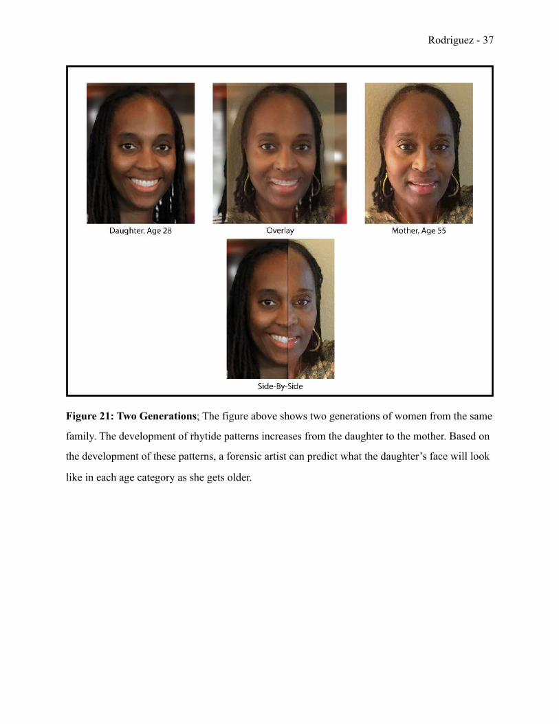
Figure 21: Two Generations; The figure above shows two generations of women from the same
family. The development of rhytide patterns increases from the daughter to the mother. Based on
the development of these patterns, a forensic artist can predict what the daughter’s face will look
like in each age category as she gets older.
Rodriguez - !38
Gender, or sex, is another variable that contributes to the structure of the human head.
It is imperative to understand the differences between the male and female skull because the
underlying structures vary in size and shape depending on the sex. A forensic artist confirmed,
“Generally speaking, anthropologists expect male skulls to be more robust with more
pronounced muscle attachment indicators” (Taylor, 2001). The bony elevated surfaces of the
skull, known as the muscle ridges, are where the muscles attach to the skull. They are usually
larger in males and include the temporal lines, which attach to the temporal bones, and the
nuchal lines, which attach to the occipital bones. This is the type of information forensic artists
must know when working on post-mortem facial reconstructions. Proper placement and size of
the underlying musculature of the skull increases the likeness of the individual being reproduced.
I observed many other differences between the male and female skull. Overall, the male
skull is larger and more angular than the female skull. The male mandible is also much larger and
the angle of the jaw is more defined. A female’s mandible is less defined and much smaller in
proportion to the skull. The supraorbital ridges and mastoid processes on the male skull are more
pronounced, whereas the same structures on the female skull are more subtle and smooth. Males
also have a sloping or receding frontal bone, but the female frontal bone is more vertical. I
realized that the external occipital protuberance (EOP), also known as the inion, is more
prominent in males as well. The American Heritage Stedman’s Medical Dictionary defines the
inion as, “The most prominent projecting point of the occipital bone at the base of the
skull” (Stedman, 2007). On a diagram of the human skull, I located the inion on the back of the
head near the lower, rear portion of the occipital bone (see Figure 23).
Rodriguez - !39
Figure 22: Differences Between the Male and Female Skull (Anterior View); The male skull
is larger and wider than the female skull. The male mandible is large, wide, and square compared
to the small, narrow, and pointed female mandible. The zygomatic arches on the male skull are
also larger than the zygomatic arches on the female skull as well.
Rodriguez - !40
Figure 23: Differences Between the Male and Female Skull (Lateral View); The male skull is
larger and wider than the female skull. The mandible on the male skull is large, wide, and square
compared to the small, narrow, and pointed mandible on the female skull. The female mandible
is more obtuse than the male mandible. The zygomatic arches on the male skull are larger and
less pointed than the zygomatic arches on the female skull. The supraorbital ridges and mastoid
processes are larger and more pronounced on the male skull as well, but subtle and smooth on
the female skull. The frontal bone on the male skull has a larger slope, while the female frontal
bone recedes less and is more vertical. The EOP is more prominent on the male skull also.
Rodriguez - !41
While studying the anatomy of the human head, I learned that there are three (3) different
head-types. Each head-type is classified by the cephalic index —which is an established standard
of head measurement. The cephalic index is specifically defined as, “The ratio of the maximum
width of the head to its maximum length, multiplied by 100” (Stedman, 2007). In the book,
Drawing the Human Head, the head is classified as a broad-headed (brachycephalic), long-
headed (dolichocephalic), or medium-headed/intermediate (mesocephalic) head-type (Hogarth,
2002). Hogarth described individuals with a brachycephalic head-type as having a wide skull and
a medium to low positioning forehead. Furthermore, he noted that their face and facial features
were usually more compact, square, and short. He observed that people with a dolichocephalic
head-type had a long and narrow skull. In addition, he noted that their forehead was either
moderately arched or highly arched. If their forehead was highly arched, he determined that their
facial structures tended to be much longer. The third head-type in the cephalic index is called the
mesocephalic head-type. Hogarth described people with this head-type as having an
intermediate-sized skull, oval-shaped face, and small facial structures.
Head-types do not identify specific individuals by age, gender, or ethnicity. However, it is
important to be aware that these head-types exists and vary across all races of people. In an
excerpt from the book, Drawing the Human Head, it says, “No one particular group of persons
can be identified by a single head type classification or facial slope. Head form variations run
through the individuals of all population groups” (Hogarth, 2002). Forensic artists should keep
this in mind and store this information for future use. It will be imperative when producing
forensic artwork of unidentified, deceased individuals with missing skull fragments or damaged
facial structures.
Race also plays a huge role in the structure of the human head and face. In anthropology
race is defined as, “Any of the traditional divisions of humankind —the commonest being the
Caucasian, Mongoloid, and Negroid— characterized by supposedly distinctive and universal
physical characteristics” (“Race,” 2018). Generally, these physical characteristics include: skin
color, pigmentation, facial form, hair texture, and body proportions. Within the 3 divisions of
humankind, I have established ten (10) geographic races. Individuals of European, Middle-
Eastern, and Mediterranean decent are considered Caucasoid; Asian and Native American people
Rodriguez - !42
are considered Mongoloid; People of African, Melanesian, Micronesian, Polynesian, and
Australian Aborigines decent are considered Negroid.
Within these 3 divisions, I discovered many physical differences in face and head from
the lateral view (see Figure 24, Figure 25, Figure 26, and Figure 27). In chapter 3 of the book,
Forensic Art and Illustration, the author wrote, “In profile, many Caucasoid skulls exhibit a
somewhat flat face with zygomatic bones that retreat or slant back. Frontally, the nasal openings
tend to be longer and narrower than in Mongoloids and Negroids” (Taylor, 2001). While people-
watching at the Indianapolis International Airport (IIA), I observed the facial features of many
Caucasian passengers. I compared their facial features with the facial features of people from
other racial groups to study the differences.
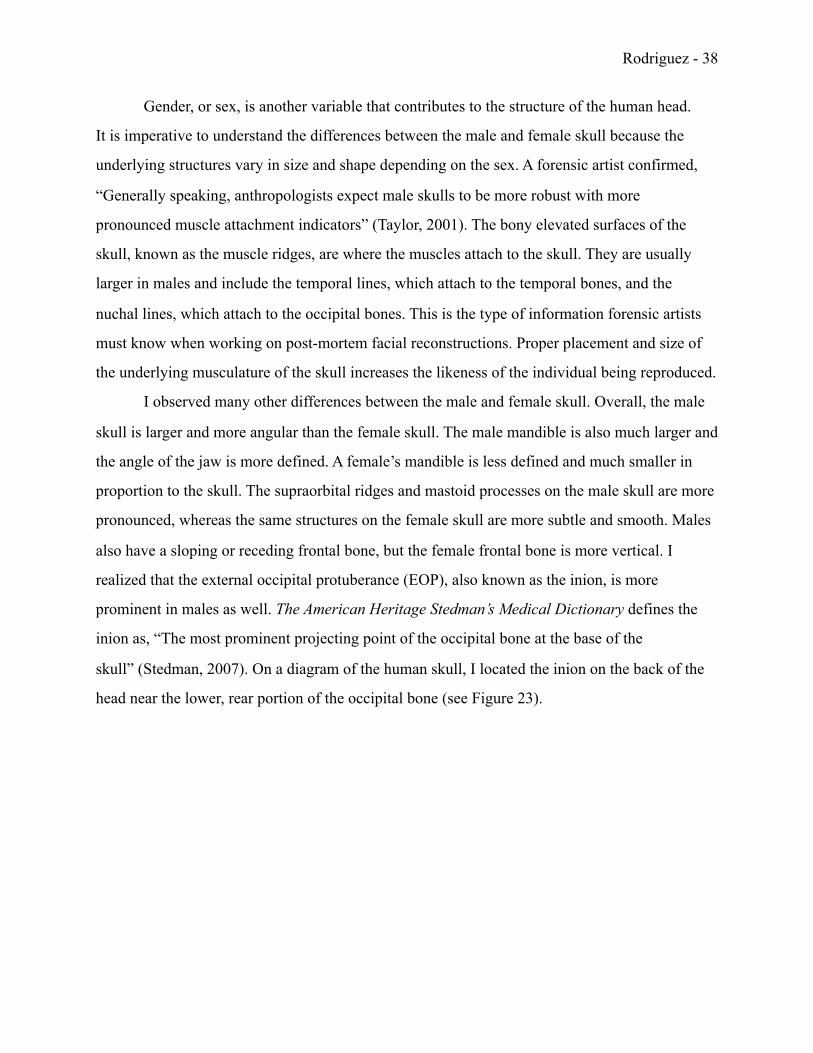
Rodriguez - !43
Figure 24: The Caucasoid Skull; Above is a picture of a white male that shows a drawing of
what his skull might look like below it. The comparison chart in Figure 27 highlights the
differences between each skull-type.
Rodriguez - !44
There are many universal characteristics of the Mongoloid race. When describing their
appearance, the author continued further and wrote, “In profile, the Mongoloid skull indicates an
often flattened face with a short cranial vault or distance from front to back. Frontally, the cheek
area is usually quite wide with projecting zygomatic bones. The width of the nose opening or
nasal aperture is usually somewhere between the Caucasoid and the Negroid. Mongoloid mouths
may also be of a size somewhere between the Caucasoid and the Negroid” (Taylor, 2001). I
identified the aforementioned physical features on many Asian passengers at the IIA. An
example of the head and skull of a Mongoloid male is shown in the figure below.
Rodriguez - !45
Figure 25: The Mongoloid Skull; Above is a picture of an Asian male that shows a drawing of
what his skull might look like below it. The comparison chart in Figure 27 highlights the
differences between each skull-type.
Rodriguez - !46
When studying the Negroid race, the author wrote, “In profile, the Negroid skull often
exhibits alveolar prognathism, which is expressed as a projection of the lower face. Frontally, the
nasal openings tend to be wider and shorter than in Caucasoids and Mongoloids, with a bridge
that is broader and flatter. Negroid mouths tend to be broader with fuller everted lips. Many
negro people also have wider set eyes than the other groups” (Taylor, 2001). I confirmed these
characteristics by looking at myself in the mirror and studying the faces of my relatives. In my
opinion, negroid facial features can be easily distinguished from those of Caucasoid and
Mongoloid decent. Not only are the underlying structures of the skull strikingly different, but
there are also obvious differences in the skin tone and hair texture of Negroid individuals as well.
Rodriguez - !47
Figure 26: The Negroid Skull; Above is a picture of a black male that shows a drawing of what
his skull might look like below it. The comparison chart in Figure 27 highlights the differences
between each skull-type.
Rodriguez - !48
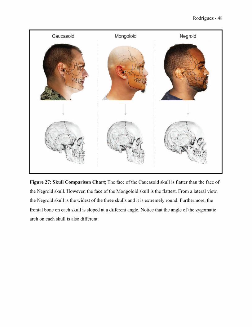
Figure 27: Skull Comparison Chart; The face of the Caucasoid skull is flatter than the face of
the Negroid skull. However, the face of the Mongoloid skull is the flattest. From a lateral view,
the Negroid skull is the widest of the three skulls and it is extremely round. Furthermore, the
frontal bone on each skull is sloped at a different angle. Notice that the angle of the zygomatic
arch on each skull is also different.
Rodriguez - !49
Conclusion:
Forensic art is a highly specialized career field for artists who have been trained
appropriately. Forensic artists should possess both artistic ability and scientific knowledge of the
human body to accurately render people of different ages, gender, and race. Understanding the
human body, written law, and law enforcement policies and procedures is crucial. Portfolios
should exhibit realistic anatomical drawings, illustrations, or sculptures to showcase both artistic
talent and scientific knowledge.
It is not easy to become a forensic artist. Full-time positions are limited, but working for a
law enforcement agency is a huge step forward in the career process. In the meantime, artists can
stay up-to-date with current forensic art methods and techniques by continuing to draw, sculpt,
and study human anatomy. Attending workshops and networking is also very helpful. This will
prepare forensic artists for employment when positions becomes available.
Overall, a career in forensic art can be extremely rewarding. Capturing a fugitive from
justice, or identifying the remains of a missing loved-one, can bring closure to a grieving family.
The impact forensic art has had on society in the last two centuries has been extraordinary. One
successful forensic artist wrote, “Many law enforcement officers have come to know the power
of forensic art. They have seen a composite drawing lead to the perpetrator they seek, or a facial
reconstruction help reveal the identity of a homicide victim. Age progressions help officers
recover abducted children and locate fugitives who have been at large for many years. Many
have witnessed the profound impact and clarity that forensic art can bring to court
proceedings” (Taylor, 2001). This is the type of impact I want my artwork to have on society.
Until that day comes, I will continue to educate other artists about this unique career field and
continue training.
Rodriguez - !50
Citations and References
American Association of Oral and Maxillofacial Surgeons. (2016, January 16). Third Molar Research News. Retrieved November 12, 2017, from https://www.aaoms.org/media/third-molar-research-news
Bailey, L. (2011, November 1). Age Progression. Retrieved November 21, 2017, from https:// www.askaforensicartist.com/age-progression/
Bender, F. (1989). Bust of John List [Clay]. Philadelphia, PA: America’s Most Wanted.
Collagen [Def. 1]. (2018). Random House Unabridged Dictionary Online. In Random House Unabridged Dictionary. Retrieved November 27, 2018, from http://www.dictionary.com/browse/collagen
Cranium Changes. (2008, January 31). Retrieved October 16, 2017, from http:// dukemagazine.duke.edu/issues/010208/depgaz13.html
Forensic Art and Facial Identification. (2015, February 11). Retrieved September 23, 2017, from https://www.dundee.ac.uk/study/pg/forensic-art-facial-identification/
Forensic Art Certification. (2017, April 13). Retrieved October 04, 2017, from https:// www.theiai.org/certifications/artist/index.php
Hogarth, B. (2002). Drawing the Human Head. New York, NY: Watson-Guptill Publications.
(n.d.) William Bradford Bishop [Age-Enhanced Photograph]. Washington, DC: Federal Bureau
of Investigation.
Rodriguez - !51
Race [Def. 3]. (2018). Random House Unabridged Dictionary Online. In Random House Unabridged Dictionary. Retrieved February 12, 2018, from http://www.dictionary.com/browse/race
Stedman, T. L. (2007). The American Heritage Stedman's Medical Dictionary. Boston, MA: Houghton Mifflin.
Taylor, K. T. (2001). Forensic Art and Illustration. Boca Raton, FL: CRC Press.
Taylor, K. T. (2001). Two-Dimensional Post-Mortem Facial Reconstruction [Drawing]. In Forensic Art And Illustration (p. 385). Boca Raton, FL: CRC Press.
Taylor, K.T. (2014). Bust of William Bradford Bishop [Clay]. Washington, DC: Federal Bureau of Investigation.
Wilkinson, C. (2008). Forensic Facial Reconstruction. Cambridge, England: Cambridge University Press.