Proc. Nati. Acad. Sci. USA Vol. 86, pp. 6264-6268, August 1989 Genetics Reordering of nine exons is necessary to form a functional actin gene in Oxytricha nova (macronuclear development/micronucleus/hypotrichs) ARTHUR F. GRESLIN, DAVID M. PRESCOTT, YOSHIO OKA*, STEPHEN H. LOUKINt, AND JAMES C. CHAPPELL Department of Molecular, Cellular, and Developmental Biology, University of Colorado, Boulder, CO 80309 Contributed by David M. Prescott, May 30, 1989 ABSTRACT During the development of a macronucleus from a micronucleus after cell mating in hypotrichs all the genes (=20,000) are excised from micronuclear chromosomes as individual small DNA molecules. Telomeres are added to the ends of each gene-sized molecule and each is ampliflied, mostly by %1000-fold, to yield a transcriptionally active macronu- cleus. As a part of the study of the excision of genes from chromosomes, we have cloned six fragments of chromosomal DNA from Oxytricha nova, each containing a full copy of an actin gene, for comparison with the structure of the actin- encoding DNA molecule in the macronucleus. All six micronu- clear actin clones had the same overall organization as judged by restriction mapping. Two micronuclear actin clones were sequenced. These differ from one another at a few nucleotide positions but both prescribe precisely the same actin polypep- tide. Both micronuclear actin genes contain nine exons sepa- rated by eight intron-like sequences. The macronuclear gene contains these nine exons without intron-like segments. Assign- ing the order 1 through 9 to the nine micronuclear exons, the order in the macronucleus is 8-7-1-2-4-3-5-9-6. In the micronuclear actin gene, all nine exons possess terminal repeat sequences. These repeat sequences provide precise directions for reordering and joining of the nine exons to yield the exon order in the macronuclear gene. Polymerase chain reaction analysis of micronuclear DNA of the related species, Oxytricha trifallax, shows that the actin gene has an unorthodox arrange- ment in this species also. The genetic apparatus of hypotrichous ciliates consists of two morphologically and functionally different nuclei, the micro- nucleus and the macronucleus. The micronucleus contains high molecular weight DNA organized into chromosomes that divide mitotically and undergo meiosis during the sexual phase of the organism's life cycle (1). In contrast, the macronucleus contains only low molecular weight DNA molecules, ranging in size from =500 base pairs (bp) to 15 kilobase pairs (kbp) with a number average size of 2200 bp (2). There are -20,000 different molecules, and each appears to contain only one transcription unit or gene-encoding region. Each of the 20,000 molecules is present, on average, in =1000 copies (3). Transcription is very active in the macronucleus but is not detectable in the micronucleus. The micronucleus is not essential for vegetative growth in at least some species of hypotrichs, including Oxytricha nova. The micronucleus functions as a germ-line nucleus. When two cells mate, the micronucleus divides meiotically, and haploid micronuclei are exchanged between the paired cells. An exchanged nucleus fuses with a stationary haploid mi- cronucleus to form a new diploid zygotic micronucleus. Shortly thereafter the old macronuclei and the remaining haploid micronuclei are destroyed. After conjugation, the new diploid micronucleus divides mitotically without cell division. One of the two new micronuclei develops to a new macronucleus. Our long-term objective is to find out how hypotrichs excise their genes from chromosomes during macronuclear development. We know that genes occur in micronuclear chromosomes singly or in small clusters separated by long spacers (4, 5). The formation of a macronucleus from a micronucleus is a complex process that includes polyteniza- tion of the chromosomes, destruction of the polytene chro- mosomes, degradation of all repetitive sequences and 90- 95% of all unique sequences, elimination of intron-like se- quences (ILSs), and addition of telomeric sequences to the gene-sized DNA molecules (6). A gene-sized molecule pos- sesses a coding region, a short leader sequence, and a trailer sequence. MATERIALS AND METHODS DNA Cloning and Sequencing. A library of micronuclear DNA molecules was constructed in phage A47.1 as described (7). A deletion series of subclones was generated for double- stranded DNA sequencing by the Erase-A-Base system (Promega). Dideoxynucleotide sequencing of double- stranded DNA was performed using the modified T7 DNA polymerase Sequenase (United States Biochemical) accord- ing to manufacturer's directions. Southern Hybridization. Micronuclear DNA was digested with BAL-31 Fast Form (International Biotechnologies) for 20 min at 30°C, according to manufacturer's directions. Digested micronuclear DNA and cloned DNAs were elec- trophoresed in 1% agarose gels at 5 V/cm for 2.5 hr in 50 mM Tris borate, pH 8.0/2 mM EDTA. DNA was transferred to nitrocellulose overnight with 1Ox SSC as a transfer medium (lx SSC is 0.15 M NaCl/0.015 M sodium citrate, pH 7.4). Blots were prehybridized in 1 M NaCl/50 mM Tris-Cl, pH 8.0/1 mM EDTA/10x Denhardt's solution/0.1% SDS/ sheared herring DNA (200 mg/ml) at 68°C for 4 hr (lx Denhardt's solution = 0.02% polyvinylpyrrolidone/0.02% Ficoll/0.02% bovine serum albumin). Hybridization was carried out in a fresh prehybridization solution at 68°C for 16 hr. Post-hybridization washes were at a final stringency of 0.2x SSC/0.1% SDS at 68°C for 1 hr. Filters were air-dried and autoradiographed. The appropriate restriction fragment to serve as a probe for each hybridization was isolated by electrophoresis and labeled with 32P by random-hexamer- primer extension to a specific activity of >108 dpm/,tg. Polymerase Chain Reaction (PCR) and Sequencing. A PCR was performed on 1 Ag of native micronuclear DNA for 30 Abbreviations: PCR, polymerase chain reaction; ILS, intron-like sequence. *Present address: Mitsubishi-Kasei Institute of Life Sciences, 11, Minaiooya, Machida-Dhi, Tokyo 194, Japan. tPresent address: Laboratory of Molecular Biology, University of Wisconsin, 1525 Linden Drive, Madison, WI 53706. 6264 The publication costs of this article were defrayed in part by page charge payment. This article must therefore be hereby marked "advertisement" in accordance with 18 U.S.C. §1734 solely to indicate this fact.
Transcript
Proc. Nati. Acad. Sci. USAVol. 86, pp. 6264-6268, August 1989Genetics
Reordering of nine exons is necessary to form a functional actingene in Oxytricha nova
ARTHUR F. GRESLIN, DAVID M. PRESCOTT, YOSHIO OKA*, STEPHEN H. LOUKINt, AND JAMES C. CHAPPELLDepartment of Molecular, Cellular, and Developmental Biology, University of Colorado, Boulder, CO 80309
Contributed by David M. Prescott, May 30, 1989
ABSTRACT During the development of a macronucleusfrom a micronucleus after cell mating in hypotrichs all thegenes (=20,000) are excised from micronuclear chromosomesas individual small DNA molecules. Telomeres are added to theends of each gene-sized molecule and each is ampliflied, mostlyby %1000-fold, to yield a transcriptionally active macronu-cleus. As a part of the study of the excision of genes fromchromosomes, we have cloned six fragments of chromosomalDNA from Oxytricha nova, each containing a full copy of anactin gene, for comparison with the structure of the actin-encoding DNA molecule in the macronucleus. All six micronu-clear actin clones had the same overall organization as judgedby restriction mapping. Two micronuclear actin clones weresequenced. These differ from one another at a few nucleotidepositions but both prescribe precisely the same actin polypep-tide. Both micronuclear actin genes contain nine exons sepa-rated by eight intron-like sequences. The macronuclear genecontains these nine exons without intron-like segments. Assign-ing the order 1 through 9 to the nine micronuclear exons, theorder in the macronucleus is 8-7-1-2-4-3-5-9-6. In themicronuclear actin gene, all nine exons possess terminal repeatsequences. These repeat sequences provide precise directionsfor reordering and joining of the nine exons to yield the exonorder in the macronuclear gene. Polymerase chain reactionanalysis of micronuclear DNA of the related species, Oxytrichatrifallax, shows that the actin gene has an unorthodox arrange-ment in this species also.
The genetic apparatus of hypotrichous ciliates consists oftwomorphologically and functionally different nuclei, the micro-nucleus and the macronucleus. The micronucleus containshigh molecular weight DNA organized into chromosomesthat divide mitotically and undergo meiosis during the sexualphase of the organism's life cycle (1). In contrast, themacronucleus contains only low molecular weight DNAmolecules, ranging in size from =500 base pairs (bp) to 15kilobase pairs (kbp) with a number average size of2200 bp (2).There are -20,000 different molecules, and each appears tocontain only one transcription unit or gene-encoding region.Each of the 20,000 molecules is present, on average, in =1000copies (3). Transcription is very active in the macronucleusbut is not detectable in the micronucleus. The micronucleusis not essential for vegetative growth in at least some speciesof hypotrichs, including Oxytricha nova.The micronucleus functions as a germ-line nucleus. When
two cells mate, the micronucleus divides meiotically, andhaploid micronuclei are exchanged between the paired cells.An exchanged nucleus fuses with a stationary haploid mi-cronucleus to form a new diploid zygotic micronucleus.Shortly thereafter the old macronuclei and the remaininghaploid micronuclei are destroyed. After conjugation, the
new diploid micronucleus divides mitotically without celldivision. One of the two new micronuclei develops to a newmacronucleus.Our long-term objective is to find out how hypotrichs
excise their genes from chromosomes during macronucleardevelopment. We know that genes occur in micronuclearchromosomes singly or in small clusters separated by longspacers (4, 5). The formation of a macronucleus from amicronucleus is a complex process that includes polyteniza-tion of the chromosomes, destruction of the polytene chro-mosomes, degradation of all repetitive sequences and 90-95% of all unique sequences, elimination of intron-like se-quences (ILSs), and addition of telomeric sequences to thegene-sized DNA molecules (6). A gene-sized molecule pos-sesses a coding region, a short leader sequence, and a trailersequence.
MATERIALS AND METHODSDNA Cloning and Sequencing. A library of micronuclear
DNA molecules was constructed in phage A47.1 as described(7). A deletion series of subclones was generated for double-stranded DNA sequencing by the Erase-A-Base system(Promega). Dideoxynucleotide sequencing of double-stranded DNA was performed using the modified T7 DNApolymerase Sequenase (United States Biochemical) accord-ing to manufacturer's directions.
Southern Hybridization. Micronuclear DNA was digestedwith BAL-31 Fast Form (International Biotechnologies) for20 min at 30°C, according to manufacturer's directions.Digested micronuclear DNA and cloned DNAs were elec-trophoresed in 1% agarose gels at 5 V/cm for 2.5 hr in 50 mMTris borate, pH 8.0/2 mM EDTA. DNA was transferred tonitrocellulose overnight with 1Ox SSC as a transfer medium(lx SSC is 0.15 M NaCl/0.015 M sodium citrate, pH 7.4).Blots were prehybridized in 1 M NaCl/50 mM Tris-Cl, pH8.0/1 mM EDTA/10x Denhardt's solution/0.1% SDS/sheared herring DNA (200 mg/ml) at 68°C for 4 hr (lxDenhardt's solution = 0.02% polyvinylpyrrolidone/0.02%Ficoll/0.02% bovine serum albumin). Hybridization wascarried out in a fresh prehybridization solution at 68°C for 16hr. Post-hybridization washes were at a final stringency of0.2x SSC/0.1% SDS at 68°C for 1 hr. Filters were air-driedand autoradiographed. The appropriate restriction fragmentto serve as a probe for each hybridization was isolated byelectrophoresis and labeled with 32P by random-hexamer-primer extension to a specific activity of >108 dpm/,tg.
Polymerase Chain Reaction (PCR) and Sequencing. A PCRwas performed on 1 Ag of native micronuclear DNA for 30
Abbreviations: PCR, polymerase chain reaction; ILS, intron-likesequence.*Present address: Mitsubishi-Kasei Institute of Life Sciences, 11,Minaiooya, Machida-Dhi, Tokyo 194, Japan.
tPresent address: Laboratory of Molecular Biology, University ofWisconsin, 1525 Linden Drive, Madison, WI 53706.
6264
The publication costs of this article were defrayed in part by page chargepayment. This article must therefore be hereby marked "advertisement"in accordance with 18 U.S.C. §1734 solely to indicate this fact.
Proc. Natl. Acad. Sci. USA 86 (1989) 6265
cycles with Thermus aquaticus (Taq) I polymerase (Perkin-Elmer/Cetus) essentially according to the manufacturer'sinstructions. For each successive cycle of replication, DNAwas denatured at 950C for 1 min, annealed to primers (100;kg/ml) at 420C for 2 min, and extended at 720C for 3 min. ThePCR product was separated from the remaining micronuclearDNA and primers by electrophoresis on a 1% agarose gel. Asecond round of amplification was performed on the PCRproduct for 25 cycles at the same temperatures with primer1 at 100 Ag/ml and primer 2 at 1 ,ug/ml. This PCR product waspurified on a NENsorb column (New England Nuclear) andsequenced essentially as above except that the primer (primer3) was 32P-5'-end-labeled and the DNA labeling step wasomitted. A 12% polyacrylamide sequencing gel was used.
RESULTSArrangement of Sequences in Cloned Micronuclear and
Macronuclear Actin Genes. The primary objective was tocompare the macronuclear actin gene with the micronuclearcopy from which it was derived. For this purpose the actingene is defined as the 1532-bp molecule plus two 36-basetelomeres that encodes the actin polypeptide in the macro-nucleus of 0. nova. The gene consists of a 1128-bp actin-encoding region, a 191-bp leader sequence, a 213-bp trailersequence, and a telomere sequence of 36 bases on each end.The complete sequence and other properties of the clonedmacronuclear actin gene have been described (ref. 8 andEMBL/GenBank data base, accession no. M22480).
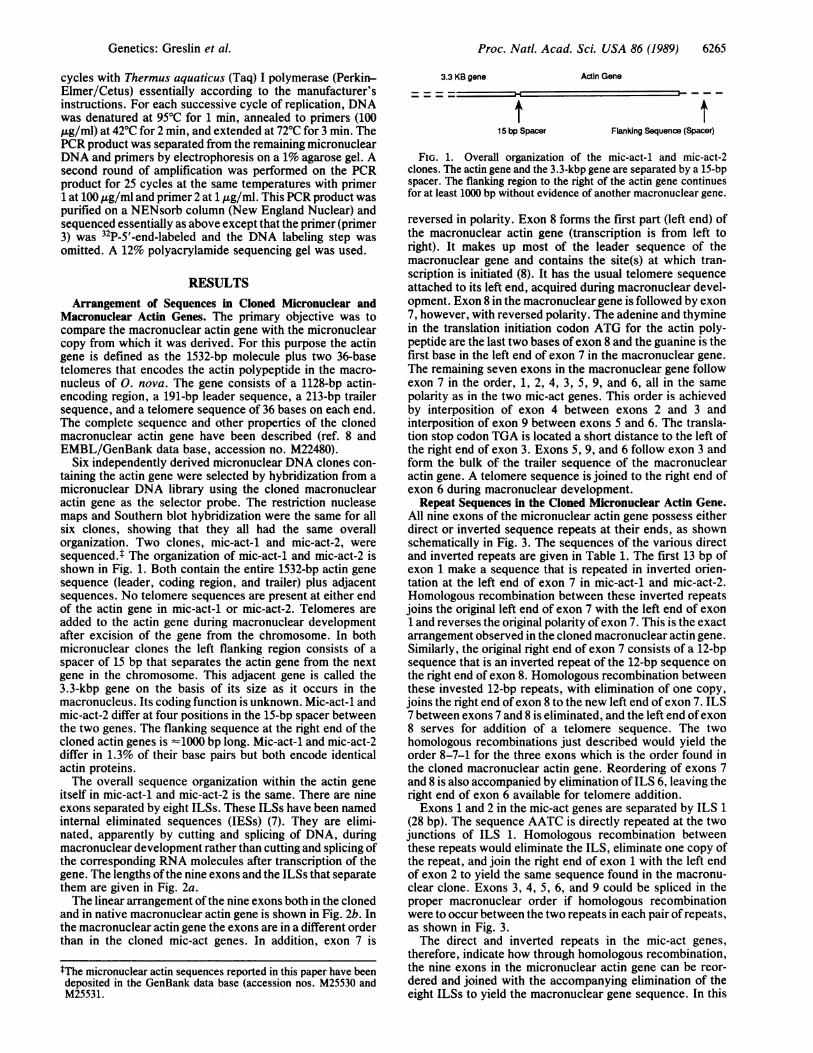
Six independently derived micronuclear DNA clones con-taining the actin gene were selected by hybridization from amicronuclear DNA library using the cloned macronuclearactin gene as the selector probe. The restriction nucleasemaps and Southern blot hybridization were the same for allsix clones, showing that they all had the same overallorganization. Two clones, mic-act-1 and mic-act-2, weresequenced.t The organization of mic-act-1 and mic-act-2 isshown in Fig. 1. Both contain the entire 1532-bp actin genesequence (leader, coding region, and trailer) plus adjacentsequences. No telomere sequences are present at either endof the actin gene in mic-act-1 or mic-act-2. Telomeres areadded to the actin gene during macronuclear developmentafter excision of the gene from the chromosome. In bothmicronuclear clones the left flanking region consists of aspacer of 15 bp that separates the actin gene from the nextgene in the chromosome. This adjacent gene is called the3.3-kbp -gene on the basis of its size as it occurs in themacronucleus. Its coding function is unknown. Mic-act-1 andmic-act-2 differ at four positions in the 15-bp spacer betweenthe two genes. The flanking sequence at the right end of thecloned actin genes is '1000 bp long. Mic-act-1 and mic-act-2differ in 1.3% of their base pairs but both encode identicalactin proteins.The overall sequence organization within the actin gene
itself in mic-act-1 and mic-act-2 is the same. There are nineexons separated by eight ILSs. These ILSs have been namedinternal eliminated sequences (IESs) (7). They are elimi-nated, apparently by cutting and splicing of DNA, duringmacronuclear development rather than cutting and splicing ofthe corresponding RNA molecules after transcription of thegene. The lengths ofthe nine exons and the ILSs that separatethem are given in Fig. 2a.-The linear arrangement of the nine exons both in the cloned
and in native macronuclear actin gene is shown in Fig. 2b. Inthe macronuclear actin gene the exons are in a different orderthan in the cloned mic-act genes. In addition, exon 7 is
3.3 KB gene
t- -
15 bp Spacer
Actin Gene
Flanking Sequence (Spacer)
FIG. 1. Overall organization of the mic-act-1 and mic-act-2clones. The actin gene and the 3.3-kbp gene are separated by a 15-bpspacer. The flanking region to the right of the actin gene continuesfor at least 1000 bp without evidence of another macronuclear gene.
reversed in polarity. Exon 8 forms the first part (left end) ofthe macronuclear actin gene (transcription is from left toright). It makes up most of the leader sequence of themacronuclear gene and contains the site(s) at which tran-scription is initiated (8). It has the usual telomere sequenceattached to its left end, acquired during macronuclear devel-opment. Exon 8 in the macronuclear gene is followed by exon7, however, with reversed polarity. The adenine and thyminein the translation initiation codon ATG for the actin poly-peptide are the last two bases ofexon 8 and the guanine is thefirst base in the left end of exon 7 in the macronuclear gene.The remaining seven exons in the macronuclear gene followexon 7 in the order, 1, 2, 4, 3, 5, 9, and 6, all in the samepolarity as in the two mic-act genes. This order is achievedby interposition of exon 4 between exons 2 and 3 andinterposition of exon 9 between exons 5 and 6. The transla-tion stop codon TGA is located a short distance to the left ofthe right end of exon 3. Exons 5, 9, and 6 follow exon 3 andform the bulk of the trailer sequence of the macronuclearactin gene. A telomere sequence is joined to the right end ofexon 6 during macronuclear development.Repeat Sequences in the Cloned Micronuclear Actin Gene.
All nine exons of the micronuclear actin gene possess eitherdirect or inverted sequence repeats at their ends, as shownschematically in Fig. 3. The sequences of the various directand inverted repeats are given in Table 1. The first 13 bp ofexon 1 make a sequence that is repeated in inverted orien-tation at the left end of exon 7 in mic-act-1 and mic-act-2.Homologous recombination between these inverted repeatsjoins the original left end of exon 7 with the left end of exon1 and reverses the original polarity ofexon 7. This is the exactarrangement observed in the cloned macronuclear actin gene.Similarly, the original right end of exon 7 consists of a 12-bpsequence that is an inverted repeat of the 12-bp sequence onthe right end of exon 8. Homologous recombination betweenthese invested 12-bp repeats, with elimination of one copy,joins the right end of exon 8 to the new left end of exon 7. ILS7 between exons 7 and 8 is eliminated, and the left end ofexon8 serves for addition of a telomere sequence. The twohomologous recombinations just described would yield theorder 8-7-1 for the three exons which is the order found inthe cloned macronuclear actin gene. Reordering of exons 7and 8 is also accompanied by elimination ofILS 6, leaving theright end of exon 6 available for telomere addition.Exons 1 and 2 in the mic-act genes are separated by ILS 1
(28 bp). The sequence AATC is directly repeated at the twojunctions of ILS 1. Homologous recombination betweenthese repeats would eliminate the ILS, eliminate one copy ofthe repeat, and join the right end of exon 1 with the left endof exon 2 to yield the same sequence found in the macronu-clear clone. Exons 3, 4, 5, 6, and 9 could be spliced in theproper macronuclear order if homologous recombinationwere to occur between the two repeats in each pair ofrepeats,as shown in Fig. 3.The direct and inverted repeats in the mic-act genes,
therefore, indicate how through homologous recombination,the nine exons in the micronuclear actin gene can be reor-dered and joined with the accompanying elimination of theeight ILSs to yield the macronuclear gene sequence. In this
tThe micronuclear actin sequences reported in this paper have beendeposited in the GenBank data base (accession nos. M25530 andM25531.
Genetics: Greslin et al.
Proc. Natl. Acad. Sci. USA 86 (1989)
MICRONUCLEAR ACTIN GENE
a 4
0(3.3 kbp Gene I|| Actin Gene
15 bpSpacer
1476 bp
2446 bp
28 bpILS 1
n
3 4 5 6 7 8 9150 bp 37 bp 113 bp 9o bp 60 bp 194 bp 15 bp=1 -I - -
III I11 bp 109 bp 12 bp 16 bp 15 bpILS 2 ILS 3 ILS 5 ILS 6 ILS 8
L8 bp 11 bp Flanking sequenceILS4 ILS 7
MACRONUCLEAR DEVELOPMENT
Telomere ATG
8 7 2
TGA Telomere
4 3 5 96
1604 bp
MATURE MACRONUCLEAR ACTIN GENE
FIG. 2. Scale drawing showing the order and relative sizes of actin exons and ILSs in the micronuclear actin gene (a) and the macronuclearactin gene (b). Each rectangle depicts one exon. The size of each exon (in bp) was determined by counting from the first unique base pair inan exon through the direct repeat at the right end of the exon. Exon 7 was counted from right to left. ATG of the start codon is at the junctionof exons 7 and 8 in the macronuclear gene.
scheme, the origin of every one of the 1532 bp of themacronuclear gene is precisely accounted for.We have determined (8) the sequence of a macronuclear
actin gene. That sequence matches perfectly the actin se-quence generated by reordering of the nine exons in mic-act-2. Therefore, we conclude that mic-act-2 is processed toyield a macronuclear actin gene. We do not yet know whethermic-act-1 is processed to yield a macronuclear actin gene.Order of the Nine Exons in the Actin Gene in Native
Micronuclear DNA. Although the same unorthodox arrange-ment of exons was found by restriction nuclease mapping inall six micronuclear actin clones, the unorthodox arrange-ment could be the result of a complicated and consistentlyrepeated artifact ofcloning. To test the possibility ofa cloningartifact, a portion of the actin gene in native micronuclearDNA was amplified by the PCR (see Fig. 4). The sequence ofthis PCR product was determined and compared to the
a
sequence of the cloned DNA. The sequence of the PCRproduct generated in this way matched the sequence ofclones mic-act-1 and mic-act-2. Thus, the order ILS 5, exon6, inverted exon 7, ILS 7, and left end of exon 8 wasconfirmed for native uncloned micronuclear DNA. We con-clude that the unorthodox arrangement ofexons in the clonedmic-act genes is not an artifact of cloning and that theunorthodox arrangement is present in native uncloned mi-cronuclear DNA.Because of the orientation of the primers used in this PCR,
any orthodox (macronuclear) arrangement present would notbe detected. However, to test the possibility that there maybe actin sequence arrangements other than the arrangementfound in the micronuclear DNA clones and PCR products,the following was done: Nine micrograms of micronuclearDNA was treated with BAL-31, a procedure that reliablyremoves contaminating macronuclear actin sequences.
3' TAS 5' TAS
1 2 3 4 5 6 7 8 9
~~~~-zu Ud- [=
b
C
5' TAS
^ 8 1 4 5 67 2 3 9
I LN&N t:::1VAna 11 r --I
9 :mlN K.E.:.:l RMW_ mmff::
5TAS
8
3'TAS
3'TAS
7 1 2 4 3 5 9 6 +
FIG. 3. (a) Arrangement of the direct and inverted repeats located at the ends of each exon. The blocks filled with various patterns representpairs of repeats. (b) All exons can be arranged in the order of the macronuclear gene by aligning a given repeat with the other member of itspair. (c) Homologous crossing-over between the two members of each pair of repeats yields a complete macronuclear actin gene (except fortelomeres) with ILSs removed. The polarity of exon 7 is shown by a horizontal arrow. TAS, telomere addition site.
b
6266 Genetics: Greslin et al.
I
Proc. Natl. Acad. Sci. USA 86 (1989) 6267
Table 1. Direct and inverted repeats bordering each exon
Direct and inverted repeats located at the ends of the nine exonsin mic-act-1 and mic-act-2. The sequence at the left end of exon 7 isan inverted repeat of the sequence at the left end of exon 1, and thesequence at the right end of exon 7 is an inverted repeat of thesequence at the right end of exon 8. Homologous crossing-overbetween each pair of repeats reorders the exons into the order foundin the macronuclear gene-i.e., 8-7-1-2-4-3-5-9-6.
Three micrograms of the BAL-31-treated DNA was digestedwith Kpn I, and the remainder was digested with Kpn I plusDra I. The locations of the Kpn I and Dra I sites are shownin Fig. 4a. One lane of Kpn I-digested DNA and two lanes ofKpn I-Dra I-digested DNA were separated by electrophore-sis on a 1% agarose gel and transferred to nitrocellulose.These blots of uncloned micronuclear DNA were tested withthree probes prepared from digests of the cloned macronu-clear actin gene (Fig. 4b). Probe 1 is specific for the 5' end ofthe macronuclear actin gene, probe 2 is specific for a regionin the middle ofthe actin gene and to the left of the single KpnI site in the macronuclear actin gene, and probe 3 is specificfor the 3' end of the actin gene. The probes were checked forspecificity by Southern hybridization to blots of DNA ofcloned mic-act-2 digested with Kpn I plus Dra I. All probeshybridized only to the bands predicted from the map in Fig.4a. Probe 2 hybridized to a 4.5-kbp Kpn I fragment andprobes 1 and 3 hybridized to a 960-bp Kpn I-Dra I fragment.The blot of Kpn I-digested uncloned micronuclear DNA
was hybridized with probe 2. The uncloned micronuclearDNA digested with Kpn I plus Dra I was hybridized with
probe 1 and probe 3 in separate experiments. The resultingautoradiogram of the Southern hybridization showed thatonly the bands predicted from endonuclease mapping andsequence determination of the micronuclear DNA cloneswere detected (see Fig. 4a). Probe 1 and probe 3, whichrepresent the 5' and 3' end of the macronuclear actin gene,respectively, hybridized exclusively to the 960-bp Kpn I-DraI fragment predicted from the sequence analysis of mic-act-1and mic-act-2. Probe 2 hybridized only to the 4.5-kbp Kpn Ifragment predicted from restriction endonuclease analysis ofthe mic-act genes. Since probe 1 hybridized only to the sameband to which probe 3 hybridized and did not hybridize to theband to which probe 2 hybridized, we conclude that noorthodox arrangement of actin exons is present in micronu-clear DNA. If there were a micronuclear actin gene with theorthodox arrangement of exons as seen in the macronuclearactin gene, then both probe 1 and probe 2 would havehybridized to the same Kpn I-Kpn I restriction fragment.Furthermore, since no unpredicted bands were found, thereare no sequence arrangements other than the arrangementfound in the micronuclear DNA clones and the PCR productobtained with micronuclear DNA.
Micronuclear Actin Gene in Oxytricha trifalax. It is con-ceivable that during the 8-year interval since our strain of O.nova last mated that the micronuclear actin genes haverearranged into the unorthodox order we have detected anddo not represent the form in cells with a normal micronucleuscapable of successful mating. Therefore, we examined mi-cronuclear actin DNA in 0. trifallax, for which we havemating strains (a gift from Robert Hammersmith, Ball StateUniversity). Two clones of 0. trifallax were grown andmated, with >90% survival of exconjugants. Micronucleiwere isolated from one of the parental strains and the DNAwas used in a PCR. Primers for the reaction were synthesizedusing the sequence of the macronuclear actin gene publishedfor a similar species, O.fallax (9). Primers were chosen to testfor the presence of an unorthodox arrangement of the actingene in micronuclear DNA. A PCR with these primers usingtotal macronuclear DNA of 0. trifallax or a cloned macro-nuclear actin gene from 0. fallax predictably yielded no
a
Kpn I
Flanking sequence
3' ProbesTHREE
MATURE MACRONUCLEAR ACTIN GENE
FIG. 4. Relationship of the three hybridization probes (solid thick bars) derived from cloned macronuclear actin DNA to the two restrictionfragments of actin-specific native (uncloned) micronuclear DNA produced by Kpn I plus Dra I digestion. Probes 1 and 3 are derived from the5' and 3' ends of the macronuclear actin gene, respectively. Probe 2 is derived from the Bgl II-HindIII fragment located to the left of the KpnI site in the macronuclear actin gene. If the exon arrangement of the macronuclear actin gene was the same in native micronuclear DNA, thenprobe 1 would hybridize to the same Kpn I-Kpn I restriction fragment in the micronuclear actin gene as probe 2 (see b); however, it does not.Instead, probe 1 hybridizes to the same restriction fragment as probe 3 and only to that fragment. Therefore, the arrangement of exons in nativemicronuclear actin DNA is different from the arrangement in the cloned macronuclear actin gene. The line labeled PCR product indicates theregion from ILS 5 to the left end of exon 8 that was amplified from native micronuclear DNA by a PCR and sequenced. The capitol T over exon
8 in a indicates the transcription start site, and the arrow shows the direction of transcription that would occur if micronuclear DNA were tobe transcribed.
Spacer
b
5'
Bgl 11Hpa IT- >
8 78
ONE
PCR Product
Hind III Kpn
4 32
TWO
5 9 6
Genetics: Gresfin et al.
Proc. Natl. Acad. Sci. USA 86 (1989)
product. A PCR with these primers using total 0. trifallaxmicronuclear DNA or total 0. trifallax micronuclear DNAdigested with BAL-31 to remove macronuclear DNA con-tamination yielded an abundant product of -630 bp. A PCRcould only yield such a product if the exons containing theprimer sequences were in an unorthodox arrangement inmicronuclear DNA of 0. trifallax. Since this shows anunorthodox order of exons in 0. trifallax, we conclude thatthe unorthodox order in 0. nova is not an artifact that aroseduring the many years since its last mating.
DISCUSSIONThe actin gene or genes in the micronucleus of 0. novaconsist of nine exons, arranged in an unorthodox fashion.The exons are reordered to form a functional actin geneduring macronuclear development. These statements arebased of the following observations: (i) Six micronuclearDNA clones containing complete micronuclear actin geneswere isolated. All six were shown by restriction mapping andSouthern hybridization to have the same unorthodox ar-rangement. Two micronuclear actin genes, mic-act-1 andmic-act-2, were defined by minor differences in sequences.(ii) The unorthodox arrangement of the nine exons is not acloning artifact because the same arrangement was shown tobe present in uncloned micronuclear DNA using the PCRmethod. This was confirmed with three segments of thecloned macronuclear actin gene as hybridization probes totest the order of exons in uncloned micronuclear DNA. (iii)No orthodox versions of the actin gene could be found inuncloned micronuclear DNA using the three segments of thecloned macronuclear actin gene as search probes. (iv) Mac-ronuclear actin genes contain only the orthodox arrangementof exons. (v) An unorthodox arrangement of the actin gene isnot unique to 0. nova but also occurs in 0. trifallax.The presence ofdirect and inverted repeat sequences at the
ends of the nine exons in the micronuclear actin geneindicates a precise scheme for generating the macronuclearactin gene for the micronuclear gene. Recombination be-tween the two members of each pair of repeats would createthe orthodox order and eliminate all ILSs, as shown sche-matically in Fig. 3. For exons 1 and 2, a direct repeat of 4 bp(AATC) occurs at each junction with the ILS between themand after removal of the ILS; one of these repeats remains inthe junction between the two exons. The remaining sevenILSs are bordered by longer repeats, ranging from 9 to 13 bp,and these ILSs separate exons that become reordered. Inthese seven cases the removal of the ILS is accompanied bya transposition ofan exon tojoin (presumably by homologousrecombination) an exon other than the one originally adja-cent. Thus, short direct repeats are present in the one casewhere two adjacent exons are joined (exons 1 and 2), and theintervening ILS is removed. Where exons are transposed,longer repeat sequences lie at the ILS junctions.
Construction of the macronuclear actin gene could con-ceivably occur by transcription of the micronuclear actingene in the developing macronucleus. The transcript could beprocessed to remove ILSs and reorder exons, followed byconversion into DNA by reverse transcriptase (10, 11). Thiswould require transcription of portions of both strands of themicronuclear actin DNA. In generating the macronucleargene the coding strand is derived from one strand of exons1-6, 8, and 9 and from the opposite strand for exon 7.Therefore, at least portions of both strands of the micronu-clear gene must be transcribed and ILSs must be removed.Eight RNA segments transcribed from one strand and oneexon in a transcript of the opposite strand must be splicedtogether to produce anRNA suitable for reverse transcriptioninto a DNA molecule to which telomeres are added to formthe macronuclear version of the actin gene. We favor aDNA-processing model since it seems molecularly less de-manding than the RNA model.Micronuclear and macronuclear versions of three genes in
0. nova have been analyzed by Ribas-Aparicio et al. (10).They have ILSs with short direct repeats at theirjunctions inthe micronuclear versions of the genes. The short directrepeats presumably signal correct excision and splicing, andILS 1 in the actin genes described in this paper is similarlyorganized. However, Ribas-Aparicio et al. (10) found no signof reordering of exons in any of the three micronuclear genesto make the macronuclear versions. Two extensions of thepresent work are to test for unorthodoxy among other genesin 0. nova and to test whether unorthodoxy occurs inhypotrichs generally.
We thank Alan Zahler and Giovanni Bosco for their helpfuldiscussions. This work is supported by a contract from the NationalFoundation for Cancer Research and by Research Grant #5 RO1GM19199 from the National Institute of General Medical Sciences toD.M.P.
1. Prescott, D. M. (1983) Mod. Cell Biol. 2, 329-352.2. Swanton, M. T., Heumann, J. M. & Prescott, D. M. (1980)
Chromosoma 77, 217-227.3. Lauth, M. R., Spear, B. B., Heumann, J. M. & Prescott,
D. M. (1976) Cell 7, 66-74.4. Boswell, R. E., Jahn, C. L., Greslin, A. F. & Prescott, D. M.
(1983) Nucleic Acids Res. 11, 3651-3663.5. Klobutcher, L. A., Vailonis-Walsh, A. M., Cahill, K. & Ribas-
Aparicio, R. M. (1986) Mol. Cell. Biol. 6, 3606-3613.6. Ammermann, D. (1974) Chromosoma 45, 401-429.7. Klobutcher, L. A., Jahn, C. L. & Prescott, D. M. (1984) Cell
36, 1045-1055.8. Greslin, A. F., Loukin, S. H., Oka, Y. & Prescott, D. M.
(1988) DNA 7, 529-536.9. Kaine, B. P. & Spear, B. B. (1982) Nature (London) 295,
430-432.10. Ribas-Aparicio, R. M., Sparkowski, J. J., Proulx, A. E.,
Mitchell, J. D. & Klobutcher, L. A. (1987) Genes Dev. 1,323-336.