Chapter 15 Formation of net-like structures Net-like structures are common in almost every higher organism. The vascular system, the lymphatic system, the nervous system, the tracheae of insects, the veins of leaves and those of insect wings are examples (Fig.15.1). Such net-like structures can be used to supply a tissue with nutritional substances, such as oxygen and water. The filamentous elements of a net consist of either linearly arranged, differentiated cells or of long fibers formed out of single cells. Filaments can provide information or mechanical stability and they can be used to remove certain substances from an area. A net-like structure with all its ramifications is certainly not formed by the interpretation of positional information. This would require an enormous number of threshold values in each cell. Moreover, in many net systems, regulatory processes have been observed. For instance, new tracheae grow into a field of artificially evoked oxygen deficiency (Wigglesworth, 1954). K¨ uhn (1948) found a mutant insect with a missing vein in the wing. The remaining veins were rearranged to compensate for the missing vein; there was no large gap in the pattern. Some substances are known to have an influence on the formation of a net: the plant hormone auxin in leaves (Jost, 1942), the nerve growth factor (Levi-Montalcini, 1964) on adrenergic nerves, a tumor angiogenesis factor (Folkmann, et al., 1971, Folkman, 1976) on blood vessels. These findings indicate that elongation and branching are locally controlled processes. The question remains, as to what biochemical interactions govern the formation of elongated structures. How, for instance, can the elongation of a nerve be directed towards a particular target area? How is the very small surface area selected in which a new branch is initiated? Or, as in the case of leaves, how can the differentiation of cells into members of the vascular system proceed along a line, such that a certain distance from other vascular elements is maintained? How are these processes encoded in the genes? The model I have proposed for the generation of net-like structures (Mein- hardt, 1976) is based on the repetition of two steps: (i) localization of the elonga- tion of a filament, and (ii) the elongation itself. The localization can be achieved by pattern formation based on short range activation coupled with long range inhibition, as described above. In regard to the orientation of elongation, let us assume that it is the purpose of a net to remove some substrate such as auxin or 163
Transcript
Chapter 15
Formation of net-like structures
Net-like structures are common in almost every higher organism. The vascularsystem, the lymphatic system, the nervous system, the tracheae of insects, theveins of leaves and those of insect wings are examples (Fig.15.1). Such net-likestructures can be used to supply a tissue with nutritional substances, such asoxygen and water. The filamentous elements of a net consist of either linearlyarranged, differentiated cells or of long fibers formed out of single cells. Filamentscan provide information or mechanical stability and they can be used to removecertain substances from an area. A net-like structure with all its ramificationsis certainly not formed by the interpretation of positional information. Thiswould require an enormous number of threshold values in each cell. Moreover, inmany net systems, regulatory processes have been observed. For instance, newtracheae grow into a field of artificially evoked oxygen deficiency (Wigglesworth,1954). Kuhn (1948) found a mutant insect with a missing vein in the wing. Theremaining veins were rearranged to compensate for the missing vein; there wasno large gap in the pattern. Some substances are known to have an influence onthe formation of a net: the plant hormone auxin in leaves (Jost, 1942), the nervegrowth factor (Levi-Montalcini, 1964) on adrenergic nerves, a tumor angiogenesisfactor (Folkmann, et al., 1971, Folkman, 1976) on blood vessels. These findingsindicate that elongation and branching are locally controlled processes. Thequestion remains, as to what biochemical interactions govern the formation ofelongated structures. How, for instance, can the elongation of a nerve be directedtowards a particular target area? How is the very small surface area selected inwhich a new branch is initiated? Or, as in the case of leaves, how can thedifferentiation of cells into members of the vascular system proceed along a line,such that a certain distance from other vascular elements is maintained? Howare these processes encoded in the genes?
The model I have proposed for the generation of net-like structures (Mein-hardt, 1976) is based on the repetition of two steps: (i) localization of the elonga-tion of a filament, and (ii) the elongation itself. The localization can be achievedby pattern formation based on short range activation coupled with long rangeinhibition, as described above. In regard to the orientation of elongation, let usassume that it is the purpose of a net to remove some substrate such as auxin or
163
164 CHAPTER 15. FORMATION OF NET-LIKE STRUCTURES
Figure 15.1: Examples for net-like structures. (a) Dentritic trees of three nerve cells in aganglion of the blowfly Calliphora (Hausen et al., 1980, Figure courtesy K. Hausen). (b) bloodvessels in the allantois of the developing chicken. (c) Tracheal system in an abdominal segmentof an insect (drawn after Wigglesworth, 1954, see also Fig. 15.3). (d) Skeleton of a poplar leaf.All other cells has been removed by microorganisms.
nerve growth factor from its surroundings. It could also be the remedy of somedeficiency, such as the supply of oxygen mediated by insect tracheae. If auto-catalysis depends on this substrate, the elongation will be oriented towards theincreasing substrate concentration. The local high signal concentration causesan elongation of the filament which, in turn, causes a shift of the signal. Longfilaments are formed as a trail behind a wandering filament-inducing signal. Themechanism will be explained in some detail for the special case in which filamentsare formed by ordered differentiation, within a field of undifferentiated cells. Ageneralization will be given later.
15.1 Formation of a filament
We wish to translate this idea into a mathematical model which can be interpretedon a molecular basis. We have to supplement the activator-inhibitor mechanismwith a description of the differentiation process and a mechanism for the activatorshift. We have seen (Eq.3.2) that a sharp local maximum can be generated bythe interaction of an activator a and an inhibitor h
∂a
∂t=
ca2s
h− µa + Da∆a + ρ0y (15.1a)
∂h
∂t= ca2s− νh + Dh∆h + ρ1y (15.1b)
or∂h
∂t= ca2 − νh + Dh∆h (15.2b′)
(The new terms s, ρ0y and ρ1y will be explained below; Da∆a and Dh∆hdenote the generalized diffusion terms for more than one dimension.) The localhigh activator concentration would be the signal for a cell to differentiate, i.e.,to switch irreversibly from one state to another. The state of differentiation can
15.1. FORMATION OF A FILAMENT 165
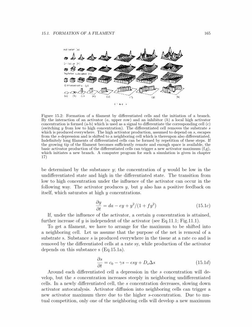
Figure 15.2: Formation of a filament by differentiated cells and the initiation of a branch.By the interaction of an activator (a, upper row) and an inhibitor (h) a local high activatorconcentration is formed (a-b) which is used as a signal to differentiate the corresponding cell (c)(switching y from low to high concentration). The differentiated cell removes the substrate swhich is produced everywhere. The high activator production, assumed to depend on s, escapesfrom the s-depression and is shifted to a neighboring cell which is thereupon also differentiated.Indefinitely long filaments of differentiated cells can be formed by repetition of these steps. Ifthe growing tip of the filament becomes sufficiently remote and enough space is available, thebasic activator production of the differentiated cells can trigger a new activator maximum (f,g),which initiates a new branch. A computer program for such a simulation is given in chapter17)
be determined by the substance y; the concentration of y would be low in theundifferentiated state and high in the differentiated state. The transition fromlow to high concentration under the influence of the activator can occur in thefollowing way. The activator produces y, but y also has a positive feedback onitself, which saturates at high y concentrations.
∂y
∂t= da− ey + y2/(1 + fy2) (15.1c)
If, under the influence of the activator, a certain y concentration is attained,further increase of y is independent of the activator (see Eq.11.1; Fig.11.1).
To get a filament, we have to arrange for the maximum to be shifted intoa neighboring cell. Let us assume that the purpose of the net is removal of asubstrate s. Substance s is produced everywhere in the tissue at a rate co and isremoved by the differentiated cells at a rate sy, while production of the activatordepends on this substance s (Eq.15.1a).
∂s
∂t= c0 − γs− εsy + Ds∆s (15.1d)
Around each differentiated cell a depression in the s concentration will de-velop, but the s concentration increases steeply in neighboring undifferentiatedcells. In a newly differentiated cell, the s concentration decreases, slowing downactivator autocatalysis. Activator diffusion into neighboring cells can trigger anew activator maximum there due to the higher s-concentration. Due to mu-tual competition, only one of the neighboring cells will develop a new maximum
166 CHAPTER 15. FORMATION OF NET-LIKE STRUCTURES
and even the previously active cell will be inhibited. The result is a shift of theactivator maximum into a neighboring cell which subsequently becomes differen-tiated itself. The next cell to be activated will be the one in front of the tip ofthis incipient filament. It is the adjacent cell with the highest s-concentration,because it has the least contact with the s-removing differentiated cells. By rep-etition of this process - shift of the signal, differentiation and shift again - longfilaments of differentiated cells can be formed (Fig.15.2). The structures whichcan be generated by this simple mechanism have features similar to those of bio-logical networks. For example, bifurcations and lateral branches can be formed,the density of filaments can be regulated according to local demand, filamentscan be oriented towards a target area and a damaged net can be repaired.
15.2 Formation of lateral branches
To form a net, individual filaments have to branch repetitively. A branch canbe formed either by bifurcation at the growing tip or by the formation of anew growth point along an existing filament. According to the model, as thelength of a filament increases, inhibition arising mainly from the high activatorconcentration at the growing tip may no longer be sufficient to suppress the basic(activator- independent, oy in Eq.15.1a, Fig.15.2g) activator production of thecells of the filament. By autocatalysis, a new activator maximum may be formedalong the filament, but, since the concentration of s is higher in the environmentof the filament, the activator maximum is immediately shifted to a cell at theside of the filament and a branch is initiated.
15.3 Limitation of maximum net density
Net density will increase, since any branch can give rise to other branches. Theultimate net density can be controlled in two ways. In the first case, suppose theactivator, but not the inhibitor, is dependent on the concentration of s. Then, asnet density increases both the average s concentration and the maximum activa-tor concentration decrease. Once a certain net density is reached, the activatormaximum will be too low to induce further cell differentiation. The final net den-sity will be proportional to the local production of s; net density will increase aslong as more of the substance to be removed is present (see Fig.15.8). This modelmay apply to the growth of tracheae into a region of experimentally induced oxy-gen deficiency (Wigglesworth, 1954). Alternatively, if production of both theactivator and the inhibitor are dependent on s (Eq.15.1b), then elongation andbranching will be independent of the absolute s concentration. Here, final netdensity can be controlled by production of a basic inhibitor by the differenti-ated cells (ρ1y in Eq.15.1b). This creates a background inhibitor concentrationin proportion to local net density. Filament elongation or the formation of newbranches will cease if activator production is suppressed when the backgroundinhibitor concentration rises above a certain level. This type of regulation leads
15.4. FINDING A TARGET CELL 167
Figure 15.3: Repair of a damaged net. (a) The tracheal system in the ventral abdomen of anormal, undamaged bug. (b) Oxygen supply to the fourth segment is disrupted by cuttingthe corresponding trachea (broken line, arrow in Fig (a). (c) Fourteen days later, tracheoles ofthe third segment have migrated into the oxygen-deprived segment (after Wigglesworth, 1954).In this process, patches of oxygen deprived epithelial cells send out cell processes which makeconnections with the tracheae. By retraction of the cell processes, the tracheae are pulled intoan area of oxygen deficiency (Wigglesworth, 1959). Simulation. A complete net (d). Afterremoval of the filaments in the lower half (e), new veins grow into the area (f). After completeregeneration (g), the newly formed part of the pattern look similar but not identical to theoriginal net. The high activator concentration may be the signal for the epithelial cells toattract tracheae (differentiated cells: open squares, activated cells: filled squares).
to s-independent spacing of the net.
15.4 How a growing filament finds a particular target cell
According to the theory proposed here, elongation proceeds in the direction ofthe highest concentration of s. In the case of homogeneous s-production, that isusually located in front of the filament tip. If, on the contrary, the substance s isproduced only in a particular area, the filament will follow the resulting gradientin s concentration upwards to the target area.
15.5 Regeneration of a net
Destruction of a filament of a net frequently leads to branching of nearby fila-ments, which repair the damage. Wigglesworth (1954), for instance, cut a tracheawhich supplied a particular segment of a bug with oxygen. Tracheae from neigh-boring segments subsequently migrated across the segment border (Fig.15.3) andmaintained the oxygen supply. The proposed model reproduces this regenerativecapability. In an area without filaments, s is no longer removed. The increasings concentration attracts new branches (Fig.15.3) and the damage is repaired.
168 CHAPTER 15. FORMATION OF NET-LIKE STRUCTURES
Figure 15.4: Avoidance orientation and the formation of reconnections. The basic principle ofthe proposed model of line formation consists of repulsion between the differentiated cells andthe differentiation-inducing signal. The elongation of a filament will be oriented away fromother filaments into the largest available free space. (a) A record of this avoidance reactioncan be seen in this maple leaf. However, reconnections (anastomosis) between veins are alsopossible. (b) According to the model, two growing tips (arrow heads) show a strong mutualrepulsion, whereas the repulsion which an existing filament exerts onto a growing tip is moremoderate. Connections are possible as the strong withdrawing movement (dashed lines) oftwo growing tips overrides the weaker repulsion arising from an existing filament. The numberof reconnection depends therefore on the repulsion of an existing line and of how strong thetendency is to make a line straight.
15.6 Formation of reconnections
In leaves most of the finer veins end blindly, but some of them connect with otherveins to form closed loops (anastomosis). In the model, filament elongation isdirected towards the largest available unfilamented space; a growing filament willtherefore keep its distance from existing ones. A result of this mutual avoidanceduring growth can be seen in the final pattern of a leaf (Fig.15.4a). Under themodel, this repulsion results from two different inhibitory factors on activatorproduction. The inhibitor centered around a growing tip and used to keep theactivator localized is a very strong factor. A weaker factor results from the de-pletion of the substance along an existing filament, which provides the stimulusfor the shift of the activator peak away from the differentiated cells. A growingtip is strongly repelled by another growing tip, but it is only much more weaklyrepelled by an existing filament. This disparity in repulsive forces allows theavoidance mechanism occasionally to be overcome. Thus, when the weaker re-pulsion of an existing filament is overcome by the stronger mutual repulsion oftwo growing tips, reconnection of filaments is made possible. An example of sucha reconnection is sketched in Fig.15.4b.
According to Avary (1933), reconnections in the tobacco leaf are formed onlyafter the transition in terms of marginal to intercalary growth. That is under-standable from the model since, during intercalary growth, two activator peakscan arise quite close together. As they develop more fully, strong repulsion willresult from the increasing influence of their mutual inhibitors. They can thus beforced to elongate in directions that bring them into contact with other, olderfilaments. The midvein and the main lateral branches are formed before inter-calary growth begins, which explains why it is only branches of higher order thatusually form reconnections. For a complete description of plant venation, onehas to take into account that the veins not only remove the auxin from the sur-
15.7. VARIATION IN PATTERN FORMATION 169
Figure 15.5: Influence of random fluctuation on pattern formation. (a) Two leaves of thesame tree. Their pattern formation is presumably controlled by the same genetic information.Nevertheless, the pattern is only similar, not identical. (b-d) and (e-g) Two simulations withthe same parameters and the same initial condition but with different random fluctuation (3%) in the constant c, Eq.15.1a,b. The model reaction determines only properties of the overallpattern such as average net density. Fine details are influenced by small local differences.
rounding tissue but that they transport it in a polar fashion towards the roots.In a young crossvein, the polarity of the active transport is not completely fixed.Different parts of a vein can pump towards each other (Sachs, 1975). This wouldlead to local accumulations of auxin which, in turn, attracts other veins leadingalso to reconnections. The active transport of auxin itself seems to be also anautocatalytic process (Hertel and Flory, 1968). Mitchison (1980) has proposed amodel for leaf venation which is based essentially on the transport of auxin.
The mode of reconnection described in Fig.15.4 is only possible in a two-dimensional system. If a third dimension is available, the deflected filament wouldavoid the existing branches by passing underneath or above. Three-dimensionalnetworks consisting of only one cell type, such as tracheae or lymph capillaries,usually end blindly. Extensive reconnections in three dimensions are possible,however, if two cell types are involved, as in the case of veins and arteries. Eachcell type can form its own network by the repulsive interaction described above.Growth of the capillaries towards one other can occur if one cell type produces asubstance which accelerates the activator production of the other cell type.
15.7 Variation in pattern formation
In the model, very complex patterns can be generated by the interactions of veryfew substances. These interactions can easily be encoded by genes. The ques-tion may arise as to how reproducible such patterns would be. The parametersdetermine only the general features of the pattern, such as average net density,the distance between branching points or the straightness of the lines. The finedetails depend on external influences or even on random variations. For instance,during the initiation of a new branch, small differences between two neighboringsites determine to which site the activator maximum will escape and therefore
170 CHAPTER 15. FORMATION OF NET-LIKE STRUCTURES
Figure 15.6: Simulation of a dichotomously branching leaf pattern. The simple dichotomousleaf pattern shows only bifurcation of the growing veins, without later lateral branching. Sucha pattern can be seen in the Ginkgo and some ferns (Fig 15.7). Its simulation requires onlytwo controlling substances. Local high activator (a, top row) concentration is formed by au-tocatalysis. Inhibitory action results from the depletion of the substrate s (a,b). The localhigh activator concentration irreversibly differentiates (b) the corresponding cells (switchingy from low to high concentrations). Since the differentiated cells also remove the substrate,the activator maximum wanders away from the differentiated cells (c). If enough free space isavailable, the activator maximum can split (see also 5.1), leading to a bifurcation (d-e).
toward which site the new branch will grow. In a leaf, for instance, it does notmatter whether the first branch leads to the left or to the right. Usually, suchdetails are insignificant and can arise at random. However, once a branch hasbeen made, let us say to the left, the resulting asymmetry strongly influencessubsequent branching. Due to the presence of the new branch, the concentrationof s will drop on the left site and the next branch will extend to the right, and soon. Since each decision depends to such an extent on the previous one, the overallpattern is reproducible. The random element in the process of pattern formationimplies that two patterns generated by the same mechanism will be similar butnot identical. This can be observed in nature. For example, leaves from the sametree are not identical, even though they are surely developed under the control ofthe same genetic information. Similarly, the details of neuronal branching differamong genetically identical individuals of the water flea Daphnia (Macagno et al.,1973). In Fig.15.5, two simulations are provided, starting with the same initialcondition but allowing different random fluctuations. The resulting patterns aresimilar but not identical.
15.8 Formation of a dichotomous branching pattern
One may ask whether this mechanism of line formation is the simplest possible.It is not. In this model, two inhibitory actions are involved in line formation,
15.8. FORMATION OF A DICHOTOMOUS BRANCHING PATTERN 171
Figure 15.7: Comparison of branching patterns of leaves with their simulations. (a) An evolu-tionarily older form of branching is dichotomous, such that a growing line can split into two,but lateral branching is not possible. (b) A simulation of dichotomous branching is possibleunder the assumption of only two controlling substances (see Fig. 15.6), in which the forkingpattern is reproduced. (c) Lateral branching in a leaf of Fittonia verschaffelti; by a caprice ofnature, the major veins appear white. (d) In the simulation initially one differentiated cell wasassumed to be present (arrow). The first vein is oriented towards the largest available space, inthe diagonal. The first lateral branches try to grow out at 90o but are repelled by the marginsand establish, therefore, an angle of 45o with the midvein (see Fig. 15.2). The following lateralbranches are repelled by the first and appear, therefore, also at 45o but branches of higher ordergrow out at 90o . The margins are avoided since the inhibitor cannot diffuse past the margin; itis, therefore, accumulated here. Reconnections are occasionally possible between higher orderbranches. Whereas the main lateral branches are quite straight, the higher order branches aremore curved since they are frequently deflected by other growing tips. The simulation of aleaf with 29x29 cells can be only a crude approximation of the reality (even such a relativelysimple simulation requires 28 h on a relatively fast PTP 11/40 computer). Nonetheless, itdoes demonstrate that such a complex pattern can be formed by the interaction of only a fewsubstances.
one to localize activation at the growing tip, and the other to determine thedirection in which the center of activation will migrate. As discussed in chapter5, the inhibitor may be replaced by a substrate which is depleted during activatorproduction. Therefore, both tasks, the formation and the shift of the activatorpeak, can be mediated by one and the same substances. Including the activator,only two substances would be sufficient to control differentiation (Eq.15.2).
∂a
∂t= ca2s− µa + Da∆a (15.2a)
∂s
∂t= c0 − ca2s− γs− εsy + Ds∆s (15.2b)
172 CHAPTER 15. FORMATION OF NET-LIKE STRUCTURES
∂y
∂t= da− ey + y2/(1 + fy2) (15.2c)
A pattern formed according to this interaction is given in Fig.15.6. The maindifference from the pattern formation discussed above is that here lateral branch-ing is not possible. To provide sufficient drive, the differentiated cells have toremove such a substantial amount of s that the formation of secondary activatorpeaks along an existing filament is no longer possible. The activator maximumat the growing tip can still divide into two maxima, allowing binary fission ofan extending filament. It seems that this is the evolutionarily older dichotomousbranching pattern of leaf vascularization which can still be seen in the leaves ofsome ferns and the Ginkgo tree. If this view is correct, the evolutionary stepfrom the dichotomous (Fig.15.7a) to the common leaf pattern (Fig.15.7b) wouldinvolve separation of the two inhibitory effects by the “invention” of a separateinhibitory substance. The evolutionary advantage of lateral branching is that itopens the possibility for intercalary growth. New branches can be extended intothe expanding spaces, providing necessary nourishment to the tissue. In addition,damage to any particular vein is less disruptive to the tissue as a whole, since thenetwork has closed loops and other pathways are available.
15.9 Filaments formed by oriented cell division or by ex-tensions of single cells
Elongation of a line by accretion of newly formed, differentiated cells is only one ofseveral possible modes of line formation that can be simulated under the proposedtheory. The high activator concentration at the filament tip could control celldivision, while activator increase in front of the tip could orient the process. Inthis case, a network is formed by organized proliferation of the constituent cells.
In both the nervous system and the tracheal system, the elements of the fila-ments consist of highly extended single cells. The formation of such a net can beexplained by the model, under the assumption that a local high activator concen-tration is formed on the cell surface. This can act as a stimulus for the expansionof pseudopodes and the formation of a growth cone. As in the orientation of achemotactic sensitive cell (see Fig.5.6), the precise localization of the activatorpeak during the fiber elongation is facilitated by periodic formation and decayof the signal. Harrison (1910) has shown that the growth of a nerve fiber is nota continuous process but, rather, that phases of fast elongation alternate withphases of searching for a new direction. Several pseudopodes may be sent out atthe same time, although most of them will later be retracted.
A closer look at the branching pattern of nerves reveals features similar tothose seen in leaves. When the cells branch, the branches are typically at 90o,and individual branches maintain distance from one another. In the growth ofa nerve, there again appear to be two types of inhibition involved. The firstlocalizes the growth cone and suppresses the development of additional growthcones in surrounding elements. It also allows the selection of a very small area
15.10. KNOWN SUBSTANCES 173
Figure 15.8: Regulation of the density of a net. (a,b) Experiments by Folkman et al. (1971),redrawn after Folkman (1976). A piece of tumor tissue (T) and cartilage (C) are grafted intothe cornea of a rabbit. After 20 days, some capillary growth is induced by the tumor but newvessels are inhibited in the vicinity of the cartilage. If the cartilage is inactivated by boiling, thetumor become vascularized (b), 30 days after the operation, the eye will be overgrown by thetumor. (c-e) According to the model, the density of a net can be regulated by the concentrationof a substance s which is produced everywhere in the tissue and which is removed by the net. (c)“Normal” net density. (d) A two-fold higher s production in the upper half area leads to a muchhigher vessel density. This may be the situation in a tumor, with its excessive vascularisation.(c) As (b), but an inhibitor-producing cell layer is assumed in the center of the field; no vesselscan grow through the inhibited area, but can circumvent it (Eq.15.1 with 15.1b’).
where a new branch is actually initiated. The second inhibition results fromdepletion of the substance, s, which is possibly a nerve growth factor. This leadsto orientation of each branch away from its neighbors; it is a mechanism for themutual repulsion of nerve fibers. Genetically identical or similar individuals showthe same general pattern of branching, but there are large differences in the finestructure of the branching (Macagno et al., 1973). Of course, nerve cells do morethan merely branch toward their targets; they also demonstrate spatial orderingof their connections. Any of the pattern generating mechanisms discussed abovecan supply the required spatial cues. Such spatial patterning can be used toset up a graded surface property in a field of the nerve cells. Together withthe processes of competition and branching here described, this produces modelsthat can generate many of the patterns seen in the retinotectal systems of lowervertebrates (Fraser, 1980; Gierer, 1981b).
15.10 Known substances which influence the formation ofnets
As already mentioned, there are several substances known which influence theformation of particular nets. Some have been identified and others have beenonly partially purified. All these substances are comparable to the proposed
174 CHAPTER 15. FORMATION OF NET-LIKE STRUCTURES
shift- substance s, whereas none of the activator-inhibitor type are known. Itseems likely that this is because substances of the shift-type s must be constantlyproduced or present in all the cells into which the filaments should grow. Incontrast, the proposed activator-inhibitor substances are produced very locallyand possibly only during short time intervals. Further difficulties in observingthese substances result from the strong mutual influence of their production rates(see p. 50).
In leaves, additional vascular elements are formed after application of auxin toan injury (Jost, 1942). Auxin is known to be actively transported from the leavesto the roots. The veins remove auxin from their environment. Similarly, nervegrowth factor NGF (Levi-Montalcini, 1964), which is necessary for the outgrowthof adreneric nerves, is actively transported from filaments to the cell body (seeThoenen and Barde, 1980). The highest NGF-concentration that a particularnerve will encounter is presumably that at its end, since more surface elementsare available to remove NGF along a fiber. As long as no other constraints areimposed, this would lead to linear elongation of the fiber. The valley of NGFcentered along each fiber would cause fibers of the same type to keep a certaindistance from each other. If higher concentrations of NGF were present in aparticular area, this area would attract growing fibers. According to the model,NGF and auxin would be co-factors for the activation of fiber elongation, forinstance in the formation of a growth cone. The activation itself has to haveautocatalytic properties, with an element of lateral inhibition.
The rapid growth of a tumor is only possible if it is extremely well nourished bya plethora of newly formed vessels (Algire et al., 1945). Obviously, a malignanttumor must be able to overcome the body’s control of blood vessel formation.Thus, an understanding of the control of vessel density is of great importance.Folkman et al. (1971) have isolated, from different tumors, a tumor angiogenesisfactor (TAF) which induces ingrowth of vessels into normally avascular areas,such as the cornea or the epidermis (Fig.15.8a,b). According to the model, thedensity of the net can be controlled by the substance s. Examples are given inFig.15.8. Low, medium and high s-concentrations lead to corresponding densitiesof the filaments. These densities may correspond to the situation of an avasculartissue, of a normal tissue and of a tumor. TAF may itself be the substance,or it may induce the synthesis of a substance, which is removed by the vesselsand which is a co-factor in the local activation of elongation or the originationof a new branch. On the other hand, Folkman et al. (1971) have isolated afactor from avascular cartilage which suppresses the ingrowth of vessels. Thisfactor could be the proposed inhibitor, since an externally supplied inhibitor cansuppress activator production and so suppress elongation and branching, despitethe presence of high s-concentration (Fig.15.8).
Chapter 16
Summary and conclusion: Howto achieve the spatialorganization of a developingembryo
Different models have been discussed for particular developmental systems andit may be worthwhile to summarize by showing how these mechanisms may belinked together to allow a reproducible development of an organism.
In the formation of the primary embryonic axis a process must be involvedwhich is able to generate a pattern from more or less homogeneous initial condi-tions. A reaction in which a short ranging autocatalysis is coupled with a longranging inhibition is able to generate such a pattern in a very reliable way. Thismechanism accounts also for the re-establishment of an “organizing region” afteran experimental interference or its unspecific induction. Small asymmetries inthe environment of the maturing egg or of the early embryo can orientate the de-veloping embryo in a predictable way. Influences as weak as gravitation (Kochavand Eyal-Giladi, 1971) are sufficient for such an orientation.
If the first pattern is involved to orient, for instance, the antero-posterior axis,a second pattern would be required to organize the dimension perpendicular tothe first, for instance, the dorso-ventral dimension. For the developing organism,it is absolutely essential that both patterns are perpendicular to each other (or atleast not parallel). This can be accomplished by an appropriate coupling betweenthe two systems, for instance if a border between “dorsal” and “ventral” is thecondition to form the most anterior or posterior structure (or vice versa, see Fig.12.8, 12.9).
Patterns formed by reaction-diffusion mechanisms are necessarily transientsince they depend on the size and the geometry of the fields. Both size and ge-ometry change during development. The graded concentration profiles createdby reaction-diffusion mechanisms can act as morphogen and provide positionalinformation for the cells. The cells change their (internal) state of determinationin a systematic way until it corresponds to the external signal - the local mor-
175
176 CHAPTER 16. SUMMARY AND CONCLUSION
phogen concentration. This process may be in fact an oscillatory process. Thecell may alternate between two states and this allows a “gate”-like transitionfrom one structure-controlling gene to the next. It allows a counting of spatialstructures on the gene level. Sequences of similar but not identical structuressuch as the somites or segments can be formed. Each element of such a sequenceis necessarily subdivided into two parts. For instance an insect segment consistsfrom the beginning of an anterior and a posterior compartment.
The determination of adjacent cells into different developmental pathways im-plies that borders are formed. Such borders enable further “fine”-structuring.Different cells on both sides of the border may co-operate through substancesdiffusing across the border, to produce together new morphogen molecules. Thehighest concentration is centred over the common boundary and the local con-centration is a measure for the distance to that particular border. As we haveseen, many experiments concerning the limb formation in vertebrates or of ap-pendages in insects are explicable under this assumption. The dependence of theformation of new structures on borders between pre-existing structures assuresa correct spatial relationship of newly formed and existing structures. An armcannot grow out of the hip region or the body cannot carry two left arms.
On principle, this mode of finer and finer subdivision of a developing embryocan be continued many times over. The interpretation of each sub-pattern createsnew borders which, in turn, produce the pattern for the next subdivision. As faras we know for the insects, this mechanism is used twice; once for the primarysubdivision into segments and secondly to form the segments of appendages. Inthe next further subdivision, the sequence of elements within a particular legsegment is formed. Such a intrasegmental pattern seems to be not controlledby a local morphogen concentration but is generated by a mutual activation ofneighboring structures. The different elements of the sequence stabilize eachother on long range but exclude each other locally. Such a sequence of elementsis dynamically stable over a substantial range of sizes and, after an injury, it canbe repaired in a self- regulatory process.
Autocatalysis and lateral inhibition, early in development the driving forceto initiate pattern formation, seems also to play an essential role during laterdevelopment. It allows the spacing of repetitive structures such as bristles, hairs,feathers, stomatas or leaves. Depending on the mode of growth and initiation,the spacing can be more or less regular but in any case a minimum and maximumdistance between the structures is maintained. This mechanism allows the selec-tion of a small region out of a larger possible area. The precise location where avein of a leaf should branch can be, for instance, selected in this way. Since thistype of pattern formation is based on competition, small external influences candetermine which region will dominate over the others.
The mechanisms of pattern formation and cell determination discussed abovedo not by themselves represent a complete theory of development. For instance,the very important question about how cell proliferation is controlled has beenalmost completely neglected in this book. Once an adequate theory of growthcontrol is developed, interactions of growth, pattern formation and cell differen-
177
tiation could be explicitly incorporated into the models.It was our intention to show that the emergence of pattern during development
can be explained by relatively simple, coupled biochemical interactions. All theingredients used, such as diffusion and the mutual activation and inhibition ofbiochemical reactions, are known to exist in other biochemical systems. Explana-tions of a variety of phenomena have been given without additional unreasonableassumptions. Although unequivocal biochemical evidence of the existence of suchpattern-forming mechanisms awaits future investigation, calculations have shownthe internal consistency of the theory. Many models initially considered werefound, by computer simulation, to be unable to account in a quantitative way forsome initially chosen basic experimental observations. However, after a modelconsistent with these particular experiments was developed, it was also found of-ten to be able to account for phenomena for which it was not originally designed.This of course, does not prove the validity of the model. Nonetheless, it doessuggest that the models are close to what actually occurs in development.