This article was downloaded by: [University of Windsor] On: 06 July 2014, At: 16:07 Publisher: Taylor & Francis Informa Ltd Registered in England and Wales Registered Number: 1072954 Registered office: Mortimer House, 37-41 Mortimer Street, London W1T 3JH, UK Journal of Experimental Nanoscience Publication details, including instructions for authors and subscription information: http://www.tandfonline.com/loi/tjen20 Formation of stable and strong green luminescent ZnO/Cd(OH) 2 core-shell nanostructure by sol–gel method Rupali Mishra a b , Raghvendra S. Yadav a b , Sharda S. Sanjay c , Avinash C. Pandey a b & Chitra Dar a a Department of Physics , University of Allahabad , Allahabad-211002 , India b Nanophosphor Application Centre, University of Allahabad , Allahabad-211002 , India c Department of Chemistry , Ewing Christian College , Allahabad , India Published online: 08 Feb 2010. To cite this article: Rupali Mishra , Raghvendra S. Yadav , Sharda S. Sanjay , Avinash C. Pandey & Chitra Dar (2010) Formation of stable and strong green luminescent ZnO/Cd(OH) 2 core- shell nanostructure by sol–gel method, Journal of Experimental Nanoscience, 5:1, 17-25, DOI: 10.1080/17458080903115395 To link to this article: http://dx.doi.org/10.1080/17458080903115395 PLEASE SCROLL DOWN FOR ARTICLE Taylor & Francis makes every effort to ensure the accuracy of all the information (the “Content”) contained in the publications on our platform. However, Taylor & Francis, our agents, and our licensors make no representations or warranties whatsoever as to the accuracy, completeness, or suitability for any purpose of the Content. Any opinions and views expressed in this publication are the opinions and views of the authors, and are not the views of or endorsed by Taylor & Francis. The accuracy of the Content should not be relied upon and should be independently verified with primary sources of information. Taylor and Francis shall not be liable for any losses, actions, claims, proceedings, demands, costs, expenses, damages, and other liabilities whatsoever or howsoever caused arising directly or indirectly in connection with, in relation to or arising out of the use of the Content. This article may be used for research, teaching, and private study purposes. Any substantial or systematic reproduction, redistribution, reselling, loan, sub-licensing, systematic supply, or distribution in any form to anyone is expressly forbidden. Terms &
Transcript
This article was downloaded by: [University of Windsor]On: 06 July 2014, At: 16:07Publisher: Taylor & FrancisInforma Ltd Registered in England and Wales Registered Number: 1072954 Registeredoffice: Mortimer House, 37-41 Mortimer Street, London W1T 3JH, UK
Journal of Experimental NanosciencePublication details, including instructions for authors andsubscription information:http://www.tandfonline.com/loi/tjen20
Formation of stable and strong greenluminescent ZnO/Cd(OH)2 core-shellnanostructure by sol–gel methodRupali Mishra a b , Raghvendra S. Yadav a b , Sharda S. Sanjay c ,Avinash C. Pandey a b & Chitra Dar aa Department of Physics , University of Allahabad ,Allahabad-211002 , Indiab Nanophosphor Application Centre, University of Allahabad ,Allahabad-211002 , Indiac Department of Chemistry , Ewing Christian College , Allahabad ,IndiaPublished online: 08 Feb 2010.
To cite this article: Rupali Mishra , Raghvendra S. Yadav , Sharda S. Sanjay , Avinash C. Pandey& Chitra Dar (2010) Formation of stable and strong green luminescent ZnO/Cd(OH)2 core-shell nanostructure by sol–gel method, Journal of Experimental Nanoscience, 5:1, 17-25, DOI:10.1080/17458080903115395
To link to this article: http://dx.doi.org/10.1080/17458080903115395
PLEASE SCROLL DOWN FOR ARTICLE
Taylor & Francis makes every effort to ensure the accuracy of all the information (the“Content”) contained in the publications on our platform. However, Taylor & Francis,our agents, and our licensors make no representations or warranties whatsoever as tothe accuracy, completeness, or suitability for any purpose of the Content. Any opinionsand views expressed in this publication are the opinions and views of the authors,and are not the views of or endorsed by Taylor & Francis. The accuracy of the Contentshould not be relied upon and should be independently verified with primary sourcesof information. Taylor and Francis shall not be liable for any losses, actions, claims,proceedings, demands, costs, expenses, damages, and other liabilities whatsoever orhowsoever caused arising directly or indirectly in connection with, in relation to or arisingout of the use of the Content.
This article may be used for research, teaching, and private study purposes. Anysubstantial or systematic reproduction, redistribution, reselling, loan, sub-licensing,systematic supply, or distribution in any form to anyone is expressly forbidden. Terms &
Journal of Experimental NanoscienceVol. 5, No. 1, February 2010, 17–25
Formation of stable and strong green luminescent ZnO/Cd(OH)2core-shell nanostructure by sol–gel method
Rupali Mishraab*, Raghvendra S. Yadavab, Sharda S. Sanjayc, Avinash C. Pandeyab
and Chitra Dara
aDepartment of Physics, University of Allahabad, Allahabad-211002, India;bNanophosphor Application Centre, University of Allahabad, Allahabad-211002, India;cDepartment of Chemistry, Ewing Christian College, Allahabad, India
(Received 12 November 2008; final version received 13 June 2009)
Highly stable and strong green luminescent ZnO/Cd(OH)2 core-shell nano-particles have been synthesised by simple sol–gel route. X-ray diffraction (XRD)and energy dispersive analysis of X-rays confirm the formation of ZnO/Cd(OH)2core-shell nanoparticles. The XRD and UV–visible spectroscopy shows that ZnOcore size can be efficiently engineered by varying initial precursor ratio. Thephotoluminescence emission spectra showed the remarkably stable and enhancedvisible (green) emission from suspended ZnO/Cd(OH)2 nanoparticles in com-parison with bare ZnO nanoparticles. It was postulated that Cd(OH)2 layer at thesurface of ZnO nanoparticles prevent the agglomeration of nanoparticles andefficiently assist the trapping of hole at the surface site, a first step necessary forvisible emission.
Keywords: core-shell nanoparticles; green luminescence; surface state
1. Introduction
ZnO has a wide band gap of 3.37 eV at room temperature and a large exciton bindingenergy of 60meV. The luminescent properties of ZnO have received considerable attentiondue to its potential application in diverse areas, such as ultraviolet (UV) light emitters,lasers, varistors, transparent high power electronics, chemical sensors and phosphors forflat panel display devices [1–4].
The discovery of quantum size effect in nanometre sized particles has led to moreinterest in ZnO material. To date, the most successful approach to obtain quantum-sizedZnO has been the sol–gel route [5]. The ZnO nanocrystals prepared by this methodexhibits remarkably high green luminescence as compared with that of UV luminescence.These properties are considered to provide a major advantage in the development of fullcolour displays and biological fluorescence labelling applications. However, ZnOnanocrystals tend to aggregate or undergo Ostwald ripening owing to their high surfaceenergy [6]. As a result, such nanocrystals are unstable in dispersions or in powder form
during storage [7]. Dijken et al. [8] reported in their studies of ZnO nanoparticle that as thesize increases intensity of UV emission increases while that of green emission intensitydecreases. The strategies to control the particle sizes include encapsulating an organiccapping agent on the nanoparticle surfaces such as alkylthiols, polymers micelles [9,10] orinorganic SiO2 shells [11]. Generally any attempt to stabilise ZnO nanocrystals by usingcapping agents diminishes green emission and enhances UV emission due to surfacepassivation [10,12,13]. Thus it seems experimental challenge to synthesise stable and stronggreen luminescent ZnO nanocrystals.
Dijken et al. [14] have also proposed a model to explain the luminescence property ofZnO nanoparticle on the basis of steady and time resolved luminescence measurements onthe suspensions of ZnO nanoparticles synthesised by solution precipitation route. With thehelp of the model we can outline the necessary conditions to obtain stable and strong greenemission from ZnO nanocrystals as
(1) Quenching the growth of ZnO nanocrystals at the smaller particle size to increasethe probability of tunnelling process.
(2) Efficient trapping of hole at the surface site.
In this work we have proposed the synthesis of ZnO/Cd(OH)2 core-shell nanoparticlesfor stable and strong green emission and also explained how ZnO/Cd(OH)2 nanostructuresatisfies the above outlined conditions for green emission.
2. Experimental details
2.1. Chemicals
Zinc acetate dihydrate [Zn(CH3COO)2 � 2H2O], cadmium acetate dihydtrate[Cd(CH3COO)2 � 2H2O] from Merck (Mumbai, India) and lithium hydroxide monohy-drate (LiOH �H2O) from Thomas Baker (Mumbai, India) were used as precursors, andabsolute ethanol and n-Heptane (Merck, India) were used as solvents directly without anyfurther purification.
2.2. Sample preparation
The synthesis of bare and core-shell ZnO nanocrystals were carried out by the sol–gelmethod of Bahnemann et al. and Spanhel et al. with few modifications under roomtemperature conditions [5,15]. The preparation procedure was as follows: 0.14M lithiumhydroxide was dissolved in 50mL ethanol in an ultrasonic bath to obtain a clear solution.Then 0.1M zinc acetate was dissolved in 75mL boiling ethanol (75�C) at atmosphericpressure under refluxing, and the solution was allowed to cool at room temperature. Thehydroxide-containing solution was then added drop wise, under vigorous stirring, to thezinc acetate solution, resulting in a transparent sol, which when exposed to UV radiation,exhibits green luminescence. Following the same procedure, core-shell nanostructures weresynthesised by taking 0.lM (cadmium acetateþ zinc acetate) solution in place of zincacetate solution. In this case also we get a transparent sol of core-shell nanoparticles.Both bare and core-shell nanoparticle solutions were precipitated by addition ofexcess n-Heptane. The final ZnO nanoparticle and core-shell nanoparticle powderedsamples were obtained by centrifugation of the precipitate and heated at 100�C for 12 h.
18 R. Mishra et al.
Retracted
Dow
nloa
ded
by [
Uni
vers
ity o
f W
inds
or]
at 1
6:07
06
July
201
4
The core-shell nanoparticles were synthesised for 5, 10, 15, 20, 25 and 30% concentrationsof cadmium in an initial precursor solution. The bare ZnO and core-shell nanoparticleswere named as ZnO_0, ZnO_5, ZnO_10, ZnO_15, ZnO_20, ZnO_25 and ZnO_30.
2.3. Characterisation
All the measurements for characterisation were performed at room temperature. TheX-ray diffractometry (XRD) for the crystal structure of the dried powder samples wascarried out with Rigaku D/MAX-2200 H/PC diffractometer. The optical absorption andphotoluminescence spectra of nanoparticles were examined with a Perkin-Elmer Lambda35 UV–Visible spectrometer and Perkin-Elmer LS 55 spectrophotometer, respectively. Forphotoluminescence study we have taken a fixed amount sol (10 ml) in a cuvette filled withethanol.
3. Results and discussions
It is known from the basic chemistry that CdO is formed only at temperature above 200�C,and below 200�C synthesis by the acetate route leads to only CdCO3 or Cd(OH)2. Thus,incorporation of Cd in ZnO is possible only when the reaction temperature is above 200�C;however, we have performed all the reactions at the room temperature and precipitates aredried at temperatures below 100�C. Therefore, this reaction condition rules out thepossibility of Zn1�xCdxO alloy formation.
As shown in Figure 1, the X-ray diffraction (XRD) patterns of all the powderednanoparticles show peaks corresponding to ZnO hexagonal phase without any shift and noappearance of any peaks corresponding to any other compound. This shows the possibilityof formation of ZnO core-shell nanoparticle as the XRD pattern of core-shell nanoparticleshows peaks corresponding to core only. Also the energy dispersive analysis of X-rays(EDAX) spectrum of samples shows that zinc, cadmium and oxygen are the mainelemental components. Thus by the combined XRD and EDAX analyses and also with thehelp of basic chemistry we can say that ZnO/Cd(OH)2 core-shell nanostructure is formedin this case. By analysing the expected reaction procedure step-by-step, it is clear thatinitially only Cd(OH)2 and basic zinc acetate complex is formed, when the cadmiumacetate and zinc acetate is refluxed in the ethanolic medium. When LiOH is added in thereaction medium at room temperature, nucleation of ZnO starts in the presence ofCd(OH)2, an ideal condition for capping of ZnO with Cd(OH)2. In this way ZnO/Cd(OH)2core-shell nanoparticle is formed as a final product.
As the concentration of cadmium increases the XRD peak broadening also increases,this indicates that ZnO core size decreases as Cd to Zn ratio increases. By applying Debye–Scherrer formula, we can calculate the sizes of ZnO core in different core-shellnanoparticles as it is known that II–VI semiconductor core/shell nanocrystals such asCdS:Mn/ZnS [16] or CdSe/CdS [17] exhibited no XRD peak broadening upon thedeposition of the shell layer. The size of ZnO core are 13.8 nm, 11.8 nm, 9.5 nm, 8.6 nm,6.4 nm for ZnO_0, ZnO_5, ZnO_10, ZnO_15, ZnO_30 core-shell nanoparticle,respectively.
The UV–Visible absorption spectra (Figure 2) of the ZnO bare and core-shellnanoparticles show the blue shift in absorption edge. It means increase in band gap as
Journal of Experimental Nanoscience 19
Retracted
Dow
nloa
ded
by [
Uni
vers
ity o
f W
inds
or]
at 1
6:07
06
July
201
4
300 350 400 450−0.3
0.0
0.3
0.6
0.9
1.2
1.5
1.8
2.1
2.4
2.7
3.0
3.3
Inte
nsity
(a.
u.)
Wavelength (nm)
(a)
(b)
(c)
(d)
(e)
(f)
(g)
Figure 2. UV–visible absorption spectra of ZnO bare and core-shell nanoparticle suspensions:(a) ZnO_0, (b) ZnO_5, (c) ZnO_10, (d) ZnO_15, (e) ZnO_20, (f ) ZnO_25, (g) ZnO_30.
the amount of Cd to Zn salt increases because of continuous decrease in ZnO core size.The increase in band gap by decreasing ZnO core size is observed due to well-knownquantum confinement effect [18,19]. ZnO is a direct band gap semiconductor, andtherefore its absorption coefficient is related to the excitation energy (Eexc¼ h�) byð�h�Þ2¼ constant (h�¼Eg), where Eg is the band gap energy [20]. Thus we can calculatethe band gap by ð�h�Þ2 versus energy h� plot (tauplot). After calculating band gap, we candetermine the sizes of ZnO core by using a relation between band gap and radius ofnanocrystal [21].
The ZnO core sizes calculated using absorption spectra are consistent with sizecalculated with XRD spectra using the Debye–Scherrer formula (Table 1). These resultsshow that by varying the initial precursor ratio we can efficiently stop the growth ofnanoparticles at the smaller particle size desired for green emission.
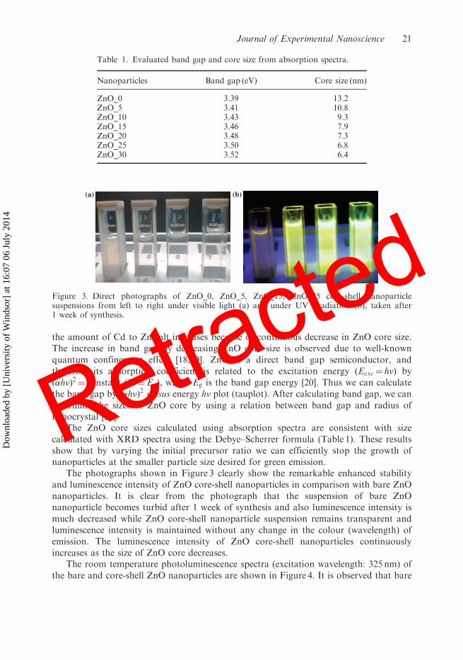
The photographs shown in Figure 3 clearly show the remarkable enhanced stabilityand luminescence intensity of ZnO core-shell nanoparticles in comparison with bare ZnOnanoparticles. It is clear from the photograph that the suspension of bare ZnOnanoparticle becomes turbid after 1 week of synthesis and also luminescence intensity ismuch decreased while ZnO core-shell nanoparticle suspension remains transparent andluminescence intensity is maintained without any change in the colour (wavelength) ofemission. The luminescence intensity of ZnO core-shell nanoparticles continuouslyincreases as the size of ZnO core decreases.
The room temperature photoluminescence spectra (excitation wavelength: 325 nm) ofthe bare and core-shell ZnO nanoparticles are shown in Figure 4. It is observed that bare
(b)(a)
Figure 3. Direct photographs of ZnO_0, ZnO_5, ZnO_15, ZnO_25 core-shell nanoparticlesuspensions from left to right under visible light (a) and under UV irradiation (b), taken after1 week of synthesis.
Table 1. Evaluated band gap and core size from absorption spectra.
and core-shell nanoparticles exhibit a broad emission band in the green region only;however, a very small peak in the UV region is also observed from bare ZnO nanoparticles(not clearly shown in the figure because of high luminescence intensity in visible regionfrom other core-shell nanoparticles). The intensity of green emission increasescontinuously from ZnO_0 to ZnO_25 nanoparticles. For ZnO_30 core-shell nanoparticle,a drastic increase in luminescence intensity is observed due to much controlled core sizeand nearly complete passivation by Cd(OH)2 shell. The shift in the green emission peakfrom 528 nm for ZnO_0 to 517 nm for ZnO_30 core-shell nanoparticle is also observed dueto decreased particle size leading to increased band gap Eg [8]. This explanation assumesthat the energy of shallow surface defect level will shift with changes in the valence bandedges, but that deep defect levels associated with oxygen vacancies will not shift [14].
As Cd and Zn belonging to the same group have very much similar property, so CdII
can easily sit at the position of ZnII at the surface of ZnO nanoparticle, by increasingcadmium salt concentration, more and more Cd(OH)2 molecules are absorbed at thesurface of ZnO nanoparticle. ZnII and CdII, being in the same group element, can betreated similar to the surface of ZnO nanoparticle from the luminescence point of view;that means, it does not introduce any new luminescence characteristic in ZnOnanoparticle.
Now, how Cd(OH)2 at the surface of ZnO nanoparticle supports the visible emission.It has been shown by several authors [22,23] that if a molecule or functional groupis absorbed at the surface of a ZnO nanoparticle it dominantly controls the emissionproperty of the ZnO nanoparticle. Sakohara et al. [22] have reported in their studies thatthe ZnO nanoparticle synthesised by sol–gel method are not chemically pure but haveacetate groups, which originate from the reagent materials absorbed on the surface of
700
−500
50100150200250300350400450500550600650700750800
12
×
Inte
nsity
(a.
u.)
Wavelength (nm)
(a)(b)(c)(d)(e)(f)
300 400 500 600
Figure 4. Photoluminescence spectra from suspended bare and core-shell ZnO nanoparticles:(a) ZnO_0, (b) ZnO_5, (c) ZnO_15, (d) ZnO_20, (e) ZnO_25, (f ) ZnO_30.
22 R. Mishra et al.
Retracted
Dow
nloa
ded
by [
Uni
vers
ity o
f W
inds
or]
at 1
6:07
06
July
201
4
the nanoparticle. The acetate groups consist of a mixture of unidentate, chelate andbridging type structures. In their report, unidendate complex is believed to trap the photo-generated holes formed during UV excitation, and this phenomenon intensifies the visibleluminescence, while the bidentate type is capable of capturing the electron, and it quenchesthe visible luminescence. The coverage of the ZnO quantum dot surface with Zn(OH)2shell has been reported to decrease the intensity of the excitonic emission and enhance thevisible emission [23]. Similarly Norberg and Gamelin [24] showed a direct correlationbetween green emission intensities with surface hydroxide concentration and they proposethe identity of the O2� surface trap to be surface OH�. They showed that surface-absorbedOH� ions behave like O2� surface sites and trap the hole at the surface site resulting invisible emission. As mentioned above, there is no difference between CdII and ZnII at thesurface of a ZnO nanoparticle, thus the ZnO/Cd(OH)2 core-shell nanoparticle can besimply treated as the ZnO nanoparticle having a shell of hydroxyl functional group on itssurface. Thus in the view of above studies, we can also propose the hydroxyl ion present atthe surface of ZnO nanoparticle as the fast trapping centre of the hole at the surface site, afirst step necessary for the visible emission according to Dijken’s model. The higher theconcentration of hydroxyl ions at the surface, the higher the probability of surfacetrapping of holes at the surface site and the greater the visible luminescence intensity andthe lower the excitonic emission intensity.
4. Conclusions
In summary, we have prepared a series of ZnO/Cd(OH)2 core-shell nanoparticles by thesol–gel method having continually decreasing core size and increasing effective passivationby Cd(OH)2 molecules. The Cd(OH)2 shell controls the growth of ZnO nanoparticles andalso increases the green emission intensity. This work proposes a method to synthesisehighly luminescent and stable green emitting ZnO nanoparticles.
Acknowledgements
This work was financially supported by the Department of Science and Technology, Ministry ofScience, Government of India, India. The authors would like to thank Dr Ramesh Chandra, IIT,Roorkee, India, for his assistance in carrying out EDAX measurements.
References
[1] X.P. Shen, A.H. Yuan, Y.M. Hu, Y. Jiang, Z. Xu, and Z. Hu, Fabrication, characterization and
field emission properties of large-scale uniform ZnO nanotube arrays, Nanotechnology 16 (2005),
pp. 2039–2043.
[2] E. Monroy, F. Omnes, and F. Calle, Wide-bandgap semiconductor ultraviolet photodetectors,
Semicond. Sci. Technol. 18 (2003), pp. R33–R51.[3] Y.R. Ryu, T.S. Lee, J.A. Lubguman, H.W. White, and Y.S. Park, ZnO devices: Photodiodes and
p-type field-effect transistors, Appl. Phys. Lett. 87 (2005), p. 153504.[4] X. Han, G. Wang, Q. Wang, L. Cao, R. Liu, and J.G. Hau, Ultraviolet lasing and time resolved
photo luminescence of well-aligned ZnO nanorod arrays, Appl. Phys. Lett. 87 (2005), p. 223106.
Journal of Experimental Nanoscience 23
Retracted
Dow
nloa
ded
by [
Uni
vers
ity o
f W
inds
or]
at 1
6:07
06
July
201
4
[5] L. Spanhel and M.A. Anderson, Semiconductor clusters in the sol-gel process: Quantized
aggregation, gelation, and crystal growth in concentrated zinc oxide colloids, J. Am. Chem. Soc.
113 (1991), pp. 2826–2833.[6] G. Oskam, Z. Hu, R.L. Penn, N. Pesika, and P.C. Searson, Coarsening of metal oxide
nanoparticles, Phys. Rev. E 66 (2002), p. 01403.[7] D.V. Talapin, A.L. Rogach, E.V. Shevehenko, A. Kornowski, M. Haase, and H. Weller,
Dynamic distribution of growth rates within the ensembles of colloidal II–VII semiconductor
nanocrystals as factors governing their photoluminescence efficiency, J. Am. Chem. Soc. 124
(2002), p. 5782.
[8] A. van Dijken, J. Makkinje, and A. Meijerink, The influence of particle size on the
luminescence quantum efficiency of nanocrystalline ZnO particles, J. Lumin. 92 (2001),
pp. 323–328.[9] N.S. Pesika, Z. Hu, K.J. Stebe, and P.C. Searson, The quenching of growth of ZnO nanoparticles
by adsorption of octanethiol, J. Phys. Chem. B 106 (2002), pp. 6985–6990.[10] L. Guo, S.H. Yang, C.L. Yang, P. Yu, J. Wang, and G. Wong, Highly monodisperse polymer-
capped ZnO nanoparticles: Preparation and optical properties, Appl. Phys. Lett. 76 (2000),
p. 2901.[11] M. Abdullah, S. Shibamoto, and K. Okuyama, Synthesis of ZnO/SiO2 nanocomposites emitting
specific luminescence colors, Opt. Mater. 26 (2004), pp. 95–100.[12] S. Mahamuni, K. Borgohain, B.S. Bendre, V.J. Leppert, and S.H. Risbud, Spectroscopic and
structural characterization of electrochemically grown ZnO quantum dots, J. Appl. Phys. 85
(1999), p. 2861.
[13] N.S. Norberg and D.R. Gamelin, Influence of surface modification on the luminescence of
colloidal ZnO nanocrystals, J. Phys. Chem. B 109 (2005), pp. 20810–20816.
[14] A. van Dijken, E.A. Meulenkamp, D. Vanmaekelbergh, and A. Meijerink, The kinetics of the
radiative and nonradiative processes in nanocrystalline ZnO particles upon photoexcitation,
J. Phys. Chem. B 104 (2000), pp. 1715–1723.[15] D.W. Bahnemann, C. Kormann, and M.R. Hoffmann, Preparation and characterization of
quantum size zinc oxide: A detailed spectroscopic study, J. Phys. Chem. B 91 (1987),
pp. 3789–3798.[16] H. Yang and P.H. Holloway, Efficient and photostable ZnS-passivated CdS:Mn luminescent
nanocrystals, Adv. Funct. Mater. 14 (2004), pp. 152–156.[17] X. Peng, M.C. Schlamp, A.V. Kadavanich, and A.P. Alivisatos, Epitaxial growth of highly
luminescent CdSe/CdS core/shell nanocrystals with photostability and electronic accessibility,
J. Am. Chem. Soc. 119 (1997), pp. 7019–7029.[18] H. Yang and P.H. Holloway, Efficient and photostable ZnS-passivated CdS:Mn luminescent
nanocrystals, Adv. Funct. Mater. 14 (2004), pp. 152–156.[19] X. Peng, M.C. Schlamp, A.V. Kadavanich, and A.P. Alivisatos, Epitaxial growth of highly
luminescent CdSe/CdS core/shell nanocrystals with photostability and electronic accessibility,
J. Am. Chem. Soc. 119 (1997), pp. 7019–7029.
[20] L.E. Brus, A simple model for the ionization potential, electron affinity, and aqueous redox
potentials of small semiconductor crystallites, J. Chem. Phys. 79 (1983), p. 5566.
[21] L.E. Brus, Electron–electron and electron–hole interactions in small semiconductor
crystallites: The size dependence of the lowest excited electronic state, J. Chem. Phys. 80
(1984), p. 4403.[22] S. Monticone, R. Tufeu, and A.V. Kanaev, Complex nature of the UV and visible fluorescence of
colloidal ZnO nanoparticles, J. Phys. Chem. B 102 (1998), pp. 2854–2862.[23] Y. Kayanuma, Quantum-size effects of interacting electrons and holes in semiconductor
microcrystals with spherical shape, Phys. Rev. B 38 (1998), p. 9797.
24 R. Mishra et al.
Retracted
Dow
nloa
ded
by [
Uni
vers
ity o
f W
inds
or]
at 1
6:07
06
July
201
4
[24] S. Sakohara, M. Ishida, and M.A. Anderson, Visible luminescence and surface properties ofnanosized ZnO colloids prepared by hydrolyzing zinc acetate, J. Phys. Chem. B 102 (1998),pp. 10169–10175.
[25] H. Zhou, H. Alves, D.M. Hofmann, W. Kriegseis, and B.K. Mayer, Behind the excitonic
emission of ZnO quantum dots: ZnO/Zn(OH)2 core-shell structure, Appl. Phys. Lett. 80 (2002),pp. 210–212.
[26] N.S. Norberg and D.R. Gamelin, Influence of surface modification on the luminescence of
colloidal ZnO nanocrystals, J. Phys. Chem. B 109 (2005), pp. 20810–20816.