1 Fossilized iron bacteria reveal pathway to biological 2 origin of banded iron formation 3 Ernest Chi Fru 1,2* , Magnus Ivarsson 1,2 , Stephanos P Kilias 3 , Stefan Bengtson 1,2 , 4 Veneta Belivanova 1 , Federica Marone 4 , Danielle Fortin 5 , Curt Broman 6 , Marco 5 Stampanoni 4,7 6 7 1 Swedish Museum of Natural History, Department of Palaeobiology and 2 Nordic 8 Centre for Earth Evolution (NordCEE), Box 50007, 105 05 Stockholm, Sweden. 9 3 Department of Economic Geology and Geochemistry, Faculty of Geology and 10 Geoenvironment, National and Kapodistrian University of Athens University of 11 Athens, Panepistimiopolis, Zographou, 15784, Athens, Greece. 4 Swiss Light Source, 12 Paul Scherrer Institute, CH-5232 Villigen, Switzerland. 5 Department of Earth 13 Sciences, University of Ottawa, Ottawa, Ontario, Canada. 6 Department of Geological 14 Sciences, Stockholm University, Sweden. 7 Institute for Biomedical Engineering, 15 University and ETH Zürich, CH-8092 Zürich, Switzerland. 16 17 *Correspondence should be addressed to: [email protected]18 19 20 21
Transcript
1 Fossilized iron bacteria reveal pathway to biological 2
origin of banded iron formation 3 Ernest Chi Fru1,2*, Magnus Ivarsson1,2, Stephanos P Kilias3, Stefan Bengtson1,2, 4 Veneta Belivanova1, Federica Marone4, Danielle Fortin5, Curt Broman6, Marco 5
Stampanoni4,7 6 7 1Swedish Museum of Natural History, Department of Palaeobiology and 2Nordic 8 Centre for Earth Evolution (NordCEE), Box 50007, 105 05 Stockholm, Sweden. 9 3Department of Economic Geology and Geochemistry, Faculty of Geology and 10 Geoenvironment, National and Kapodistrian University of Athens University of 11 Athens, Panepistimiopolis, Zographou, 15784, Athens, Greece. 4Swiss Light Source, 12 Paul Scherrer Institute, CH-5232 Villigen, Switzerland. 5Department of Earth 13 Sciences, University of Ottawa, Ottawa, Ontario, Canada. 6Department of Geological 14 Sciences, Stockholm University, Sweden. 7Institute for Biomedical Engineering, 15 University and ETH Zürich, CH-8092 Zürich, Switzerland. 16 17 *Correspondence should be addressed to: [email protected] 18 19 20 21
2
Debates on the formation of banded iron formations (BIFs) in ancient 22 ferruginous oceans are dominated by a dichotomy between abiotic and biotic 23 iron cycling. This is fuelled by difficulties in unraveling the exact processes 24 involved in their formation. Here we provide fossil environmental evidence for 25 anoxygenic photoferrotrophic deposition of analogue banded iron rocks in 26 shallow marine waters associated with an Early Quaternary hydrothermal vent 27 field on Milos Island, Greece. Trace metal, major and rare earth elemental 28 compositions suggest the deposited rocks closely resemble BIFs of Precambrian 29 origin. Well-preserved microbial fossils in combination with chemical data imply 30 that band formation was linked to periodic massive encrustation of anoxygenic 31 phototrophic biofilms by iron oxyhydroxide alternating with abiotic silica 32 precipitation. The data implicate cyclic anoxygenic photoferrotrophy and their 33 fossilization mechanisms in the construction of micro-skeletal fabrics that result 34 in the formation of characteristic BIF bands of varying silica and iron oxides 35 ratios. 36 37 It is generally believed that banded iron formations (BIFs), the bulk of which was 38 formed in late Archaean/Palaeoproterozoic marine basins, occurred in stratified water 39 columns deep in the ocean and on continental shelf margins1-4. Soluble ferrous iron, 40 supplied from mid-ocean ridges and hydrothermal vents, was oxidized by various 41 processes to ferric iron and deposited in association with varying silica ratios as 42 BIFs1-4. But the mechanism that drove and sustained the massive and sporadic 43 deposition of BIFs throughout much of the Precambrian remains a mystery, despite 44 decades of research. 45
3
The low Archaean oxygen levels (~10-5 of present-day concentrations or even 46 less5) are generally considered insufficient to have supported broad-scale BIF 47 generation1,6. Instead, biotic, anoxygenic photoferrotrophic precipitation according to 48 the equation, 4Fe2+ + CO2 + 11H2O + light → [CH2O] + 4Fe(OH)3 + 8H+, has been 49 proposed6-14. With the exemption of proxies such as iron isotopes12,13, there exists no 50 direct environmental evidence—neither in ancient nor modern ecosystems—51 demonstrating how photoferrotrophs could have accounted for vast-scale biological 52 BIF deposition, including the formation of their spectacular banded consistency. 53
Cape Vani, located on the NW of Milos Island, in the Hellenic (Aegean) 54 Volcanic Arc hosts an Early Quaternary shallow ferruginous marine microbial deposit 55 that was supported by chemical energy released from focused and diffused 56 hydrothermal vents15,16. Biogeochemical evidence indicates this deposit occurred in a 57 restricted shallow marine basin at the foot of an andesite continental shelf, where 58 calm waters resulted in water column stratification and the development of local 59 anoxia15. We provide new biogeochemical data supporting this fact and independently 60 illustrate that these conditions resemble those that supported the deposition of some 61 Precambrian BIFs, with the iron sourced from seafloor hydrothermal vents and 62 hotspots. 63 64 Results 65 Geology and geochemistry. The Cape Vani rocks (Fig. 1. See supplementary Fig. S1 66 for description of the geology of the field site), display remarkable BIF-like patterns 67 (Fig. 2), including alternating micrometric to millimetric iron oxide and silica bands 68 (supplementary Fig. S2). Similar to Precambrian BIFs, silica/iron ratios in these rocks 69 are inversely related (Fig. 3a), while a low range of TiO2 and Al2O3 and K2O 70
4
concentrations suggest little terrigenous sedimentary input17-19. Heavy rare earth 71 element (HREE) enrichment occurs in both the ferruginous and siliceous components 72 of the rock relative to light REE (LREE) (Fig. 3b) and are comparable to reported 73 Precambrian BIF profiles17-19. Together with a pronounced positive Eu anomaly, these 74 observations hint on a hydrothermal origin of the deposits as opposed to seawater 75 derived sediments that are often LREE enriched relative to HREE18. Calculated 76 Eu/Eu* and Ce/Ce* anomalies (North American Shale Composite (NASC) 77 normalized) of ~0.23 and ~2.56, respectively, lie within the reported Precambrian BIF 78 range, while Ce depletion in relation to the other REEs supports anoxic depositional 79 conditions17-19. Sedimentary Ce levels are generally expected to increase under 80 oxidizing conditions because unlike most REEs, Ce3+ is oxidized to Ce4+ and co-81 precipitated in association with iron oxyhydro(oxides)17-19. Furthermore, the 82 occurrence and preservation of MnO (Fig. 3c) in concentrations comparable to 83 Mesoarchean Witwatersrand Supergroup BIFs19, supports a reduced sedimentary 84 environment. 85
Cr in oceanic sediments have been mostly associated with a terrigenous origin, 86 resulting from the dissolution of ultramafic rocks and soils20,21. In oxygenated 87 conditions, the released Cr3+ is oxidized by Mn(III) and (IV) to stable Cr6+, which 88 under reducing conditions is transformed by microbial activity and Fe2+ to Cr3+ and 89 preserved in association with iron oxyhydro(oxides)20,21. Instead, we found that Cr3+ 90 was consistently below detection limit, a situation similar to low Cr3+ concentrations 91 reported for much of Archaean Agloma-type BIFs deposited close to deep submarine 92 volcanic arcs and spreading ridges21. After ~2.5 Ga, Superior-type BIF facies (near-93 shore continental shelf deposits) are reported to have experienced a sudden spike in 94 Cr concentrations, which was linked to clastic detrital input resulting from oxygen-95
5
driven weathering of crustal rocks20,21. Our Cr data thus support the idea of low 96 oxygen tension in the water column where the Cape Vani marine deposits were 97 formed and low levels of continental crust detrital delivery into the basin. 98
The haematite signal varied in the Cape Vani deposits as a function of silica 99 concentrations while trace elemental compositions followed the same general trends 100 regardless of the silica to iron ratio (Fig. 3d), indicating that the rocks were deposited 101 under persistent/consistent biogeochemical conditions for extended periods. The high 102 silica load particularly suggests an ocean saturated with hydrothermally-derived 103 amorphous silica, similar to inferences made for Precambrian oceans from chemical 104 analysis of BIFs1,17,18. 105
No organic carbon signal was obtained by Raman spectroscopy—a BIF feature 106 often attributed to remineralization of organic matter to dissolved inorganic carbon 107 through respiration22. The largely low levels of whole rock total carbon supports this 108 hypothesis, while the close to zero total sulphur concentrations suggest a non-109 sulphidic depositional environment (Fig. 3c). Therefore the Cape Vani deposit could 110 be classified as a cherty, low carbon, haematite-rich BIF analogue, formed under 111 reducing conditions. The recorded low oxygen concentrations would have promoted 112 anaerobic chemoautotrophic iron oxidation19. 113 114 Fossil iron bacteria. From the haematite-rich jasper bands (Fig. 2, supplementary 115 Fig. S2) we report remarkably well-preserved microbial fossils (Fig. 4a-e, 116 supplementary Fig.S3), which on the basis of distinct morphological features, 117 including: spherical/ovoidal cells adhering to >100 µm long stalk appendages, tubular 118 forks (Fig. 4a-c), exospore-like features (Fig. 4b, d) and fossilization mechanism, we 119 interpret as fossil relatives of the anoxygenic photoferrotroph, Rhodomicrobium 120
6
vanielii 7,8,23,24. Similar to their modern counterparts, these microorganisms grew by 121 polar budding, forming vast networks of cells connected end-to-end by stalk 122 appendages, when transforming Fe2+ to Fe3+ during anoxygenic photosynthesis. The 123 fossil structures are encrusted with haematite, the only oxyhydro(oxide) present on 124 these biological structures and as free deposits (Fig. 4f). The predominance of 125 haematite, points to circumneutral depositional/diagenetic conditions, where 126 haematite tends to be abundant between pH 7 and 8, with ferrihydrite as precursor25. 127
Most aerobic iron-oxidizing appendage bacteria escape entombment during 128 growth on iron by discarding their stalks26, which probably explains why it is 129 common to identify such structures in ancient sedimentary iron-rich rocks without 130 associated cells27. However, Rhodomicrobium vanielii becomes completely encrusted 131 during anaerobic phototrophic growth on Fe, leading to the arrest of cell growth and 132 Fe metabolism7,8. This likely accounts for the remarkable preservation we report here 133 and for the fact that the cells and their stalks were heavily encrusted by haematite. 134 135 Discussion 136 Three major mechanisms are currently implicated in the oxidation of Fe2+ to Fe3+: 137 abiotic O2 precipitation1,26, UV-driven photochemical Fe2+ oxidation1,2 and microbial 138 transformation1,3,6-14,28. In addition to the low Archaean O2 levels, recent experimental 139 studies have rejected photochemical Fe2+ oxidation as a potential major contributor to 140 BIF formation1,3. Instead, it has been suggested through microbial culture studies and 141 biogeochemical extrapolations that BIF deposition in the stratified Precambrian iron-142 rich oceans was entirely possible under the activity of the iron oxidizing bacteria1,3 143 and most especially with the involvement of anoxygenic photoferrotrophs12-14. 144 However it has never been demonstrated in the natural environment how this was 145
7
possible—i.e. microbial transformation of Fe2+ → Fe3+ → BIFs—or how the activity 146 of these microorganisms could have accounted for the alternating bands. In fact this 147 process has never been shown to form BIFs, not in nature or in the laboratory. 148
We show that the fossilized haematite-encrusted structures formed 3D skeletons 149 that tethered the iron-rich sections of the rock (Fig. 4, supplementary Fig. S3), which 150 can be better visualised with 3D visual aids in the synchrotron radiation X-ray 151 microscopy tomographic stereo renderings presented in Figures 3c-e. Supplementary 152 videos S1 and S2 give a deeper appreciation of depth. We did not observe such 153 geometries in portions of the rock populated by single cells, suggesting that a strong 154 prerequisite for the formation of these 3D micro-fabrics was that the cells were 155 immobilized in the stalk production phase and fossilized in their original spatial 156 position, which is possible with Rhodomicrobium vanielii7,8. 157
In a stratified ocean, these photoferrotrophic biofilm gels would have formed 158 below the chemocline, its depth and size dependent on light penetration. Following 159 biofilm encrustation and death, soluble Fe2+ in this zone would have remained 160 suspended in the anoxic water column in the absence of any major oxidant. 161 Continuous hydrothermal venting would have increased this soluble Fe2+ pool, until a 162 new biofilm succession became available to precipitate this soluble Fe2+ pool to Fe3+. 163 Cyclic lapse in intense photoferrotrophic Fe2+ oxidation would have enabled the 164 precipitation of the silica-rich bands, relative to the ratio of silica to Fe2+ oxidized 165 (Fig. 5a). This model is consistent with the fact that dense fossil biofilm populations 166 encrusted with haematite tended to alternate with the chert-rich bands, which mostly 167 lacked dense biofilms characteristic of the photoferrotrophic fossils and with lower 168 haematite content (Fig. 5b, c). These in situ observations implicate biofilm density in 169 both banding visibility and biogeochemistry. Nonetheless, the persistent high 170
8
concentration of silica even in the iron-rich bands, suggests that the silica load in the 171 water column was likely constant, but that the rate of Fe3+ precipitation determined 172 the ratios of silica to iron deposited in the various bands. Therefore silica precipitation 173 and sedimentation rates were likely much slower than biological Fe3+ formation. 174 These results demonstrate that cyclic growth and death of biofilms, after encrustation, 175 allowed the incorporation of different ratios of silica and iron in the alternating bands 176 (Fig. 3a, 5). 177
Additionally, if photoferrotrophy was the main mechanism for Fe3+ 178 precipitation in the ancient oceans, variations in solar irradiation, including changes in 179 Earth’s orbital eccentricity and obliquity, all of which influence water temperatures, 180 light penetration and depth of the thermocline, would have exerted strong temporal 181 and spatial variations on intense photoferrotrophic Fe3+ precipitation28. Experimental 182 evidence suggests that low temperatures indeed promote Si precipitation over 183 microbial Fe2+ transformation and vice versa29. We suggest that cyclic blooming of 184 anoxygenic photoferrotrophy, related to Fe2+ oxidation followed by rapid encrustation 185 and fossilization by Fe3+, was key to BIF formation and banding (Fig. 5). 186
It is possible that with the emergence of oxygen oases in the Archaean oceans30-187 35 and the rise of atmospheric oxygen during the Great Oxidation Event (GOE), ~2.45 188 billion years ago5, rapid abiotic transformation of Fe2+ to Fe3+ by oxygen could have 189 contributed to significant BIF formation31. This is feasible, especially if iron had a 190 continental source, or if Fe2+ upwelling from submarine hydrothermal sources reached 191 the oxic surface waters above the chemocline. However, it has been suggested that in 192 the redox stratified Proterozoic oceans, following the GOE, anoxygenic 193 photoferrotrophic activity below the chemocline might have hindered or reduced 194 potential contact of submarine-derived Fe2+ with the overlying oxic surface waters14. 195
9
Specifically, an origin below the chemocline of late Palaeoproterozoic BIFs has been 196 suggested—implicating biotic processes in their formation4. Our model supports and 197 is favoured by these various hypotheses. Furthermore, the Archaean BIFs were 198 deposited under anaerobic conditions, where anoxygenic photoferrotrophy activity 199 was likely important4,36. 200
The high demand for oxygen required for the abiotic deposition of BIFs at the 201 scale recorded in the anoxic Archaean world is problematic. Proponents of biological 202 processes linked to the oxygen deposition model suggest that primitive Archaean 203 oxygenic photosynthetic bacteria thrived during periodic Fe2+ availability36, supplying 204 oxygen to microaerophilic Fe2+ oxidizers capable of precipitating Fe3+ (ref. 38). 205 Although feasible, clear in situ evidence for this process or the scale of it and what 206 caused the proposed periodicity in iron availability is lacking. Band development in 207 BIFs has been related to basinal silicification due to evaporitic and climatic 208 fluctuations1, while periodic volcanism is said to have increased oceanic hydrothermal 209 iron flux 1,4,38. The latter process supports our hypothesis for a hydrothermal origin of 210 the Cape Vani BIF analogues. However, combined, these suggestions do not reliably 211 account for the periodicity of the fine alternating BIF banding texture. Neither do they 212 explain why iron would be preferentially sedimented over silica and vice versa. 213
As we have demonstrated, microorganisms when coupled to our model, could 214 easily have controlled both the deposition of BIFs and band formation at different 215 scales. Conveniently, the scale of their activity could have been coupled to increased 216 volcanic release of iron and climatic fluctuations. Microbial growth cycles are 217 universally affected by changes in environmental conditions—including nutrient 218 availability and temperatures28,29. 219
A scenario much difficult to explain involves the role of microaerophilic iron 220
10
oxidizers in vast deposition of Archaean BIFs. These microorganisms have a high 221 requirement for oxidizing huge quantities of iron to generate small amounts of 222 cellular energy26. As a consequence, a persistent and tremendous oxygen supply 223 source would have been required over the course of this entire process to enable large 224 scale BIF formation in the predominantly anoxic Archaean oceans. 225
Perhaps oxygen exerted a stronger impact on BIF formation after the GOE, both 226 through abiotic rapid oxygen precipitation of iron and biotic microaerophilic bacterial 227 iron oxidation4,19. At this time, the oceans had become stratified and predominated by 228 deep-water reducing conditions. Our model provides a clear example of how 229 anaerobic biological processes might have continued to contribute towards BIF 230 deposition below the chemocline, in the presence of an elevated oxygen atmosphere 231 and overlying oxic surface waters. This would have been the case, especially if the 232 oceans were relatively calm. 233
As we show, the Cape Vani BIF analogue was deposited under anoxic 234 conditions, below an atmosphere of present-day atmospheric oxygen concentrations. 235 It is thus likely that with lesser oxygen levels present in the early 236 Archaean/Palaeoproterozoic oceans, this process might have been able to contribute 237 to significant BIF deposition. There is evidence of a chemocline reaching >100 m 238 depths already by 2.5 Ga19,33 and the ability of anoxygenic photoferrotrophs to utilize 239 light in the wavelengths reaching this depth14. It is therefore possible that these 240 microorganisms could have formed vast and uninterrupted biofilms just below this 241 layer, enough to generate extensive BIF deposits. In addition, biological Fe3+ 242 precipitation involves both active and passive processes26. Thus in the case where 243 surplus Fe3+ was being precipitated by oxygen in the overlying oxic surface waters, it 244 could have been passively adsorbed by the biofilms below the chemocline during Fe3+ 245
11
rain-down. Such an alternate hypothesis could still be explained by our banding 246 model, if the encrusted and denser biofilms sedimented faster. This model is possible, 247 particularly if iron supply to the oxic surface waters occurred in pulses. However, our 248 data show no evidence for this process. 249
The Cape Vani BIF depositional conditions are similar to those in the early 250 oceans, but the basin was limited in size—laterally (~1 km) and probably vertically 251 too (depth, unknown), compared to Superior type BIF deposits, stretching hundreds of 252 kilometers. In the predominantly oxic Early Quaternary atmosphere, the small scale of 253 this basin would have exerted a major force on the development of typical 254 Precambrian ocean conditions (suitable redox and adequate levels of silica and iron 255 concentrations) over extended periods. In the largeness of the open ocean, these 256 circumstances, which likely influenced band periodicity and thickness, would have 257 been diluted. Hence we assume this basin to be a scaled down version of the 258 Precambrian oceans, where processes where also scaled down accordingly. 259
The Cape Vani deposits and their microbial fossils are extremely well 260 preserved, with apparently no regional contact deformation and metamorphism15,16, 261 making them a remarkable new analogue system for unraveling the in situ 262 biogeochemical processes that might have orchestrated the large-scale deposition of 263 BIFs. If indeed these are young BIF analogues, then the geochemical data provide 264 new insights on the likely chemical constitution of BIFs prior to diagenesis, 265 deformation and metamorphism billions of year later. 266
We conclude that anoxygenic photoferrotrophy might have played a crucial role 267 in the biogeochemical transformation of the early geobiosphere and that microbial 268 activity and their fossilization mechanisms are not only directly implicated in iron-269
12
oxyhydro(oxides) deposition, but might also have been vital for the formation of 270 micro-fabrics that enhanced the structural integrity of BIFs. 271 272 Methods 273 Optical microscopy. Reflected light microscopy and imaging was performed on 274 doubly polished sections (~150-200 µm thick) with an Olympus BX51 microscope 275 fitted with an Olympus DP71 Camera. Images were acquired with an Olympus 276 cellSens® 1.7 Digital Imaging software. 277 278 Raman spectroscopy. The thin sections were analysed with a laser Raman confocal 279 spectrometer (Horiba instrument LabRAM HR 800), equipped with a multichannel 280 air-cooled CCD detector and an Ar-ion laser (1 = 514 nm) as the excitation source. A 281 power output of 8 mW was focused on the sample. The instrument was integrated 282 with an Olympus BX51 microscope. The wavelength of the laser beam focused on 1 283 mm specimen spots with a 100× objective was changed from 1 = 514 nm to λ = 514 284 nm and the diameter from 1 mm to μm. The spectral resolution was ~0.3 cm-1. The 285 instrument was calibrated with a neon lamp and the Raman line (520.7 cm-1) of a 286 silicon wafer. Instrument control and data acquisition was made with LabSpec 5 287 software. 288 289 Synchrotron Radiation X-Ray Tomographic Microscopy. Thin sections were 290 reduced in size and mounted on a 3 mm wide brass peg and analysed on the 291 TOMCAT beamline at the Swiss Light Source at the Paul Scherrer Institute40. The X-292 ray energy was optimized for maximum contrast at 13–20 keV. 1501 projections were 293 acquired equiangularly over 180°, online post-processed and rearranged into flat- and 294
13
darkfield-corrected sinograms. Reconstruction was performed on a Linux PC farm 295 using highly optimized routines based on the Fourier Transform method40. A 20× lens 296 was used, resulting in voxel stacks with a voxel size of 0.37 µm. The data derived 297 from the scans were then analysed and rendered using Avizo® 298 <http://www.vsg3d.com/> software. 299 300 Elemental analysis of rock samples. AcmeLabs® (http://acmelab.com) conducted 301 analyses on 0.2 g of whole rock samples. Eu/Eu* and Ce/Ce* anomalies were 302 calculated using the North American Composite Shale (NASC) reference standard as 303 previously described for BIFs42. 304 305 Sample storage. The microbial fossil specimens are deposited at the Swedish 306 Museum of Natural History under the reference numbers: SMNH X5036 and SMNH 307 X5037. The sawn rock samples are deposited at the Museum of Palaeontology and 308 Geology, National and Kapodistrian University of Athens (N.K.U.A.) and given 309 reference numbers: AMPG 936 and AMPG 935. 310 311 References 312
1. Posth, N. R., Konhauser, K. O., Kappler, A. Encyclopedia of Geobiology (eds 313 Reitner, J. & Thiel, V.) 92–103 (Springer, 2011). 314
2. Braterman, P. S., Graham C.-.S. A., Sloper, R. W. Photo-oxidation of hydrated 315 Fe2+-significance for banded iron formations. Nature 303, 5913, 163–164 316 (1983). 317
3. Konhauser, K. O., Hamade, T. Riaswell, R. Morris, R. C., Ferris F. G., 318 Southam, G. Canfied, D. E. Could bacteria have formed the Precambrian 319
14
banded iron formations? Geology 12, 1097–1082 (2002). 320 4. Bekker, A., Slack, J. F., Planavsky, N., Krapež, B., Hofmann, A., Konhauser, 321
K. O., Rouxel, O. J. Iron Formation: The Sedimentary Product of a Complex 322 Interplay among Mantle, Tectonic, Oceanic, and Biospheric Processes. Econ. 323 Geol. 105, 467–508 (2010). 324
5. Canfield, D. E. The early history of atmospheric oxygen: Homage to Robert 325 A. Garrels. Annu. Rev. Earth Plan. Sci. 33, 1–36 (2005). 326
6. Crowe, S. A. et al. Photoferrotrophs thrive in an Archean Ocean analogue. 327 Proc. Natl. Acad. Sci. U. S. A. 105, 15938–15943 (2008). 328
7. Heising, S., Schink, B. Phototrophic oxidation of ferrous iron by a 329 Rhodomicrobium vannielii strain. Microbiology 144, 2263–2269 (1998). 330
8. Straub, K. L., Benz, M., Schink, B. Iron metabolism in anoxic environments at 331 near neutral pH. FEMS Microbiol. Ecol. 34, 181–186 (2001). 332
9. Widdel, F., Schnell, S., Heising, S., Ehrenreich, A., Assmus, B., Schink, B. 333 Ferrous iron oxidation by anoxygenic phototrophic bacteria. Nature 362, 834–334 836 (1993). 335
10. Ehrenreich, A., Widdel, F. Anaerobic oxidation of ferrous iron by purple 336 bacteria, a new type of phototrophic metabolism. Appl. Environ. Microbiol. 337 60, 4517–4526 (1994). 338
11. Heising, S., Richter, L., Ludwig, W., Schink, B. Chlorobium ferrooxidans sp. 339 nov., a phototrophic green sulfur bacterium that oxidizes ferrous iron in 340 coculture with a ‘Geospirillum’ sp. strain. Arch. Microbiol. 172, 116–124 341 (1999). 342
12. Dauphas, N. et al. Clues from Fe isotope variations on the origin of Early 343 Archean BIFs from Greenland. Science, 306, 2077–2080 (2004). 344
15
13. Czaja, A. D. et al. Biological Fe oxidation controlled deposition of banded 345 iron formation in the ca. 3770 Ma Isua Supracrustal Belt (West Greenland). 346 Earth Planet. Sci. Lett. 363, 192–203 (2013). 347
14. Kappler, A., Pasquero, C., Konhauser, K. O., Newman, D. K. Deposition of 348 banded iron formations by anoxygenic phototrophic Fe(II)-oxidizing bacteria. 349 Geology 33, 865–868 (2005). 350
15. Liakopoulos, A., Glasby, G. P., Papavassiliou, C. T., Boulegue, J. Nature 351 and origin of the Vani manganese deposit, Milos, Greece: an overview. Ore 352 Geol. Rev. 18, 181–209 (2001). 353
16. Kilias, S. P. Microbial Mats in Siliciclastic Depositional Systems Through 354 Time, SEPM (eds Noffke, N., Chafetz, H.). Soc. Sed. Geol. Special Pub. 101, 355 97–100 (2011). 356
17. Planavsky, N. et al. Rare earth element and yttrium compositions of Archean 357 and Paleoproterozoic Fe formations revisited: New perspectives on the 358 significance and mechanisms of deposition. Geochim. Cosmochim. Acta 74, 359 6387–6405 (2010). 360
18. Smith, A. J. B., Beukes, N. J., Gutzmer, J. The composition and depositional 361 environments of Mesoarchean iron formations of the West Rand group of the 362 Witwatersrand Supergroup, South Africa. Econ. Geol. 108, 111–134 (2013). 363
19. Planavsky, N. et al. Iron oxidizing microbial ecosystems thrived in late 364 Paleoproterozoic redox-stratified oceans. Earth Plan. Sci. 286, 230–242 365 (2009). 366
20. Frei, R., Gaucher, A., Poulton, S. W., Canfield, D. E. Fluctuations in 367 Precambrian atmospheric oxygenation recorded by chromium isotopes. Nature 368 461, 250–254 (2009). 369
16
21. Konhauser et al. Aerobic bacterial pyrite oxidation and acid rock drainage 370 during the great oxidation event. Nature 478, 369–373 (2011). 371
22. Bontognalia, T. R. R., Fischerb, W. W., Föllmi, K. B. Siliciclastic associated 372 banded iron formation from the 3.2 Ga Moodies Group, Barberton Greenstone 373 Belt, South Africa. Precam. Res. 226, 116–124 (2013). 374
23. Faith-Anthony, A, O., Ibrahim, N., Hamzah A. Application of Electron 375 Microscopy and Energy Dispersive X-Ray Spectroscopy in the 376 Characterization of Rhodomicrobium vannielii. J. Ad. Mic. Res. 6, 46–52 377 (2011). 378
24. Whittenbury, R., Dow, C. S. Morphogenesis and differentiation in 379 Rhodomicrobium vannielii and other budding and prosthecate bacteria. Bact. 380 Rev. 41, 754–808 (1977). 381
25. Schwertmann, U., Murad, E. Effect of pH on the formation of goethite and 382 haematite from ferrihydrite. Clay Min. 31, 277–284 (1983). 383
26. Chi Fru, E., Piccinelli, P., Fortin, D. Insights into the global microbial 384 community structure associated with iron oxyhydroxide minerals deposited in 385 the aerobic biogeosphere. Geomicrobiol. J. 7, 587–610 (2012). 386
27. Little, C. T., Glynn S. E. J., Mills R. A. Four-hundred and ninety-million year 387 record of bacteriogenic iron oxide precipitation at seafloor hydrothermal vents. 388 Geomicrobiol. J. 21, 415–429 (2004). 389
28. Ji, J. et al. Centennial blooming of anoxygenic phototrophic bacteria in 390 Qinghai Lake linked to solar and monsoon activities during the last 18,000 391 years. Quat. Sci. Rev. 28, 1304–1308 (2009). 392
29. Posth, N. R., Hegler, F. Konhauser, K. O., Kappler, A. Alternating Si and Fe 393 deposition caused by temperature fluctuations in Precambrian oceans. Nat. 394
17
Geosci. 1, 703–708 (2008). 395 30. Anbar, A. D. et al. A whiff of oxygen before the Great Oxidation Event? 396
Science 317, 1903–1906 (2007). 397 31. Czaja, A. D. et al. Evidence for free oxygen in the Neoarchean ocean based on 398
32. Kaufman, A. J. et al. Late Archean biospheric oxygenation and atmospheric 401 evolution. Science 317, 1900–1903 (2007). 402
33. Kendall, B. et al. Pervasive oxygenation along late Archaean ocean margins. 403 Nat. Geosci. 3, 647–652 (2010). 404
34. Cloud, P. Significance of the Gunflint (Precambrian) microflora: 405 photosynthetic oxygen may have had important local effects before becoming 406 a major atmospheric gas. Science 148, 27–35 (1965). 407
35. Cloud, P. Paleoecological significance of the banded iron formation. Econ. 408 Geol. 68, 1135–1143 (1973). 409
36. Fralick, P., Pufahl, P. K. Iron formation in Neoarchean Deltaic Successions 410 and the microbially mediated deposition of transgressive systems tracts. J. 411 Sediment. Res. 76, 1057–1066 (2006). 412
37. Holm, N. G. The 13C/12C ratios of siderite and organic matter of a modern 413 metalliferous hydrothermal sediment and their implications for banded iron 414 formations. Chem. Geol. 77, 41–45 (1989). 415
38. Isley, A. E., Abbott, D. H. Plume-related mafic volcanism and the deposition 416 of banded iron formation. J. Geophy. Res. 104, 15461–15477 (1999). 417
39. Downs, R. T. The RRUFF Project: an integrated study of the chemistry, 418 crystallography, Raman and infrared spectroscopy of minerals. Program and 419
18
Abstracts of the 19th General Meeting of the International Mineralogical 420 Association in Kobe, Japan. O03-13, (2006). 421
40. Marone, F., Stampanoni, M. Regridding reconstruction algorithm for real time 422 tomographic imaging. J. Synchrotron Rad. 19, 1029–1037 (2012). 423
41. Kato, Y. et al. Rare Earth element variations in mid-Archean banded iron 424 formations: implications for the chemistry of ocean continent and plate 425 tectonics. Geochim. Cosmochim. Acta 62, 3475–3497 (1998). 426
427 Acknowledgements 428 We would like to thank Stefan Ohlsson at NRM for laboratory assistance. We 429 acknowledge funding from The Swedish Research Council (Contract No. 2012-4364), 430 Swedish National Space Board (Contract No. 83/10), Per-Erik Lindahls stipend, The 431 Royal Swedish Academy of Sciences, The Danish National Research Foundation 432 (DNRF53), The Paul Scherrer Institute and The Special Account for Research Grants, 433 N.K.U.A (70/4/8646). Ernest Chi Fru was funded by an individual Marie Curie 434 fellowship (Contract No. PIEF.GA-2010-276475) from the European Union. 435 436 Author Contributions 437 E.C.F., M.I., S.B and S.P.K. conducted the design and analyses. M.I. and S.P.K. 438 performed fieldwork. M.I. prepared thin sections and acquired light micrographs. 439 S.B., F.M., M.S. and V.B. acquired Synchrotron X-Ray Radiation data at the Swiss 440 Light Source, Paul Scherrer Institute. K.B. performed Raman spectroscopy. All 441 authors contributed to data analysis. E.C.F., M.I., S.B., D.F. and S.P.K. wrote paper. 442 443 Additional Information 444
19
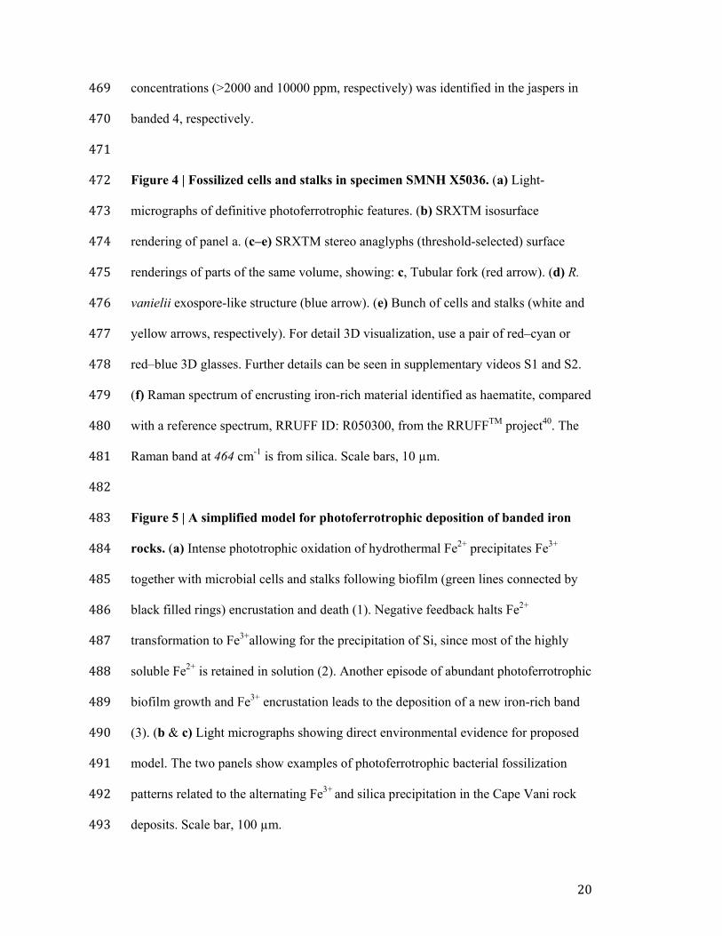
Supplementary Information accompanies this paper at http://www.nature.com/ 445 naturecommunications 446 Competing financial interests: The authors declare no competing financial interests. 447 448 Figure legends 449 450 Figure 1 | Location and geologic features of Cape Vani. Insert, satellite map 451 showing the location of Milos Island (black circle on unbroken white line). Gray 452 circle on unbroken white line indicates Santorini. Broken white line shows the 453 Hellenic Trench, where the African plate is subducted under the Euroasian plate to 454 give rise to the Hellenic (Aegean) Volcanic Arc (unbroken white line). 455
456 Figure 2 | Stratigraphy of the Cape Vani deposit. (a) the lithofacies association, 457 constituting the fossiliferous Cape Vani banded Fe-rich sedimentary chert, NW Milos 458 Island. (b) Sawed rock sample of the investigated chert. Details are found in 459 supplementary Fig. S1. 460 461 Figure 3 | Whole rock chemistry. (a) behavior of silica, haematite, Al2O3 and TiO2. 462 (b) NASC normalized Rare Earth Element (REE) fractionation profiles. Minimum 463 detection limit (MDL) varied from 0.01-0.1 ppm. c, Behavior of major elements. 464 MDL was 0.002 for Cr2O3 to 0.04 wt% for Fe2O3. Cr2O3 was consistently below the 465 detection limit. (d) Trace elemental composition. MDL varied from 0.1 to 1 ppm, 466 with the exception of Ni that had an MDL of 20 ppm and was below the MDL. Bi, Sn 467 and Se were below MDL (0.1, 1, 0.5, respectively). An unusually high Sb and Pb 468
20
concentrations (>2000 and 10000 ppm, respectively) was identified in the jaspers in 469 banded 4, respectively. 470 471 Figure 4 | Fossilized cells and stalks in specimen SMNH X5036. (a) Light-472 micrographs of definitive photoferrotrophic features. (b) SRXTM isosurface 473 rendering of panel a. (c–e) SRXTM stereo anaglyphs (threshold-selected) surface 474 renderings of parts of the same volume, showing: c, Tubular fork (red arrow). (d) R. 475 vanielii exospore-like structure (blue arrow). (e) Bunch of cells and stalks (white and 476 yellow arrows, respectively). For detail 3D visualization, use a pair of red–cyan or 477 red–blue 3D glasses. Further details can be seen in supplementary videos S1 and S2. 478 (f) Raman spectrum of encrusting iron-rich material identified as haematite, compared 479 with a reference spectrum, RRUFF ID: R050300, from the RRUFFTM project40. The 480 Raman band at 464 cm-1 is from silica. Scale bars, 10 µm. 481 482 Figure 5 | A simplified model for photoferrotrophic deposition of banded iron 483 rocks. (a) Intense phototrophic oxidation of hydrothermal Fe2+ precipitates Fe3+ 484 together with microbial cells and stalks following biofilm (green lines connected by 485 black filled rings) encrustation and death (1). Negative feedback halts Fe2+ 486 transformation to Fe3+allowing for the precipitation of Si, since most of the highly 487 soluble Fe2+ is retained in solution (2). Another episode of abundant photoferrotrophic 488 biofilm growth and Fe3+ encrustation leads to the deposition of a new iron-rich band 489 (3). (b & c) Light micrographs showing direct environmental evidence for proposed 490 model. The two panels show examples of photoferrotrophic bacterial fossilization 491 patterns related to the alternating Fe3+ and silica precipitation in the Cape Vani rock 492 deposits. Scale bar, 100 µm. 493
Gre
ece!
Crete!
Turkey!
Mediterranean sea!
Aegean sea!
Black sea!Adriatic sea!
not in scale
Millimeter-thick Mn(-Fe) crust !
Thick- to thin-bedded parallel- !and cross-stratified sandstone !
Well sorted thick-bedded, granule- !to pebble-bearing sandstone !
Thick-bedded, pebble to cobble !conglomerate with !