52
Fowl cholera
| Date post: | 13-Jul-2015 |
| Category: |
Education |
| Upload: | ossama-motawae |
| View: | 264 times |
| Download: | 2 times |
Fowl cholera
Plan of Talk
Introduction
Predisposing factors
Incidence and distribution
Etiology
Transmission
Clinical signs
Post mortem lesions
Diagnosis
Treatment
Prevention
Synonyms
Avian Cholera
Avian Pasteurellosis
Avian Hemorrhagic septicemia
Kolera Unggas (INA)
Cont. …
Fowl cholera is a contagious, bacterial disease that affectsdomestic and wild birds worldwide, caused by Pasteurellamultocida type A.
It usually occurs as a septicemia of sudden onset with highmorbidity and mortality, but chronic and asymptomaticinfections also occur.
Cont. …
Turkeys and waterfowl are more susceptible than chickens.
Older chickens are more susceptible than young ones.
Cont. …
Pathogenic Pasteurella species are:
1. Pasteurella multocida type A– Fowl Cholera in chicken.
2. Pasteurella multocida type B– Septicemia epizootica or Hemorrhagic septicemia in ruminant.
3. Pasteurella haemolytica– Pneumonia Pasteurellosis in Cattle.
Plan of Talk
Introduction
Predisposing factors
Incidence and distribution
Etiology
Transmission
Clinical signs
Post mortem lesions
Diagnosis
Treatment
Prevention
Predisposing Factors
Fowl Cholera is closely related to some stress factor;
1. Change of weather, fluctuation of temp, humidity….ect.
2. Move to new cage.
3. Debeaking.
4. Alteration of food suddenly.
5. Exhaustion.
6. Over crowding.
7. Transport in long time with lack of drink.
Plan of Talk
Introduction
Predisposing factors
Incidence and distribution
Etiology
Transmission
Clinical signs
Post mortem lesions
Diagnosis
Treatment
Prevention
Incidence & Distribution
Fowl cholera occurs sporadically or enzootically in mostcountries.
It sometimes causes high mortality; at other times, losses arenominal.
Cont. …
Fowl cholera is more prevalent in late summer, fall, andwinter.
Chickens become more susceptible as they reach maturity.
Cont. …
Acute from;
Alberts and Graham reported a loss of 68% within 6 days in aflock of 52 month old turkeys.
Vaught et al. reported that more than 1,000 wild geese diedof FC in one night.
Chronic from;
In studying the chronic respiratory form in chickens, Hall et al.observed that mortality was low, but infection persisted for atleast 4 years.

Plan of Talk
Introduction
Predisposing factors
Incidence and distribution
Etiology
Transmission
Clinical signs
Post mortem lesions
Diagnosis
Treatment
Prevention
Etiology
The genus Pasteurella sensustricto includes at least 11species.
Only 7 species have been associated with avian hosts.
Among these 7 species, P. multocida is considered thecausative agent of fowl cholera.
Cont. …
Pasteurella multocida
Small, gram-negative.
Non-motile rod with a capsule that may exhibit pleomorphism(the ability of some bacteria to alter their shape or size inresponse to environmental conditions) after repeatedsubculture.
The organism is susceptible to ordinary disinfectants, sunlight,drying, and heat.

Plan of Talk
Introduction
Predisposing factors
Incidence and distribution
Etiology
Transmission
Clinical signs
Post mortem lesions
Diagnosis
Treatment
Prevention
Transmission
Transmission occur through:
1. Oral
2. Inhalation
Cont. …
Indirect contact:
Through food/drink, tools/materials which were contaminatedby the agents, animals transmitted and wind.
Direct Contact:
Through discharges and feces.
Cont. …
Chronically infected birds and asymptomatic carriers areconsidered to be major sources of infection.
Wild birds may introduce the organism into a poultry flock,but mammals (including rodents, pigs, dogs, and cats) mayalso carry the infection.
Plan of Talk
Introduction
Predisposing factors
Incidence and distribution
Etiology
Transmission
Clinical signs
Post mortem lesions
Diagnosis
Treatment
Prevention

Clinical Signs
Clinical findings vary greatly depending on the course of disease:
1. Acute
2. Chronic
Cont. …
In acute fowl cholera,
1. Sudden surges in mortality, without previous signs.
2. Fever.
3. Loss of appetite.
4. Ruffled feathers.
5. Mucous discharge from the mouth.
6. Green watery diarrhea.
7. Respiratory difficulty.
8. Blue or purple coloration of skin and swelling of comb andwattles.
9. Pneumonia is particularly common in turkeys.
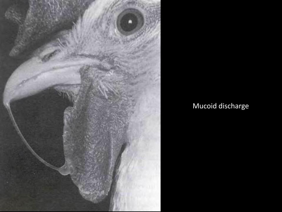
Mucoid discharge

Swollen wattlesBlue/ purple coloration
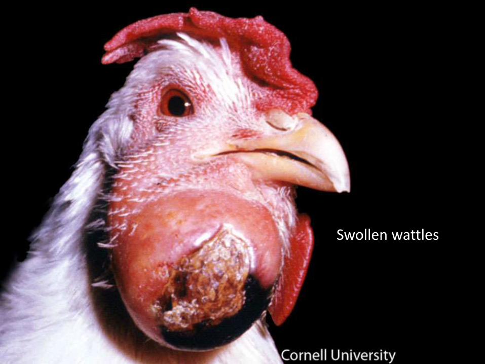
Swollen wattles
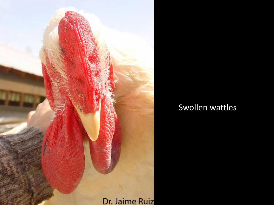
Swollen wattles
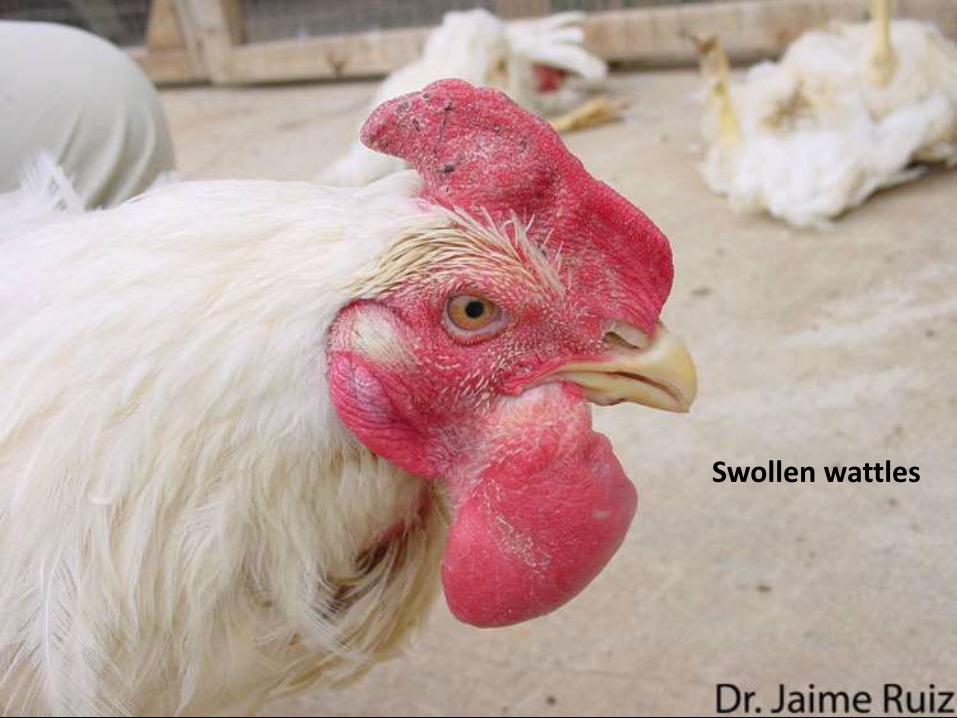
Swollen wattles
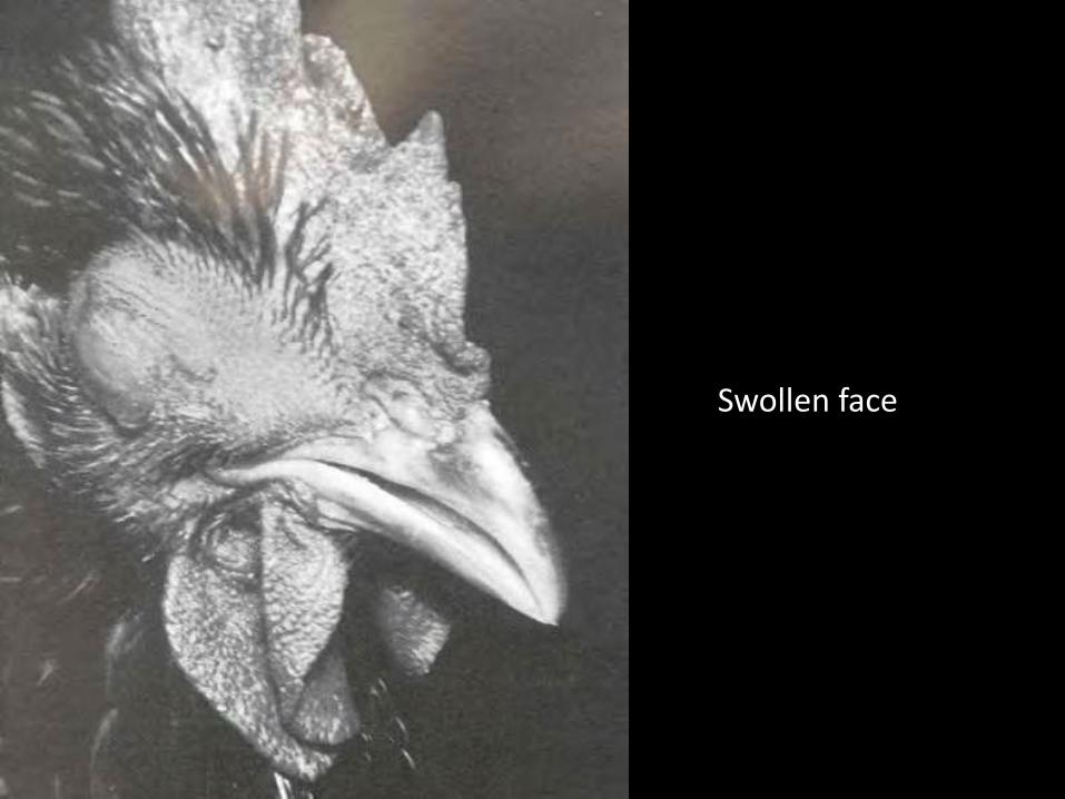
Swollen face
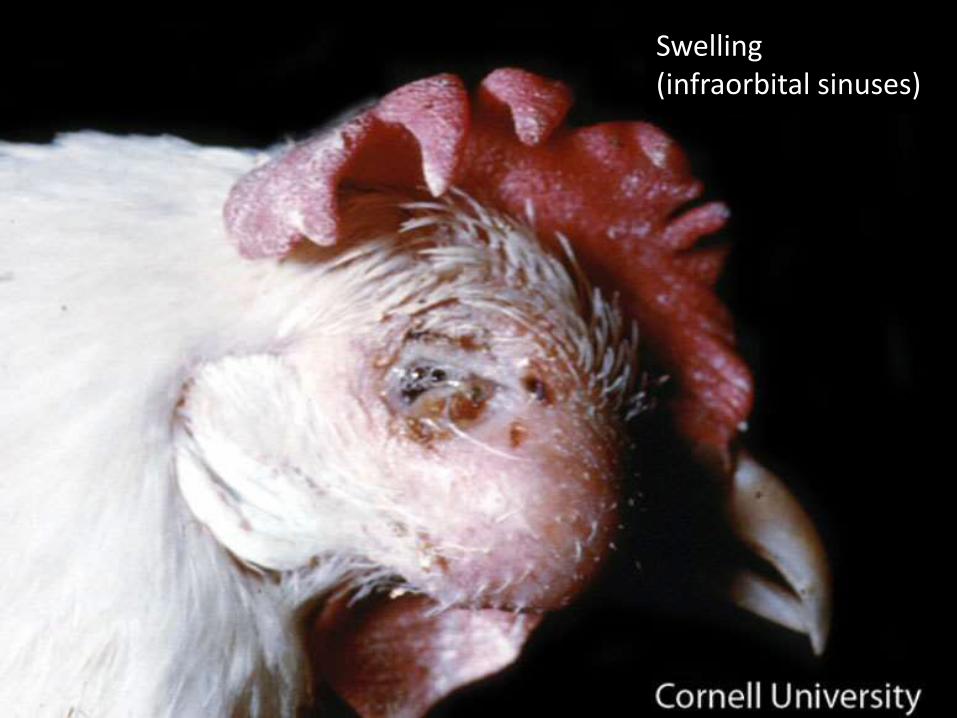
Swelling (infraorbital sinuses)
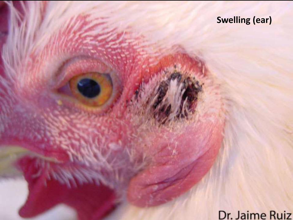
Swelling (ear)
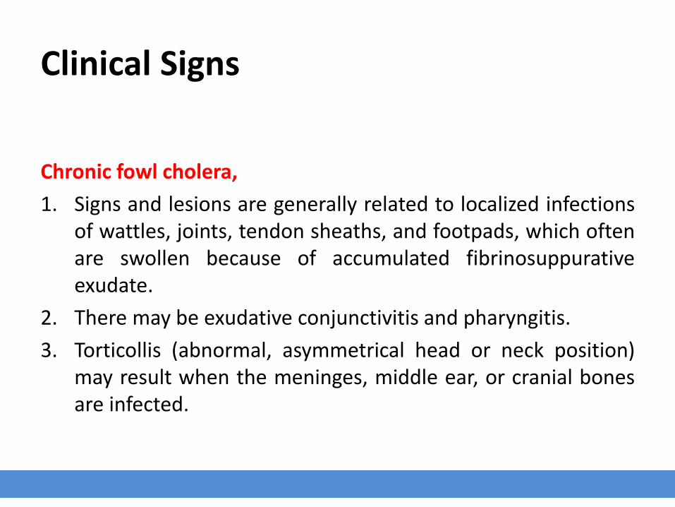
Clinical Signs
Chronic fowl cholera,
1. Signs and lesions are generally related to localized infectionsof wattles, joints, tendon sheaths, and footpads, which oftenare swollen because of accumulated fibrinosuppurativeexudate.
2. There may be exudative conjunctivitis and pharyngitis.
3. Torticollis (abnormal, asymmetrical head or neck position)may result when the meninges, middle ear, or cranial bonesare infected.
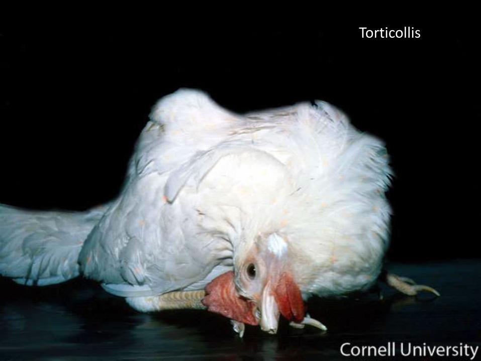
Torticollis

Plan of Talk
Introduction
Predisposing factors
Incidence and distribution
Etiology
Transmission
Clinical signs
Post mortem lesions
Diagnosis
Treatment
Prevention
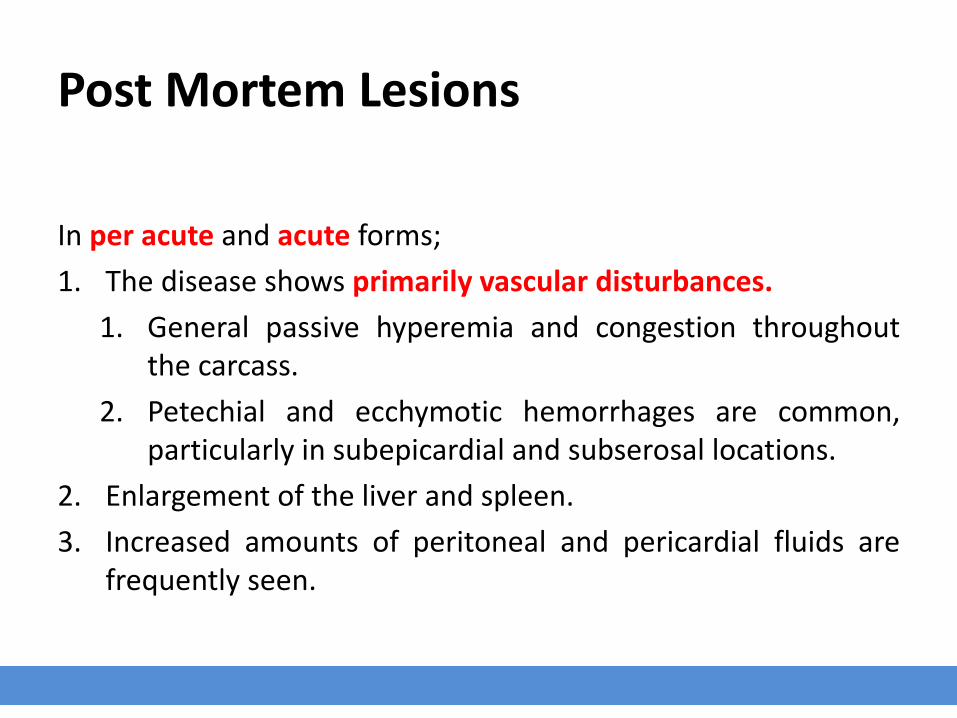
Post Mortem Lesions
In per acute and acute forms;
1. The disease shows primarily vascular disturbances.
1. General passive hyperemia and congestion throughoutthe carcass.
2. Petechial and ecchymotic hemorrhages are common,particularly in subepicardial and subserosal locations.
2. Enlargement of the liver and spleen.
3. Increased amounts of peritoneal and pericardial fluids arefrequently seen.
Cont. …
In sub acute forms;
1. Multiple, small, necrotic foci may be disseminatedthroughout the liver and spleen.
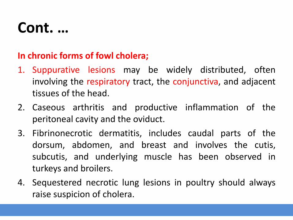
Cont. …
In chronic forms of fowl cholera;
1. Suppurative lesions may be widely distributed, ofteninvolving the respiratory tract, the conjunctiva, and adjacenttissues of the head.
2. Caseous arthritis and productive inflammation of theperitoneal cavity and the oviduct.
3. Fibrinonecrotic dermatitis, includes caudal parts of thedorsum, abdomen, and breast and involves the cutis,subcutis, and underlying muscle has been observed inturkeys and broilers.
4. Sequestered necrotic lung lesions in poultry should alwaysraise suspicion of cholera.
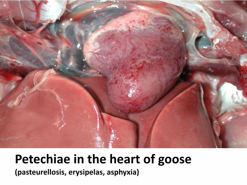
Petechiae in the heart of goose(pasteurellosis, erysipelas, asphyxia)
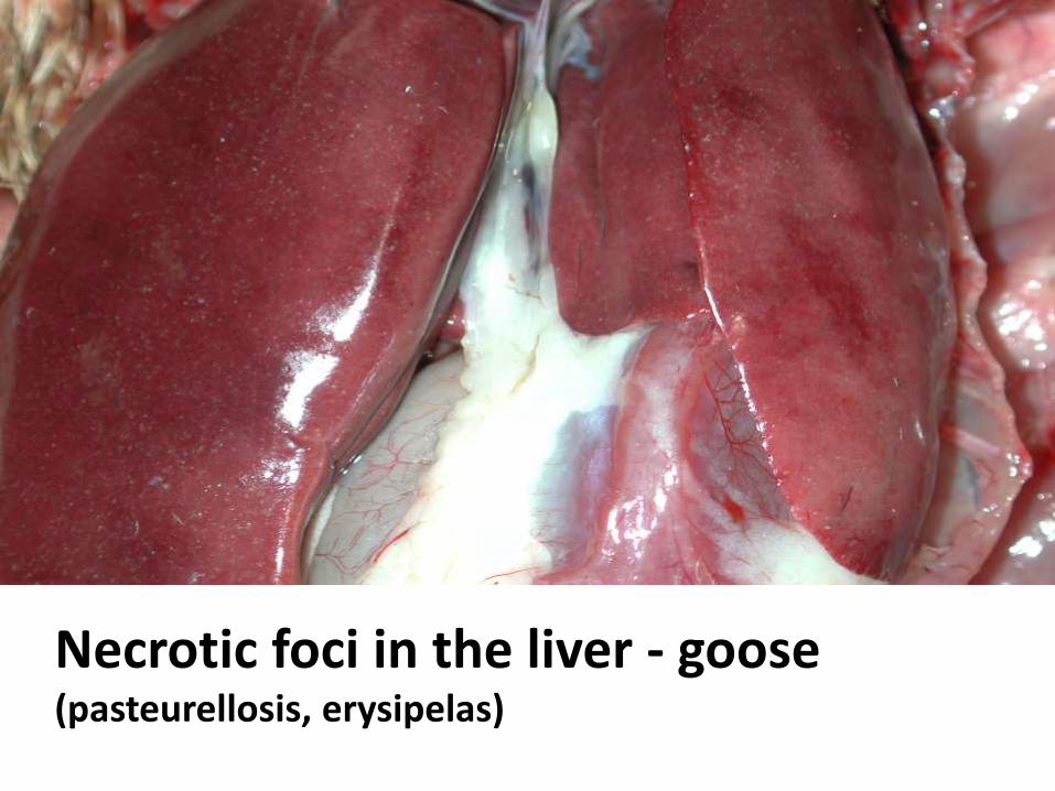
Necrotic foci in the liver - goose(pasteurellosis, erysipelas)

Liver - Multiple, small, necrotic foci
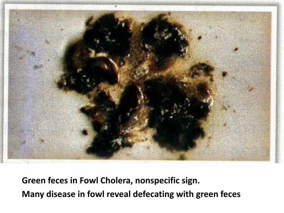
Green feces in Fowl Cholera, nonspecific sign.
Many disease in fowl reveal defecating with green feces
Plan of Talk
Introduction
Predisposing factors
Incidence and distribution
Etiology
Transmission
Clinical signs
Post mortem lesions
Diagnosis
Treatment
Prevention
Diagnosis
Although the history, signs, and lesions may aid diagnosis, Pmultocida should be isolated, characterized, and identified forconfirmation.

Plan of Talk
Introduction
Predisposing factors
Incidence and distribution
Etiology
Transmission
Clinical signs
Post mortem lesions
Diagnosis
Treatment
Prevention
Treatment
A number of drugs will lower mortality from fowl cholera;however, deaths may resume when treatment is discontinued,showing that treatment does not eliminate P multocida from aflock.

Cont. …
Sensitivity testing often aids in drugselection and is important becauseof the emergence of multi-resistantstrains.
Cont. …
Sulfas should be used with caution in breeders because ofpotential toxicity.
Penicillin is often effective for sulfa-resistant infections.
High levels of tetracycline antibiotics in the feed (0.04%),drinking water, or administered parenteral may be useful.
Norfloxacin administered via drinking water is also effectiveagainst fowl cholera.
In ducks, a combined injection of streptomycin anddihydrostreptomycin can be effective.
Plan of Talk
Introduction
Predisposing factors
Incidence and distribution
Etiology
Transmission
Clinical signs
Post mortem lesions
Diagnosis
Treatment
Prevention
Prevention
Eradication of infection requires:
1. Depopulation, cleaning and disinfection of buildings andequipment.
2. The premise should then be kept free of poultry for fewweeks.
3. High level of biosecurity.
4. Rodents, wild birds, pets, and other animals that may becarriers of P multocida and must be excluded from poultryhouses.
Vaccination – Live Vaccines
Attenuated live vaccines are available for administration:
1. In drinking water to turkeys.
2. By wing-web inoculation to chickens.
Vaccination of chickens and turkeys with live P. multocidavaccines induces protection against heterologous serotypechallenge.
Cont. …
Three live vaccines available for use in the United States are:
1. CU (Clemson University), a strain of low virulence
2. M-9, a mutant of CU with very low virulence
3. PM-1, a mutant of CU intermediate in virulence between CUand M-9.
Cont. …
The use of live FC vaccines stimulates an effective immuneresponse but has the disadvantage of potentially resulting inmortality in the vaccinated birds.
If the mortality post vaccination becomes excessive, it can bereduced by the administration of an antibiotic.
This should be avoided, if possible, until at least 4 dayspost-vaccination when there will be at least partialimmunity induced by the vaccine.
Vaccination – Killed Vaccines
Commercially produced bacterins are available.
Bacterins usually contain whole cells of serotypes 1, 3, and 4emulsified in an oil adjuvant.


