Editors: Johannes Kleinheinz, Christophe Meyer Coordination: Michel Loose, Thomas Pfluger Fractures of the Mandibular Condyle Basic Considerations and Treatment London, Berlin, Chicago, Tokyo, Barcelona, Beijing, Istanbul, Milan, Moscow, New Delhi, Paris, Prague, São Paulo, and Warsaw IBRA International Bone Research Association
Transcript
Editors: Johannes Kleinheinz, Christophe MeyerCoordination: Michel Loose, Thomas Pfluger
Fractures of the Mandibular CondyleBasic Considerations and Treatment
London, Berlin, Chicago, Tokyo, Barcelona, Beijing, Istanbul, Milan, Moscow, New Delhi, Paris, Prague, São Paulo, and Warsaw
IBRA International BoneResearch Association
v
ForewordTo dedicate such a large volume to such a small bone—the mandibular condyle—may seem surprising. Withoutattempting to be encyclopedic, this book provides anoverview of the techniques most commonly employedfor the treatment of condylar neck and head fractures.They were described by the authors at the First Interna-tional Symposium on Fractures of the Condyle, held on29 and 30 November 2007 in Strasbourg, France.
The polemics surrounding the treatment of condylarfractures have been raging for some fifty years. It was JeanDelaire (Nantes, France) who launched the modern de-bate in 1960, noting the poor results of treatment in termsof articular function and physiology. Delaire recom-mended a method of functional treatment, or articularreeducation, which has since conquered France, Europeand, later, the world. The pertinence of his reflectionsand the clear improvement of functional outcome wereevident. The functional treatment approach (with orwithout appliances) pioneered by Delaire remains themost widely used condylar fracture treatment in theworld.
Surgical treatment has primarily focused on condylarneck fractures. Access to this small bone, which is deeplyseated and covered by branches of the facial nerve, isconsidered to be very difficult. Courageous pioneers inthe 1920s developed an access principle at a time whenonly pins and steel wires were available. The moderntechnique of compression screw osteosynthesis was firstdescribed by Petzel around 1980, and has since been im-proved, defended and publicized by Uwe Eckelt of Dres-den, Germany in an ongoing crusade. I am also person-ally indebted to Uwe Eckelt, whom I visited in 1993,regarding the improvements I later developed to thistechnique. The modified technique permits osteosyn-thesis by a direct cutaneous approach, using a rectangu-lar plate and, later, a trapezoidal plate subsequently de-veloped by the engineers at Medartis in cooperationwith Christophe Meyer, Besançon, France, in 2001. Thismuch more direct method of osteosynthesis simplifies
the procedure and renders it more accessible to a greaternumber of persons.
The initially used endobuccal access route has al-ways been very difficult. However, it still has many advo-cates, who currently use an endoscope for improvedfracture visualization. This has led to the developmentof specific osteosynthesis materials, which will be de-scribed here by Constantin A. Landes (Frankfurt, Ger-many) and Günter Lauer (Dresden, Germany).
Up to the present, none of the available surgical tech-niques has become predominant. Some surgeons alwaysremain unconvinced because the majority of techniquesare still rather difficult to perform, the functional resultsare “sufficient” but not always excellent, and the risks offacial paralysis or obvious scarring are often dissuasive.
Nevertheless, the use of osteosynthesis has steadilyprogressed because anatomical reconstruction has un-deniable advantages: early, pain-free and efficient reed-ucation and facial rehabilitation of better quality.
For most authors, fractures of the condylar head re-main confined to the domain of functional reeducation.However, it has been demonstrated that shortening of thelateral pterygoid muscle irretrievably reduces its function-al capacity. It is Michael Rasse (Innsbruck, Austria) who,from 1984 on, is credited with recommending osteosyn-thesis by a direct cutaneous access route to the articula-tion, achieving a functional outcome of excellent quality.
This book describes most of the techniques current-ly used for the treatment of condylar neck and head frac-tures. This review is complete but not exhaustive. The dif-ficulty consists in offering a simple, reproducible andstandardized technique which can one day help establishosteosynthesis of the mandibular condyles, which still hasthe reputation of being a particularly difficult surgery.
Professor Astrid Wilk, Department of Plastic, Oral andMaxillofacial Surgery, Strasbourg University Hospital,Strasbourg (France)
vii
Preface“The idea must become reality, or it will remain a soapbubble” (Berthold Auerbach). This book is the ideaturned into reality originating from the InternationalSymposium for Condylar Fracture Osteosynthesiswhich was organized by Professor Astrid Wilk in co-operation with the International Bone Research Asso-ciation (IBRA) in Strasbourg in November 2007. Weused the experience and expertise in treatment ofcondylar fractures which manifested there to compilethis book.
This compendium comprises state-of-the-artknowledge, experience, statements and strategies regarding treatment of condylar fractures and wassupplemented by basic research topics. It is thoughtas a support in individual decision-making in daily routine.
How long will this book be topical? Hopefully, areprint will be necessary soon as this would prove therapid progress in this field which can lead to new ideasmaterialising to the benefit of the patient and not turn-ing into soap bubbles.
First of all, we would like to thank the authors whodemonstrated their dedication and discipline in workingout their contributions to the book and also patiencewhen it came to amend the manuscripts. Furthermore,our special thanks go to our editorial colleagues ThomasPfluger and Michel Loose for their help and support. Onthe part of the Quintessence Publishing Group we thankMr. Bernd Burkart and Ms. Ina Steinbrück for their pro-fessional assistance.
Christophe Meyer and Johannes Kleinheinz, editors
ix
Prof. Dr. Peter HyckelFriedrich-Schiller-Universität, Gesichtschirurgie/ Plastische Chirurgie D-07740 Jena, Germany
Prof. Dr. Dr. Tateyuki IizukaKlinik und Poliklinik für Schädel-, Kiefer- und Gesichtschirurgie, Inselspital, Universität Bern CH-3010 Bern, Switzerland
Prof. Dr. Jean-Luc KahnInstitut d'Anatomie Normale Faculté de Médicine4 rue Kirschleger, F-67095 Strasbourg Cedex, France
Prof. Dr. Dr. Johannes KleinheinzUniversitätsklinikum Münster Waldeyerstrasse 30 D-48149 Münster, Germany
Univ. Doz. Dr. Clemens Klug Medizinische Universität Wien, Universitätsklinik fürMund-, Kiefer- und Gesichtschirurgie Währinger Gürtel 18-20, A-1190 Wien, Austria
Dr. Dr. Constantin LandesZentrum der Zahn-, Mund- und Kieferheilkunde,Johann Wolfgang Goethe UniversitätTheodor-Stern-Kai 7, D-60596 Frankfurt am Main, Germany
Prof. Dr. Dr. Günter LauerKlinik und Poliklinik für Mund-, Kiefer- und Gesichts-chirurgie, Universitätsklinikum Carl Gustav CarusFetscherstr. 74, D-01307 Dresden, Germany
Dr. Olivier Lieger Department Klinische Forschung, InselspitalPostfach 43, CH-3010 Bern, Switzerland
Dr. Tim W. Lloyd Eastman Dental Hospital, University College Hospital 235 Euston Road, NW1 2BU London, Great Britain
Me. Marie-Reine BollackHôpitaux Universitaires de Strasbourg,Service de Stomatologie Chirurgie Maxillo-Faciale1 place de'l Hôpital, F-67091 Strasbourg Cedex, France
Prof. Pierre Bourjat6 rue St. Florent, D-67200 Strasbourg, France
Dr. Luigi CalifanoUniversity of Naples Federico II, Department of Maxillofacial Surgery Via Pansini n° 5, I-80131 Napoli, Italy
Prof. Dr. Dr. Uwe EckeltKlinik und Poliklinik für Mund-, Kiefer- und Gesichts-chirurgie, Universitätsklinikum der TU Dresden Fetscherstr. 74, D-01309 Dresden, Germany
Prof. Edward Ellis III, DDS, MS Division of Oral and Maxillofacial Surgery, University of Texas Southwestern, Medical Center 5323 Harry Hines Blvd, CS3.104, Dallas, TX 75390-9109, USA
Dr. Francesco FabbrocinoUniversity of Naples Federico II, Department of Structural EngineeringVia Pansini n° 5, CI-80131 Napoli, Italy
Prof. Joël FerriService de Chirurgie Maxillo-Faciale et Stomatologie,Centre Hospitalier Régional Universitaire de LilleF-59039 Lille Cedex, France
Dr. Wock HallermannKlinik und Poliklinik für Schädel-, Kiefer- und Gesichtschirurgie Inselspital Universität Bern CH-3010 Bern, Switzerland
PD Dr. Ariane HohoffUniversitätsklinikum Münster, Poliklinik für KieferorthopädieWaldeyerstr. 30, D-48149 Münster, Germany
Editors andContributors
Editors and Contributors
x
Dr. Maurizio Mattarocci A. Cardarelli Hospital, Department of Maxillofacial SurgeryVia A. Cardarelli 9, I-80131 Napoli, Italy
Prof. Christophe MeyerHôpital Jean Minjoz, Service de stomatologie et de chirurgie maxillo-faciale Boulevard FlemingF-25030 Besançon Cedex, France
Univ.-Prof. Dr. med. Dr. med. dent. Andreas Neff Klinik und Poliklinik für Mund-, Kiefer- und Gesichtschirurgie Standort MarburgGeorg-Voigt-Str. 3, D-35033 Marburg, Germany
Dr. Salvatore ParascandoloA. Cardarelli Hospital, Department of Maxillofacial SurgeryVia A. Cardarelli 9, I-80131 Napoli, Italy
Dr. Mario PasquinoUniversity of Naples Federico II, Department of Structural EngineeringI-80131 Napoli, Italy
Dr. Gwenaël Raoul Service de Chirurgie Maxillo-Faciale et Stomatologie,Centre Hospitalier Régional Universitaire de LilleF-59039 Lille Cedex, France
Univ.-Prof. Dr. Michael Rasse Medizinische Universität InnsbruckChristoph-Probst-Platz, Innrain 52 A-6020 Innsbruck, Austria
Prof. Dr. Dr. em. Dieter SchumannFriedrich-Schiller-UniversitätBachstraße 18, D-07743 Jena, Germany
DI Dr. Rudolf SeemannMedizinische Universität Wien, Universitätsklinik fürMund-, Kiefer- und GesichtschirurgieWähringer Gürtel 18-20, A-1190 Wien, Austria
Dr. Eric Solyom Service de Chirurgie Maxillo-Faciale et Stomatologie,Centre Hospitalier Régional Universitaire de LilleF-59039 Lille Cedex, France
Dr. Alessia SpinziaUniversity of Naples Federico II, Department of Maxillofacial SurgeryVia Pansini n° 5, I-80131 Napoli
PD Dr. Thomas StammUniversitätsklinikum Münster, Poliklinik für Kieferorthopädie Waldeyerstr. 30, D-48149 Münster, Germany
Dr. Hanna Thorén MD, DDS, PhDUniversity of Helsinki Institute of Dentistry, Department of Oral and Maxillofacial SurgeryKasarmikatu 11-13, BOX 41FIN-00014 University of Helsinki, Finland
Dr. Gaylord Throckmorton Division of Oral and Maxillofacial Surgery, University of Texas, Southwestern Medical Center 5323 Harry Hines Blvd, CS3.104, Dallas, TX 75390-9109
Prof. Dr. Dr. Arne WagnerPraxis Prof. Arne WagnerSolingergasse 5, A-1190 Wien, Austria
DDr. Felix WanschitzMedizinische Universität Wien, Universitätsklinik fürMund-, Kiefer- und GesichtschirurgieWähringer Gürtel 18-20, A-1190 Wien, Austria
Prof. Dr. Dr. Dieter WeingartKatharinenhospital, Klinik für Kiefer- undGesichtschirurgie Kriegsbergstr. 60, D-70174 Stuttgart, Germany
Dr. Kai Wermker Klinik und Poliklinik für Mund-, Kiefer- und Gesichtschirurgie, Universitätsklinikum MünsterWaldeyerstr. 30, D-48149 Münster, Germany
Prof. Dr. Astrid WilkHôpitaux Universitaires de Strasbourg,Service de Stomatologie, Chirurgie Maxillo-Faciale1 place de'l Hôpital, B.P. Nr. 426 F-67091 Strasbourg Cedex, France
Dr. Axel WissService de Chirurgie Maxillo-Faciale et Stomatologie,Centre Hospitalier Régional Universitaire de LilleF-59039 Lille Cedex, France
Table of ContentsForeword VAstrid WilkPreface VIIJohannes Kleinheinz, Christophe Meyer
Basics 11 Embryology, development and growth of the condyle 3
Michael Rasse2 The temporomandibular joint 15
Jean-Luc Kahn3 Physiology of the temporomandibular joint 21
Thomas Stamm4 Incidence, etiology and classification of condylar fractures 29
Kai Wermker5 History of treatment concepts 41
Uwe Eckelt6 Biomechanics 47
Christophe Meyer7 Clinical features of condylar fractures 65
Astrid Wilk8 Imaging of condylar fractures 73
Pierre Bourjat9 Microtrauma to the mandibular joint and condylar hyperplasia 81
Dieter Schumann, Peter Hyckel10 Treatment of mandibular condylar process fractures: biological
considerations 87Edward Ellis, Gaylord Throckmorton, Olivier Lieger, Tim W. Lloyd
Functional treatment of condylar fractures 10511 Orthopedic treatment 107
Ariane Hohoff12 Functional treatment of condylar fractures 121
Joël Ferri, Gwenaël Raoul, Axel Wiss, Eric Solyom
xi
Table of Contents
xii
Surgical treatment of condylar neck fractures 13113 Surgical approaches to the condylar neck 133
13.1 The transparotideal approach 135Dieter Weingart
Michael RasseClinic of Craniomaxillofacial and Oral Surgery, Medical University,
Innsbruck, Austria
PhylogenesisMammals differ from reptiles, amphibians and fish in thatthey developed a new jaw articulation. During the courseof evolution, the primary jaw articulation of non-mam-malian vertebrates was replaced by a secondary jaw artic-ulation. Studies suggest that the primary jaw joint wasincorporated in the chain of ossicles of the middle ear.The primary jaw joint is composed of bones derived fromthe skeleton of the branchial arches, as are other parts ofthe hearing apparatus. The skeleton of the branchialarches is derived from preformed cartilage that eitherremains as cartilage, undergoes ossification, or disap-pears during ontogenesis.9,31 In higher and primitive ver-tebrates, parts of the jaws are also derived from bones ofdesmal origin. The number of bones that form the cra-nium and the jaws generally decreases over the course ofphylogenesis.9,31,56
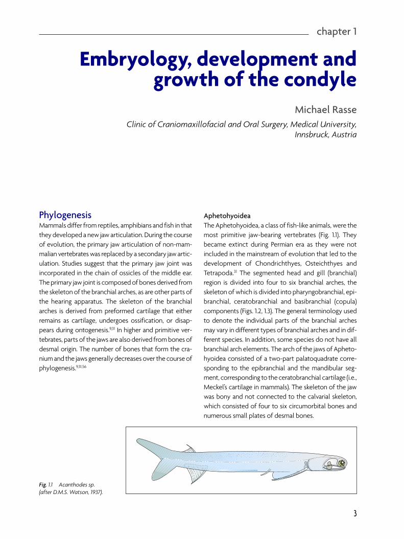
Aphetohyoidea The Aphetohyoidea, a class of fish-like animals, were themost primitive jaw-bearing vertebrates (Fig. 1.1). Theybecame extinct during Permian era as they were notincluded in the mainstream of evolution that led to thedevelopment of Chondrichthyes, Osteichthyes andTetrapoda.31 The segmented head and gill (branchial)region is divided into four to six branchial arches, theskeleton of which is divided into pharyngobranchial, epi-branchial, ceratobranchial and basibranchial (copula)components (Figs. 1.2, 1.3). The general terminology usedto denote the individual parts of the branchial archesmay vary in different types of branchial arches and in dif-ferent species. In addition, some species do not have allbranchial arch elements. The arch of the jaws of Apheto-hyoidea consisted of a two-part palatoquadrate corre-sponding to the epibranchial and the mandibular seg-ment, corresponding to the ceratobranchial cartilage (i.e.,Meckel’s cartilage in mammals). The skeleton of the jawwas bony and not connected to the calvarial skeleton,which consisted of four to six circumorbital bones andnumerous small plates of desmal bones.
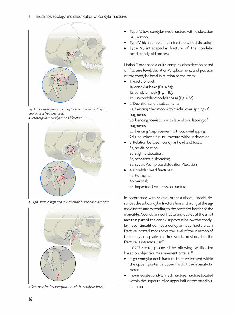
c Subcondylar fracture (fracture of the condylar base)
Fig. 4.3 Classification of condylar fractures according toanatomical fracture level.a Intracapsular condylar head fracture
b High, middle-high and low fracture of the condylar neck
• Type IV, low condylar neck fracture with dislocationi.e. luxation
• Type V, high condylar neck fracture with dislocation • Type VI, intracapsular fracture of the condylar
head/condyloid process
Lindahl23 proposed a quite complex classification basedon fracture level, deviation/displacement, and positionof the condylar head in relation to the fossa:• 1, Fracture level:
1a, condylar head (Fig. 4.3a); 1b, condylar neck (Fig. 4.3b); 1c, subcondylar/condylar base (Fig. 4.3c)
• 2, Deviation and displacement: 2a, bending/deviation with medial overlapping offragments; 2b, bending/deviation with lateral overlapping offragments; 2c, bending/displacement without overlapping; 2d, undisplaced fissural fracture without deviation
• 3, Relation between condylar head and fossa: 3a, no dislocation; 3b, slight dislocation; 3c, moderate dislocation; 3d, severe/complete dislocation/luxation
In accordance with several other authors, Lindahl de-scribes the subcondylar fracture line as starting at the sig-moid notch and extending to the posterior border of themandible. A condylar neck fracture is located at the smalland thin part of the condylar process below the condy-lar head. Lindahl defines a condylar head fracture as afracture located at or above the level of the insertion ofthe condylar capsule; in other words, most or all of thefracture is intracapsular.23
In 1997, Krenkel proposed the following classificationbased on objective measurement criteria. 19
• High condylar neck fracture: fracture located withinthe upper quarter or upper third of the mandibularramus
• Intermediate condylar neck fracture: fracture locatedwithin the upper third or upper half of the mandibu-lar ramus
4 Incidence, etiology and classification of condylar fractures
36
Classification
37
• Low condylar neck fracture: fracture located bet-ween the lower half of the mandibular ramus and themandibular angle
Note: The third type frequently is not classified as acondylar fracture, but as a simple fracture of the mandi-bular ramus.
Ellis et al. classified condylar fractures as follows: 11
• Condylar head fracture: an intracapsular fracturelocated at the border between the condylar head andneck (Fig. 4.3 a)
• Condylar neck fracture: fracture located below thecondylar head but on or above the lowest point ofthe sigmoid notch
• Condylar base fracture: fracture in which the fractureline is located below the lowest point of the sigmoidnotch
They also differentiated between • No detectable dislocation on X-rays and correct posi-
tion of the condylar head• Slight dislocation in which most of the condylar head
remains within the articular fossa and the degree ofangulation/bending of the condylar process is < 20°
• Severe/maximum dislocation: the condylar head ispositioned on the articular tubercle or more anteri-orly, and the degree of angulation/bending is > 20°
Neff et al.28 extended the classification of Spiessl andSchroll40 and discriminated type V and VI diacapitularfractures of the condylar head/condyloid process intothe following types: • Type A (VI A): displacement of the medial parts of the
condylar head; the vertical dimension is intact with-out contraction and the fracture is restrained/sup-ported
• Type B (VI B): fracture involving the lateral parts of thecondyle with a loss of vertical dimension, a lack ofrestraint, contracted fracture appearance, and in-volvement of the lateral pole of the condylar headand lateral ligament
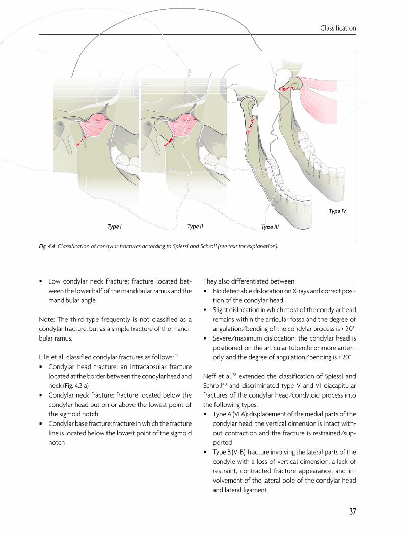
Fig. 4.4 Classification of condylar fractures according to Spiessl and Schroll (see text for explanation).
Type I Type II Type III
Type IV
13.4 Retromandibular approach
156
adjustment can be facilitated by pulling the mandibledownwards. After aligning the fragment, stable plate orscrew osteosynthesis is carried out (Fig. 13.66). The pos-terior border of the ramus and the mandibular notchserve as reference lines for correct three-dimensionalrepositioning.
Wound closure is performed in layers after checkingmandibular mobility and dental occlusion. A wound drain(suction drain) is inserted close to the fracture line, withthe exit placed at the lower border of the incision line.The first step is refixation of the pterygomasseteric sling,followed by closure of the subcutaneous and cutaneouslayers. The skin sutures are removed one week later. Aftercompletion of wound healing, the scar is hardly visible(Fig. 13.67).
Comparison with other approachesThe retromandibular approach differs from other ap-proaches in that the parotid gland is bypassed dorsallyalong anatomical structures without a need for dissec-tion of any additional tissues or organs. With this ap-proach, the incision is more cranial than in a submandi-bular approach and more dorsal than in a transparotidapproach. At this point, it is important to remember thebasic difference between incision and dissection. A retro-mandibular incision has also used for the transparotidapproach.5, 7 This must be taken into account when dif-ferent approaches are compared, especially in regard tocomplication rates.
Fig. 13.61 a and b The incision line lies between the sternoclei-domastoid muscle and the ascending mandibular ramus.
Fig. 13.62 Capsule of the parotid gland
b
a
Comparison with other approaches
157
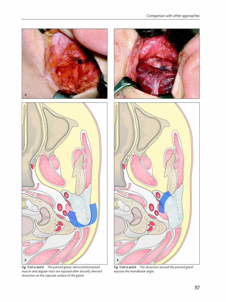
Fig. 13.63 a and b The parotid gland, sternocleidomastoidmuscle and angular tract are exposed after dorsally directeddissection on the capsular surface of the gland.
Fig. 13.64 a and b The dissection around the parotid glandexposes the mandibular angle.