20
FRACTURES OF THE PROXIMAL HUMERUS Presented by Mahsa Mehdizade Dr. Mardani Porsina Hospital Spring 1392
| Date post: | 30-Dec-2015 |
| Category: |
Documents |
| Upload: | noble-valenzuela |
| View: | 71 times |
| Download: | 7 times |

FRACTURES OF THE PROXIMAL HUMERUS
Presented by
Mahsa Mehdizade
Dr. Mardani
Porsina Hospital
Spring 1392

Incidence
Proximal humerus fxs comprise 4-5% of all fxs.
Minimal displacement 80%
Two-part fxs 10%
Three-part fxs 3%
Four-part fxs 4%
Articular surface fxs 3%

Anatomy
Comprised of four segments:
Humeral head
Greater tuberosity
Lesser tuberosity
Humeral shaft

Neurovascular SupplyAnterior and posterior humeral circumflex arteriesArcuate artery-continuation of the ant humeral circumflex and supplies most of the humeral head.Axillary nerve-most commonly injured


Forces on Segments
Greater tuberosity is displaced superiorly and posteriorly by the supraspinatus and external rotators.
Lesser tuberosity is displaced medially by the subscapularis.
The shaft is displaced medially by the pectoralis major.

Mechanism of Injury
Elderly, osteoporotic, usually female: fall on outstretched arm.
Young adults: high-energy trauma; usually more severe fxs and dislocations
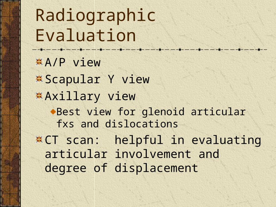
Radiographic Evaluation
A/P view
Scapular Y view
Axillary viewBest view for glenoid articular fxs and dislocations
CT scan: helpful in evaluating articular involvement and degree of displacement


ClassificationsNeer-four parts: greater and lesser tuberosities; shaft; humeral head.
A part is displaced only if >1cm of displacement or 45 degrees of angulation is present.At least 2 views must be obtained
AO-emphasizes the vascular supply to the articular segment
Three types:• Type A: Extraarticular unifocal fxs• Type B: Extraarticular bifocal fxs• Type C: Articular fxs
Not commonly used

Neer Classification

Treatment OptionsClosed reduction
ImmobilizationEarly ROM if stable
External stabilizationPercutaneous pinsExternal fixatorIlizarov frame
Open reduction and internal fixation
Screw fixationTension bandingButtress platingFix-Clip system
Intramedullary fixation
Rush rods
Ender’s nails
Nails with interlocking screws
Excisional arthroplasty
Hemiarthroplasty


Fractures to Consider for Closed Treatment
Minimally displaced 2 part fx’s (or positional reduction of significant displacement)
GT fractures should be <5mm).
Minimally displaced 3- and 4-part fractures

Fractures to Consider for ORIF
Displaced GT fx (> 5 mm)
LT fx with involvement of articular surface
Displaced or unstable surgical neck fx
Displaced anatomic neck fx in young pt.
Displaced, reconstructible 3- and 4-part fractures

Fractures to Consider Hemiarthroplasty
Young/Middle agenonreconstructable articular surface (severe head split) or extruded anatomic neck
Elderlymany 4 parts
some severe 3 parts
most 3,4 part fracture dislocations
most head splits

Potential ComplicationsNeurologic injury
Brachial plexus-Stableforth reported an incidence of 6.1%Axillary-common
Vascular injuryStableforth also reported a 4.9% incidence of arterial injury with displaced fxs; most commonly the axillary arteryAn intact radial pulse doe not exclude an arterial injury so keep it in mind.

Complications cont.Avascular necrosis
Hagg and Lungberg reported an incidence of 3 – 14% with 3- part fxs and 13 – 34% with 4-part fxs, using closed reduction.
Nonunion (uncommon)Malunion – often associated with AVNAdhesive capsulitisMyositis ossificansInfection


